
Previous issues
- Page Path
- HOME > Browse articles > Previous issues
- Volume 47 (3); August 2022
-


Research Articles
- Effects of different topical anesthetics on pain from needle insertion and injection, and the influence of anxiety in patients awaiting endodontic treatment
- Fatih Aksoy, Samet Tosun
- Restor Dent Endod 2022;47(3):e25. Published online June 7, 2022
- DOI: https://doi.org/10.5395/rde.2022.47.e25
-
 Abstract
 PDF
PDF PubReader
 ePub
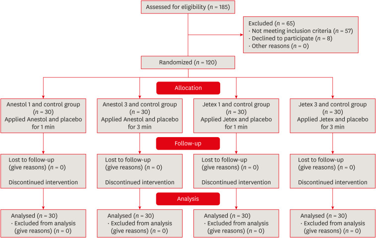
ePub Objectives This study aimed to evaluate the effects of 5% lidocaine and 2.5% lidocaine/2.5% prilocaine topical anesthetic on pain during needle insertion and infiltration injection in the labial mucosa of anterior maxillary teeth, and to assess the relationship between patients’ anxiety and pain scores.
Materials and Methods The Modified Dental Anxiety Scale questionnaire was applied and recorded. Patients were randomly divided into 4 groups (
n = 30), as follows: G1 group: 5% lidocaine and placebo for 1 minute, G2 group: 2.5% lidocaine/2.5% prilocaine and placebo for 1 minute, G3 group: 5% lidocaine and placebo for 3 minutes, and G4 group: 2.5% lidocaine/2.5% prilocaine and placebo for 3 minutes. Before the application of topical anesthesia, one side was randomly selected as the topical anesthesia and the contralateral side as the placebo. The pain levels were measured with Visual Analog Scale (VAS) immediately after needle insertion and injection and were compared. The correlation between anxiety and pain scores was analyzed.Results Administration of 5% lidocaine for 1 minute had significantly higher pain scores for both insertion and infiltration injection than the other groups (
p < 0.05). There was a significant moderate positive correlation between dental anxiety and the injection-induced VAS pain score in the placebo side in all groups (p < 0.05).Conclusions Topical anesthetics significantly reduced the pain caused by both needle insertion and injection pain in comparison to the placebo side. The pain scores of patients with dental anxiety were lower on the topical anesthesia compared to the placebo side.
Trial Registration Thai Clinical Trials Registry Identifier:
TCTR20201217002 -
Citations
Citations to this article as recorded by
- Rapid local anesthesia in children enhanced by STAR particles: a first-in-humans, randomized clinical trial
Andrew R. Tadros, Mark R. Prausnitz, Eric I. Felner
Drug Delivery and Translational Research.2026; 16(2): 539. CrossRef - Evaluation of Articaine Infiltration Location on the Success Rate of Mandibular Lateral Incisor Anesthesia: A Prospective Crossover Randomized Clinical Trial Study
Alireza Adl, Fahime Alimardani, Fereshte Sobhnamayan
Journal of Endodontics.2026; 52(2): 175. CrossRef - Comparison of topical ethyl chloride spray and subcutaneous 1% mepivacaine for local anesthesia in single-rod etonogestrel implant insertion: an observational study
Rosana Garrido-Santamaría, Jesús Martínez-Tofé, Noelia Navas-Echazarreta, Antonio Rodríguez-Calvo, Michał Czapla, Ignacio Larrayoz-Roldán, Raúl Juárez-Vela, Antonio Martinez-Sabater, Regina Ruiz De Viñaspre-Hernandez
Frontiers in Medicine.2026;[Epub] CrossRef - The Case for Transdermal Lidocaine 7.5% in the Management of Localized Pain: A Biopharmaceutical Review
Alexandra LaStella
Scriptum Pharmacologia .2025;[Epub] CrossRef - A Framework for the Modulation and Alleviation of Pain Sensations: A Narrative Review
Rushita Dobariya, Niraj Kinariwala, Nirav Parekh, Dhruvi Gangani, Devshree Dave, Hasti Maru, Nandani Mangukiya, Siddhi Singh
Cureus.2025;[Epub] CrossRef - Phacoemulsification Techniques and Their Effects on Corneal Endothelial Cells and Visual Acuity: A Review of "Direct-Chop" and "Stop-and-Chop" Approaches Under Topical Anesthesia
Devwrath Upasani, Sachin Daigavane
Cureus.2024;[Epub] CrossRef - Local anaesthetics in pediatric dental practice (literature review)
E. V. Ekimov, G. I. Skripkina, A. Zh. Garifullina, N. V. Chumichkin
Pediatric dentistry and dental prophylaxis.2023; 23(3): 211. CrossRef
- Rapid local anesthesia in children enhanced by STAR particles: a first-in-humans, randomized clinical trial
- 4,400 View
- 62 Download
- 4 Web of Science
- 7 Crossref

- Association between cigarette smoking and the prevalence of post-endodontic periapical pathology: a systematic review and meta-analysis
- Néstor Ríos-Osorio, Hernan Darío Muñoz-Alvear, Fabio Andrés Jiménez-Castellanos, Sara Quijano-Guauque, Oscar Jiménez-Peña, Herney Andrés García-Perdomo, Javier Caviedes-Bucheli
- Restor Dent Endod 2022;47(3):e27. Published online June 13, 2022
- DOI: https://doi.org/10.5395/rde.2022.47.e27
-
Abstract
PDFPubReaderePub
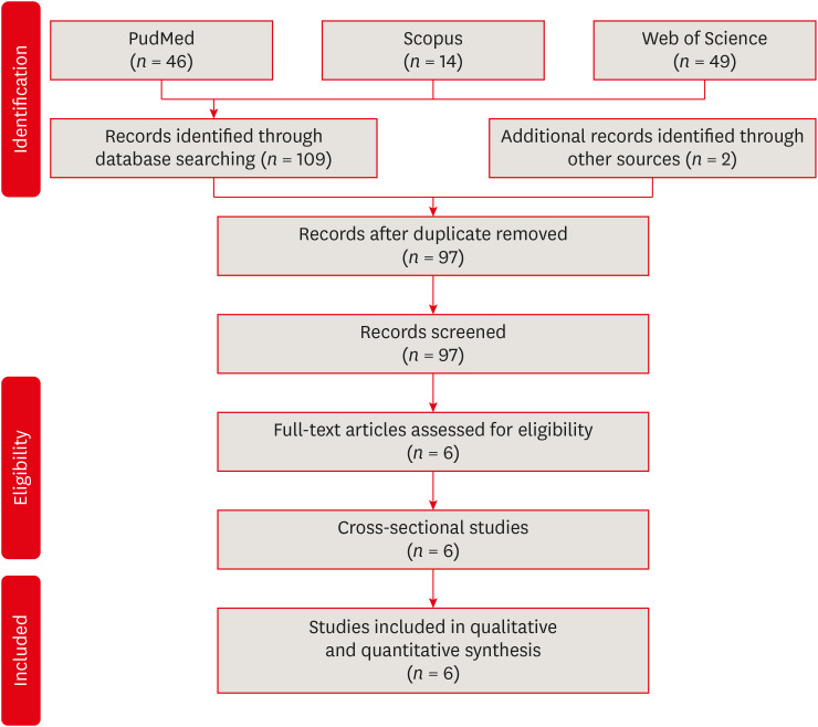
Objectives This systematic review and meta-analysis aimed to assess the association of cigarette smoking with the prevalence of post-endodontic apical periodontitis in humans.
Materials and Methods We searched through PubMed/Medline, Web of Science, and Scopus from inception to December 2020. Risk of bias was performed by using the Newcastle-Ottawa Scale for cross-sectional, cohort, and case-control studies. We performed the statistical analysis in Review Manager 5.3 (RevMan 5.3).
Results 6 studies met the inclusion criteria for qualitative and quantitative synthesis. Statistical analysis of these studies suggests that there were no differences in the prevalence of post endodontic apical periodontitis (AP) when comparing non-smokers
vs smoker subjects regarding patients (odds ratio [OR], 0.68; 95% confidence interval [CI], 0.31–1.49; I2 = 58%) and teeth (OR, 1.71; 95% CI, 0.99–2.93; I2 = 72%).Conclusions Our findings suggest that there was no association between cigarette smoking and post-endodontic apical periodontitis, as we did not find statistical differences in the prevalence of post-endodontic AP when comparing non-smokers
vs smoker subjects. Therefore, smoking should not be considered a risk factor associated with endodontic failure.-
Citations
Citations to this article as recorded by- The role of smoking as a risk indicator for apical periodontitis and endodontic status: a cross-sectional study of a portuguese adult sample
Isabel Silva Martins, Natália Pestana de Vasconcelos, Américo Santos Afonso, Ana Cristina Braga, Irene Pina-Vaz
Odontology.2026;[Epub] CrossRef - RISK FACTORS FOR CHRONIC APICAL PERIODONTITIS ACCORDING TO THE CASE-CONTROL STUDY
N. Bagryantseva
Vrach.2026; : 43. CrossRef
- The role of smoking as a risk indicator for apical periodontitis and endodontic status: a cross-sectional study of a portuguese adult sample
- 4,003 View
- 49 Download
- 1 Web of Science
- 2 Crossref
- Chitosan-induced biomodification on demineralized dentin to improve the adhesive interface
- Isabella Rodrigues Ziotti, Vitória Leite Paschoini, Silmara Aparecida Milori Corona, Aline Evangelista Souza-Gabriel
- Restor Dent Endod 2022;47(3):e28. Published online June 15, 2022
- DOI: https://doi.org/10.5395/rde.2022.47.e28
-
Abstract
PDFPubReaderePub
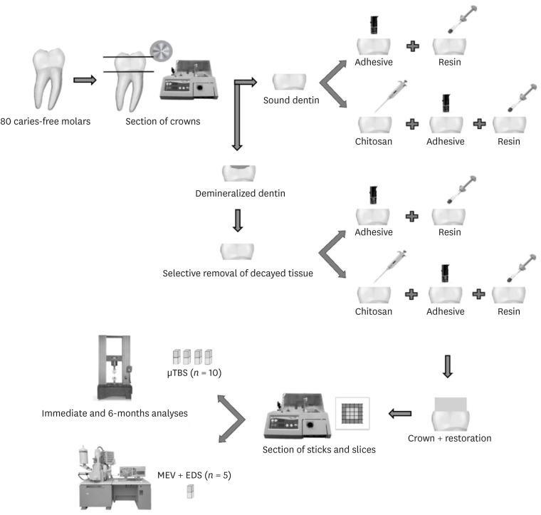
Objectives Metalloproteinase-inhibiting agents, such as chitosan, can prevent collagen degradation in demineralized dental substrates, thereby improving the adhesive interface. This study evaluated the bond strength (BS) and chemical and morphological characterization of the adhesive interface after applying chitosan solution to demineralized dentin.
Materials and Methods The 80 third molars were selected. Forty teeth underwent caries induction using the pH cycling method. The teeth were divided according to the treatment: distilled water (control) and 2.5% chitosan solution. The surfaces were restored using adhesive and composite resins. Half of the specimens in each group were aged, and the other half underwent immediate analyses. The teeth were sectioned and underwent the microtensile bond strength test (µTBS), and chemical and morphological analyses using energy-dispersive spectroscopy and scanning electron microscopy, respectively. Data analysis was performed using 3-way analysis of variance.
Results For µTBS, sound dentin was superior to demineralized dentin (
p < 0.001), chitosan-treated specimens had higher bond strength than the untreated ones (p < 0.001), and those that underwent immediate analysis had higher values than the aged specimens (p = 0.019). No significant differences were observed in the chemical or morphological compositions.Conclusions Chitosan treatment improved bond strength both immediately and after aging, even in demineralized dentin.
-
Citations
Citations to this article as recorded by- The impact of sodium fluoride addition to an adhesive system on micro tensile bond strength of ceramic occlusal veneer
Doaa Shawky Abdelmenam, Waleed Mohamed Elshahawy, Rania Khedr Ahmed
Odontology.2026;[Epub] CrossRef - Recent advances in medical applications of chitosan-based biomaterials
Dinesh Kumar Sharma
International Journal of Polymeric Materials and Polymeric Biomaterials.2025; 74(11): 1027. CrossRef - Push-Out Bond Strength of Different Luting Cements Following Post Space Irrigation with 2% Chitosan: An In Vitro Study
Shimaa Rifaat, Ahmed Rahoma, Hind Muneer Alharbi, Sawsan Jamal Kazim, Shrouq Ali Aljuaid, Basmah Omar Alakloby, Faraz A. Farooqi, Noha Taymour
Prosthesis.2025; 7(1): 18. CrossRef - Bioinspired Dentin Biomodification: Current Evidence and Emerging Approaches
Priyanka S R, Sharath Pare
International Journal of Innovative Science and Research Technology.2025; : 219. CrossRef - A synergistic approach to tooth remineralization using nano-chitosan, fluoride, and pulsed magnetic field
Alaa M. Khalil, Samar A. Abbassy, Mona Mohy ElDin, Sherif Kandil, Ahmed M. El-Khatib
Scientific Reports.2025;[Epub] CrossRef - Chitosan-based Nano/Biomaterials in Bone Tissue Engineering and Regenerative Medicine: Recent Progress and Advances
Taha Jafari, Seyed Morteza Naghib, M. R. Mozafari
Current Organic Synthesis.2025; 22(4): 457. CrossRef - Influence of Non-Staining Chitosan-Based Nano-Silver Fluoride on Shear Bond Strengths of Dental Restorations
Bennett T. Amaechi, Sima Abdollahi, Tejal Gohil, Amos C. Obiefuna, Temitayo Omoniyi, Temitope O. Omosebi, Thais S. Phillips, Noha Elhabashi
Journal of Composites Science.2025; 9(10): 518. CrossRef - Does dentin pretreatment with chitosan improve the bond strength of restorative material? A systematic review and meta-analysis of in vitro studies
Luísa Valente Gotardo Lara Alves, Nathália Mancioppi Cerqueira, Amanda Pelegrin Candemil, André Luis Faria-e-Silva, Manoel Damião Sousa-Neto, Aline Evangelista Souza-Gabriel
International Journal of Adhesion and Adhesives.2024; 128: 103553. CrossRef - Comparative Evaluation of Apical Leakage in Root Canal Obturation Using AH Plus Sealer, Bioceramic Sealer, and Bioceramic Sealer Incorporated With Chitosan Nanoparticles: An In Vitro Study
Sushmita Rane, Varsha Pandit, Sanpreet S Sachdev, Shivani Chauhan, Rishabh Mistry, Barun Kumar
Cureus.2024;[Epub] CrossRef - Aesthetic impact of resin infiltration and its mechanical effect on ceramic bonding for white spot lesions
Jiaen Shu, Yijia Huang, Xueying Ma, Zhonghua Duan, Pei Wu, Sijing Chu, Yuqiong Wu, Yuhua Wang
BMC Oral Health.2024;[Epub] CrossRef - Effect of Incorporating Chitosan to Resin Modified Glass Ionomer Cement on Shear Bond Strength to Dentin (An In vitro Comparative Study)
Aya Tahseen Khudhair, Muna Saleem Khalaf
Journal of International Society of Preventive and Community Dentistry.2024; 14(3): 225. CrossRef - Biomodification of eroded and abraded dentin with epigallocatechin-3-gallate (EGCG)
Bruna Dantas Abreu, Renata Siqueira Scatolin, Silmara Aparecida Milori Corona, Fabiana Almeida Curylofo Zotti
Journal of the Mechanical Behavior of Biomedical Materials.2023; 147: 106158. CrossRef - Chitosan-Based Biomaterials for Tissue Regeneration
Yevgeniy Kim, Zharylkasyn Zharkinbekov, Kamila Raziyeva, Laura Tabyldiyeva, Kamila Berikova, Dias Zhumagul, Kamila Temirkhanova, Arman Saparov
Pharmaceutics.2023; 15(3): 807. CrossRef - Er:YAG laser in selective caries removal and dentin treatment with chitosan: a randomized clinical trial in primary molars
Rai Matheus Carvalho Santos, Renata Siqueira Scatolin, Sérgio Luiz de Souza Salvador, Aline Evangelista Souza-Gabriel, Silmara Aparecida Milori Corona
Lasers in Medical Science.2023;[Epub] CrossRef - Effect of Dentin Surface Pretreatment With Chitosan Nanoparticles on Immediate and Prolonged Shear Bond Strength of Resin Composite: An in Vitro Study
Shaymaa Ali Abdul-Razzaq, Muna Saleem Khalaf
Dental Hypotheses.2023; 14(3): 84. CrossRef - MODERN TRENDS AND PERSPECTIVES OF THE DEVELOPMENT OF ADHESIVE DENTISTRY. INNOVATIVE TECHNIQUES FOR THE APPLICATION OF ADHESIVE SYSTEMS
Oleksandr O. Pompii, Viktor A. Tkachenko, Tetiana M. Kerimova, Elina S. Pompii
Wiadomości Lekarskie.2023; 76(12): 2721. CrossRef
- The impact of sodium fluoride addition to an adhesive system on micro tensile bond strength of ceramic occlusal veneer
- 3,185 View
- 92 Download
- 15 Web of Science
- 16 Crossref
- Resin infiltrant protects deproteinized dentin against erosive and abrasive wear
- Ana Theresa Queiroz de Albuquerque, Bruna Oliveira Bezerra, Isabelly de Carvalho Leal, Maria Denise Rodrigues de Moraes, Mary Anne S. Melo, Vanara Florêncio Passos
- Restor Dent Endod 2022;47(3):e29. Published online July 1, 2022
- DOI: https://doi.org/10.5395/rde.2022.47.e29
-
Abstract
PDFPubReaderePub
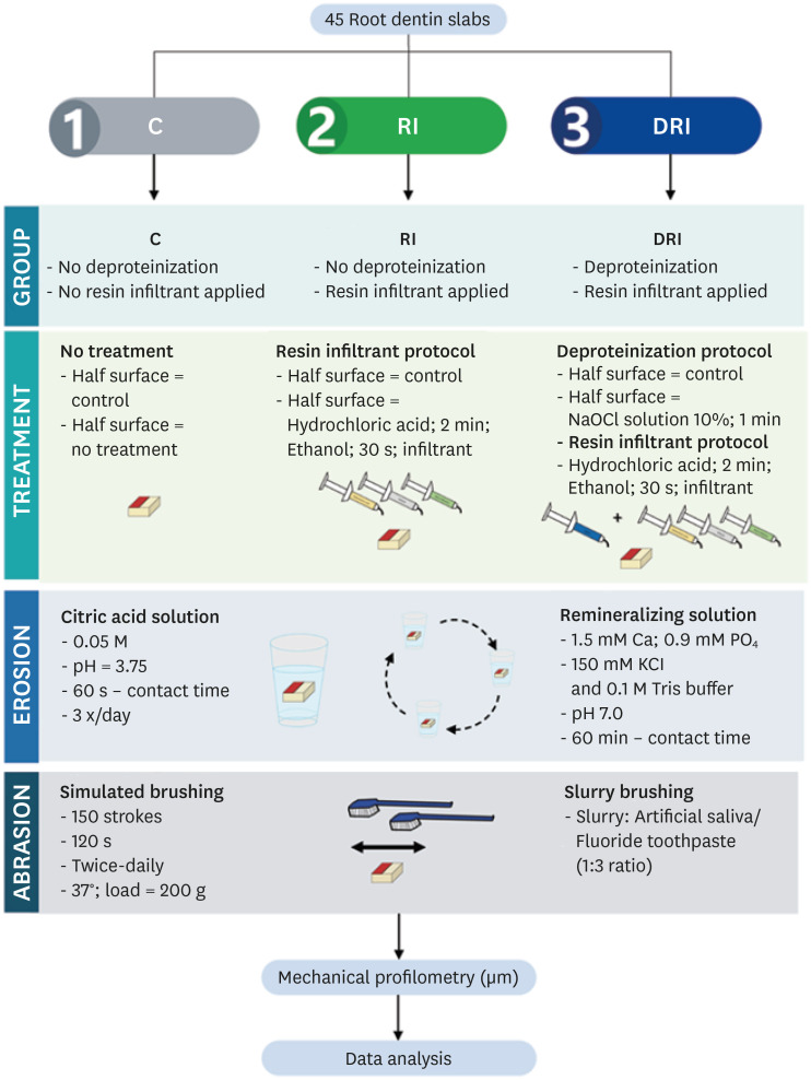
Objectives This study aimed to investigate the anti-erosive/abrasive effect of resin infiltration of previous deproteinized dentin.
Materials and Methods Dentin slabs were randomly assigned to 3 groups (
n = 15): Control (no deproteinization; no resin infiltrant applied), RI (no deproteinization; resin infiltrant applied), and DRI (deproteinization; resin infiltrant applied). After undergoing the assigned treatment, all slabs were subjected to anin vitro cycling model for 5 days. The specimens were immersed in citric acid (0.05 M, pH = 3.75; 60 seconds; 3 times/day) and brushed (150 strokes). Between the challenges, the specimens were exposed to a remineralizing solution (60 minutes). The morphological alterations were analyzed by mechanical profilometry (µm) and scanning electron microscopy (SEM). Data were submitted to one-way analysis of variance (ANOVA) and Tukey tests (p < 0.05).Results Control and RI groups presented mineral wear and did not significantly differ from each other (
p = 0.063). DRI maintained a protective layer preserving the dentin (p < 0.001). After erosive/abrasive cycles, it was observed that in group RI, only 25% of the slabs partially evidenced the presence of the infiltrating, while, in the DRI group, 80% of the slabs presented the treated surface entirely covered by a resin-component layer protecting the dentin surface as observed in SEM images.Conclusions The removal of the organic content allows the resin infiltrant to efficiently protect the dentin surface against erosive/abrasive lesions.
-
Citations
Citations to this article as recorded by- Acidic/abrasive challenges on simulated non-carious cervical lesions development and morphology
Giovanna C. Denucci, Ian Towle, Cecilia P. Turssi, George J. Eckert, Anderson T. Hara
Archives of Oral Biology.2025; 169: 106120. CrossRef - Physio‐Mechanic and Microscopic Analyses of Bioactive Glass‐Based Resin Infiltrants
Syed Zubairuddin Ahmed, Abdul Samad Khan, Wejdan Waleed Nasser, Methayel Abdulrahman Alrushaid, Zahrah Mohammed Alfaraj, Moayad Mohammed Aljeshi, Asma Tufail Shah, Budi Aslinie Md Sabri, Sultan Akhtar, Mohamed Ibrahim Abu Hassan
Microscopy Research and Technique.2025; 88(2): 595. CrossRef - Resin Infiltration Treatment of Developmental Enamel Defects in a Patient With Hydrocephalus and Cerebral Palsy: A Case Report on the Impact on the Maternal Caregiver
Eduarda Martins Fontes Cantarella de Almeida, Anna Luísa Araujo Pimenta, Francisco Wanderley Garcia de Paula‐Silva, Fabricio Kitazono de Carvalho, Laurindo Borelli‐Neto, Susanne Effenberger, Fernanda de Carvalho Panzeri, Silmara Aparecida Milori Corona, K
Special Care in Dentistry.2025;[Epub] CrossRef
- Acidic/abrasive challenges on simulated non-carious cervical lesions development and morphology
- 3,092 View
- 52 Download
- 3 Web of Science
- 3 Crossref
- Effect of intracanal cryotherapy on postoperative pain after endodontic treatment: systematic review with meta-analysis
- Fernanda Garcias Hespanhol, Ludmila Silva Guimarães, Lívia Azeredo Alves Antunes, Leonardo Santos Antunes
- Restor Dent Endod 2022;47(3):e30. Published online July 4, 2022
- DOI: https://doi.org/10.5395/rde.2022.47.e30
-
Abstract
PDFPubReaderePub
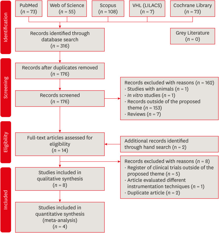
Objectives This study aimed to evaluate the effectiveness of final irrigation with cold saline solution after endodontic treatment compared with saline solution at room temperature against postoperative pain following endodontic treatment.
Materials and Methods A broad search was performed in the PubMed, Web of Science, Scopus, Cochrane Library, Virtual Health Library (LILACS), and Grey Literature databases. Two independent reviewers performed data extraction, risk of bias using the Cochrane methodology, and certainty of evidence using the Grading of Recommendations, Assessment, Development and Evaluations (GRADE) approach.
Results Eight studies were included in qualitative synthesis. Intracanal cryotherapy favored the reduction of postoperative pain in the systematic review. Four studies were included in meta-analyses. The meta-analysis showed that intracanal cryotherapy reduced postoperative pain in teeth with symptomatic apical periodontitis (SAP) at 24 hours. There was no association between intracanal cryotherapy and control (room temperature) groups in teeth with normal periapical tissue with respect to postoperative pain at 24 hours and 48 hours.
Conclusions Intracanal cryotherapy was effective in reducing postoperative pain after endodontic treatment in teeth with SAP.
-
Citations
Citations to this article as recorded by- Postoperative Pain After Endodontic Treatment in HIV‐Positive Patients Under HAART: A Prospective Observational Cohort Study
Marcos Felipe Iparraguirre Nuñovero, Marco Antonio Hungaro Duarte, Luciana Reis Azevedo Alanis, Bruno Cavalini Cavenago, Ulisses Xavier da Silva Neto, Everdan Carneiro
International Endodontic Journal.2026; 59(5): 788. CrossRef - Effect of low-temperature intracanal sodium hypochlorite on root surface temperature reduction and organic tissue dissolution: an in vitro study
Marcos Felipe Iparraguirre Nuñovero, Marco Antonio Hungaro Duarte, Ulisses Xavier da Silva Neto, Vânia Portela Ditzel Westphalen, Pedro Cesar Gomes Titato, Bruno Cavalini Cavenago, Everdan Carneiro
Scientific Reports.2026;[Epub] CrossRef - Effectiveness of intracanal cryotherapy in reducing post-endodontic pain in irreversible pulpitis: a systematic review and meta-analysis
Maged Mohamed, Asmaa Abdelmajeed, Muhammad Salah-Uddin Anwar Laithy, Dina Abozaid
Scientific Reports.2026;[Epub] CrossRef - Post-Operative Pain After Endodontic Instrumentation, Irrigation and Obturation: An Umbrella Review of Systematic Reviews Published from 2016 to 2025
Fausto Zamparini, Andrea Spinelli, Gioia Quadrini, Maria Giovanna Gandolfi, Carlo Prati
Journal of Clinical Medicine.2026; 15(12): 4775. CrossRef - Impact of intracanal cryotherapy on postoperative pain in symptomatic apical periodontitis: A systematic review and meta-analysis of randomized clinical trials
Nishtha K. Patel, Prerak Doshi, Shaily R. Dalal, Pooja R. Kesharani, Shilpa S. Shah, Mohil H. Kale
Endodontology.2025; 37(2): 101. CrossRef - Evaluation of Post‐Endodontic Pain Reduction Using Intracanal Cryotherapy in Symptomatic Apical Periodontitis
Anam Fayyaz Bashir, Ussamah Waheed Jatala, Muhammad Amber Fareed, Sheryar Sheryar, Saadia Ahmad Chattha, Saima Razaq Khan, Shahzad Ahmad, Shazia Iqbal, Muhammad Sohail Zafar, Shahzad Ali
Australian Endodontic Journal.2025; 51(3): 677. CrossRef - Comparing cryotherapy and ketorolac tromethamine against room-temperature saline irrigation using interleukin-8 levels and post-operative pain within single-visit endodontic treatment of symptomatic irreversible pulpitis superimposed by apical periodontit
Yousra Khaled Ezzat, Alaa Diab, Olfat Shaker, Sarah Abouelenien
BMC Oral Health.2025;[Epub] CrossRef - Determining Efficacy of Intracanal Cryotherapy on Post Endodontic Pain in Irreversible Pulpitis
Anam Fayyaz Bashir, Ussamah Waheed Jatala, Moeen ud din Ahmad, Muhammad Talha Khan, Saima Razzaq Khan, Aisha Arshad Butt
Pakistan Journal of Health Sciences.2024; : 68. CrossRef - The effect of intracanal cryotherapy with and without foraminal enlargement on pain prevention after endodontic treatment: a randomized clinical trial
Marcos Felipe Iparraguirre Nuñovero, Marco Antonio Hungaro Duarte, André Vinícius Kaled Segato, Ulisses Xavier da Silva Neto, Vania Portela Ditzel Westphalen, Everdan Carneiro
Scientific Reports.2024;[Epub] CrossRef - Effect of cryotherapy duration on experimentally induced connective tissue inflammationin vivo
Jorge Vera, Mayra Alejandra Castro-Nuñez, María Fernanda Troncoso-Cibrian, Ana Gabriela Carrillo-Varguez, Edgar Ramiro Méndez Sánchez, Viviana Sarmiento, Lourdes Lanzagorta-Rebollo, Prasanna Neelakantan, Monica Romero, Ana Arias
Restorative Dentistry & Endodontics.2023;[Epub] CrossRef - Evaluation of knowledge and awareness of pediatric oral health among school teachers of Hazaribag before and after oral health education.
Vipin Ahuja, Annapurna Ahuja, Nilima Thosar
F1000Research.2023; 12: 1292. CrossRef
- Postoperative Pain After Endodontic Treatment in HIV‐Positive Patients Under HAART: A Prospective Observational Cohort Study
- 4,780 View
- 103 Download
- 10 Web of Science
- 11 Crossref
- Cytotoxicity of two self-adhesive resin cements and their interference in the phagocytic activity of murine macrophages
- Danilo Couto da Silva, Leonardo Gomes Vaz, Warley Luciano Fonseca Tavares, Leda Quercia Vieira, Ricardo Reis de Oliveira, Antônio Paulino Ribeiro Sobrinho
- Restor Dent Endod 2022;47(3):e31. Published online July 14, 2022
- DOI: https://doi.org/10.5395/rde.2022.47.e31
-
Abstract
PDFPubReaderePub
Objectives This study aimed to evaluate
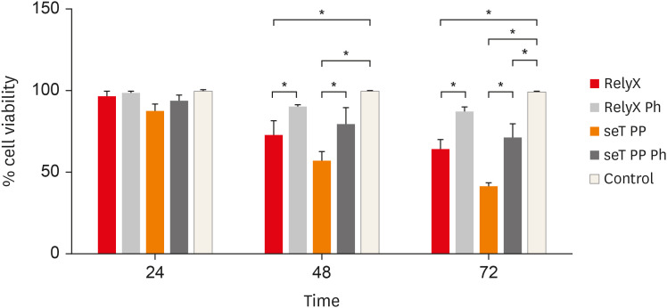
in vitro the effects of the self-adhesive resin cements RelyX U200 (3M ESPE) and seT PP (SDI Limited) on murine macrophages and the interference of the photoactivation.Materials and Methods Cell viability assays, cell adherence, yeast phagocytosis of
Saccharomyces boulardii and production of reactive oxygen species (ROS) were performed in the presence of capillaries containing the respective self-adhesive cement when photoactivated or not.Results After long periods of contact, both types of cements, when not photoactivated, are more cytotoxic for macrophages. The seT PP cement when only chemically activated seems to interfere more negatively in the process of phagocytosis of yeasts
S. boulardii. Both types of cements interfere in the cell adhesion process, independent of photoactivation. None of the types of cements tested was able to induce the production of ROS.Conclusions Our results highlight the great importance of the photoactivation of self-adhesive resin cements in the dental clinic, since RelyX U200, when photoactivated, presented the best results within the evaluated parameters.
-
Citations
Citations to this article as recorded by- Influence of Preheating Self-Adhesive Cements on the Degree of Conversion, Cell Migration, and Cell Viability
Henrique Cantarelli, Fernando Antonio Costa Xavier, Fernando Freitas Portella, Keiichi Hosaka, Eduardo Galia Reston, Louis Hardan, Rim Bourgi, Celso Afonso Klein-Junior
Applied Mechanics.2024; 5(3): 553. CrossRef - Dental Luting Cements: An Updated Comprehensive Review
Artak Heboyan, Anna Vardanyan, Mohmed Isaqali Karobari, Anand Marya, Tatevik Avagyan, Hamid Tebyaniyan, Mohammed Mustafa, Dinesh Rokaya, Anna Avetisyan
Molecules.2023; 28(4): 1619. CrossRef
- Influence of Preheating Self-Adhesive Cements on the Degree of Conversion, Cell Migration, and Cell Viability
- 2,777 View
- 26 Download
- 5 Web of Science
- 2 Crossref
- Influence of inorganic composition and filler particle morphology on the mechanical properties of self-adhesive resin cements
- Marina Rodrigues Santi, Rodrigo Barros Esteves Lins, Beatriz Ometto Sahadi, Giovanna Corrêa Denucci, Gabriela Soffner, Luís Roberto Marcondes Martins
- Restor Dent Endod 2022;47(3):e32. Published online July 14, 2022
- DOI: https://doi.org/10.5395/rde.2022.47.e32
-
Abstract
PDFPubReaderePub
Objectives This study aimed to evaluate the influence of inorganic composition and filler particle morphology on the mechanical properties of different self-adhesive resin cements (SARCs).
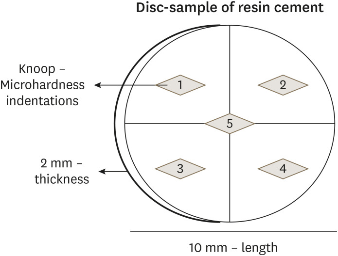
Materials and Methods Three SARCs including RelyX Unicem-2 (RUN), Maxcem Elite (MAX), and Calibra Universal (CAL) were tested. Rectangular bar-shaped specimens were prepared for flexural strength (FS) and flexural modulus (FM) and determined by a 3-point bending test. The Knoop microhardness (KHN) and top/bottom microhardness ratio (%KHN) were conducted on the top and bottom faces of disc-shaped samples. Sorption (Wsp) and solubility (Wsl) were evaluated after 24 hours of water immersion. Filler morphology was analyzed by scanning electron microscopy and X-ray energy dispersive spectroscopy (EDS). FS, FM, %KHN, Wsp, Wsl, and EDS results were submitted to 1-way analysis of variance and Tukey’s
post-hoc test, and KHN also to pairedt -test (α = 0.05).Results SARC-CAL presented the highest FS value, and SARC-RUN presented the highest FM. SARC-MAX and RUN showed the lowest Wsp and Wsl values. KHN values decreased from top to bottom and the SARCs did not differ statistically. Also, all resin cements presented carbon, aluminum, and silica in their composition. SARC-MAX and RUN showed irregular and splintered particles while CAL presented small and regular size particles.
Conclusions A higher mechanical strength can be achieved by a reduced spread in grit size and the filler morphology can influence the KHN, as well as photoinitiators in the composition. Wsp and Wsl can be correlated with ions diffusion of inorganic particles.
-
Citations
Citations to this article as recorded by- Strategic Adhesion and Dental Tissue Conservation: Contemporary Perspectives on Interfacial Bond Longevity and Minimally Invasive Restorative Designs
Cristiana Cuzic, Mihai Rominu, Horatiu Urechescu, Alisia Pricop, Ovidiu Stefan Cuzic, Raul Rotar, Marius Octavian Pricop, Anca Jivanescu
Biomedicines.2026; 14(6): 1391. CrossRef - Effect of Luting Cement on Marginal and Internal Adaptation of Novel Ceramic-Reinforced Polymer Crowns: A Micro-CT Study
Naluemol Sriprasert, Nantawan Krajangta, Thanakorn Wasanapiarnpong, Pavinee Padipatvuthikul Didron, Thanasak Rakmanee
Polymers.2026; 18(14): 1714. CrossRef - Comparative Evaluation of Color Stability in Bioactive and Conventional Resin Cements Under Thermal Stress Conditions
Alaa Turkistani, Hanin E. Yeslam
Biomimetics.2025; 10(7): 432. CrossRef - Assessment of fit accuracy and retentive strength of additively manufactured zirconia crowns luted to Ti‐base abutments with different resin cements: An in vitro study
Rafat Sasany, Sultan Merve Uçar, Burak Yilmaz
Journal of Prosthodontics.2025;[Epub] CrossRef - Bioactive Resin Cement Color Stability and Restoration Thickness as Determinants of the Final Shade in a Glass–Ceramic CAD/CAM Material
Hanin E. Yeslam, Alaa Turkistani
Journal of Functional Biomaterials.2025; 16(9): 319. CrossRef - Light transmittance through resin-matrix composite onlays adhered to resin-matrix cements or flowable composites
Rita Fidalgo-Pereira, Susana O. Catarino, Óscar Carvalho, Nélio Veiga, Orlanda Torres, Annabel Braem, Júlio C.M. Souza
Journal of the Mechanical Behavior of Biomedical Materials.2024; 151: 106353. CrossRef - Effects of a relined fiberglass post with conventional and self-adhesive resin cement
Wilton Lima dos Santos Junior, Marina Rodrigues Santi, Rodrigo Barros Esteves Lins, Luís Roberto Marcondes Martins
Restorative Dentistry & Endodontics.2024;[Epub] CrossRef - Dental Resin-Based Luting Materials—Review
Aleksandra Maletin, Milica Jeremić Knežević, Daniela Đurović Koprivica, Tanja Veljović, Tatjana Puškar, Bojana Milekić, Ivan Ristić
Polymers.2023; 15(20): 4156. CrossRef - A Scoping Review on the Polymerization of Resin-Matrix Cements Used in Restorative Dentistry
Rita Fidalgo-Pereira, Orlanda Torres, Óscar Carvalho, Filipe S. Silva, Susana O. Catarino, Mutlu Özcan, Júlio C. M. Souza
Materials.2023; 16(4): 1560. CrossRef
- Strategic Adhesion and Dental Tissue Conservation: Contemporary Perspectives on Interfacial Bond Longevity and Minimally Invasive Restorative Designs
- 2,998 View
- 32 Download
- 8 Web of Science
- 9 Crossref
- Proximity of maxillary molar apexes to the cortical bone surface and the maxillary sinus
- Han Shin Lee, Dokyung Kim, Sung Kyo Kim
- Restor Dent Endod 2022;47(3):e33. Published online August 8, 2022
- DOI: https://doi.org/10.5395/rde.2022.47.e33
-
Abstract
PDFPubReaderePub
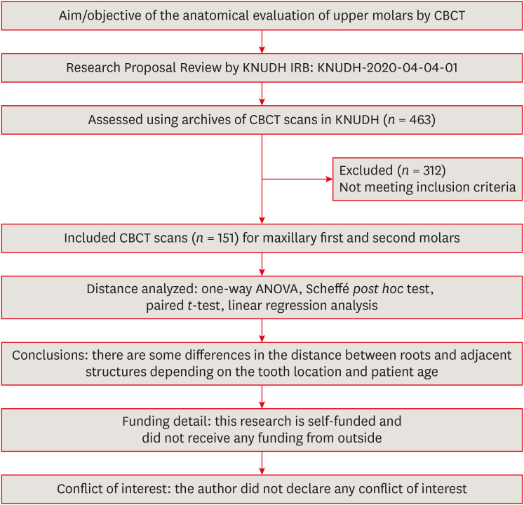
Objectives This study aimed to analyze the proximity of maxillary molar roots to their overlying cortical bone surfaces and the maxillary sinus.
Materials and Methods Cone-beam computed tomographic images of 151 patients with completely erupted upper molars that had 3 separate roots were studied. The following distances were measured: from the root apex to the cortical plate and maxillary sinus floor, and from the apical 3-mm level of the root to the cortical plate. Differences between groups were analyzed with 1-way analysis of variance and the Scheffé
post hoc test, the significance of differences between cone-beam computed tomography views with the pairedt -test, and the significance of differences among age groups with linear regression analysis. The significance level was set atp < 0.05.Results The mesiobuccal and distobuccal root apexes of maxillary second molars were more distant from the buccal cortical plate than the maxillary first molars (
p < 0.05). The apical 3-mm level of the mesiobuccal root of the first molar was closer to the buccal cortical bone than the second molar (p < 0.05). In the maxillary first molars, the thickness of the buccal cortical bone decreased in all roots with age (p < 0.05). In all root apexes of both molars, the difference in the vertical level between the maxillary sinus floor and the root apex increased with age (p < 0.05).Conclusions Awareness of the anatomical profile of maxillary molar apices in relation to the cortical bones and maxillary sinus will be beneficial for apical surgery.
-
Citations
Citations to this article as recorded by- Proximity of maxillary molar palatal roots to adjacent structures for endodontic microsurgery: a cone-beam computed tomography study
Xiaoxiang Huang, Jun Xu, Benxiang Hou, Ying Wang
BMC Oral Health.2025;[Epub] CrossRef - Periapical bone loss configuration in sub-Saudi patients afflicted with periapical abscesses: A 3D cone-beam computed tomography analysis
Swati A. Srivastava, Rahaf A. Alawajy, Rehab Abdelaziz, Elzahraa A. Eldwakhly, Selma A. Saadaldin, Rahaf A. Almohareb, Fahda Nabeel Algahtani, Mai Salah Soliman, Manal M. Abdelhafeez
Saudi Endodontic Journal.2025; 15(2): 144. CrossRef
- Proximity of maxillary molar palatal roots to adjacent structures for endodontic microsurgery: a cone-beam computed tomography study
- 4,190 View
- 35 Download
- 1 Web of Science
- 2 Crossref
- Outcome of endodontic treatments performed by Brazilian undergraduate students: 3- to 8-year follow up
- Jéssica Gabriele da Rocha, Isabella Marian Lena, Jéssica Lopes Trindade, Gabriela Salatino Liedke, Renata Dornelles Morgental, Carlos Alexandre Souza Bier
- Restor Dent Endod 2022;47(3):e34. Published online August 18, 2022
- DOI: https://doi.org/10.5395/rde.2022.47.e34
-
Abstract
PDFPubReaderePub
Objectives This study aimed to evaluate the success rate of endodontic treatments performed by undergraduate students and the factors associated with the outcome.
Materials and Methods A follow-up of 3 to 8 years after root canal filling was carried out in 91 patients. At the follow-up visits, medical and dental history questionnaires were applied along with clinical and radiographic examinations. Data collected in the clinical exam included: the presence of pain, swelling, sinus tract, mobility, tenderness to palpation and percussion, periodontal probing profile, and type/quality of coronal restoration. Postoperative and follow-up radiographs were digitalized and analyzed by 2 trained and calibrated examiners to assess periapical healing. The treatment outcome was based on strict clinical and radiographic criteria and classified as success (absence of any clinical and radiographic sign of apical periodontitis) or failure (other combination). Logistic regression was used to investigate the impact of clinical and radiographic variables on endodontic treatment outcomes at a 5% significance level.
Results The success rate of endodontic treatments was 60.7%. The only risk factor significantly associated with failure was the presence of a periapical lesion on the postoperative radiograph (odds ratio, 3.35; 95% confidence interval, 1.17–9.54).
Conclusions The success rate of endodontic treatments performed by undergraduate students was low and was jeopardized by the presence of a periapical lesion on the postoperative radiograph.
-
Citations
Citations to this article as recorded by- Outcomes of root canal treatment performed by undergraduate students: A systematic review and meta‐analysis
Philip Y.‐H. Chien, Sepanta Hosseinpour, Ove A. Peters, Christine I. Peters
International Endodontic Journal.2026; 59(6): 929. CrossRef - Effect of quality of radiographs taken during root canal treatment on technical quality of root canal fillings and endodontic outcome
Jia Min Ng, Yan Yee Lee, Prashanti Chippagiri, Elaheh Ahanin, Abhishek Parolia
Restorative Dentistry & Endodontics.2025; 50(1): e3. CrossRef - Factors Influencing the Long-Term Survival and Success of Endodontically Treated and Retreated Teeth: An Ambispective Study at an Educational Hospital
Reem Barakat, Rahaf Almohareb, Ghaliah Alsawah, Hadeel Busuhail, Shahad A. Alshihri, Ghadah T. Alrashid, Ghadeer Y. Alotaibi, Mamata Hebbal
Journal of Clinical Medicine.2025; 14(21): 7826. CrossRef - A bibliometric comparison of undergraduate and postgraduate endodontic education publications: The topics, trends, and challenges
Jinglan Zhang, Xiaowei Liu, Lei Yang, Yiran Wang, Dingming Huang, Xuelian Tan
Journal of Dental Education.2023; 87(12): 1661. CrossRef
- Outcomes of root canal treatment performed by undergraduate students: A systematic review and meta‐analysis
- 5,892 View
- 52 Download
- 6 Web of Science
- 4 Crossref
Case Report
- Persistent pain after successful endodontic treatment in a patient with Wegener’s granulomatosis: a case report
- Ricardo Machado, Jorge Aleixo Pereira, Filipe Colombo Vitali, Michele Bolan, Elena Riet Correa Rivero
- Restor Dent Endod 2022;47(3):e26. Published online June 9, 2022
- DOI: https://doi.org/10.5395/rde.2022.47.e26
-
Abstract
PDFPubReaderePub
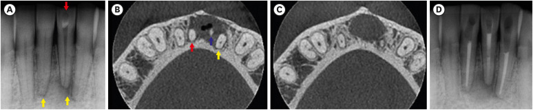
Wegener’s granulomatosis (WG) is a condition with immune-mediated pathogenesis that can present oral manifestations. This report describes the case of a patient diagnosed with WG 14 years previously, who was affected by persistent pain of non-odontogenic origin after successful endodontic treatment. A 39-year-old woman with WG was diagnosed with pulp necrosis and apical periodontitis of teeth #31, #32, and #41, after evaluation through a clinical examination and cone-beam computed tomography (CBCT). At the first appointment, these teeth were subjected to conventional endodontic treatment. At 6- and 12-month follow-up visits, the patient complained of persistent pain associated with the endodontically treated teeth (mainly in tooth #31), despite complete remission of the periapical lesions shown by radiographic and CBCT exams proving the effectiveness of the endodontic treatments, thus indicating a probable diagnostic of persistent pain of non-odontogenic nature. After the surgical procedure was performed to curette the lesion and section 3 mm of the apical third of tooth #31, the histopathological analysis suggested that the painful condition was likely associated with the patient's systemic condition. Based on clinical, radiographic, and histopathological findings, this unusual case report suggests that WG may be related to non-odontogenic persistent pain after successful endodontic treatments.
-
Citations
Citations to this article as recorded by- Toothaches of Non-odontogenic Origin
Davis C. Thomas, Tanvee Somaiya, Ahana Ajayakumar, Vaishnavi Prabhakar
Dental Clinics of North America.2026; 70(1): 209. CrossRef - Prevalence of persistente pain after endodontic treatment
Edmundo Duarte Martins, Allya Francisca Marques Borges, Lidiane Oliveira Leão, Bianca Marques de Mattos de Araujo, José Stechman Neto, Tatiana Carvalho Kowaltschuk, Camila de Castro Corrêa Corrêa, Cristiano Miranda de Araújo, Karinna Veríssimo Meira Tave
Brazilian Journal of Oral Sciences.2026; 25: e269272. CrossRef
- Toothaches of Non-odontogenic Origin
- 6,820 View
- 84 Download
- 2 Crossref
 First
First Prev
Prev

