Search
- Page Path
- HOME > Search
- Resolvin E1 incorporated carboxymethyl chitosan scaffold accelerates repair of dental pulp stem cells under inflammatory conditions: a laboratory investigation
- Hemalatha P Balasubramanian, Nandini Suresh, Vishnupriya Koteeswaran, Velmurugan Natanasabapathy
- Restor Dent Endod 2025;50(4):e40. Published online November 28, 2025
- DOI: https://doi.org/10.5395/rde.2025.50.e40
-
 Abstract
 PDF
PDF Supplementary Material
 PubReader
 ePub
ePub - Objectives
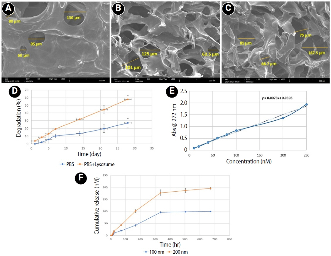
This study fabricated and characterized a resolvin E1 (RvE1)-loaded carboxymethyl chitosan (CMC) scaffold and determined its cytotoxicity and mineralization potential on inflamed human dental pulp stem cells (hDPSCs).
Methods
CMC scaffold incorporated with two concentrations of RvE1 (100 and 200 nM) was fabricated and characterized. The scaffolds’ porosity, drug release kinetics, and degradation were assessed. The impact of RvE1 on inflamed hDPSCs proliferation, proinflammatory gene expression (tumor necrosis factor alpha [TNF-α]), alkaline phosphatase activity, and alizarin red S staining was evaluated.
Results
Scanning electron microscopy analysis demonstrated a highly porous interconnected microstructure. Release kinetics showed gradual RvE1 release peaking at day 14. Cumulative degradation of the CMC scaffold at 28 days was 57.35%. Inflamed hDPSCs exposed to 200 nM RvE1-CMC scaffold exhibited significantly improved viability compared to 100 nM. Both RvE1-CMC scaffolds significantly suppressed the expression of TNF-α at 7 days. Alkaline phosphatase activity was enhanced by both RvE1 concentrations on days 7 and 14. Alizarin red staining revealed superior mineralization potential of 200 nM RvE1 on days 14 and 21.
Conclusions
This study concludes 200 nM RvE1-CMC scaffold is a promising therapy for inflamed pulp conditions, enhancing cell proliferation and biomineralization potential in inflamed hDPSCs.
- 499 View
- 22 Download

- Stress distribution of restorations in external cervical root resorption under occlusal and traumatic loads: a finite element analysis
- Padmapriya Ramanujam, Paul Kevin Abishek Karthikeyan, Vignesh Srinivasan, Selvakarthikeyan Ulaganathan, Velmurugan Natanasabapathy, Nandini Suresh
- Restor Dent Endod 2025;50(2):e21. Published online May 21, 2025
- DOI: https://doi.org/10.5395/rde.2025.50.e21
-
 Abstract
 PDF
PDF PubReader
 ePub
ePub - Objectives
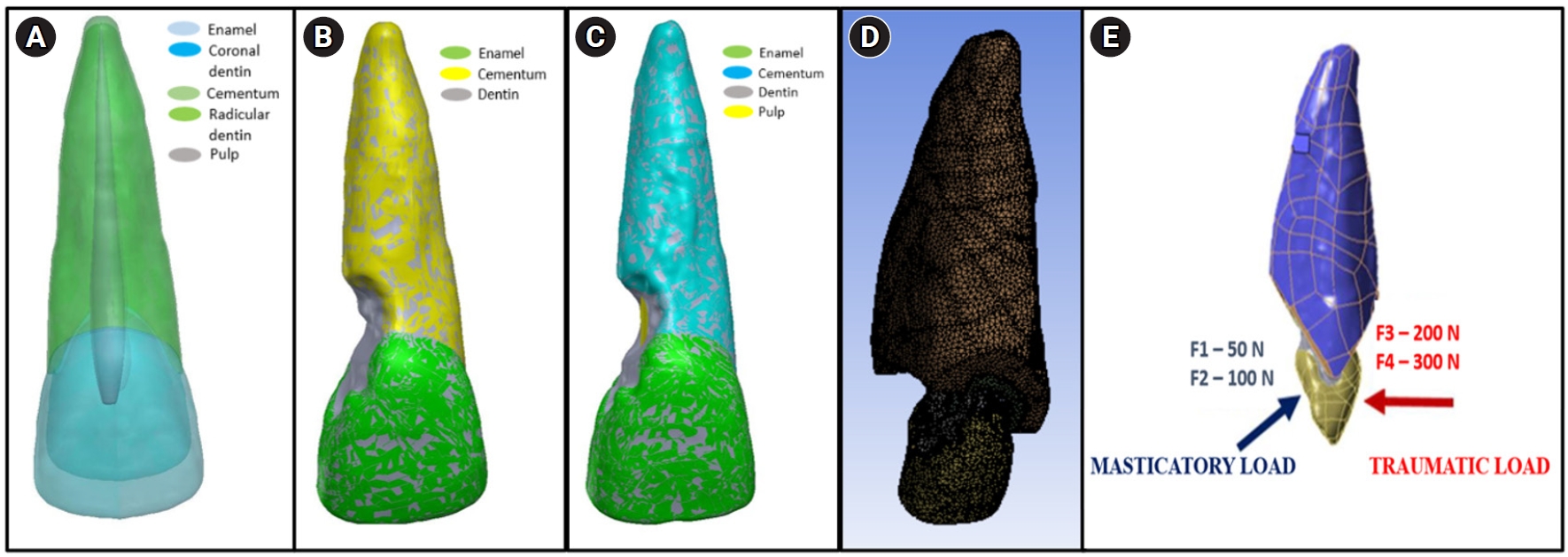
This study analyzed the stress distribution in a maxillary central incisor with external cervical resorptive defect restored with different restorative materials under normal masticatory and traumatic loading conditions using finite element analysis.
Methods
Cone-beam computed tomography of an extracted intact incisor and created resorptive models (Patel’s 3D classification-2Bd and 2Bp) in the maxillary central incisor was performed for finite element models. The 2Bd models were restored either with glass ionomer cement (GIC)/Biodentine (Septodont) or a combination of both with composite resin. 2Bp models were restored externally with a combination technique and internally with root canal treatment. The other model was external restoration with GIC and internal with fiber post. Two masticatory loads were applied at 45˚ to the palatal aspect, and two traumatic loads were applied at 90˚ to the buccal aspect. Maximum von Mises stresses were calculated, and stress distribution patterns were studied.
Results
In 2Bd models, all restorative strategies decreased stress considerably, similar to the control model under all loads. In 2Bp models, the dentin component showed maximum stress at the deepest portion of the resorptive defect, which transfers into the adjacent pulp space. In 2Bp defects, a multilayered restoration externally and root canal treatment internally provides better stress distribution compared to the placement of a fiber post.
Conclusions
Increase in load, proportionally increased von Mises stress, despite the direction or angulation of the load. Multilayered restoration is preferred for 2Bd defects, and using an internal approach of root canal treatment is suggested to restore 2Bp defects.
- 1,855 View
- 128 Download

- Biomineralization of three calcium silicate-based cements after implantation in rat subcutaneous tissue
- Ranjdar Mahmood Talabani, Balkees Taha Garib, Reza Masaeli, Kavosh Zandsalimi, Farinaz Ketabat
- Restor Dent Endod 2021;46(1):e1. Published online December 2, 2020
- DOI: https://doi.org/10.5395/rde.2021.46.e1
-
 Abstract
 PDF
PDF PubReader
 ePub
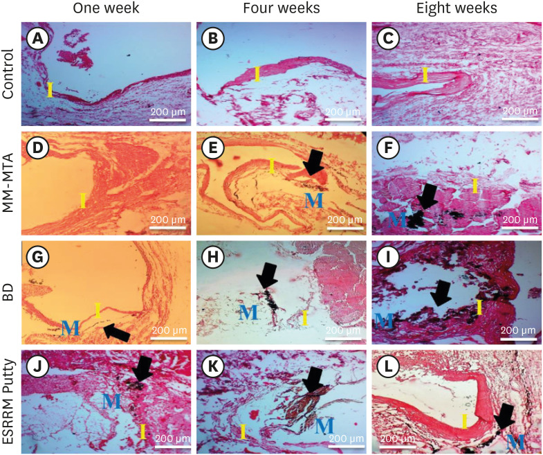
ePub Objectives The aim of this study was to evaluate the dystrophic mineralization deposits from 3 calcium silicate-based cements (Micro-Mega mineral trioxide aggregate [MM-MTA], Biodentine [BD], and EndoSequence Root Repair Material [ESRRM] putty) over time after subcutaneous implantation into rats.
Materials and Methods Forty-five silicon tubes containing the tested materials and 15 empty tubes (serving as a control group) were subcutaneously implanted into the backs of 15 Wistar rats. At 1, 4, and 8 weeks after implantation, the animals were euthanized (
n = 5 animals/group), and the silicon tubes were removed with the surrounding tissues. Histopathological tissue sections were stained with von Kossa stain to assess mineralization. Scanning electron microscopy and energy-dispersive X-ray spectroscopy (SEM/EDX) were also used to assess the chemical components of the surface precipitates deposited on the implant and the pattern of calcium and phosphorus distribution at the material-tissue interface. The calcium-to-phosphorus ratios were compared using the non-parametric Kruskal-Wallis test at a significance level of 5%.Results The von Kossa staining showed that both BD and ESRRM putty induced mineralization starting at week 1; this mineralization increased further until the end of the study. In contrast, MM-MTA induced dystrophic calcification later, from 4 weeks onward. SEM/EDX showed no statistically significant differences in the calcium- and phosphorus-rich areas among the 3 materials at any time point (
p > 0.05).Conclusions After subcutaneous implantation, biomineralization of the 3-calcium silicate-based cements started early and increased over time, and all 3 tested cements generated calcium- and phosphorus-containing surface precipitates.
-
Citations
Citations to this article as recorded by- Evaluating Retrieval-Augmented Large Language Models on External Cervical Resorption: A Comparative Study of Gemini and NotebookLM
Marc Garcia-Font, Nicolás Dufey-Portilla, Fernando Durán-Sindreu, José Antonio González Sánchez, Gustavo Rodríguez Millán, Venkateshbabu Nagendrababu, Paul M.H. Dummer, Francesc Abella Sans
Journal of Endodontics.2025;[Epub] CrossRef - Antibacterial, biocompatible, and mineralization‐inducing properties of calcium silicate‐based cements
Taimy Cruz Hondares, Xiaoxiao Hao, Yanfang Zhao, Yuyin Lin, Dobrawa Napierala, Janice G. Jackson, Ping Zhang
International Journal of Paediatric Dentistry.2024; 34(6): 843. CrossRef - Bioactive potential of Bio‐C Pulpo is evidenced by presence of birefringent calcite and osteocalcin immunoexpression in the rat subcutaneous tissue
Marcela Borsatto Queiroz, Rafaela Nanami Handa Inada, Camila Soares Lopes, Juliane Maria Guerreiro‐Tanomaru, Estela Sasso‐Cerri, Mário Tanomaru‐Filho, Paulo Sérgio Cerri
Journal of Biomedical Materials Research Part B: Applied Biomaterials.2022; 110(10): 2369. CrossRef
- Evaluating Retrieval-Augmented Large Language Models on External Cervical Resorption: A Comparative Study of Gemini and NotebookLM
- 2,258 View
- 19 Download
- 3 Web of Science
- 3 Crossref

-
Comparative evaluation of the bond strength of self-adhering and bulk-fill flowable composites to MTA Plus, Dycal, Biodentine, and TheraCal: an
in vitro study - Aakrati Raina, Asheesh Sawhny, Saurav Paul, Sridevi Nandamuri
- Restor Dent Endod 2020;45(1):e10. Published online January 8, 2020
- DOI: https://doi.org/10.5395/rde.2020.45.e10

-
 Abstract
 PDF
PDF PubReader
 ePub
ePub Objectives This study aimed to compare the shear bond strength (SBS) of a self-adhering flowable composite (Dyad Flow) and a bulk-fill flowable composite (Smart Dentin Replacement [SDR]) to several pulp-capping materials, including MTA Plus, Dycal, Biodentine, and TheraCal.
Materials and Methods Eighty acrylic blocks with 2-mm-deep central holes that were 4 mm in diameter were prepared and divided into 2 groups (
n = 40 each) according to the composite used (Dyad Flow or SDR). They were further divided into 4 sub-groups (n = 10 each) according to the pulp-capping agent used. SBS was tested using a universal testing machine at a crosshead speed of 1 mm/min. Data were analyzed using 2-way analysis of variance. Ap value of < 0.05 was considered to indicate statistical significance.Results A statistically significant difference (
p = 0.040) was found between Dyad Flow and SDR in terms of bond strength to MTA Plus, Dycal, Biodentine, and TheraCal.Conclusions Among the 8 sub-groups, the combination of TheraCal and SDR exhibited the highest SBS.
-
Citations
Citations to this article as recorded by- Shear Bond Strength of Liner Materials to Caries-Free and Caries-Affected Dentin
ZK Greene, NR Smith, T Gomes, NC Lawson
Operative Dentistry.2025; 50(3): 324. CrossRef - Shear Bond Strength of Biointeractive Restorative Materials to NeoMTA Plus and Biodentine
Zübeyde Uçar Gündoğar, Gül Keskin, Merve Yaman Küçükersen
Polymers.2025; 17(22): 3070. CrossRef - Hygroscopic bioactive light-cured composite promoting dentine bridge formation
Yunzi Long, Guibin Huang, Siyi Liu, Liju Xu, Ailing Li, Dong Qiu, Yanmei Dong
Regenerative Biomaterials.2024;[Epub] CrossRef - Comparative evaluation of shear bond strength and modes of failure of five different reinforced glass ionomer restorative cements to TheraCal LC: An in vitro study
Kalyani Gajanan Umale, Vandana Jaykumar Gade, Ambar W. Raut
Journal of Conservative Dentistry and Endodontics.2024; 27(2): 200. CrossRef - Evaluation of the Effect of Chitosan-Based Irrigation Solutions on the Bond Strength of Mineral Trioxide Aggregate to Bulk-Fill Composite
Arzu Şahin Mantı, Bağdagül Helvacıoğlu Kıvanç
Journal of Functional Biomaterials.2024; 15(12): 370. CrossRef - Radiopacity evaluations of the novel calcium-silicate and glass-Ionomer-based materials
Yeşim Şeşen Uslu, Elif Çelebi, Meriç Berkman
Journal of Health Sciences and Medicine.2024; 7(2): 192. CrossRef - Effect of Er Cr YSGG laser etching procedure on the bond strength of different calcium silicate cements
Yesim Sesen Uslu, Hakan Yasin Gönder, Pinar Sesen, Gizem Gunduz Bektaş
Lasers in Dental Science.2024;[Epub] CrossRef - The micro‐shear bond strength of new endodontic tricalcium silicate‐based putty: An in vitro study
Merve Yeniçeri Özata, Seda Falakaloğlu, Gianluca Plotino, Özkan Adıgüzel
Australian Endodontic Journal.2023; 49(1): 124. CrossRef - Analysis of the bond strength between conventional, putty or resin‐modified calcium silicate cement and bulk fill composites
İ Ipek, B Karaağaç Eskibağlar, Ş Yildiz, O Ataş, M Ünal
Australian Dental Journal.2023; 68(4): 265. CrossRef - Effect of Different Adhesive Strategies on the Microshear Bond Strength of Calcium-Silicate-Based Materials
Aliye Tuğçe Gürcan, Soner Şişmanoğlu, Görkem Sengez
Journal of Advanced Oral Research.2022; 13(2): 191. CrossRef - BULK FİLL KOMPOZİT REZİN RESTORATİF MATERYALLER
Merve NEZİR, Suat ÖZCAN
Atatürk Üniversitesi Diş Hekimliği Fakültesi Dergisi.2022; : 1. CrossRef - Effect of Bioinductive Cavity Liners on Shear Bond Strength of Dental Composite to Dentin
Saba Tohidkhah, Elham Ahmadi, Mahdi Abbasi, Reza Morvaridi Farimani, Ladan Ranjbar Omrani, Victor Feitosa
BioMed Research International.2022;[Epub] CrossRef - Bond Strength of Adhesive Systems to Calcium Silicate-Based Materials: A Systematic Review and Meta-Analysis of In Vitro Studies
Louis Hardan, Davide Mancino, Rim Bourgi, Alejandra Alvarado-Orozco, Laura Emma Rodríguez-Vilchis, Abigailt Flores-Ledesma, Carlos Enrique Cuevas-Suárez, Monika Lukomska-Szymanska, Ammar Eid, Maya-Line Danhache, Maryline Minoux, Youssef Haïkel, Naji Kharo
Gels.2022; 8(5): 311. CrossRef - How do imaging protocols affect the assessment of root-end fillings?
Fernanda Ferrari Esteves Torres, Reinhilde Jacobs, Mostafa EzEldeen, Karla de Faria-Vasconcelos, Juliane Maria Guerreiro-Tanomaru, Bernardo Camargo dos Santos, Mário Tanomaru-Filho
Restorative Dentistry & Endodontics.2022;[Epub] CrossRef - Evaluation of Shear Bond Strength of Resin‐Based Composites to Biodentine with Three Types of Seventh‐Generation Bonding Agents: An In Vitro Study
Huda Abbas Abdullah, Zahraa Abdulaali Al-Ibraheemi, Zanbaq Azeez Hanoon, Julfikar Haider, Boonlert Kukiattrakoon
International Journal of Dentistry.2022;[Epub] CrossRef - Evaluation of the Bond Strength of Different Pulp Capping Materials to Dental Adhesive Systems: An In Vitro Study
Sema Yazici Akbiyik, Elif Pınar Bakir, S¸eyhmus Bakir
Journal of Advanced Oral Research.2021; 12(2): 286. CrossRef - Differential Gene Expression Changes in Human Primary Dental Pulp Cells Treated with Biodentine and TheraCal LC Compared to MTA
Ok Hyung Nam, Ho Sun Lee, Jae-Hwan Kim, Yong Kwon Chae, Seoung-Jin Hong, Sang Wook Kang, Hyo-Seol Lee, Sung Chul Choi, Young Kim
Biomedicines.2020; 8(11): 445. CrossRef
- Shear Bond Strength of Liner Materials to Caries-Free and Caries-Affected Dentin
- 2,298 View
- 43 Download
- 17 Crossref

- A micro-computed tomography evaluation of voids using calcium silicate-based materials in teeth with simulated internal root resorption
- Vildan Tek, Sevinç Aktemur Türker
- Restor Dent Endod 2020;45(1):e5. Published online November 29, 2019
- DOI: https://doi.org/10.5395/rde.2020.45.e5
-
 Abstract
 PDF
PDF PubReader
 ePub
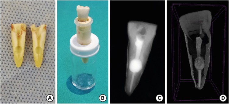
ePub Objectives The obturation quality of MTA, Biodentine, Total Fill BC root canal sealer (RCS), and warm gutta-percha (WGP) in teeth with simulated internal root resorption (IRR) was evaluated by using micro-computed tomography.
Materials and Methods Standardized IRR cavities were created using 40 extracted maxillary central incisor teeth and randomly assigned into 4 groups (
n = 10). IRR cavities were filled with MTA, Biodentine, Total Fill BC RCS (bulk-fill form) and WGP + Total Fill BC RCS. Percentage of voids between resorptive cavity walls and obturation material (external void), and inside the filling materials (internal voids) were measured.Results Total Fill BC sealer in the bulk-fill form presented significantly highest values of external and internal void percentages (
p < 0.05). Biodentine showed a significantly lowest external void percentage (p < 0.05). WGP + Total Fill BC RCS presented significantly lower values of internal void percentages than all groups (p < 0.05), except Biodentine (p > 0.05).Conclusion None of the filling materials were created void-free obturation in resorption cavities. Biodentine may favor its application in teeth with IRR over Angelus MTA and bulk-fill form of Total Fill BC.
-
Citations
Citations to this article as recorded by- Removal of AH Plus Bioceramic Sealer from Artificial Internal Resorption Cavities Using Different Irrigation Activation Systems
Mine Büker, Meltem Sümbüllü, Emine Şimşek, Fadime Sena Sezer
Cumhuriyet Dental Journal.2025; 28(3): 383. CrossRef - Functional and Bioactive Performance of Premixed Bioceramic Sealers with Warm Obturation: A Scoping Review
Patryk Wiśniewski, Stanisław Krokosz, Małgorzata Pietruska, Anna Zalewska
Gels.2025; 11(11): 932. CrossRef - Evaluation of the effectiveness of different supplemental cleaning techniques in the retreatment of roots with small simulated internal resorption cavities: an in vitro comparative study
Sine Güngör Us, Özgür Uzun, Nazlı Merve Güngör
BMC Oral Health.2025;[Epub] CrossRef - Evaluation of Different Techniques and Materials for Filling in 3-dimensional Printed Teeth Replicas with Perforating Internal Resorption by Means of Micro–Computed Tomography
Angelo J.S. Torres-Carrillo, Helena C. Assis, Rodrigo E. Salazar-Gamarra, Leonardo Moreira Teodosio, Alice C. Silva-Sousa, Jardel F. Mazzi-Chaves, Priscila B. Ferreira-Soares, Manoel D. Sousa-Neto, Fabiane C. Lopes-Olhê
Journal of Endodontics.2024; 50(2): 205. CrossRef - Three-Dimensional Measurement of Obturation Quality of Bioceramic Materials in Filling Artificial Internal Root Resorption Cavities Using Different Obturation Techniques: An In Vitro Comparative Study
Ammar M. Sharki, Ahmed H. Ali
Journal of Endodontics.2024; 50(7): 997. CrossRef - Evaluation of calcium hydroxide root canal filling materials by cone beam computed tomography and three-dimensional modeling
Asel Usdat Ozturk, Ekin Dogan, Venus Seyedoskuyi, Berk Senguler, Asli Topaloglu-Ak
Folia Medica.2024; 66(2): 250. CrossRef - Clinical applications of calcium silicate‐based materials: a narrative review
S Küçükkaya Eren
Australian Dental Journal.2023;[Epub] CrossRef - A critical analysis of research methods and experimental models to study root canal fillings
Gustavo De‐Deus, Erick Miranda Souza, Emmanuel João Nogueira Leal Silva, Felipe Gonçalves Belladonna, Marco Simões‐Carvalho, Daniele Moreira Cavalcante, Marco Aurélio Versiani
International Endodontic Journal.2022; 55(S2): 384. CrossRef - An Updated Review on Properties and Indications of Calcium Silicate‐Based Cements in Endodontic Therapy
Fateme Eskandari, Alireza Razavian, Rozhina Hamidi, Khadije Yousefi, Susan Borzou, Zohaib Khurshid
International Journal of Dentistry.2022;[Epub] CrossRef - Efficacy Of Calcium Silicate-Based Sealers In Root Canal Treatment: A Systematic Review
Hattan Mohammed Omar Baismail, Mohammed Ghazi Moiser Albalawi, Alaa Mofareh Thoilek Alanazi, Muhannad Atallah Saleem Alatawi, Badr Soliman Alhussain
Annals of Dental Specialty.2021; 9(1): 87. CrossRef
- Removal of AH Plus Bioceramic Sealer from Artificial Internal Resorption Cavities Using Different Irrigation Activation Systems
- 2,295 View
- 26 Download
- 10 Crossref

- The push-out bond strength of BIOfactor mineral trioxide aggregate, a novel root repair material
- Makbule Bilge Akbulut, Durmus Alperen Bozkurt, Arslan Terlemez, Melek Akman
- Restor Dent Endod 2019;44(1):e5. Published online January 28, 2019
- DOI: https://doi.org/10.5395/rde.2019.44.e5
-
 Abstract
 PDF
PDF PubReader
 ePub
ePub Objectives The aim of this
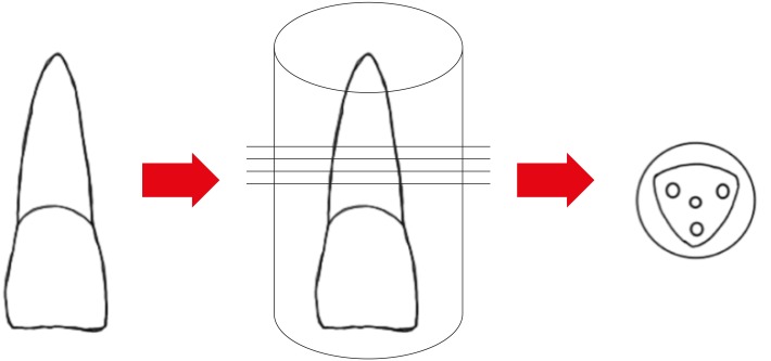
in vitro study was to evaluate the push-out bond strength of a novel calcium silicate-based root repair material-BIOfactor MTA to root canal dentin in comparison with white MTA-Angelus (Angelus) and Biodentine (Septodont).Materials and Methods The coronal parts of 12 central incisors were removed and the roots were embedded in acrylic resin blocks. Midroot dentin of each sample was horizontally sectioned into 1.1 mm slices and 3 slices were obtained from each root. Three canal-like standardized holes having 1 mm in diameter were created parallel to the root canal on each dentin slice with a diamond bur. The holes were filled with MTA-Angelus, Biodentine, or BIOfactor MTA. Wet gauze was placed over the specimens and samples were stored in an incubator at 37°C for 7 days to allow complete setting. Then samples were subjected to the push-out test method using a universal test machine with the loading speed of 1 mm/min. Data was statistically analyzed using Friedman test and
post hoc Wilcoxon signed rank test with Bonferroni correction.Results There were no significant differences among the push-out bond strength values of MTA-Angelus, Biodentine, and BIOfactor MTA (
p > 0.017). Most of the specimens exhibited cohesive failure in all groups, with the highest rate found in Biodentine group.Conclusions Based on the results of this study, MTA-Angelus, Biodentine, and BIOfactor MTA showed similar resistances to the push-out testing.
-
Citations
Citations to this article as recorded by- Examination of the Bond Strength of Retrograde Filling in Teeth with Failed Apical Resection After Retreatment
Sevda Tok, Leyla Benan Ayranci
Applied Sciences.2025; 15(7): 3441. CrossRef - Comparative Analysis of Physicocomechanical Properties of MTA and Biodentine with Addition of Graphene Oxide to MTA and Biodentine: An In-vitro Study
Tanvi Arvind Jagtap, Budhabhushan A. Sonvane, Guruprasad Handal, Jayashri Nimba Bhangare, Kedar Vilas Saraf, Abhishek Mulay
Journal of Pharmacy and Bioallied Sciences.2025; 17(Suppl 1): S608. CrossRef - Influence of Incubation Duration on Bond Strength and Microhardness of Calcium Silicate‐Based Materials
Emine Şimşek, Makbule Bilge Akbulut
Australian Endodontic Journal.2025; 51(2): 438. CrossRef - Comparative evaluation of push-out bond strength after root perforation repair using recently introduced bioceramic and calcium silicate-based materials – An in vitro study
Gurinder Kaur, Deepak Kurup, Deepyanti Dubey, Ajit Hindlekar, Ganesh Ranganath Jadhav, Priya Mittal, Siddharth Shinde
Endodontology.2025; 37(2): 194. CrossRef - Comparative Evaluation of Push-out Bond Strength of Conventional Mineral Trioxide Aggregate, Biodentine, and Two Novel Antibacterial-enhanced Mineral Trioxide Aggregates
Sanjeev Khanagar, Suman Panda, Prabhadevi C Maganur, Ganesh Jeevanandan, Satish Vishwanathaiah, Ather A Syed, Sara Kalagi, Arokia RS Merlin, Vignesh Ravindran, Aram AlShehri
The Journal of Contemporary Dental Practice.2024; 25(2): 168. CrossRef - Influence of Phase Composition and Morphology on the Calcium Ion Release of Several Classical and Hybrid Endodontic Cements
Ivanka Dimitrova, Galia Gentscheva, Ivanka Spassova, Daniela Kovacheva
Materials.2024; 17(22): 5568. CrossRef - The Effect of Two Different MTA (Mineral Trioxide Aggregate) On Thermal Insulation
Gizem Akkus, Ecem Salmaz, Didem Oner Ozdas
The Open Dentistry Journal.2024;[Epub] CrossRef - Comparison of push‐out bond strength and apical microleakage of different calcium silicate‐based cements after using EDTA, chitosan and phytic acid irrigations
Tutku Koçak Şahin, Murat Ünal
Microscopy Research and Technique.2024; 87(9): 2072. CrossRef - In vitro evaluation of the physical characteristics and push-out bond strength of new experimental nano-MTA
Nada Omar, Yousra Aly, Haidy N. Salem
Bulletin of the National Research Centre.2024;[Epub] CrossRef - Interfacial characteristics of BIOfactor MTA and Biodentine with dentin
Makbule Bilge Akbulut, Şeref Nur Mutlu, Mehmet Ali Soylu, Emine Şimşek
Microscopy Research and Technique.2023; 86(2): 258. CrossRef - Systemic effect of calcium silicate-based cements with different radiopacifiers-histopathological analysis in rats
Osman Ataş, Kubra Bılge, Semsettin Yıldız, Serkan Dundar, Ilknur Calik, Asime Gezer Ataş, Alihan Bozoglan
PeerJ.2023; 11: e15376. CrossRef - The push-out bond strength of three root canal materials used in primary teeth: in vitro study
Hazal Özer, Merve Abaklı İnci, Sevcihan Açar Tuzluca
Frontiers in Dental Medicine.2023;[Epub] CrossRef - Effects of different irrigation protocols on push-out bond strength of pre-mixed calcium silicate-based cements
Sabiha Ceren İlisulu, Aliye Tugce Gürcan, Soner Sismanoglu
Journal of the Australian Ceramic Society.2023; 59(5): 1381. CrossRef - Micro-Computed Tomographic Evaluation of the Sealing Quality and Bond Strength of Different MTA Apical Plugs
Taibe Tokgöz Kaplan, Murat Selim Botsalı
European Journal of Therapeutics.2023; 30(1): 29. CrossRef - Kan kontaminasyonunun farklı kök ucu dolgu materyallerinin dentine bağlanma dayanımına etkisi
Şeyma Nur GERÇEKCİOĞLU, Melike BAYRAM, Emre BAYRAM
Acta Odontologica Turcica.2023; 40(1): 9. CrossRef - Tooth Discoloration Effect of BIOfactor Mineral Trioxide Aggregate: A 6-Month In Vitro Study
Şeref Nur Mutlu, Makbule Bilge Akbulut
Applied Sciences.2023; 13(15): 8914. CrossRef - Comparative Evaluation of the Push-Out Bond Strength of Root-End Filling Materials by Using Different Condensation Methods
Pelin Tüfenkçi, Sevinç Sevgi, Ayşenur Öncü, Fatma Semra Sevimay, Berkan Çelikten
Cyprus Journal of Medical Sciences.2022; 7(7): 115. CrossRef - Effect of Different Adhesive Strategies on the Microshear Bond Strength of Calcium-Silicate-Based Materials
Aliye Tuğçe Gürcan, Soner Şişmanoğlu, Görkem Sengez
Journal of Advanced Oral Research.2022; 13(2): 191. CrossRef - BIOfactor MTA’nın Radyoopasitesinin Dijital Radyografi ile Değerlendirilmesi
Şeref Nur MUTLU, Makbule Bilge AKBULUT
Selcuk Dental Journal.2022; 9(2): 520. CrossRef - Morphological and Chemical Analysis of Different Types of Calcium Silicate‐Based Cements
Okba Mahmoud, Nashwan Abdullah Al-Afifi, Mohideen Salihu Farook, Maysara Adnan Ibrahim, Saaid Al Shehadat, Mohammed Amjed Alsaegh, Sandrine Bittencourt Berger
International Journal of Dentistry.2022;[Epub] CrossRef - Influence of Blood Contamination on Push-Out Bond Strength of Three Calcium Silicate-Based Materials to Root Dentin
Cristina Rodrigues Paulo, Joana A. Marques, Diana B. Sequeira, Patrícia Diogo, Rui Paiva, Paulo J. Palma, João Miguel Santos
Applied Sciences.2021; 11(15): 6849. CrossRef - An In vitro comparative evaluation of effect of novel irrigant Qmix and 17% ethylenediaminetetraacetic acid on the push-out bond strength of biodentine and endosequence bioceramic root repair material
VandanaJ Gade, Aparajita Gangrade, JaykumarR Gade, Neelam Rahul
Journal of the International Clinical Dental Research Organization.2021; 13(2): 124. CrossRef - A micro-computed tomographic study using a novel test model to assess the filling ability and volumetric changes of bioceramic root repair materials
Fernanda Ferrari Esteves Torres, Jader Camilo Pinto, Gabriella Oliveira Figueira, Juliane Maria Guerreiro-Tanomaru, Mario Tanomaru-Filho
Restorative Dentistry & Endodontics.2021;[Epub] CrossRef - Micro-computed tomographic evaluation of the flow and filling ability of endodontic materials using different test models
Fernanda Ferrari Esteves Torres, Juliane Maria Guerreiro-Tanomaru, Gisselle Moraima Chavez-Andrade, Jader Camilo Pinto, Fábio Luiz Camargo Villela Berbert, Mario Tanomaru-Filho
Restorative Dentistry & Endodontics.2020;[Epub] CrossRef
- Examination of the Bond Strength of Retrograde Filling in Teeth with Failed Apical Resection After Retreatment
- 1,872 View
- 11 Download
- 24 Crossref

- Fibre reinforcement in a structurally compromised endodontically treated molar: a case report
- Renita Soares, Ida de Noronha de Ataide, Marina Fernandes, Rajan Lambor
- Restor Dent Endod 2016;41(2):143-147. Published online February 22, 2016
- DOI: https://doi.org/10.5395/rde.2016.41.2.143
-
 Abstract
 PDF
PDF PubReader
 ePub
ePub The reconstruction of structurally compromised posterior teeth is a rather challenging procedure. The tendency of endodontically treated teeth (ETT) to fracture is considerably higher than vital teeth. Although posts and core build-ups followed by conventional crowns have been generally employed for the purpose of reconstruction, this procedure entails sacrificing a considerable amount of residual sound enamel and dentin. This has drawn the attention of researchers to fibre reinforcement. Fibre-reinforced composite (FRC), designed to replace dentin, enables the biomimetic restoration of teeth. Besides improving the strength of the restoration, the incorporation of glass fibres into composite resins leads to favorable fracture patterns because the fibre layer acts as a stress breaker and stops crack propagation. The following case report presents a technique for reinforcing a badly broken-down ETT with biomimetic materials and FRC. The proper utilization of FRC in structurally compromised teeth can be considered to be an economical and practical measure that may obviate the use of extensive prosthetic treatment.
-
Citations
Citations to this article as recorded by- Performance of direct and indirect onlay restorations for structurally compromised teeth
Khaled Abid Althaqafi
The Journal of Prosthetic Dentistry.2025; 133(6): 1513. CrossRef - Endodontically Treated Teeth with Fiber-Reinforced Composite Resins
Ridhima Gupta, Ashwini B. Prasad, Deepak Raisingani, Deeksha Khurana, Prachi Mital, Vaishali Moryani
Journal of Dental Research and Review.2022; 9(4): 310. CrossRef - Survival and success of endocrowns: A systematic review and meta-analysis
Raghad A. Al-Dabbagh
The Journal of Prosthetic Dentistry.2021; 125(3): 415.e1. CrossRef - Short fiber‐reinforced composite restorations: A review of the current literature
Sufyan Garoushi, Ausama Gargoum, Pekka K. Vallittu, Lippo Lassila
Journal of Investigative and Clinical Dentistry.2018;[Epub] CrossRef
- Performance of direct and indirect onlay restorations for structurally compromised teeth
- 1,852 View
- 42 Download
- 4 Crossref

- Evaluation of reparative dentin formation of ProRoot MTA, Biodentine and BioAggregate using micro-CT and immunohistochemistry
- Jia Kim, Young-Sang Song, Kyung-San Min, Sun-Hun Kim, Jeong-Tae Koh, Bin-Na Lee, Hoon-Sang Chang, In-Nam Hwang, Won-Mann Oh, Yun-Chan Hwang
- Restor Dent Endod 2016;41(1):29-36. Published online January 4, 2016
- DOI: https://doi.org/10.5395/rde.2016.41.1.29

-
 Abstract
 PDF
PDF PubReader
 ePub
ePub Objectives The purpose of this study was to assess the ability of two new calcium silicate-based pulp-capping materials (Biodentine and BioAggregate) to induce healing in a rat pulp injury model and to compare them with mineral trioxide aggregate (MTA).
Materials and Methods Eighteen rats were anesthetized, cavities were prepared and the pulp was capped with either of ProRoot MTA, Biodentine, or BioAggregate. The specimens were scanned using a high-resolution micro-computed tomography (micro-CT) system and were prepared and evaluated histologically and immunohistochemically using dentin sialoprotein (DSP).
Results On micro-CT analysis, the ProRoot MTA and Biodentine groups showed significantly thicker hard tissue formation (
p < 0.05). On H&E staining, ProRoot MTA showed complete dentin bridge formation with normal pulpal histology. In the Biodentine and BioAggregate groups, a thick, homogeneous hard tissue barrier was observed. The ProRoot MTA specimens showed strong immunopositive reaction for DSP.Conclusions Our results suggest that calcium silicate-based pulp-capping materials induce favorable effects on reparative processes during vital pulp therapy and that both Biodentine and BioAggregate could be considered as alternatives to ProRoot MTA.
-
Citations
Citations to this article as recorded by- Evaluation of microhardness, monomer conversion, and antibacterial properties of an experimental pulp-capping material containing collagen–hydroxyapatite nanocomposite and/or chlorhexidine
Hacer Balkaya, Sezer Demirbuğa, Fatih Duman, Ahmet Ceylan, Ömer Aydın
Odontology.2026; 114(1): 204. CrossRef - Clinical applications and classification of calcium silicate-based cements based on their history and evolution: a narrative review
Kenta Tsuchiya, Salvatore Sauro, Hidehiko Sano, Jukka P. Matinlinna, Monica Yamauti, Shuhei Hoshika, Yu Toida, Rafiqul Islam, Atsushi Tomokiyo
Clinical Oral Investigations.2025;[Epub] CrossRef - Impact of diode laser irradiation along with Biodentine on dental pulp stem cell proliferation and pluripotent gene expression
Ladan Alborzy, Sedighe Sadat Hashemikamangar, Mahshid Hodjat, Nasim Chiniforush, Behnaz Behniafar
Scientific Reports.2025;[Epub] CrossRef - The effect of different treatment methods on apical closure and treatment success in immature permanent first molars with reversible pulpitis
Muhammed ALAGOZ, Sera SIMSEK DERELIOĞLU
BMC Oral Health.2025;[Epub] CrossRef - Histological evaluation of pulp response to alendronate and Biodentine as pulp capping agents: an animal study
Thangavel Boopathi, Sekar Manimaran, Joseline Charles Kerena, Mathew Sebeena, Kumaravadivel Karthick, Natesan Thangaraj Deepa
Restorative Dentistry & Endodontics.2024;[Epub] CrossRef - Comparative Clinical and Radiographic Success Rate of Bioceramic Premix vs Biosilicate-based Medicament as Indirect Pulp Treatment Materials in Primary Molars: A Double-blind Randomized Trial with a Follow-up of 12 Months
Aditi Mathur, Meenakshi Nankar, Sunnypriyatham Tirupathi, Payal Kothari, Rashmi Chauhan, Ashrita Suvarna
International Journal of Clinical Pediatric Dentistry.2024; 17(7): 748. CrossRef - Effects of mineral trioxide aggregate and methyl sulfonyl methane on pulp exposure via RUNX2 and RANKL pathways
Altar Ateş, Ayca Kurt, Tolga Mercantepe
Odontology.2024; 112(3): 895. CrossRef - Evaluation of biocompatibility and bioactive potential of Well-Root PT by comparison with ProRoot MTA and Biodentine
Yong Kwon Chae, Ju Ri Ye, Ok Hyung Nam
Journal of Dental Sciences.2024; 19(4): 2218. CrossRef - Dentine Remineralisation Induced by “Bioactive” Materials through Mineral Deposition: An In Vitro Study
Marta Kunert, Ireneusz Piwonski, Louis Hardan, Rim Bourgi, Salvatore Sauro, Francesco Inchingolo, Monika Lukomska-Szymanska
Nanomaterials.2024; 14(3): 274. CrossRef - Different pulp capping agents and their effect on pulp inflammatory response: A narrative review
Mustafa Tariq Mutar, Anas F Mahdee
The Saudi Dental Journal.2024; 36(10): 1295. CrossRef - Clinical application of calcium silicate-based bioceramics in endodontics
Xinyuan Wang, Yizhi Xiao, Wencheng Song, Lanxiang Ye, Chen Yang, Yuzhen Xing, Zhenglin Yuan
Journal of Translational Medicine.2023;[Epub] CrossRef - Evaluation of the pulp response following direct pulp capping with exogenous nitric oxide and Mineral Trioxide Aggregate (MTA) a histologic study
Amirah Alnour, Ghassan Almohammad, Anas Abdo, Kinda Layous
Heliyon.2023; 9(7): e17458. CrossRef - Histological evaluation of dental pulp response to Biodentine, enamel matrix derivative (Emdogain), and mineral trioxide aggregate as direct pulp-capping agents – A randomized clinical trial
Takhellambam Premlata Devi, Amandeep Kaur, Shamurailatpam Priyadarshini, B. S. Deepak, Sumita Banerjee, Ngairangbam Sanjeeta
Journal of Medical Society.2023; 37(3): 107. CrossRef - Effect of Intracoronal Sealing Biomaterials on the Histological Outcome of Endodontic Revitalisation in Immature Sheep Teeth—A Pilot Study
Elanagai Rathinam, Sivaprakash Rajasekharan, Heidi Declercq, Christian Vanhove, Peter De Coster, Luc Martens
Journal of Functional Biomaterials.2023; 14(4): 214. CrossRef - Restorative management of the posterior tooth that has undergone a pulpotomy
Nicholas N Longridge, James S Hyde, Fadi Jarad, Sondos Albadri
Dental Update.2023; 50(11): 932. CrossRef - Direct pulp capping procedures – Evidence and practice
Rafiqul Islam, Md Refat Readul Islam, Toru Tanaka, Mohammad Khursheed Alam, Hany Mohamed Aly Ahmed, Hidehiko Sano
Japanese Dental Science Review.2023; 59: 48. CrossRef - A novel analysis of the formation and resorption changes in dental hard tissue using longitudinal in vivo micro computed tomography
Yeon-Jee YOO, Joonil HWANG, So-Hyun PARK, Jaehong HWANG, Seungryong CHO, Sun-Young KIM
Dental Materials Journal.2023; 42(5): 708. CrossRef - Evaluation of pH and Calcium Ion Diffusion from Intracanal MTA and Bioaggregate to Simulated External Resorption Cavities Through Dentinal Tubules
Umut AKSOY, Kaan POLATOĞLU, Feridun ŞAKLAR
European Annals of Dental Sciences.2022; 49(3): 108. CrossRef - Pulpa Kuafajı ve Kuafaj Materyallerine Güncel Bir Bakış: Derleme
Dilek AKIN, Çiğdem ATALAYIN ÖZKAYA
Selcuk Dental Journal.2022; 9(2): 617. CrossRef - The Influence of New Bioactive Materials on Pulp–Dentin Complex Regeneration in the Assessment of Cone Bone Computed Tomography (CBCT) and Computed Micro-Tomography (Micro-CT) from a Present and Future Perspective—A Systematic Review
Mirona Paula Palczewska-Komsa, Bartosz Gapiński, Alicja Nowicka
Journal of Clinical Medicine.2022; 11(11): 3091. CrossRef - Evaluation of shear bond strength of e-mineral trioxide aggregate and biodentine with glass ionomer cement
Hemalatha Hiremath, Aishwarya Singh Solanki, Shivangi Trivedi, Devansh Verma
Endodontology.2022; 34(2): 127. CrossRef - Multiple growth factors accommodated degradable submicron calcium sulfate hemihydrate/porous hydroxyapatite for dentin-pulp regeneration
Chih-Wen Chi, Bharathi Priya Lohanathan, Ching-Ching Wong, Che-Lun Chen, Hsun-Chang Lin, Yu-Chih Chiang
Biomaterials Advances.2022; 140: 213045. CrossRef - THE EFFECT OF BLOOD CONTAMINATION ON SHEAR BOND STRENGTH OF CALCIUM SILICATE-BASED PULP CAPPING MATERIALS
Hasan Fatih YAVUZ, Güneş BULUT EYÜBOĞLU
Cumhuriyet Dental Journal.2022; 24(4): 371. CrossRef - Comparison of Four Dental Pulp-Capping Agents by Cone-Beam Computed Tomography and Histological Techniques—A Split-Mouth Design Ex Vivo Study
Jayanandan Muruganandhan, Govindarajan Sujatha, Saravanan Poorni, Manali Ramakrishnan Srinivasan, Nezar Boreak, Ahmed Al-Kahtani, Mohammed Mashyakhy, Hitesh Chohan, Shilpa Bhandi, A. Thirumal Raj, Alessio Zanza, Luca Testarelli, Shankargouda Patil
Applied Sciences.2021; 11(7): 3045. CrossRef - Effect of Naturally Occurring Biogenic Materials on Human Dental Pulp Stem Cells (hDPSC): an In Vitro Study.
Prasanna T. Dahake, Vinod V. Panchal, Yogesh J. Kale, Mahesh V. Dadpe, Shrikant B. Kendre, Vijay M. Kumbar
Regenerative Engineering and Translational Medicine.2021; 7(4): 506. CrossRef - Influence of Ultrasonic Activation on the Physicochemical Properties of Calcium Silicate-Based Cements
Fredson Márcio Acris De Carvalho, Yara Teresinha Corrêa Silva-Sousa, Carlos Eduardo Saraiva Miranda, Paulo Henrique Miller Calderon, Ana Flávia Simões Barbosa, Luciana Martins Domingues De Macedo, Fuad Jacob Abi Rached-Junior, Boonlert Kukiattrakoon
International Journal of Dentistry.2021; 2021: 1. CrossRef - Tailored 70S30C Bioactive glass induces severe inflammation as pulpotomy agent in primary teeth: an interim analysis of a randomised controlled trial
Yasmine Elhamouly, Rania M. El Backly, Dalia M. Talaat, Samia S. Omar, Maha El Tantawi, Karin M. L. Dowidar
Clinical Oral Investigations.2021; 25(6): 3775. CrossRef - Response of dental pulp capped with calcium-silicate based material, calcium hydroxide and adhesive resin in rabbit teeth
Cynthia Kassis, Pierre Khoury, Karim Corbani, Charbel Mansour, Louis Hardan, Ghassan Yared, Carole Chakar
Brazilian Journal of Oral Sciences.2021;[Epub] CrossRef - Effectiveness of Direct Pulp Capping Bioactive Materials in Dentin Regeneration: A Systematic Review
Ermin Nie, Jiali Yu, Rui Jiang, Xiangzhen Liu, Xiang Li, Rafiqul Islam, Mohammad Khursheed Alam
Materials.2021; 14(22): 6811. CrossRef - Immunohistochemical expression of non-collagenous extracellular matrix molecules involved in tertiary dentinogenesis following direct pulp capping: a systematic review
C. Călin, M. Sajin, V.T. Moldovan, C. Coman, S.I. Stratul, A.C. Didilescu
Annals of Anatomy - Anatomischer Anzeiger.2021; 235: 151674. CrossRef - Recent Advances in Indirect Pulp Treatment Materials for Primary Teeth: A Literature Review
Omar AES El Meligy, Afnan M Saber, Sumer M Alaki
International Journal of Clinical Pediatric Dentistry.2021; 14(6): 795. CrossRef - Chitosan-Based Accelerated Portland Cement Promotes Dentinogenic/Osteogenic Differentiation and Mineralization Activity of SHED
Hasan Subhi, Adam Husein, Dasmawati Mohamad, Nik Rozainah Nik Abdul Ghani, Asma-Abdullah Nurul
Polymers.2021; 13(19): 3358. CrossRef - Histological evaluation of the regenerative potential of a novel treated dentin matrix hydrogel in direct pulp capping
Ahmed A. Holiel, Elsayed M. Mahmoud, Wegdan M. Abdel-Fattah, Khadiga Y. Kawana
Clinical Oral Investigations.2021; 25(4): 2101. CrossRef - Minimal Intervention in Dentistry: A Literature Review on Biodentine as a Bioactive Pulp Capping Material
Naji Ziad Arandi, Mohammad Thabet, Mona Abbassy
BioMed Research International.2021;[Epub] CrossRef - Potential of tailored amorphous multiporous calcium silicate glass for pulp capping regenerative endodontics—A preliminary assessment
Jie Liu, Chao-An Chen, Xiaofei Zhu, Brian R. Morrow, Ukrit Thamma, Tia J. Kowal, Hassan M. Moawad, Matthias M. Falk, Himanshu Jain, George T.-J. Huang
Journal of Dentistry.2021; 109: 103655. CrossRef - Tomographic evaluation of direct pulp capping using a novel injectable treated dentin matrix hydrogel: a 2-year randomized controlled clinical trial
Ahmed A. Holiel, Elsayed M. Mahmoud, Wegdan M. Abdel-Fattah
Clinical Oral Investigations.2021; 25(7): 4621. CrossRef - Hard tissue formation after direct pulp capping with osteostatin and MTA in vivo
Ji-Hye Yoon, Sung-Hyeon Choi, Jeong-Tae Koh, Bin-Na Lee, Hoon-Sang Chang, In-Nam Hwang, Won-Mann Oh, Yun-Chan Hwang
Restorative Dentistry & Endodontics.2021;[Epub] CrossRef - Hydraulic cements for various intra-coronal applications: Part 1
Stephen J Bonsor, Josette Camilleri
Dental Update.2021; 48(8): 653. CrossRef - In vivo Biocompatibility and Bioactivity of Calcium Silicate-Based Bioceramics in Endodontics
Wencheng Song, Wei Sun, Lili Chen, Zhenglin Yuan
Frontiers in Bioengineering and Biotechnology.2020;[Epub] CrossRef - Evaluation of dentinogenesis inducer biomaterials: an in vivo study
Anabela B. Paula, Mafalda Laranjo, Carlos-Miguel Marto, Siri Paulo, Ana M. Abrantes, Bruno Fernandes, João Casalta-Lopes, Manuel Marques-Ferreira, Maria Filomena Botelho, Eunice Carrilho
Journal of Applied Oral Science.2020;[Epub] CrossRef - Micro-computed tomographic evaluation of a new system for root canal filling using calcium silicate-based root canal sealers
Mario Tanomaru-Filho, Fernanda Ferrari Esteves Torres, Jader Camilo Pinto, Airton Oliveira Santos-Junior, Karina Ines Medina Carita Tavares, Juliane Maria Guerreiro-Tanomaru
Restorative Dentistry & Endodontics.2020;[Epub] CrossRef - Bio-Inductive Materials in Direct and Indirect Pulp Capping—A Review Article
Marta Kunert, Monika Lukomska-Szymanska
Materials.2020; 13(5): 1204. CrossRef - Micro-computed tomographic evaluation of the flow and filling ability of endodontic materials using different test models
Fernanda Ferrari Esteves Torres, Juliane Maria Guerreiro-Tanomaru, Gisselle Moraima Chavez-Andrade, Jader Camilo Pinto, Fábio Luiz Camargo Villela Berbert, Mario Tanomaru-Filho
Restorative Dentistry & Endodontics.2020;[Epub] CrossRef - Release of Transforming Growth Factor Beta 1 from Human Tooth Dentin after Application of Either ProRoot MTA or Biodentine as a Coronal Barrier
Kunlada Wattanapakkavong, Tanida Srisuwan
Journal of Endodontics.2019; 45(6): 701. CrossRef - Effect of Leptin on Odontoblastic Differentiation and Angiogenesis: An In Vivo Study
Sung-Hyeon Choi, Ji-Hyun Jang, Jeong-Tae Koh, Hoon-Sang Chang, Yun-Chan Hwang, In-Nam Hwang, Bin-Na Lee, Won-Mann Oh
Journal of Endodontics.2019; 45(11): 1332. CrossRef - Análise da composição química dos cimentos MTA Angelus® branco, cinza e HP Repair® através de Microscopia Eletrônica de Varredura (MEV) acoplada a Espectrômetro de Energia Dispersiva (EDS)
Gabriela Duarte Rocha SARZEDA, Marcelo Santos BAHIA, Paulo Victor Teixeira DORIGUÊTTO, Karina Lopes DEVITO, Anamaria Pessoa Pereira LEITE
Revista de Odontologia da UNESP.2019;[Epub] CrossRef - Direct Pulp Capping: Which is the Most Effective Biomaterial? A Retrospective Clinical Study
Anabela Paula, Eunice Carrilho, Mafalda Laranjo, Ana M. Abrantes, João Casalta-Lopes, Maria Filomena Botelho, Carlos Miguel Marto, Manuel M. Ferreira
Materials.2019; 12(20): 3382. CrossRef - Characterization of Odontoblast-like Cell Phenotype and Reparative Dentin Formation In Vivo: A Comprehensive Literature Review
Dimitrios Tziafas
Journal of Endodontics.2019; 45(3): 241. CrossRef - Mineral trioxide aggregate and other bioactive endodontic cements: an updated overview – part I: vital pulp therapy
M. Parirokh, M. Torabinejad, P. M. H. Dummer
International Endodontic Journal.2018; 51(2): 177. CrossRef - Effects of calcium silicate cements on dental pulp cells: A systematic review
Ramy Emara, Karim Elhennawy, Falk Schwendicke
Journal of Dentistry.2018; 77: 18. CrossRef - Biodentine™ material characteristics and clinical applications: a 3 year literature review and update
S. Rajasekharan, L. C. Martens, R. G. E. C. Cauwels, R. P. Anthonappa
European Archives of Paediatric Dentistry.2018; 19(1): 1. CrossRef - The Relationship of Surface Characteristics and Antimicrobial Performance of Pulp Capping Materials
Cher Farrugia, Christie Y.K. Lung, Pierre Schembri Wismayer, Maria Teresa Arias-Moliz, Josette Camilleri
Journal of Endodontics.2018; 44(7): 1115. CrossRef - Effect of iRoot Fast Set root repair material on the proliferation, migration and differentiation of human dental pulp stem cells in vitro
Yan Sun, Tao Luo, Ya Shen, Markus Haapasalo, Ling Zou, Jun Liu, Gianpaolo Papaccio
PLOS ONE.2017; 12(10): e0186848. CrossRef - Bioactive-glass in Endodontic Therapy and Associated Microsurgery
Andrea Corrado Profeta, Gian Marco Prucher
The Open Dentistry Journal.2017; 11(1): 164. CrossRef - Influence of Biodentine® - A Dentine Substitute - On Collagen Type I Synthesis in Pulp Fibroblasts In Vitro
Frangis Nikfarjam, Kim Beyer, Anke König, Matthias Hofmann, Manuel Butting, Eva Valesky, Stefan Kippenberger, Roland Kaufmann, Detlef Heidemann, August Bernd, Nadja Nicole Zöller, Dimitrios Karamichos
PLOS ONE.2016; 11(12): e0167633. CrossRef - Effect of an Experimental Direct Pulp-capping Material on the Properties and Osteogenic Differentiation of Human Dental Pulp Stem Cells
Fan Yu, Yan Dong, Yan-wei Yang, Ping-ting Lin, Hao-han Yu, Xiang Sun, Xue-fei Sun, Huan Zhou, Li Huang, Ji-hua Chen
Scientific Reports.2016;[Epub] CrossRef
- Evaluation of microhardness, monomer conversion, and antibacterial properties of an experimental pulp-capping material containing collagen–hydroxyapatite nanocomposite and/or chlorhexidine
- 2,883 View
- 31 Download
- 56 Crossref

- Clinical and radiographical evaluation of mineral trioxide aggregate, biodentine and propolis as pulpotomy medicaments in primary teeth
- Bharti Kusum, Kumar Rakesh, Khanna Richa
- Restor Dent Endod 2015;40(4):276-285. Published online September 9, 2015
- DOI: https://doi.org/10.5395/rde.2015.40.4.276
-
 Abstract
 PDF
PDF PubReader
 ePub
ePub Objectives The purpose of this study was to evaluate the efficacy of mineral trioxide aggregate (MTA), Biodentine and Propolis as pulpotomy medicaments in primary dentition, both clinically and radiographically.
Materials and Methods A total of 75 healthy 3 to 10 yr old children each having at least one carious primary molar tooth were selected. Random assignment of the pulpotomy medicaments was done as follows: Group I, MTA; Group II, Biodentine; Group III, Propolis. All the pulpotomized teeth were evaluated at 3, 6, and 9 mon clinically and radiographically, based on the scoring criteria system.
Results The clinical success rates were found to be similar among the three groups at 3 and 6 mon where as a significant decrease in success rate was observed in Group III (84%) compared to both Group I (100%) and Group II (100%) at 9 mon. Radiographic success rates over a period of 9 mon in Groups I, II, and III were 92, 80, and 72%, respectively.
Conclusions Teeth treated with MTA and Biodentine showed more favorable clinical and radiographic success as compared to Propolis at 9 mon follow-up.
-
Citations
Citations to this article as recorded by- The Clinical Effectiveness of Propolis on the Endodontic Treatment of Permanent Teeth: A Systematic Review of Randomized Clinical Trials and Updates
Amirah Y Aldosari, Amira M Aljared, Hanin S Alqurshy, Abdullah M Alfarran, Mohanad G Alnahdi, Sarah S Alharbi, Wed S Alharbi, Faisal T Alghamdi
Cureus.2025;[Epub] CrossRef - Natural nanoparticles versus the gold standard in direct pulp capping: a randomized clinical trial
Dalia M. Elasser, Sabah M. Sobhy, Rania Rashad Omar Taha, Nevin A. Gad, Dina M. Abdel-Ghany
The Saudi Dental Journal.2025;[Epub] CrossRef - Systematic review and meta analysis of first and second generation bioceramic materials in primary dentition pulpotomies
João Albernaz Neves, Luísa Bandeira Lopes, Marta Alves Duarte, José João Mendes, Tiago Pimentel
Scientific Reports.2025;[Epub] CrossRef - EFFICACY OF BIODENTINE VERSUS MINERAL TRIOXIDE AGGREGATE IN PULPOTOMY FOR PRIMARY TEETH: A SYSTEMATIC REVIEW AND META-ANALYSIS OF RANDOMIZED CONTROLLED TRIALS
KORINA YUN-FAN LU, JENNIFER L. GIBBS, CHENG-YU WU, MARKUS B. BLATZ, XUEHAO MA, MIN-WEN FU, KEVIN SHENG-KAI MA
Journal of Evidence-Based Dental Practice.2025; 25(4): 102191. CrossRef - Calcium Silicate-Based Cements in Restorative Dentistry: Vital Pulp Therapy Clinical, Radiographic, and Histological Outcomes on Deciduous and Permanent Dentition—A Systematic Review and Meta-Analysis
Maria Teresa Xavier, Ana Luísa Costa, João Carlos Ramos, João Caramês, Duarte Marques, Jorge N. R. Martins
Materials.2024; 17(17): 4264. CrossRef - Outcome assessment methods of bioactive and biodegradable materials as pulpotomy agents in primary and permanent teeth: a scoping review
Yasmine Elhamouly, May M. Adham, Karin M L Dowidar, Rania M. El Backly
BMC Oral Health.2024;[Epub] CrossRef - Comparative Evaluation of the Efficacy of Theracal Lc, Mineral Trioxide Aggregate, and Biodentine As Direct Pulp Capping Materials in Patients With Pulpal Exposure in Posterior Teeth: A Triple Blinded Randomized Parallel Group Clinical Trial
Joyeeta Mahapatra, Pradnya P Nikhade, Aditya Patel, Nikhil Mankar, Prachi Taori
Cureus.2024;[Epub] CrossRef - Efficacy of different endodontic irrigants in the lesion sterilization and tissue repair technique in primary molars: A randomized controlled clinical trial
Anukriti Dimri, Nikhil Srivastava, Vivek Rana, Noopur Kaushik
Journal of Indian Society of Pedodontics and Preventive Dentistry.2024; 42(4): 294. CrossRef - Clinical and Radiographic Success Rates of Pulpotomies in Primary Molars Treated with Formocresol, BiodentineTM, and Endo Repair: A Randomized Clinical Trial
Elham Farokh Gisour, Farzaneh Jalali, Fatemeh Jahanimoghadam, Tania Dehesh
Pesquisa Brasileira em Odontopediatria e Clínica Integrada.2023;[Epub] CrossRef - Evaluation and comparison of mineral trioxide aggregate and cold ceramic in primary tooth pulpotomy: Clinical and radiographic study
Bita Rasteh, Leila Basir, Shirin Taravati, Masoumeh Khataminia
Journal of Family Medicine and Primary Care.2023; 12(12): 3068. CrossRef - Comparison between the Radiographic and Clinical Rates of Success for TheraCal and MTA in Primary Tooth Pulpotomy within a 12‐Month Follow‐Up: A Split‐Mouth Clinical Trial
Sedigheh Hassanpour, Naser Asl Aminabadi, Mahdi Rahbar, Leila Erfanparast, Iole Vozza
BioMed Research International.2023;[Epub] CrossRef - Biodentine™ Pulpotomy in Stage I Primary Molars: A 12-month Follow-up
Balsam Noueiri, Hitaf Nasrallah
International Journal of Clinical Pediatric Dentistry.2023; 15(6): 660. CrossRef - Omega 3 based formulations as new possible pulpotomy agents for primary teeth
Nancy M. Metwally, Amina M. El Hosary, Gamal M. El Maghraby, Maha A. El Demellawy, Mohamed Nabil, Elsayed M. Deraz
Tanta Dental Journal.2022; 19(4): 287. CrossRef - Clinical efficacy of Er:YAG laser application in pulpotomy of primary molars: a 2-year follow-up study
Junhui Wang, Yujiang Chen, Baize Zhang, Xin Ge, Xiaojing Wang
Lasers in Medical Science.2022; 37(9): 3705. CrossRef - Comparative efficacy of medicaments or techniques for pulpotomy of primary molars: a network meta-analysis
Jiehua Guo, Na Zhang, Yuzhao Cheng
Clinical Oral Investigations.2022; 27(1): 91. CrossRef - Comparative evaluation of zinc oxide-eugenol and zinc oxide with Neem oil in root canal treatment of primary teeth
Dhvani Gordhanbhai Patel, Shoba Fernandes, Yash Bafna, Krunal Choksi, Shaila Chaudhary, Priya Mishra
AYU (An International Quarterly Journal of Research in Ayurveda).2022; 43(1): 8. CrossRef - Treatment Outcomes of Pulpotomy with Propolis in Comparison with MTA in Human Primary Molars: A 24-month Follow-up Randomized Controlled Trial
Rayala Chandrasekhar, C Vinay, KS Uloopi, Kakarla Sri RojaRamya
International Journal of Clinical Pediatric Dentistry.2022; 15(S1): S3. CrossRef - Comparison of Clinical and Radiographic Success between MTA and Biodentine in Pulpotomy of Primary Mandibular Second Molars with Irreversible Pulpitis: A Randomized Double‐Blind Clinical Trial
Alireza Eshghi, Maryam Hajiahmadi, Mohammad Hossein Nikbakht, Mona Esmaeili, Murilo Baena Lopes
International Journal of Dentistry.2022;[Epub] CrossRef - Does the use of natural products for endodontic therapy in primary teeth have sufficient evidence for clinical practice? A scoping review
Filipe Colombo Vitali, Ana Cristina Andrada, Helhen Cristina da Luz Cardoso, Gesner Francisco Xavier-Junior, Cleonice da Silveira Teixeira, Loise Pedrosa Salles, Erica Negrini Lia, Carla Massignan
Clinical Oral Investigations.2022; 26(10): 6043. CrossRef - Prevalence of pain following single-visit pulpectomy with stainless steel crown done by postgraduate students in a university sitting
Ashwin Shravan Kumar, Mahesh Ramakrishnan
Journal of Advanced Pharmaceutical Technology & Research.2022; 13(Suppl 1): S177. CrossRef - Clinical and histological response of human pulp tissue to direct pulp capping with mineral trioxide aggregate, Biodentine and propolis
Zahra Nasri, MaryamZare Jahromi, Atousa Aminzadeh
Dental Research Journal.2022; 19(1): 40. CrossRef - Long-term evaluation of primary teeth molar pulpotomies with Biodentine and MTA: a CONSORT randomized clinical trial
S. Vilella-Pastor, S. Sáez, A. Veloso, F. Guinot-Jimeno, M. Mercadé
European Archives of Paediatric Dentistry.2021; 22(4): 685. CrossRef - Tailored 70S30C Bioactive glass induces severe inflammation as pulpotomy agent in primary teeth: an interim analysis of a randomised controlled trial
Yasmine Elhamouly, Rania M. El Backly, Dalia M. Talaat, Samia S. Omar, Maha El Tantawi, Karin M. L. Dowidar
Clinical Oral Investigations.2021; 25(6): 3775. CrossRef - Efficacy of laser photobiomodulation pulpotomy in human primary teeth
Chandrashekar Murugesh Yavagal, Akshaya Lal, Viplavi Vijaysinh Chavan Patil, Puja C. Yavagal, Kiran Kumar Neelakantappa, Madhu Hariharan
Journal of Indian Society of Pedodontics and Preventive Dentistry.2021; 39(4): 436. CrossRef - SÜT DİŞİ AMPUTASYON TEDAVİSİNDE GÜNCEL YAKLAŞIMLAR
Necibe Damla ŞAHİN, Volkan ARIKAN
Atatürk Üniversitesi Diş Hekimliği Fakültesi Dergisi.2021; : 1. CrossRef - The use of propolis in dentistry, oral health, and medicine: A review
Felix Zulhendri, Rafael Felitti, James Fearnley, Munir Ravalia
Journal of Oral Biosciences.2021; 63(1): 23. CrossRef - Clinical and radiographic evaluation of pulpotomy using MTA, Biodentine and Er,Cr:YSGG laser in primary teeth. A clinical study
Pandiyan Ramanandvignesh, Kumar Gyanendra, Dhillon Jatinder Kaur Goswami Mridula
Laser Therapy.2020; 29(1): 29. CrossRef - Comparative Evaluation of Success of Biodentine and Mineral Trioxide Aggregate with Formocresol as Pulpotomy Medicaments in Primary Molars: An In Vivo Study
Ritika Malhotra, Shilpa Ahuja, Dipanshu Kumar, Kapil Gandhi, Rishabh Kapoor, Kumari Surabhi
International Journal of Clinical Pediatric Dentistry.2020; 13(2): 167. CrossRef - Comparison of Partial Pulpotomy in Permanent Molars Using Different Pulp Capping Agents and Restoration Materials
Jimi Lee, Nanyoung Lee, Sangho Lee, Myeongkwan Jih
THE JOURNAL OF THE KOREAN ACADEMY OF PEDTATRIC DENTISTRY.2020; 47(2): 148. CrossRef - A Comparative Evaluation between Propolis and Mineral Trioxide Aggregate as Pulpotomy Medicaments in Primary Molars
Kavita Madan, Sudhindra Baliga, Pranjali Deulkar, Nilima Thosar, Nilesh Rathi, Meghana Deshpande, Sphurti Bane
Journal of Evolution of Medical and Dental Sciences.2020; 9(15): 1256. CrossRef - Efficacy of Biodentine and Mineral Trioxide Aggregate in Primary Molar Pulpotomies—A Systematic Review and Meta-Analysis With Trial Sequential Analysis of Randomized Clinical Trials
Venkateshbabu Nagendrababu, Shaju Jacob Pulikkotil, Sajesh K. Veettil, Peerawat Jinatongthai, James L. Gutmann
Journal of Evidence Based Dental Practice.2019; 19(1): 17. CrossRef - Efficacy of Alternative Medicaments for Pulp Treatment in Primary Teeth in the Short Term: A Meta-analysis
Joon Soo Park, Bosky Jasani, Jilen Patel, Robert P. Anthonappa, Nigel M. King
Journal of Evidence Based Dental Practice.2019; 19(4): 101309. CrossRef - The evaluation of MTA and Biodentine as a pulpotomy materials for carious exposures in primary teeth
Burcu Nihan Çelik, Merve Safa Mutluay, Volkan Arıkan, Şaziye Sarı
Clinical Oral Investigations.2019; 23(2): 661. CrossRef - Effects of MTA and Brazilian propolis on the biological properties of dental pulp cells
Bingqing Shi, Yuming Zhao, Xiaojing Yuan
Brazilian Oral Research.2019;[Epub] CrossRef - MTA and biodentine for primary teeth pulpotomy: a systematic review and meta-analysis of clinical trials
Emyr Stringhini Junior, Manuela Gouvêa Campêlo dos Santos, Luciana Butini Oliveira, Montse Mercadé
Clinical Oral Investigations.2019; 23(4): 1967. CrossRef - Comparison of the success rate of a bioactive dentin substitute with those of other root restoration materials in pulpotomy of primary teeth
Hooman Shafaee, Mehrnoosh Alirezaie, Abdolrasoul Rangrazi, Erfan Bardideh
The Journal of the American Dental Association.2019; 150(8): 676. CrossRef - BiodentineTM versus formocresol pulpotomy technique in primary molars: a 12–month randomized controlled clinical trial
Omar Abd El Sadek El Meligy, Najlaa Mohamed Alamoudi, Sulaiman Mohamed Allazzam, Azza Abdel Mohsen El-Housseiny
BMC Oral Health.2019;[Epub] CrossRef - Randomized Controlled Trial of Pulpotomy in Primary Molars using MTA and Formocresol Compared to 3Mixtatin: A Novel Biomaterial
Zahra Jamali, Vajiheh Alavi, Ebrahim Najafpour, Naser Asl Aminabadi, Sajjad Shirazi
Journal of Clinical Pediatric Dentistry.2018; 42(5): 361. CrossRef - Recent Trends in Tricalcium Silicates for Vital Pulp Therapy
Imad About
Current Oral Health Reports.2018; 5(3): 178. CrossRef - Clinical and Radiographic Evaluations of Biodentine™ Pulpotomies in Mature Primary Molars (Stage 2)
Fouad Ayoub, Hitaf Nasrallah, Balsam El Noueiri, Charles Pilipili
International Journal of Clinical Pediatric Dentistry.2018; 11(6): 496. CrossRef - Clinical and radiographic evaluation of biodentine versus calcium hydroxide in primary teeth pulpotomies: a retrospective study
Silvia Caruso, Teresa Dinoi, Giuseppe Marzo, Vincenzo Campanella, Maria Rita Giuca, Roberto Gatto, Marco Pasini
BMC Oral Health.2018;[Epub] CrossRef - How does the pulpal response to Biodentine and ProRoot mineral trioxide aggregate compare in the laboratory and clinic?
R. Careddu, H. F. Duncan
British Dental Journal.2018; 225(8): 743. CrossRef - Mineral trioxide aggregate and other bioactive endodontic cements: an updated overview – part I: vital pulp therapy
M. Parirokh, M. Torabinejad, P. M. H. Dummer
International Endodontic Journal.2018; 51(2): 177. CrossRef - Pulp treatment for extensive decay in primary teeth
Violaine Smaïl-Faugeron, Anne-Marie Glenny, Frédéric Courson, Pierre Durieux, Michele Muller-Bolla, Helene Fron Chabouis
Cochrane Database of Systematic Reviews.2018;[Epub] CrossRef - Biodentine™ material characteristics and clinical applications: a 3 year literature review and update
S. Rajasekharan, L. C. Martens, R. G. E. C. Cauwels, R. P. Anthonappa
European Archives of Paediatric Dentistry.2018; 19(1): 1. CrossRef - Microleakage and Shear Bond Strength of Biodentine at Different Setting Time
Yong Ho Song, Nanyoung Lee, Sangho Lee, Myeongkwan Jih
THE JOURNAL OF THE KOREAN ACADEMY OF PEDTATRIC DENTISTRY.2018; 45(3): 344. CrossRef - Evaluation of Biodentine Pulpotomies in Deciduous Molars with Physiological Root Resorption (Stage 3)
Fouad Ayoub, Hitaf Nasrallah, Balsam El Noueiri, Charles Pilipili
International Journal of Clinical Pediatric Dentistry.2018; 11(5): 393. CrossRef - Clinical and radiographic comparison of biodentine, mineral trioxide aggregate and formocresol as pulpotomy agents in primary molars
P. Juneja, S. Kulkarni
European Archives of Paediatric Dentistry.2017; 18(4): 271. CrossRef - Success Rates of Pulpotomies in Primary Molars Using Calcium Silicate-Based Materials: A Randomized Control Trial
Yeliz Guven, Sermin Dicle Aksakal, Nilufer Avcu, Gulcan Unsal, Elif Bahar Tuna, Oya Aktoren
BioMed Research International.2017; 2017: 1. CrossRef - Biodentine: from biochemical and bioactive properties to clinical applications
Imad About
Giornale Italiano di Endodonzia.2016; 30(2): 81. CrossRef
- The Clinical Effectiveness of Propolis on the Endodontic Treatment of Permanent Teeth: A Systematic Review of Randomized Clinical Trials and Updates
- 2,452 View
- 20 Download
- 50 Crossref

-
In vitro cytotoxicity of four calcium silicate-based endodontic cements on human monocytes, a colorimetric MTT assay - Sedigheh Khedmat, Somayyeh Dehghan, Jamshid Hadjati, Farimah Masoumi, Mohammad Hossein Nekoofar, Paul Michael Howell Dummer
- Restor Dent Endod 2014;39(3):149-154. Published online April 30, 2014
- DOI: https://doi.org/10.5395/rde.2014.39.3.149
-
 Abstract
 PDF
PDF PubReader
 ePub
ePub Objectives This study was performed to evaluate the cytotoxicity of four calcium silicate-based endodontic cements at different storage times after mixing.
Materials and Methods Capillary tubes were filled with Biodentine (Septodont), Calcium Enriched Mixture (CEM cement, BioniqueDent), Tech Biosealer Endo (Tech Biosealer) and ProRoot MTA (Dentsply Tulsa Dental). Empty tubes and tubes containing Dycal were used as negative and positive control groups respectively. Filled capillary tubes were kept in 0.2 mL microtubes and incubated at 37℃. Each material was divided into 3 groups for testing at intervals of 24 hr, 7 day and 28 day after mixing. Human monocytes were isolated from peripheral blood mononuclear cells and cocultered with 24 hr, 7 day and 28 day samples of different materials for 24 and 48 hr. Cell viability was evaluated using an MTT assay.
Results In all groups, the viability of monocytes significantly improved with increasing storage time regardless of the incubation time (
p < 0.001). After 24 hr of incubation, there was no significant difference between the materials regarding monocyte viability. However, at 48 hr of incubation, ProRoot MTA and Biodentine were less cytotoxic than CEM cement and Biosealer (p < 0.01).Conclusions Biodentine and ProRoot MTA had similar biocompatibility. Mixing ProRoot MTA with PBS in place of distilled water had no effect on its biocompatibility. Biosealer and CEM cement after 48 hr of incubation were significantly more cytotoxic to on monocyte cells compared to ProRoot MTA and Biodentine.
-
Citations
Citations to this article as recorded by- Comprehensive review of composition, properties, clinical applications, and future perspectives of calcium-enriched mixture (CEM) cement: a systematic analysis
Saeed Asgary, Mahtab Aram, Mahta Fazlyab
BioMedical Engineering OnLine.2024;[Epub] CrossRef - Biocompatibility of mineral trioxide aggregate and biodentine as root-end filling materials: an in vivo study
Mohamed Nabeel, Ashraf M. Abu-Seida, Abeer A. Elgendy, Hossam M. Tawfik
Scientific Reports.2024;[Epub] CrossRef - Apoptotic effects of biodentine, calcium-enriched mixture (CEM) cement, ferric sulfate, and mineral trioxide aggregate (MTA) on human mesenchymal stem cells isolated from the human pulp of exfoliated deciduous teeth
Bahareh NAZEMI SALMAN, Mahshid MOHEBBI RAD, Ehsan SABURI
Minerva Dental and Oral Science.2024;[Epub] CrossRef - Evaluation of the Effects of Retro-Cavity Preconditioning with or Without Ethylenediaminetetraacetic Acid on Root Surface pH and Dislodgement Resistance of NeoMTA2 and Mineral Trioxide Aggregate Flow Retro-Fills: An Ex Vivo Investigation
Sedigheh Khedmat, Seyyed Ali Abaee, Hadi Assadian, Antonio Signore, Stefano Benedicenti
Journal of Functional Biomaterials.2024; 16(1): 3. CrossRef - Bone morphogenetic proteins in biomineralization of two endodontic restorative cements
Tamara A. Souza, Mirna M. Bezerra, Paulo G. B. Silva, José J. N. Costa, Rayssa F. L. A. Carneiro, Janice O. F. Barcelos, Bruno C. Vasconcelos, Hellíada V. Chaves
Journal of Biomedical Materials Research Part B: Applied Biomaterials.2021; 109(3): 348. CrossRef - Cytotoxicity and Bioactivity of Mineral Trioxide Aggregate and Bioactive Endodontic Type Cements: A Systematic Review
Uma Dixit, Rucha Shivajirao Bhise Patil, Rupanshi Parekh
International Journal of Clinical Pediatric Dentistry.2021; 14(1): 30. CrossRef - Physicochemical and Biological Properties of Mg-Doped Calcium Silicate Endodontic Cement
Kyung-Hyeon Yoo, Yong-Il Kim, Seog-Young Yoon
Materials.2021; 14(8): 1843. CrossRef - Comparative evaluation of the effect of cold ceramic and MTA-Angelus on cell viability, attachment and differentiation of dental pulp stem cells and periodontal ligament fibroblasts: an in vitro study
Sedigheh Khedmat, Pegah Sarraf, Ehsan Seyedjafari, Parisa Sanaei-rad, Faranak Noori
BMC Oral Health.2021;[Epub] CrossRef - Cytotoxicity of universal dental adhesive systems: Assessment in vitro assays on human gingival fibroblasts
Stefano Pagano, Guido Lombardo, Stefania Balloni, Maria Bodo, Stefano Cianetti, Antonella Barbati, Azadeh Montaseri, Lorella Marinucci
Toxicology in Vitro.2019; 60: 252. CrossRef - The effect of desiccation on water sorption, solubility and hygroscopic volumetric expansion of dentine replacement materials
Ruba Mustafa, Ruwaida Z. Alshali, Nick Silikas
Dental Materials.2018; 34(8): e205. CrossRef - Evaluation of the Viability of Rat Periodontal Ligament Cells after Storing at 0℃/2 MPa Condition up to One Week: In Vivo MTT Method
Sun Mi Jang, Sin-Yeon Cho, Eui-Seong Kim, Il-Young Jung, Seung Jong Lee
Journal of Korean Dental Science.2016; 9(1): 1. CrossRef - Cytotoxic effects of one‐step self‐etching adhesives on an odontoblast cell line
Yoon Lee, So‐Youn An, Yoon‐Jung Park, Frank H. Yu, Joo‐Cheol Park, Deog‐Gyu Seo
Scanning.2016; 38(1): 36. CrossRef - Influence of Biodentine® - A Dentine Substitute - On Collagen Type I Synthesis in Pulp Fibroblasts In Vitro
Frangis Nikfarjam, Kim Beyer, Anke König, Matthias Hofmann, Manuel Butting, Eva Valesky, Stefan Kippenberger, Roland Kaufmann, Detlef Heidemann, August Bernd, Nadja Nicole Zöller, Dimitrios Karamichos
PLOS ONE.2016; 11(12): e0167633. CrossRef - Evaluation of cytotoxicity and gelatinases activity in 3T3 fibroblast cell by root repair materials
Varol Basak, Tuna Elif Bahar, Karsli Emine, Kasimoglu Yelda, Koruyucu Mine, Seymen Figen, Nurten Rustem
Biotechnology & Biotechnological Equipment.2016; 30(5): 984. CrossRef - In VitroCytotoxicity of Calcium Silicate-Based Endodontic Cement as Root-End Filling Materials
Selen Küçükkaya, Mehmet Ömer Görduysus, Naciye Dilara Zeybek, Sevda Fatma Müftüoğlu
Scientifica.2016; 2016: 1. CrossRef - Cytotoxic effects of mineral trioxide aggregate, calcium enrichedmixture cement, Biodentine and octacalcium pohosphate onhuman gingival fibroblasts
Eshagh A. Saberi, Narges Farhadmollashahi, Faroogh Ghotbi, Hamed Karkeabadi, Roholla Havaei
Journal of Dental Research, Dental Clinics, Dental Prospects.2016; 10(2): 75. CrossRef - Cytotoxicity and Initial Biocompatibility of Endodontic Biomaterials (MTA and Biodentine™) Used as Root-End Filling Materials
Diana María Escobar-García, Eva Aguirre-López, Verónica Méndez-González, Amaury Pozos-Guillén
BioMed Research International.2016; 2016: 1. CrossRef - Cytotoxicity and osteogenic potential of silicate calcium cements as potential protective materials for pulpal revascularization
Eduardo A. Bortoluzzi, Li-na Niu, Chithra D. Palani, Ahmed R. El-Awady, Barry D. Hammond, Dan-dan Pei, Fu-cong Tian, Christopher W. Cutler, David H. Pashley, Franklin R. Tay
Dental Materials.2015; 31(12): 1510. CrossRef
- Comprehensive review of composition, properties, clinical applications, and future perspectives of calcium-enriched mixture (CEM) cement: a systematic analysis
- 1,757 View
- 6 Download
- 18 Crossref

- Cytotoxicity and physical properties of tricalcium silicate-based endodontic materials
- Young-Eun Jang, Bin-Na Lee, Jeong-Tae Koh, Yeong-Joon Park, Nam-Eok Joo, Hoon-Sang Chang, In-Nam Hwang, Won-Mann Oh, Yun-Chan Hwang
- Restor Dent Endod 2014;39(2):89-94. Published online March 21, 2014
- DOI: https://doi.org/10.5395/rde.2014.39.2.89
-
 Abstract
 PDF
PDF PubReader
 ePub
ePub Objectives The aim of this study was to evaluate the cytotoxicity, setting time and compressive strength of MTA and two novel tricalcium silicate-based endodontic materials, Bioaggregate (BA) and Biodentine (BD).
Materials and Methods Cytotoxicity was evaluated by using a 2,3-bis(2-methoxy-4-nitro-5-sulfophenyl)-5-((phenylamino)carbonyl)-2H-tetrazolium hydroxide (XTT) assay. Measurements of 9 heavy metals (arsenic, cadmium, chromium, copper, iron, lead, manganese, nickel, and zinc) were performed by inductively coupled plasma-mass spectrometry (ICP-MS) of leachates obtained by soaking the materials in distilled water. Setting time and compressive strength tests were performed following ISO requirements.
Results BA had comparable cell viability to MTA, whereas the cell viability of BD was significantly lower than that of MTA. The ICP-MS analysis revealed that BD released significantly higher amount of 5 heavy metals (arsenic, copper, iron, manganese, and zinc) than MTA and BA. The setting time of BD was significantly shorter than that of MTA and BA, and the compressive strength of BA was significantly lower than that of MTA and BD.
Conclusions BA and BD were biocompatible, and they did not show any cytotoxic effects on human periodontal ligament fibroblasts. BA showed comparable cytotoxicity to MTA but inferior physical properties. BD had somewhat higher cytotoxicity but superior physical properties than MTA.
-
Citations
Citations to this article as recorded by- Effect of Vital Pulp Therapy Biomaterials on Tooth Discolouration: A Review of the Literature
Maedeh Gilvari Sarshari, Kiana Shakeri, Ardavan Parhizkar, Naresh Kasoju
International Journal of Biomaterials.2025;[Epub] CrossRef - Evaluation of the physical properties of bromelain-modified biodentine for direct pulp capping
Paridhi Agrawal, Manoj Chandak, Aditya Patel, Jay Bhopatkar
BMC Oral Health.2024;[Epub] CrossRef - Evaluation of bioactivity, biocompatibility, and antibacterial properties of tricalcium silicate bone cement modified with wollastonite/ fluorapatite glass and glass-ceramic
H.K. Abd El-Hamid, A.M. Fayad, R.L. Elwan
Ceramics International.2024; 50(14): 25322. CrossRef - Evaluation of the chemical, physical, and biological properties of a newly developed bioceramic cement derived from cockle shells: an in vitro study
Monthip Wannakajeepiboon, Chankhrit Sathorn, Chatvadee Kornsuthisopon, Busayarat Santiwong, Thanakorn Wasanapiarnpong, Pairoj Linsuwanont
BMC Oral Health.2023;[Epub] CrossRef - Strength of a cement-based dental material: Early age testing and first micromechanical modeling at mature age
Petr Dohnalík, Christian Hellmich, Gilles Richard, Bernhard L. A. Pichler
Frontiers in Bioengineering and Biotechnology.2023;[Epub] CrossRef - Calcium silicate and calcium aluminate cements for dentistry reviewed
Carolyn Primus, James L. Gutmann, Franklin R. Tay, Anna B. Fuks
Journal of the American Ceramic Society.2022; 105(3): 1841. CrossRef - Biomimetic Approaches in Clinical Endodontics
Naresh Kumar, Nazrah Maher, Faiza Amin, Hani Ghabbani, Muhammad Sohail Zafar, Francisco Javier Rodríguez-Lozano, Ricardo E. Oñate-Sánchez
Biomimetics.2022; 7(4): 229. CrossRef - Effect of different manipulations on the physical, chemical and microstructural characteristics of Biodentine
Mariana Domingos Pires, Joana Cordeiro, Isabel Vasconcelos, Mariana Alves, Sérgio André Quaresma, António Ginjeira, Josette Camilleri
Dental Materials.2021; 37(7): e399. CrossRef - Minimal Intervention in Dentistry: A Literature Review on Biodentine as a Bioactive Pulp Capping Material
Naji Ziad Arandi, Mohammad Thabet, Mona Abbassy
BioMed Research International.2021;[Epub] CrossRef - Chitosan-Based Accelerated Portland Cement Promotes Dentinogenic/Osteogenic Differentiation and Mineralization Activity of SHED
Hasan Subhi, Adam Husein, Dasmawati Mohamad, Nik Rozainah Nik Abdul Ghani, Asma-Abdullah Nurul
Polymers.2021; 13(19): 3358. CrossRef - Material Pulp Cells and Tissue Interactions
Nastaran Meschi, Biraj Patel, Nikita B. Ruparel
Journal of Endodontics.2020; 46(9): S150. CrossRef - Biological Effects of Tricalcium Silicate Nanoparticle-Containing Cement on Stem Cells from Human Exfoliated Deciduous Teeth
Yoonsun Jung, Ji-Young Yoon, Kapil Dev Patel, Lan Ma, Hae-Hyoung Lee, Jongbin Kim, Jung-Hwan Lee, Jisun Shin
Nanomaterials.2020; 10(7): 1373. CrossRef - Physicochemical, mechanical and cytotoxicity evaluation of chitosan-based accelerated portland cement
Hasan Subhi, Adam Husein, Dasmawati Mohamad, Asma-Abdullah Nurul
Journal of Materials Research and Technology.2020; 9(5): 11574. CrossRef - Tricalcium silicate cements: osteogenic and angiogenic responses of human bone marrow stem cells
Mohamed R. W. Ali, Manal Mustafa, Asgeir Bårdsen, Athanasia Bletsa
European Journal of Oral Sciences.2019; 127(3): 261. CrossRef - Bioactive tri/dicalcium silicate cements for treatment of pulpal and periapical tissues
Carolyn M. Primus, Franklin R. Tay, Li-na Niu
Acta Biomaterialia.2019; 96: 35. CrossRef - Effect of phytic acid on the setting times and tensile strengths of calcium silicate‐based cements
Ozgur Uyanik, Emre Nagas, Selen Kucukkaya Eren, Zafer C. Cehreli, Pekka K. Vallittu, Lippo V.J. Lassila
Australian Endodontic Journal.2019; 45(2): 241. CrossRef - Effects of four novel root-end filling materials on the viability of periodontal ligament fibroblasts
Makbule Bilge Akbulut, Pembegul Uyar Arpaci, Ayce Unverdi Eldeniz
Restorative Dentistry & Endodontics.2018;[Epub] CrossRef - Biodentine™ material characteristics and clinical applications: a 3 year literature review and update
S. Rajasekharan, L. C. Martens, R. G. E. C. Cauwels, R. P. Anthonappa
European Archives of Paediatric Dentistry.2018; 19(1): 1. CrossRef - Root perforations: a review of diagnosis, prognosis and materials
Carlos Estrela, Daniel de Almeida Decurcio, Giampiero Rossi-Fedele, Julio Almeida Silva, Orlando Aguirre Guedes, Álvaro Henrique Borges
Brazilian Oral Research.2018;[Epub] CrossRef - Effects of chelating agent and acids on Biodentine
V Ballal, JN Marques, CN Campos, CO Lima, RA Simão, M Prado
Australian Dental Journal.2018; 63(2): 170. CrossRef - Biological interactions of a calcium silicate based cement (Biodentine™) with Stem Cells from Human Exfoliated Deciduous teeth
Eirini Athanasiadou, Maria Paschalidou, Anna Theocharidou, Nikolaos Kontoudakis, Konstantinos Arapostathis, Athina Bakopoulou
Dental Materials.2018; 34(12): 1797. CrossRef - Retention of BioAggregate and MTA as coronal plugs after intracanal medication for regenerative endodontic procedures: an ex vivo study
Suzan Abdul Wanees Amin, Shaimaa Ismail Gawdat
Restorative Dentistry & Endodontics.2018;[Epub] CrossRef - Management of Dens Invaginatus Type II Associated with Immature Apex and Large Periradicular Lesion Using Platelet-rich Fibrin and Biodentine
Shruti Goel, Ruchika Roongta Nawal, Sangeeta Talwar
Journal of Endodontics.2017; 43(10): 1750. CrossRef - Brain aluminium accumulation and oxidative stress in the presence of calcium silicate dental cements
K Demirkaya, B Can Demirdöğen, Z Öncel Torun, O Erdem, E Çırak, YM Tunca
Human & Experimental Toxicology.2017; 36(10): 1071. CrossRef - Calcium silicate‐based cements: composition, properties, and clinical applications
Alaa E. Dawood, Peter Parashos, Rebecca H.K. Wong, Eric C. Reynolds, David J. Manton
Journal of Investigative and Clinical Dentistry.2017;[Epub] CrossRef - Biological response of commercially available different tricalcium silicate‐based cements and pozzolan cement
Serhat Köseoğlu, Tuğba Pekbağr?yan?k, Ebru Kucukyilmaz, Mehmet Sağlam, Sukru Enhos, Ayşe Akgün
Microscopy Research and Technique.2017; 80(9): 994. CrossRef - Modified tricalcium silicate cement formulations with added zirconium oxide
Xin Li, Kumiko Yoshihara, Jan De Munck, Stevan Cokic, Pong Pongprueksa, Eveline Putzeys, Mariano Pedano, Zhi Chen, Kirsten Van Landuyt, Bart Van Meerbeek
Clinical Oral Investigations.2017; 21(3): 895. CrossRef - Cytotoxic effects of mineral trioxide aggregate, calcium enrichedmixture cement, Biodentine and octacalcium pohosphate onhuman gingival fibroblasts
Eshagh A. Saberi, Narges Farhadmollashahi, Faroogh Ghotbi, Hamed Karkeabadi, Roholla Havaei
Journal of Dental Research, Dental Clinics, Dental Prospects.2016; 10(2): 75. CrossRef - The effect of working time on the displacement of Biodentine™ beneath prefabricated stainless steel crown: a laboratory study
Alaa E. Dawood, David J. Manton, Peter Parashos, Rebecca H. K. Wong
Journal of Investigative and Clinical Dentistry.2016; 7(4): 391. CrossRef - Evaluation of reparative dentin formation of ProRoot MTA, Biodentine and BioAggregate using micro-CT and immunohistochemistry
Jia Kim, Young-Sang Song, Kyung-San Min, Sun-Hun Kim, Jeong-Tae Koh, Bin-Na Lee, Hoon-Sang Chang, In-Nam Hwang, Won-Mann Oh, Yun-Chan Hwang
Restorative Dentistry & Endodontics.2016; 41(1): 29. CrossRef
- Effect of Vital Pulp Therapy Biomaterials on Tooth Discolouration: A Review of the Literature
- 1,690 View
- 6 Download
- 30 Crossref

- Biodentine-a novel dentinal substitute for single visit apexification
- Gurudutt Nayak, Mohammad Faiz Hasan
- Restor Dent Endod 2014;39(2):120-125. Published online March 21, 2014
- DOI: https://doi.org/10.5395/rde.2014.39.2.120
-
 Abstract
 PDF
PDF PubReader
 ePub
ePub Use of an apical plug in management of cases with open apices has gained popularity in recent years. Biodentine, a new calcium silicate-based material has recently been introduced as a dentine substitute, whenever original dentine is damaged. This case report describes single visit apexification in a maxillary central incisor with necrotic pulp and open apex using Biodentine as an apical barrier, and a synthetic collagen material as an internal matrix. Following canal cleaning and shaping, calcium hydroxide was placed as an intracanal medicament for 1 mon. This was followed by placement of small piece of absorbable collagen membrane beyond the root apex to serve as matrix. An apical plug of Biodentine of 5 mm thickness was placed against the matrix using pre-fitted hand pluggers. The remainder of canal was back-filled with thermoplasticized gutta-percha and access cavity was restored with composite resin followed by all-ceramic crown. One year follow-up revealed restored aesthetics and function, absence of clinical signs and symptoms, resolution of periapical rarefaction, and a thin layer of calcific tissue formed apical to the Biodentine barrier. The positive clinical outcome in this case is encouraging for the use of Biodentine as an apical plug in single visit apexification procedures.
-
Citations
Citations to this article as recorded by- Cone-Beam Computed Tomography (CBCT)-Guided Non-surgical Management of Type II Dens Invaginatus in Maxillary Lateral Incisors Using Calcium Silicate-Based Materials: A Case Series
Prerna Priya
Cureus.2026;[Epub] CrossRef - A Prospective Randomised Clinical Trial Evaluating Pulpotomy in Primary Molars With Three Bioceramic Calcium Silicate Cements: 24 Month Follow‐Up
Abhinav L. Talekar, Prasad K. Musale, Gayatri S. Chaudhari, Tayaba M. H. Silotry, William F. Waggoner
International Journal of Paediatric Dentistry.2025; 35(4): 763. CrossRef - Comparative evaluation of sealing potential of mineral trioxide aggregate, biodentine, and bio-C repair in furcation perforations: A glucose penetration study
Ashwija Shetty, Hajira Anjum Sultana, A. Srirekha, C. Champa, Suditi Pal, V. Sahithi
Journal of Conservative Dentistry and Endodontics.2025; 28(2): 144. CrossRef - Effect of Vital Pulp Therapy Biomaterials on Tooth Discolouration: A Review of the Literature
Maedeh Gilvari Sarshari, Kiana Shakeri, Ardavan Parhizkar, Naresh Kasoju
International Journal of Biomaterials.2025;[Epub] CrossRef - Evaluation of interface gaps and internal voids in MTA-based biomaterials used for apexification with micro-CT
Huda Melike Bayram
Journal of the Australian Ceramic Society.2025;[Epub] CrossRef - Management of Immature Permanent Mandibular First Molar Using NeoPutty: A Case Report
Maryam Khorasanchi, Maryam Gharechahi, Zahra Azizi
Clinical Case Reports.2025;[Epub] CrossRef - Treating apical fenestration in a previously endodontically treated tooth
K. S Rajesh, Riza Farooq, F Abdul Rajak, Pradeep Kumar
Journal of Conservative Dentistry and Endodontics.2024; 27(11): 1193. CrossRef - Influence of Bioceramic Cements on the Quality of Obturation of the Immature Tooth: An In Vitro Microscopic and Tomographic Study
Raya Al-Rayesse, Ossama Al-Jabban, Ammar Eid, Alaa Kabtoleh, Frédéric Addiego, Davide Mancino, Youssef Haikel, Naji Kharouf
Bioengineering.2024; 11(3): 213. CrossRef - Fracture Susceptibility in Non-Vital Apex Teeth Following Various Modified Apexification Procedure – An In Vitro Study
NJ Nagaraj, Peyush Pratap Singh Sikarwar, Debkant Jena, Rini Gangwal, Ipsita Mohanty, Adnan Haider Rizvi
Journal of Pharmacy and Bioallied Sciences.2024; 16(Suppl 4): S3966. CrossRef - Bioceramics in Endodontics: Updates and Future Perspectives
Xu Dong, Xin Xu
Bioengineering.2023; 10(3): 354. CrossRef - Effects of Apical Barriers and Root Filling Materials on Stress Distribution in Immature Teeth: Finite Element Analysis Study
Minna Chun, Tory Silvestrin, Roberto Savignano, Gina Delia Roque-Torres
Journal of Endodontics.2023; 49(5): 575. CrossRef - Current Bio-based Cements and Radioactive Opacifiers in Endodontic Approaches: A Review of the Materials Used in Clinical Practice
A.Najah Saud, Erkan Koç , Olcay Özdemir
European Journal of Therapeutics.2023; 29(4): 930. CrossRef - Clinical Management of External Apical Root Resorption Using Amnion Membrane Matrix and Bio Dentine
Jeong-Kui Ku, In-Woong Um, Mi-Kyoung Jun, Il-hyung Kim
Journal of Current Research in Oral Surgery.2023; 3(1): 1. CrossRef - Comparative Evaluation of Mineral Trioxide Aggregate and Biodentine Apical Plug Thickness on Fracture Resistance of Immature Teeth
Pramod Mohite, Ankita Dadarao Ramteke, Ruchika Gupta, Suvarna Patil, Divya Gupta
Annals of African Medicine.2022; 21(3): 198. CrossRef - Comparison of sealing ability of mineral trioxide aggregate, biodentine with and without bioactive glass as furcation repair materials
Shaik Afreen Kamal, Roopadevi Garlapati, Nagesh Bolla, Sayesh Vemuri, Bandaru Pydiahnaidu, Yandra Lakshmi Suvarna
Endodontology.2022; 34(1): 45. CrossRef - “BIODENTINE” THE DENTINE IN A CAPSULE AS AN APICAL BARRIER IN TRAUMATIZED MAXILLARY CENTRAL INCISOR WITH TWO YEARS FOLLOW UP.
Savita Thakur, Udai Bhanu, Gurkirat Singh Grewal
INTERNATIONAL JOURNAL OF SCIENTIFIC RESEARCH.2022; : 64. CrossRef - Comparative Efficacy of Bioceramics Apexification in Periradicular Healing and Root-end Calcific Tissue Repair in Immature Traumatized Permanent Anterior Teeth
Shalini Garg, Sumit Singla, Satyavan Gangaram Damle, Abhishek Dhindsa, Ashish Loomba, Pragati Poddar
World Journal of Dentistry.2022; 13(S2): S194. CrossRef - Amnion Membrane Matrix And Bio Dentine In The Management Of An External Apical Root Resorption
Gyanendra Pratap Singh, Shruthi H Attavar, Sivaji Kavuri
Annals of Dental Specialty.2022; 10(2): 11. CrossRef - Morphological and Chemical Analysis of Different Types of Calcium Silicate‐Based Cements
Okba Mahmoud, Nashwan Abdullah Al-Afifi, Mohideen Salihu Farook, Maysara Adnan Ibrahim, Saaid Al Shehadat, Mohammed Amjed Alsaegh, Sandrine Bittencourt Berger
International Journal of Dentistry.2022;[Epub] CrossRef - Evaluation of a Novel Tool for Apical Plug Formation during Apexification of Immature Teeth
Yasser Alsayed Tolibah, Line Droubi, Saleh Alkurdi, Mohammad Tamer Abbara, Nada Bshara, Thuraya Lazkani, Chaza Kouchaji, Ibrahim Ali Ahmad, Ziad D. Baghdadi
International Journal of Environmental Research and Public Health.2022; 19(9): 5304. CrossRef - Fracture resistance of simulated immature roots using Biodentine and fiber post compared with different canal-filling materials under aging conditions
Amr Elnaghy, Shaymaa Elsaka
Clinical Oral Investigations.2020; 24(3): 1333. CrossRef - Modified Apexification Procedure for Immature Permanent Teeth with a Necrotic Pulp/Apical Periodontitis: A Case Series
Kamolthip Songtrakul, Talayeh Azarpajouh, Matthew Malek, Asgeir Sigurdsson, Bill Kahler, Louis M. Lin
Journal of Endodontics.2020; 46(1): 116. CrossRef - Efficacy of cavity liners with/without atmospheric cold helium plasma jet for dentin remineralization
Hamid Kermanshah, Reza Saeedi, Elham Ahmadi, Ladan Ranjbar Omrani
Biomaterial Investigations in Dentistry.2020; 7(1): 120. CrossRef - APICAL MICROLEAKAGE OF VARIOUS BIOMATERIALS IN SIMULATED IMMATURE APICES
Fatih TULUMBACI, Volkan ARIKAN, Aylin AKBAY OBA, İşıl SÖNMEZ ŞAROĞLU
Selcuk Dental Journal.2019; 6(3): 247. CrossRef - Mineral trioxide aggregate and other bioactive endodontic cements: an updated overview – part II: other clinical applications and complications
M. Torabinejad, M. Parirokh, P. M. H. Dummer
International Endodontic Journal.2018; 51(3): 284. CrossRef - Recent Trends in Tricalcium Silicates for Vital Pulp Therapy
Imad About
Current Oral Health Reports.2018; 5(3): 178. CrossRef - Biodentine™ material characteristics and clinical applications: a 3 year literature review and update
S. Rajasekharan, L. C. Martens, R. G. E. C. Cauwels, R. P. Anthonappa
European Archives of Paediatric Dentistry.2018; 19(1): 1. CrossRef - Will Bioceramics be the Future Root Canal Filling Materials?
Josette Camilleri
Current Oral Health Reports.2017; 4(3): 228. CrossRef - Clinical and Molecular Perspectives of Reparative Dentin Formation
Minju Song, Bo Yu, Sol Kim, Marc Hayashi, Colby Smith, Suhjin Sohn, Euiseong Kim, James Lim, Richard G. Stevenson, Reuben H. Kim
Dental Clinics of North America.2017; 61(1): 93. CrossRef - Management of Dens Invaginatus Type II Associated with Immature Apex and Large Periradicular Lesion Using Platelet-rich Fibrin and Biodentine
Shruti Goel, Ruchika Roongta Nawal, Sangeeta Talwar
Journal of Endodontics.2017; 43(10): 1750. CrossRef - Biodentine: from biochemical and bioactive properties to clinical applications
Imad About
Giornale Italiano di Endodonzia.2016; 30(2): 81. CrossRef - Apical Closure in Apexification: A Review and Case Report of Apexification Treatment of an Immature Permanent Tooth with Biodentine
Karla Vidal, Gabriela Martin, Oscar Lozano, Marco Salas, Jaime Trigueros, Gabriel Aguilar
Journal of Endodontics.2016; 42(5): 730. CrossRef - Influence of Biodentine® - A Dentine Substitute - On Collagen Type I Synthesis in Pulp Fibroblasts In Vitro
Frangis Nikfarjam, Kim Beyer, Anke König, Matthias Hofmann, Manuel Butting, Eva Valesky, Stefan Kippenberger, Roland Kaufmann, Detlef Heidemann, August Bernd, Nadja Nicole Zöller, Dimitrios Karamichos
PLOS ONE.2016; 11(12): e0167633. CrossRef
- Cone-Beam Computed Tomography (CBCT)-Guided Non-surgical Management of Type II Dens Invaginatus in Maxillary Lateral Incisors Using Calcium Silicate-Based Materials: A Case Series
- 2,234 View
- 27 Download
- 33 Crossref

- Conservative approach of a symptomatic carious immature permanent tooth using a tricalcium silicate cement (Biodentine): a case report
- Cyril Villat, Brigitte Grosgogeat, Dominique Seux, Pierre Farge
- Restor Dent Endod 2013;38(4):258-262. Published online November 12, 2013
- DOI: https://doi.org/10.5395/rde.2013.38.4.258
-
 Abstract
 PDF
PDF PubReader
 ePub
ePub The restorative management of deep carious lesions and the preservation of pulp vitality of immature teeth present real challenges for dental practitioners. New tricalcium silicate cements are of interest in the treatment of such cases. This case describes the immediate management and the follow-up of an extensive carious lesion on an immature second right mandibular premolar. Following anesthesia and rubber dam isolation, the carious lesion was removed and a partial pulpotomy was performed. After obtaining hemostasis, the exposed pulp was covered with a tricalcium silicate cement (Biodentine, Septodont) and a glass ionomer cement (Fuji IX extra, GC Corp.) restoration was placed over the tricalcium silicate cement. A review appointment was arranged after seven days, where the tooth was asymptomatic with the patient reporting no pain during the intervening period. At both 3 and 6 mon follow up, it was noted that the tooth was vital, with normal responses to thermal tests. Radiographic examination of the tooth indicated dentin-bridge formation in the pulp chamber and the continuous root formation. This case report demonstrates a fast tissue response both at the pulpal and root dentin level. The use of tricalcium silicate cement should be considered as a conservative intervention in the treatment of symptomatic immature teeth.
-
Citations
Citations to this article as recorded by- Bioceramic Pulpotomy and Direct Pulp
Capping on Complicated Fractures
Reattachment of Young Mature
Permanent Teeth: Case Series
A. Lavanya
The Traumaxilla.2025;[Epub] CrossRef - Clinical and radiographic outcomes of pulpotomy materials in permanent teeth: a systematic review of calcium hydroxide, MTA, biodentine, and iRoot BP plus
Anggi Putri Riandani, Arief Cahyanto, Rana Abdelbaset Lotfy Diab, Ratih Widyasari, Atia Nurul Sidiqa, Hendra Dian Adhita Dharsono, Myrna Nurlatifah Zakaria
BMC Oral Health.2025;[Epub] CrossRef - How Does Ethylenediaminetetraacetic Acid Irrigation Affect Biodentine? A Multimethod Ex Vivo Study
Katarzyna Dąbrowska, Aleksandra Palatyńska-Ulatowska, Leszek Klimek
Materials.2024; 17(6): 1230. CrossRef - Evaluation of Biodentine Tricalcium Silicate-Based Cement after Chlorhexidine Irrigation
Katarzyna Dąbrowska, Aleksandra Palatyńska-Ulatowska, Leszek Klimek
Applied Sciences.2024; 14(19): 8702. CrossRef - Evaluation of the chemical, physical, and biological properties of a newly developed bioceramic cement derived from cockle shells: an in vitro study
Monthip Wannakajeepiboon, Chankhrit Sathorn, Chatvadee Kornsuthisopon, Busayarat Santiwong, Thanakorn Wasanapiarnpong, Pairoj Linsuwanont
BMC Oral Health.2023;[Epub] CrossRef - Protección pulpar directa y posterior apexogénesis. Informe de un caso clínico / Direct pulp capping followed by apexogenesis. A clinical case report
Osvaldo Zmener, Ana C. Boetto
Revista de la Asociación Odontológica Argentina.2022;[Epub] CrossRef - The Effect of Irrigation with Citric Acid on Biodentine Tricalcium Silicate-Based Cement: SEM-EDS In Vitro Study
Katarzyna Dąbrowska, Aleksandra Palatyńska-Ulatowska, Leszek Klimek
Materials.2022; 15(10): 3467. CrossRef - The Immunomodulatory and Regenerative Effect of Biodentine™ on Human THP‐1 Cells and Dental Pulp Stem Cells: In Vitro Study
Duaa Abuarqoub, Nazneen Aslam, Rand Zaza, Hanan Jafar, Suzan Zalloum, Renata Atoom, Walhan Alshaer, Mairvat Al-Mrahleh, Abdalla Awidi, Bruna Sinjari
BioMed Research International.2022;[Epub] CrossRef - Biodentine: Material of choice for apexification
Himanshu Aeran, Mahema Sharma, Avantika Tuli
International Journal of Oral Health Dentistry.2021; 7(1): 54. CrossRef - Minimal Intervention in Dentistry: A Literature Review on Biodentine as a Bioactive Pulp Capping Material
Naji Ziad Arandi, Mohammad Thabet, Mona Abbassy
BioMed Research International.2021;[Epub] CrossRef - Biodentine Pulpotomies on Permanent Traumatized Teeth with Complicated Crown Fractures
Léa Haikal, Beatriz Ferraz dos Santos, Duy-Dat Vu, Marina Braniste, Basma Dabbagh
Journal of Endodontics.2020; 46(9): 1204. CrossRef - Influence of sodium hypochlorite and ultrasounds on surface features and chemical composition of Biodentine tricalcium silicate-based material
Aleksandra PALATYŃSKA-ULATOWSKA, Katarzyna BUŁA, Leszek KLIMEK
Dental Materials Journal.2020; 39(4): 587. CrossRef - Effects of two fast-setting pulp-capping materials on cell viability and osteogenic differentiation in human dental pulp stem cells: An in vitro study
Yan Sun, Jun Liu, Tao Luo, Ya Shen, Ling Zou
Archives of Oral Biology.2019; 100: 100. CrossRef - Healing Capacity of Autologous Bone Marrow–derived Mesenchymal Stem Cells on Partially Pulpotomized Dogs' Teeth
Mona H. El-Zekrid, Salah H. Mahmoud, Fawzy A. Ali, Mohamed E. Helal, Mohammed E. Grawish
Journal of Endodontics.2019; 45(3): 287. CrossRef - Large Periapical or Cystic Lesions in Association with Roots Having Open Apices Managed Nonsurgically Using 1-step Apexification Based on Platelet-rich Fibrin Matrix and Biodentine Apical Barrier: A Case Series
Sarang Sharma, Vivek Sharma, Deepak Passi, Dhirendra Srivastava, Shibani Grover, Shubha Ranjan Dutta
Journal of Endodontics.2018; 44(1): 179. CrossRef - Microleakage and Shear Bond Strength of Biodentine at Different Setting Time
Yong Ho Song, Nanyoung Lee, Sangho Lee, Myeongkwan Jih
THE JOURNAL OF THE KOREAN ACADEMY OF PEDTATRIC DENTISTRY.2018; 45(3): 344. CrossRef - Biodentine™ material characteristics and clinical applications: a 3 year literature review and update
S. Rajasekharan, L. C. Martens, R. G. E. C. Cauwels, R. P. Anthonappa
European Archives of Paediatric Dentistry.2018; 19(1): 1. CrossRef - Case Report: Immediate pain relief after partial pulpotomy of cariously exposed young permanent molar using mineral trioxide aggregate and root maturation, with two years follow-up
Passant Nagi, Nevine Waly, Adel Elbardissy, Mohammed Khalifa
F1000Research.2018; 7: 1616. CrossRef - Factors affecting the outcomes of direct pulp capping using Biodentine
Mariusz Lipski, Alicja Nowicka, Katarzyna Kot, Lidia Postek-Stefańska, Iwona Wysoczańska-Jankowicz, Lech Borkowski, Paweł Andersz, Anna Jarząbek, Katarzyna Grocholewicz, Ewa Sobolewska, Krzysztof Woźniak, Agnieszka Droździk
Clinical Oral Investigations.2018; 22(5): 2021. CrossRef - Mineral trioxide aggregate and other bioactive endodontic cements: an updated overview – part I: vital pulp therapy
M. Parirokh, M. Torabinejad, P. M. H. Dummer
International Endodontic Journal.2018; 51(2): 177. CrossRef - Effect of iRoot Fast Set root repair material on the proliferation, migration and differentiation of human dental pulp stem cells in vitro
Yan Sun, Tao Luo, Ya Shen, Markus Haapasalo, Ling Zou, Jun Liu, Gianpaolo Papaccio
PLOS ONE.2017; 12(10): e0186848. CrossRef - Dislodgement resistance of calcium silicate‐based materials from root canals with varying thickness of dentine
Ö. İ. Ulusoy, Y. N. Paltun, N. Güven, B. Çelik
International Endodontic Journal.2016; 49(12): 1188. CrossRef - Expression of Mineralization Markers during Pulp Response to Biodentine and Mineral Trioxide Aggregate
Mariana O. Daltoé, Francisco Wanderley G. Paula-Silva, Lúcia H. Faccioli, Patrícia M. Gatón-Hernández, Andiara De Rossi, Léa Assed Bezerra Silva
Journal of Endodontics.2016; 42(4): 596. CrossRef - Biodentine Reduces Tumor Necrosis Factor Alpha–induced TRPA1 Expression in Odontoblastlike Cells
Ikhlas A. El Karim, Maelíosa T.C. McCrudden, Mary K. McGahon, Tim M. Curtis, Charlotte Jeanneau, Thomas Giraud, Chris R. Irwin, Gerard J. Linden, Fionnuala T. Lundy, Imad About
Journal of Endodontics.2016; 42(4): 589. CrossRef - Coronal Pulpotomy Technique Analysis as an Alternative to Pulpectomy for Preserving the Tooth Vitality, in the Context of Tissue Regeneration: A Correlated Clinical Study across 4 Adult Permanent Molars
Raji Viola Solomon, Umrana Faizuddin, Parupalli Karunakar, Grandhala Deepthi Sarvani, Sevvana Sree Soumya, Jiiang H. Jeng
Case Reports in Dentistry.2015;[Epub] CrossRef - A Review on Biodentine, a Contemporary Dentine Replacement and Repair Material
Özlem Malkondu, Meriç Karapinar Kazandağ, Ender Kazazoğlu
BioMed Research International.2014; 2014: 1. CrossRef - The use of platelet rich plasma in the treatment of immature tooth with periapical lesion: a case report
Günseli Güven Polat, Ceren Yıldırım, Özlem Martı Akgün, Ceyhan Altun, Didem Dinçer, Cansel Köse Özkan
Restorative Dentistry & Endodontics.2014; 39(3): 230. CrossRef - Biodentine-a novel dentinal substitute for single visit apexification
Gurudutt Nayak, Mohammad Faiz Hasan
Restorative Dentistry & Endodontics.2014; 39(2): 120. CrossRef
- Bioceramic Pulpotomy and Direct Pulp
Capping on Complicated Fractures
Reattachment of Young Mature
Permanent Teeth: Case Series
- 1,848 View
- 4 Download
- 28 Crossref

- Effects of dentin moisture on the push-out bond strength of a fiber post luted with different self-adhesive resin cements
- Sevinç Aktemur Türker, Emel Uzunoğlu, Zeliha Yılmaz
- Restor Dent Endod 2013;38(4):234-240. Published online November 12, 2013
- DOI: https://doi.org/10.5395/rde.2013.38.4.234
-
 Abstract
 PDF
PDF PubReader
 ePub
ePub Objectives This study evaluated the effects of intraradicular moisture on the pushout bond strength of a fibre post luted with several self-adhesive resin cements.
Materials and Methods Endodontically treated root canals were treated with one of three luting cements: (1) RelyX U100, (2) Clearfil SA, and (3) G-Cem. Roots were then divided into four subgroups according to the moisture condition tested: (I) dry: excess water removed with paper points followed by dehydration with 95% ethanol, (II) normal moisture: canals blot-dried with paper points until appearing dry, (III) moist: canals dried by low vacuum using a Luer adapter, and (IV) wet: canals remained totally flooded. Two 1-mm-thick slices were obtained from each root sample and bond strength was measured using a push-out test setup. The data were analysed using a two-way analysis of variance and the Bonferroni
post hoc test withp = 0.05.Results Statistical analysis demonstrated that moisture levels had a significant effect on the bond strength of luting cements (
p < 0.05), with the exception of G-Cem. RelyX U100 displayed the highest bond strength under moist conditions (III). Clearfil SA had the highest bond strength under normal moisture conditions (II). Statistical ranking of bond strength values was as follows: RelyX U100 > Clearfil SA > G-Cem.Conclusions The degree of residual moisture significantly affected the adhesion of luting cements to radicular dentine.
-
Citations
Citations to this article as recorded by- Push-Out Bond Strength of Different Luting Cements Following Post Space Irrigation with 2% Chitosan: An In Vitro Study
Shimaa Rifaat, Ahmed Rahoma, Hind Muneer Alharbi, Sawsan Jamal Kazim, Shrouq Ali Aljuaid, Basmah Omar Alakloby, Faraz A. Farooqi, Noha Taymour
Prosthesis.2025; 7(1): 18. CrossRef - Dentin bond strength of resin luting agents under a simulated intra-oral environment
Takashi Washino, Hanemi Tsuruta, Masaomi Ikeda, Michael F. Burrow, Toru Nikaido
Asian Pacific Journal of Dentistry.2024; 24(2): 13. CrossRef - Effects of a relined fiberglass post with conventional and self-adhesive resin cement
Wilton Lima dos Santos Junior, Marina Rodrigues Santi, Rodrigo Barros Esteves Lins, Luís Roberto Marcondes Martins
Restorative Dentistry & Endodontics.2024;[Epub] CrossRef - Effect of dentin moisture on the adhesive properties of luting fiber posts using adhesive strategies
Renata Terumi JITUMORI, Rafaela Caroline RODRIGUES, Alessandra REIS, João Carlos GOMES, Giovana Mongruel GOMES
Brazilian Oral Research.2023;[Epub] CrossRef - Influence of different intraradicular chemical pretreatments on the bond strength of adhesive interface between dentine and fiber post cements: A systematic review and network meta‐analysis
Ana Luiza Barbosa Jurema, Ayla Macyelle de Oliveira Correia, Manuela da Silva Spinola, Eduardo Bresciani, Taciana Marco Ferraz Caneppele
European Journal of Oral Sciences.2022;[Epub] CrossRef - SELF ADEZİV REZİN SİMANLAR / SELF ADHESIVE RESIN CEMENTS
Kübra AMAÇ, Engin ESENTÜRK, Bilge TURHAN BAL
Atatürk Üniversitesi Diş Hekimliği Fakültesi Dergisi.2022; : 1. CrossRef - Postspace pretreatment with 17% ethylenediamine tetraacetic acid, 7% maleic acid, and 1% phytic acid on bond strength of fiber posts luted with a self-adhesive resin cement
PriyaC Yadav, Ramya Raghu, Ashish Shetty, Subhashini Rajasekhara
Journal of Conservative Dentistry.2021; 24(6): 558. CrossRef - Development and characterization of biological bovine dentin posts
Alice Gonçalves Penelas, Eduardo Moreira da Silva, Laiza Tatiana Poskus, Amanda Cypriano Alves, Isis Ingrid Nogueira Simões, Viviane Hass, José Guilherme Antunes Guimarães
Journal of the Mechanical Behavior of Biomedical Materials.2019; 92: 197. CrossRef - Evaluation of the influence of time and concentration of sodium hypochlorite on the bond strength of glass fibre post
Beau Knight, Robert M. Love, Roy George
Australian Endodontic Journal.2018; 44(3): 267. CrossRef - Test methods for bond strength of glass fiber posts to dentin: A review
F. C. Dos Santos, M. D. Banea, H. L. Carlo, S. De Barros
The Journal of Adhesion.2017; 93(1-2): 159. CrossRef - Is the bonding of self-adhesive cement sensitive to root region and curing mode?
Thaynara Faelly BOING, Giovana Mongruel GOMES, João Carlos GOMES, Alessandra REIS, Osnara Maria Mongruel GOMES
Journal of Applied Oral Science.2017; 25(1): 2. CrossRef - A Twofold Comparison between Dual Cure Resin Modified Cement and Glass Ionomer Cement for Orthodontic Band Cementation
Hanaa El Attar, Omnia Elhiny, Ghada Salem, Ahmed Abdelrahman, Mazen Attia
Open Access Macedonian Journal of Medical Sciences.2016; 4(4): 695. CrossRef - Shear bond strengths of various self-adhesive resin cements between bovine dentin and 4 types of adherends
Ah-Jin Kim, Da-Ryeong Park, Seunghan Oh, Ji-Myung Bae
Korean Journal of Dental Materials.2015; 42(4): 365. CrossRef
- Push-Out Bond Strength of Different Luting Cements Following Post Space Irrigation with 2% Chitosan: An In Vitro Study
- 1,569 View
- 4 Download
- 13 Crossref

- The effects of the fluoride concentration of acidulated buffer solutions on dentine remineralization
- Won-Sub Han, Chan-Young Lee
- J Korean Acad Conserv Dent 2009;34(6):526-536. Published online November 30, 2009
- DOI: https://doi.org/10.5395/JKACD.2009.34.6.526
-
 Abstract
 PDF
PDF PubReader
 ePub
ePub The aim of this vitro-study is to evaluate the effects of fluoride on remineralization of artificial dentine caries. 10 sound permanent premolars, which were extracted for orthodontic reason within 1 week, were used for this study. Artificial dentine caries was created by using a partially saturated buffer solution for 2 days with grounded thin specimens and fractured whole-body specimens. Remineralization solutions with three different fluoride concentration (1 ppm, 2 ppm and 4 ppm) were used on demineralized-specimens for 7 days. Polarizing microscope and scanning electron microscope were used for the evaluation of the mineral distribution profile and morphology of crystallites of hydroxyapatite.
The results were as follows :
When treated with the fluoride solutions, the demineralized dentine specimens showed remineralization of the upper part and demineralization of the lower part of the lesion body simultaneously.
As the concentration of fluoride increased, the mineral precipitation in the caries dentine increased. The mineral precipitation mainly occurred in the surface layer in 1 and 2 ppm-specimens and in the whole lesion body in 4 ppm-specimens.
When treated with the fluoride solution, the hydroxyapatite crystals grew. This crystal growth was even observed in the lower part of the lesion body which had shown the loss of mineral.
-
Citations
Citations to this article as recorded by- Infant Oral Health Care Concerning Education of Mothers – Part 2
Lehya Mounica Kadali, Viddyasagar Mopagar, Shilpa Shetty, Shridhar Shetty, Venkatesh Kodgi, Shantanu Chaudhari
Journal of Evolution of Medical and Dental Sciences.2021; 10(31): 2538. CrossRef
- Infant Oral Health Care Concerning Education of Mothers – Part 2
- 952 View
- 1 Download
- 1 Crossref


 KACD
KACD

 First
First Prev
Prev


