Search
- Page Path
- HOME > Search
- Effect of combined application of premixed bioceramic paste and diode laser in vital pulp therapy: an immunohistochemical randomized controlled split-mouth in vivo animal experiment
- Mo’men A. Salama, Dalia M. Fayyad, Mohamed I. Rabie, Manar A. A. Selim, Mahmoud F. Ahmed
- J Korean Acad Conserv Dent ;Published online January 20, 2026
- DOI: https://doi.org/10.5395/rde.2026.51.e4 [Epub ahead of print]
-
 Abstract
 PDF
PDF PubReader
 ePub
ePub - Objectives
This study aimed to evaluate the effect of premixed bioceramic paste (Well-Root PT; Vericom) compared to mineral trioxide aggregate (MTA) on the expression of the mineralization-related marker dentin sialoprotein (DSP) in dental pulp following direct pulp capping, with or without prior diode laser application.
Methods
Direct pulp exposures were performed in the upper and lower incisors of eight dogs (n = 96 teeth). Cavities (Class V) were created and received pulp capping with either Well-Root PT (n = 32), MTA (n = 32), or no capping material (Teflon disc only) (n = 32), with or without the application of a diode laser. Immunohistochemical analysis of DSP expression was conducted and quantified as the mean area percentage using ImageJ software at 2 and 8 weeks posttreatment.
Results
Both the Well-Root PT and MTA groups showed significantly increased DSP expression compared to the control group at both 2 and 8 weeks (p < 0.05). No significant difference in the mean area percentage of DSP expression was found between the Well-Root PT and MTA groups. The diode laser application did not produce a significant effect on DSP expression. Within-group comparison revealed a significant increase in DSP expression between the 2- and 8-week follow-up periods (p < 0.05).
Conclusions
Well-Root PT demonstrated comparable efficacy to MTA in promoting DSP expression, supporting its use as an effective direct pulp capping material. Diode laser application prior to capping had no effect on DSP expression in this experimental model.
- 53 View
- 2 Download

- Structural and morphological characterization of silver nanoparticles intruded mineral trioxide aggregate admixture as a chair-side restorative medicament: an in vitro experimental study
- H. Murali Rao, Rajkumar Krishnan, Chitra Shivalingam, Ramya Ramadoss
- Restor Dent Endod 2025;50(3):e30. Published online August 8, 2025
- DOI: https://doi.org/10.5395/rde.2025.50.e30
-
 Abstract
 PDF
PDF PubReader
 ePub
ePub - Objectives
The aim of this study was to create a rapid admixture of mineral trioxide aggregate (MTA) and silver nanoparticles (AgNPs) for chairside use in clinical settings to remediate the challenges associated with root canal treatment and pulp capping.
Methods
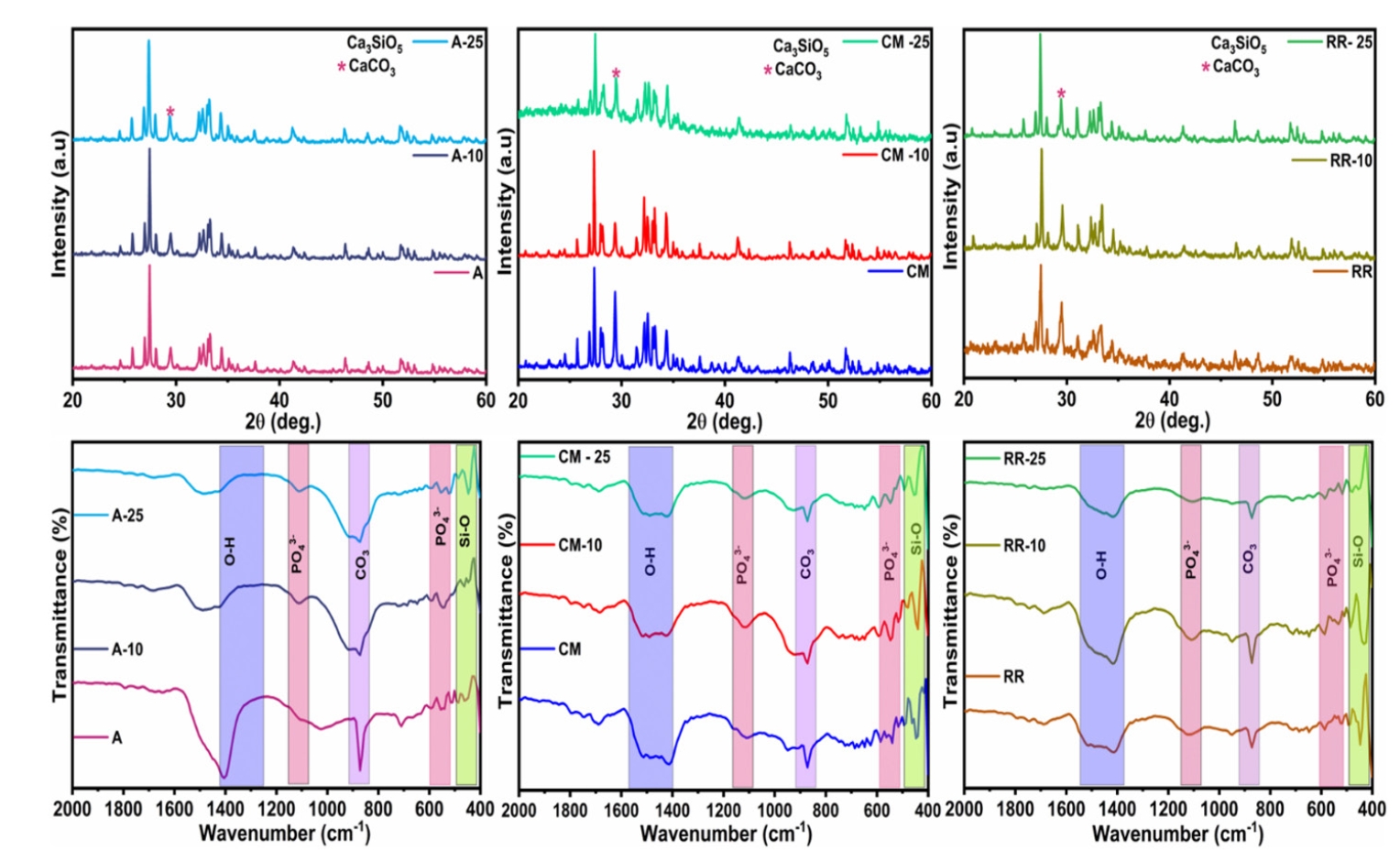
Synthesized AgNPs at ratios of 10 and 25% were added to commercially available MTA to create an admixture. The admixture was subjected to structural and morphological assessment using X-ray diffraction analysis (XRD), Fourier transform infrared (FT-IR) analysis, Raman spectroscopy, and scanning electron microscopy. Antioxidant activity was measured using the hydroxyl radical scavenging assay. A significance level of 0.05 was applied to determine statistical differences.
Results
The addition of AgNPs decreased the carbonate peak intensity in XRD and FT-IR. The rod-like morphology of MTA was changed to a flake-like morphology with the addition of AgNPs. Antibacterial efficacy enhanced proportionally with the augmentation of AgNPs concentration.
Conclusions
The creation of rapid admixture of MTA and AgNPs during chairside use in clinical settings can deliver beneficial characteristics of enhanced morphological features favoring mineralization and profound antibacterial effects to overcome the challenges associated with root canal treatment and pulp capping.
- 1,877 View
- 66 Download

- Histological evaluation of pulp response to alendronate and Biodentine as pulp capping agents: an animal study
- Thangavel Boopathi, Sekar Manimaran, Joseline Charles Kerena, Mathew Sebeena, Kumaravadivel Karthick, Natesan Thangaraj Deepa
- Restor Dent Endod 2024;49(4):e39. Published online October 29, 2024
- DOI: https://doi.org/10.5395/rde.2024.49.e39

-
 Abstract
 PDF
PDF PubReader
 ePub
ePub Objectives This study aimed to comparatively assess the histological response of the pulp toward alendronate and Biodentine in a direct pulp capping procedure.
Materials and Methods Twenty-four anterior teeth from 6 New Zealand rabbits were used in this study. Firstly, all rabbits were anesthetized according to their weight. Class V cavities were prepared on the buccal surfaces of anterior teeth. A pin-point exposure of the pulp was then made using a small, sterile round carbide bur and bleeding was arrested with a saline-soaked, sterile cotton pellet. The teeth under study were divided into 2 groups (
n = 12). The intentionally exposed pulp was capped with alendronate (Group 1) and Biodentine (Group 2), correspondingly. After 30 days, all rabbits were euthanized; the teeth under study were extracted and taken up for histological analysis.Results Biodentine showed an intact, very dense dentin bridge formation with a uniform odontoblast (OD) layer pattern and mild or absent inflammatory response whereas specimens capped with alendronate demonstrated a dense dentin bridge formation with non-uniform OD layer pattern and mild to moderate inflammatory response.
Conclusions Biodentine showed more biocompatibility than alendronate. However, alendronate can initiate reparative dentin formation and may be used as an alternative pulp capping agent.
- 3,057 View
- 129 Download

- Success rate of direct pulp capping on permanent teeth using bioactive materials: a systematic review and meta-analysis of randomized clinical trials
- Karem Paula Pinto, Gabriela Ribeiro da Silva, Cláudio Malizia Alves Ferreira, Luciana Moura Sassone, Emmanuel João Nogueira Leal da Silva
- Restor Dent Endod 2024;49(4):e34. Published online September 6, 2024
- DOI: https://doi.org/10.5395/rde.2024.49.e34
-
 Abstract
 PDF
PDF Supplementary Material
 PubReader
 ePub
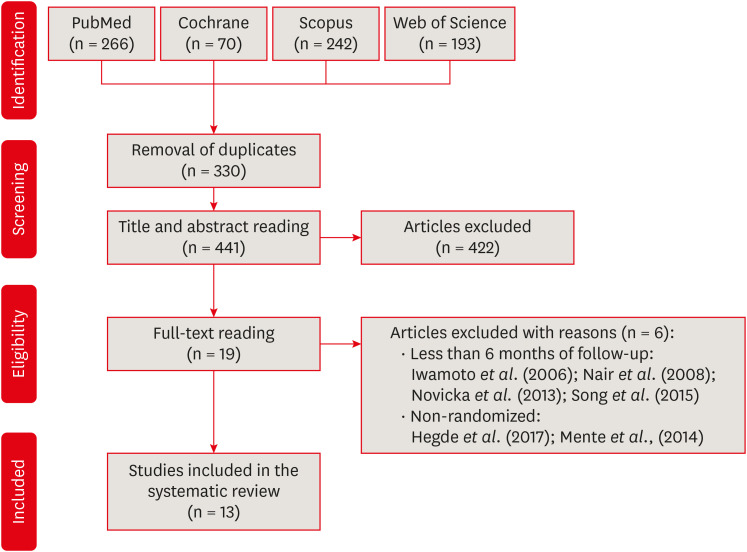
ePub This systematic review and meta-analysis aimed to evaluate the success rate of direct pulp capping (DPC) on permanent teeth, comparing the use of MTA with calcium hydroxide and calcium silicate-based cements. A systematic search was carried out in 4 databases until July 2023. The selection was based on PICOS criteria and only randomized clinical trials were included. The risk of bias was assessed using RoB-2 tool, and meta-analyses were performed using RevMan 5.3 software. The overall quality of evidence was determined using the GRADE tool. Thirteen studies were included. Meta-analyses indicated significantly higher success rate for DPC using MTA compared to calcium hydroxide, while no significant difference was observed between MTA and Biodentine, showing a success rate from 80% to 100% even after 3 years of follow-up. Five studies were classified as having high risk of bias and the GRADE assessment revealed low certainty of evidence. DPC is highly effective for permanent teeth when using MTA or Biodentine. There is a need for future well-designed randomized clinical trials to evaluate the efficacy of DPC using newer bioceramic materials.
-
Citations
Citations to this article as recorded by- Physicochemical effects of nano type-B bone substitute on pulp protective cement formulations
Njwan Fadhel SHEHAB
Dental Materials Journal.2026;[Epub] CrossRef - Photobiomodulation-assisted pulp capping using nano-hydroxyapatite and mineral trioxide aggregate: Report of two cases
Priya Pal, Rhythm Bains, Promila Verma, Vivek Kumar Bains
Journal of Healthcare Research and Education.2026; 2: 2. CrossRef - Indian Association of Conservative Dentistry and Endodontics consensus statement on deep caries management
Deepak Kumar Sharma, R. S. Mohan Kumar, Shishir Singh, Suparna Ganguly Saha, Meenal Nithin Gulve, Dipali Y. Shah, Sathish Abraham, Shruthi Nagaraja, Raksha Bhat
Journal of Conservative Dentistry and Endodontics.2025; 28(8): 714. CrossRef
- Physicochemical effects of nano type-B bone substitute on pulp protective cement formulations
- 17,739 View
- 517 Download
- 1 Web of Science
- 3 Crossref

- Stem cell-derived exosomes for dentin-pulp complex regeneration: a mini-review
- Dina A. Hammouda, Alaa M Mansour, Mahmoud A. Saeed, Ahmed R. Zaher, Mohammed E. Grawish
- Restor Dent Endod 2023;48(2):e20. Published online May 3, 2023
- DOI: https://doi.org/10.5395/rde.2023.48.e20
-
 Abstract
 PDF
PDF PubReader
 ePub
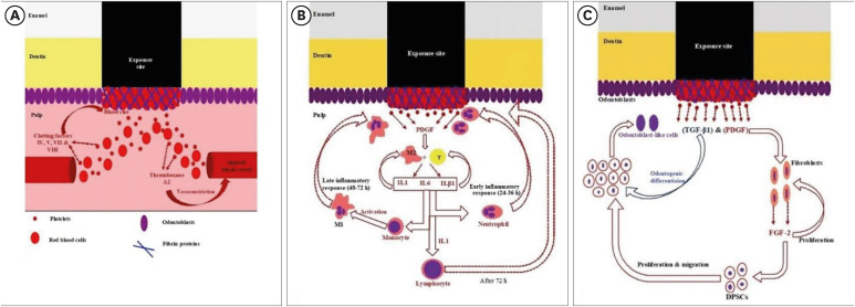
ePub This mini-review was conducted to present an overview of the use of exosomes in regenerating the dentin-pulp complex (DPC). The PubMed and Scopus databases were searched for relevant articles published between January 1, 2013 and January 1, 2023. The findings of basic
in vitro studies indicated that exosomes enhance the proliferation and migration of mesenchymal cells, as human dental pulp stem cells, via mitogen-activated protein kinases and Wingless-Int signaling pathways. In addition, they possess proangiogenic potential and contribute to neovascularization and capillary tube formation by promoting endothelial cell proliferation and migration of human umbilical vein endothelial cells. Likewise, they regulate the migration and differentiation of Schwann cells, facilitate the conversion of M1 pro-inflammatory macrophages to M2 anti-inflammatory phenotypes, and mediate immune suppression as they promote regulatory T cell conversion. Basicin vivo studies have indicated that exosomes triggered the regeneration of dentin-pulp–like tissue, and exosomes isolated under odontogenic circumstances are particularly strong inducers of tissue regeneration and stem cell differentiation. Exosomes are a promising regenerative tool for DPC in cases of small pulp exposure or for whole-pulp tissue regeneration.-
Citations
Citations to this article as recorded by- Extracellular vesicles derived from dental mesenchymal stem cells for regenerative medicine: a scoping review
Maria Emília Mota, Márcia Martins Marques, Thaís Gimenez, Suely Kunimi Kubo Ariga, Tiago Góss dos Santos, Fábio Abreu Alves, Maria Stella Moreira
Molecular Biology Reports.2026;[Epub] CrossRef - Cell Homing Strategies in Regenerative Endodontic Therapy
David Kim, Sahng G. Kim
Cells.2025; 14(3): 201. CrossRef - Impact of dental pulp cells-derived small extracellular vesicles on the properties and behavior of dental pulp cells: an in-vitro study
Dina A. Hammouda, Alaa M. Mansour, Ahmed R. Zaher, Mohammed E. Grawish
BMC Oral Health.2025;[Epub] CrossRef - Methodological Approaches for Economic Comparison of Mesenchymal Stem Cell and Exosome-based Therapies with Conventional Endodontic Treatments in Regenerative Endodontics
Madina A. Kurmanalina Kurmanalina, Nadiar M. Mussin, Aigul M. Sumanova, Violetta R. Detochkina, Maryam Mardani, Nader Tanideh, Amin Tamadon
West Kazakhstan Medical Journal.2025; 67(2): 188. CrossRef - Exosomal circ_0003057 promotes osteo/odontogenic differentiation of hDPSCs by binding with EIF4A3 through upregulated parental gene ANKH
Bingtao Wang, Yuanyuan Kong, Huixian Dong, Feng Lai, Zixin Guo, Liecong Lin, Jingyi Xu, Jingkun Zhang, Yiguo Jiang, Qianzhou Jiang
International Endodontic Journal.2025; 58(9): 1433. CrossRef - Mechanistic insights into dental stem cells‐derived exosomes in regenerative endodontics
Paras Ahmad, Nathan Estrin, Nima Farshidfar, Yufeng Zhang, Richard J. Miron
International Endodontic Journal.2025; 58(9): 1384. CrossRef - Development and characterization of an exosome-loaded biomimetic hydroxyapatite/gelatin scaffold for enhanced dental pulp regeneration
Yuen-Shan Tsai, Shih-Jung Cheng, Tsao-Li Chuang, Shu-Fang Chang, Feng-Huei Lin, Chun-Pin Lin
Journal of Dental Sciences.2025;[Epub] CrossRef - Exosomes as Promising Therapeutic Tools for Regenerative Endodontic Therapy
Qingyue Kong, Yujie Wang, Nan Jiang, Yifan Wang, Rui Wang, Xiaohan Hu, Jing Mao, Xin Shi
Biomolecules.2024; 14(3): 330. CrossRef - Role and Molecular Mechanism of miR-586 in the Differentiation of Dental Pulp Stem Cells into Odontoblast-like Cells
Gang Pan, Qianwen Zhou, Chenhua Pan, Yingxue Zhang
Cell Biochemistry and Biophysics.2024; 83(1): 507. CrossRef
- Extracellular vesicles derived from dental mesenchymal stem cells for regenerative medicine: a scoping review
- 4,712 View
- 94 Download
- 8 Web of Science
- 9 Crossref

- YouTube as a source of information about pulpotomy and pulp capping: a cross sectional reliability analysis
- Konstantinos Kodonas, Anastasia Fardi
- Restor Dent Endod 2021;46(3):e40. Published online July 6, 2021
- DOI: https://doi.org/10.5395/rde.2021.46.e40
-
 Abstract
 PDF
PDF PubReader
 ePub
ePub Objectives The purpose of this study was to critically evaluate the quality, reliability and educational content of the information of vital pulp treatment videos available on YouTube.
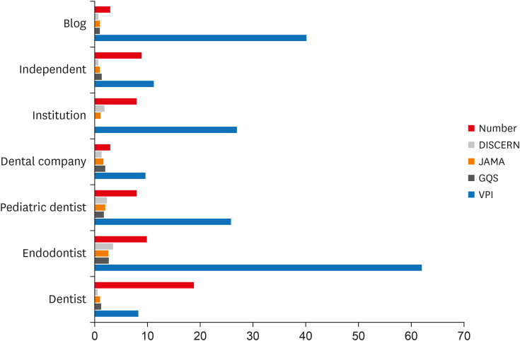
Materials and Methods The keywords “pulpotomy” and “pulp capping” were searched on YouTube on 5th July 2020, until 60 English language videos of each search term with a duration shorter than 15 minutes were acquired. Video characteristics were recorded and Video Power Index (VPI) was calculated. Reliability and educational quality of videos were evaluated using the Modified DISCERN score, the
Journal of American Medical Association (JAMA) benchmark criteria and Global Quality Scores (GQS). Videos were categorized by uploading source.Results Regarding pulpotomy, 31.7% of the videos were uploaded by specialists and 68.3% were directed by non-specialists. In the case of pulp capping, the corresponding percentages were 45% and 55%, respectively. Videos uploaded by specialists had significantly higher modified DISCERN, JAMA and GQS scores compared to those uploaded by non-specialists. Endodontists tended to have the highest reliability and VPI scores.
Conclusions YouTube videos on vital pulp treatment contain low educational quality or incomplete information. Low popularity of dental pulp capping and pulpotomy videos may be attributed to the specialized nature of these procedures. As YouTube represents an important source for patient information about different health topics, reliable informative videos should be uploaded by specialized dental professionals.
-
Citations
Citations to this article as recorded by- Assessing the Quality of YouTube® Videos on Nitrous Oxide/Oxygen Inhalation: A Multi-Dimensional Approach for Pediatric Dentists
Sanaa N. Al-Haj Ali, Nehal AlHarbi, Hessah H. Almutairi
Pesquisa Brasileira em Odontopediatria e Clínica Integrada.2025;[Epub] CrossRef - Is YouTube™ a useful resource of information about bichectomy? A cross-sectional study
H.ɪ. Durmuş, B. Ege, S. Bayazıt, M. Koparal
Annales de Chirurgie Plastique Esthétique.2025;[Epub] CrossRef - Assessing the reliability and educational value of YouTube videos on computer-controlled local anesthesia in dentistry
Hulya Cerci Akcay, Erdal Cem Kargu, Nefise Seker, Tanay Chaubal
PLOS One.2025; 20(8): e0329291. CrossRef - A content analysis of YouTube videos on interproximal enamel reduction
Weng Yan Tam, Jack Shen Tham, Smita Nimbalkar, Shilpa Gunjal, Kirti Saxena
APOS Trends in Orthodontics.2025; 0: 1. CrossRef - Comparison of YouTube, TikTok, and Instagram as digital sources for obtaining information about pulp therapy in primary and permanent teeth
Hüseyin Gürkan Güneç, Emine Kaya, Dila Nur Okumuş, Merve Gül Erence
Restorative Dentistry & Endodontics.2025; 50(3): e26. CrossRef - Evaluation of Endodontic Retreatment Videos on The Youtube Platform: Quality and Content Analysis
Birgül Özaşır, Tufan Özaşır, Derin Buğu Yüzer, Deniz İmamoğlu, Kamran Gülşahı
European Annals of Dental Sciences.2025; 52(2): 103. CrossRef - Is YouTube a reliable source for learning pre-endodontic build-up? A cross-sectional study
Merve Gökyar, İdil Özden, Hesna Sazak Öveçoğlu
Restorative Dentistry & Endodontics.2025; 50(3): e27. CrossRef - Quality of Patient-Centered eHealth Information on Erosive Tooth Wear: Systematic Search and Evaluation of Websites and YouTube Videos
Lena Holland, Amelie Friederike Kanzow, Annette Wiegand, Philipp Kanzow
Journal of Medical Internet Research.2024; 26: e49514. CrossRef - Is it safe to learn about vital pulp capping from YouTube™ videos? A content and quality analysis
Celalettin Topbaş, Tuğçe Paksoy, Ayşe Gülnihal İslamoğlu, Kemal Çağlar, Abdurrahman Kerim Kul
International Journal of Medical Informatics.2024; 185: 105409. CrossRef - Assessment of the quality of oral biopsy procedure videos shared on YouTube
A. Díaz‐Rodríguez, J. Limeres‐Posse, R. Albuquerque, V. Brailo, R. Cook, J. C. Fricain, G. Lodi, L. Monteiro, L. Silva, B. Carey, M. Diniz‐Freitas
Oral Diseases.2024; 30(5): 3081. CrossRef - İmplant üstü protezler hakkında bilgi veren internet sitelerinin okunabilirliklerinin değerlendirilmesi
Tugba TEMİZCİ
Selcuk Dental Journal.2023; 10(4): 156. CrossRef - Online Audio-Visual Information on the Treatment of OSA with Mandibular Advancement Devices: Analysis of Quality, Reliability and Contents
Serena Incerti-Parenti, Maria Lavinia Bartolucci, Elena Biondi, Andrea Fiordelli, Corrado Paganelli, Giulio Alessandri-Bonetti
Applied Sciences.2023; 13(9): 5727. CrossRef - Evaluating YouTube as a Patient Information Source for the Risks of Root Canal Treatment
Stewart McLean, Neil Cook, Alexander Rovira-Wilde, Shanon Patel, Shalini Kanagasingam
Journal of Endodontics.2023; 49(2): 155. CrossRef - Assessment of reliability and information quality of YouTube videos about root canal treatment after 2016
Myoung-jun Jung, Min-Seock Seo
BMC Oral Health.2022;[Epub] CrossRef - Is the YouTube™ a useful resource of information about orthognathic surgery?: A cross-sectional study
Seyma Bayazıt, Bilal Ege, Mahmut Koparal
Journal of Stomatology, Oral and Maxillofacial Surgery.2022; 123(6): e981. CrossRef - YoutubeTM Content Analysis as a Means of Information in Oral Medicine: A Systematic Review of the Literature
Antonio Romano, Fausto Fiori, Massimo Petruzzi, Fedora Della Vella, Rosario Serpico
International Journal of Environmental Research and Public Health.2022; 19(9): 5451. CrossRef
- Assessing the Quality of YouTube® Videos on Nitrous Oxide/Oxygen Inhalation: A Multi-Dimensional Approach for Pediatric Dentists
- 1,732 View
- 18 Download
- 14 Web of Science
- 16 Crossref

-
Hard tissue formation after direct pulp capping with osteostatin and MTA
in vivo - Ji-Hye Yoon, Sung-Hyeon Choi, Jeong-Tae Koh, Bin-Na Lee, Hoon-Sang Chang, In-Nam Hwang, Won-Mann Oh, Yun-Chan Hwang
- Restor Dent Endod 2021;46(2):e17. Published online February 25, 2021
- DOI: https://doi.org/10.5395/rde.2021.46.e17
-
 Abstract
 PDF
PDF PubReader
 ePub
ePub Objectives In recent
in vitro study, it was reported that osteostatin (OST) has an odontogenic effect and synergistic effect with mineral trioxide aggregate (MTA) in human dental pulp cells. Therefore, the aim of this study was to evaluate whether OST has a synergistic effect with MTA on hard tissue formationin vivo .Materials and Methods Thirty-two maxillary molars of Spraque-Dawley rats were used in this study. An occlusal cavity was prepared and the exposed pulps were randomly divided into 3 groups: group 1 (control; ProRoot MTA), group 2 (OST 100 μM + ProRoot MTA), group 3 (OST 10 mM + ProRoot MTA). Exposed pulps were capped with each material and cavities were restored with resin modified glass ionomer. The animals were sacrificed after 4 weeks. All harvested teeth were scanned with micro-computed tomography (CT). The samples were prepared and hard tissue formation was evaluated histologically. For immunohistochemical analysis, the specimens were sectioned and incubated with primary antibodies against dentin sialoprotein (DSP).
Results In the micro-CT analysis, it is revealed that OST with ProRoot MTA groups showed more mineralized bridge than the control (
p < 0.05). In the H&E staining, it is showed that more quantity of the mineralized dentin bridge was formed in the OST with ProRoot MTA group compared to the control (p < 0.05). In all groups, DSP was expressed in newly formed reparative dentin area.Conclusions OST can be a supplementary pulp capping material when used with MTA to make synergistic effect in hard tissue formation.
-
Citations
Citations to this article as recorded by- Pulpal responses to mineral trioxide aggregate with and without zinc oxide addition in mature canine teeth after full pulpotomy
Behnam Bolhari, Neda Kardouni Khouzestani, Hadi Assadian, Saeed Farzad-Mohajeri, Mohammad Mehdi Dehghan, Soheil Niavarzi, Behnam Dorost, Venkateshbabu Nagendrababu, Henry F. Duncan, Artak Heboyan, Antonio Signore, Stefano Benedicenti
Scientific Reports.2025;[Epub] CrossRef - Research Advancements in Peptides for Promoting Reparative Dentin Regeneration in Direct Pulp Capping: A Narrative Review
Jiawen Wang, Shuwei Qiao, Tianjia Huang, Junjie Lian, Song Zhu
International Journal of Peptide Research and Therapeutics.2025;[Epub] CrossRef - Biocompatibility and pro-mineralization effects of premixed calcium silicate-based materials on human dental pulp stem cells: An in vitro and in vivo study
Nyein Chan KO, Sonoko NODA, Yamato OKADA, Kento TAZAWA, Nobuyuki KAWASHIMA, Takashi OKIJI
Dental Materials Journal.2024; 43(5): 729. CrossRef - Osteostatin, a peptide for the future treatment of musculoskeletal diseases
Daniel Lozano, Arancha R. Gortazar, Sergio Portal-Núñez
Biochemical Pharmacology.2024; 223: 116177. CrossRef - Comparison of bioactive material failure rates in vital pulp treatment of permanent matured teeth – a systematic review and network meta-analysis
Péter Komora, Orsolya Vámos, Noémi Gede, Péter Hegyi, Kata Kelemen, Adél Galvács, Gábor Varga, Beáta Kerémi, János Vág
Scientific Reports.2024;[Epub] CrossRef - Hard tissue formation in pulpotomized primary teeth in dogs with nanomaterials MCM-48 and MCM-48/hydroxyapatite: an in vivo animal study
Sahar Talebi, Nosrat Nourbakhsh, Ardeshir Talebi, Amir Abbas Nourbakhsh, Abbas Haghighat, Maziar Manshayi, Hamid Reza Bakhsheshi, Razieh Karimi, Rahman Nazeri, Kenneth J.D. Mackenzie
BMC Oral Health.2024;[Epub] CrossRef - Reparative Mineralized Tissue Characterization by Different Bioactive Direct Pulp-capping Agents
Mrunal Shinde, Varsha Pandit, Sarita Singh, Aniket Jadhav, Sarah Marium, Smita Patil
Journal of the International Clinical Dental Research Organization.2024; 16(1): 8. CrossRef - Effects of mineral trioxide aggregate and methyl sulfonyl methane on pulp exposure via RUNX2 and RANKL pathways
Altar Ateş, Ayca Kurt, Tolga Mercantepe
Odontology.2024; 112(3): 895. CrossRef - Effects of barium titanate on the dielectric constant, radiopacity, and biological properties of tricalcium silicate-based bioceramics
Yoorina CHOI, Yun-Chan HWANG, Mi-Kyung YU, Kwang-Won LEE, Kyung-San MIN
Dental Materials Journal.2023; 42(1): 55. CrossRef - Bioactive potential of Bio‐C Pulpo is evidenced by presence of birefringent calcite and osteocalcin immunoexpression in the rat subcutaneous tissue
Marcela Borsatto Queiroz, Rafaela Nanami Handa Inada, Camila Soares Lopes, Juliane Maria Guerreiro‐Tanomaru, Estela Sasso‐Cerri, Mário Tanomaru‐Filho, Paulo Sérgio Cerri
Journal of Biomedical Materials Research Part B: Applied Biomaterials.2022; 110(10): 2369. CrossRef - The Influence of New Bioactive Materials on Pulp–Dentin Complex Regeneration in the Assessment of Cone Bone Computed Tomography (CBCT) and Computed Micro-Tomography (Micro-CT) from a Present and Future Perspective—A Systematic Review
Mirona Paula Palczewska-Komsa, Bartosz Gapiński, Alicja Nowicka
Journal of Clinical Medicine.2022; 11(11): 3091. CrossRef - A Breakthrough in the Era of Calcium Silicate-Based Cements: A Critical Review
Payal S Chaudhari, Manoj G Chandak, Akshay A Jaiswal, Nikhil P Mankar, Priyanka Paul
Cureus.2022;[Epub] CrossRef - Effectiveness of Direct Pulp Capping Bioactive Materials in Dentin Regeneration: A Systematic Review
Ermin Nie, Jiali Yu, Rui Jiang, Xiangzhen Liu, Xiang Li, Rafiqul Islam, Mohammad Khursheed Alam
Materials.2021; 14(22): 6811. CrossRef
- Pulpal responses to mineral trioxide aggregate with and without zinc oxide addition in mature canine teeth after full pulpotomy
- 2,672 View
- 36 Download
- 12 Web of Science
- 13 Crossref

- Bioactivity of endodontic biomaterials on dental pulp stem cells through dentin
- Bahar Javid, Narges Panahandeh, Hassan Torabzadeh, Hamid Nazarian, Ardavan Parhizkar, Saeed Asgary
- Restor Dent Endod 2020;45(1):e3. Published online November 4, 2019
- DOI: https://doi.org/10.5395/rde.2020.45.e3

-
 Abstract
 PDF
PDF PubReader
 ePub
ePub Objectives This study investigated the indirect effect of calcium-enriched mixture (CEM) cement and mineral trioxide aggregate (MTA), as 2 calcium silicate-based hydraulic cements, on human dental pulp stem cells (hDPSCs) through different dentin thicknesses.
Materials and Methods Two-chamber setups were designed to simulate indirect pulp capping (IPC). Human molars were sectioned to obtain 0.1-, 0.3-, and 0.5-mm-thick dentin discs, which were placed between the 2 chambers to simulate an IPC procedure. Then, MTA and CEM were applied on one side of the discs, while hDPSCs were cultured on the other side. After 2 weeks of incubation, the cells were removed, and cell proliferation, morphology, and attachment to the discs were evaluated under scanning electron microscopy (SEM). Energy-dispersive X-ray (EDXA) spectroscopy was performed for elemental analysis. Alkaline phosphatase (ALP) activity was assessed quantitatively. The data were analyzed using the Kruskal-Wallis and Mann-Whitney tests.
Results SEM micrographs revealed elongated cells, collagen fibers, and calcified nucleations in all samples. EDXA verified that the calcified nucleations consisted of calcium phosphate. The largest calcifications were seen in the 0.1-mm-thick dentin subgroups. There was no significant difference in ALP activity across the CEM subgroups; however, ALP activity was significantly lower in the 0.1-mm-thick dentin subgroup than in the other MTA subgroups (
p < 0.05).Conclusions The employed capping biomaterials exerted biological activity on hDPSCs, as shown by cell proliferation, morphology, and attachment and calcific precipitations, through 0.1- to 0.5-mm-thick layers of dentin. In IPC, the bioactivity of these endodontic biomaterials is probably beneficial.
-
Citations
Citations to this article as recorded by- Dental pulp capping materials: modulators of stem cell behavior and regenerative potential
Ali Cheayto, Sara Ayoub, Sarah Ayad Al-Tameemi, Mohammad Fayyad-Kazan
Biomedical Physics & Engineering Express.2025; 11(6): 062004. CrossRef - Effect of pulp capping materials on odontogenic differentiation of human dental pulp stem cells: An in vitro study
Mahmoud M. Bakr, Mohamed Shamel, Shereen N. Raafat, Robert M. Love, Mahmoud M. Al‐Ankily
Clinical and Experimental Dental Research.2024;[Epub] CrossRef - Effects of Growth Factors on the Differentiation of Dental Stem Cells: A
Systematic Review and Meta-analysis (Part I)
Sayna Shamszadeh, Armin Shirvani, Hassan Torabzadeh, Saeed Asgary
Current Stem Cell Research & Therapy.2024; 19(4): 523. CrossRef - The Role of Growth Factor Delivery Systems on Cellular Activities of Dental
Stem Cells: A Systematic Review (Part II)
Sayna Shamszadeh, Armin Shirvani, Saeed Asgary
Current Stem Cell Research & Therapy.2024; 19(4): 587. CrossRef - Comprehensive review of composition, properties, clinical applications, and future perspectives of calcium-enriched mixture (CEM) cement: a systematic analysis
Saeed Asgary, Mahtab Aram, Mahta Fazlyab
BioMedical Engineering OnLine.2024;[Epub] CrossRef - Evaluation of dental pulp stem cells behavior after odontogenic differentiation induction by three different bioactive materials on two different scaffolds
Basma Ahmed, Mai H. Ragab, Rania A. Galhom, Hayam Y. Hassan
BMC Oral Health.2023;[Epub] CrossRef - Characterization of Dental Pulp Stem Cell Responses to Functional Biomaterials Including Mineralized Trioxide Aggregates
Sejin Bae, Bueonguk Kang, Hyungbin Lee, Harrison Luu, Eric Mullins, Karl Kingsley
Journal of Functional Biomaterials.2021; 12(1): 15. CrossRef - Incorporation of amoxicillin-loaded microspheres in mineral trioxide aggregate cement: an in vitro study
Fábio Rocha Bohns, Vicente Castelo Branco Leitune, Isadora Martini Garcia, Bruna Genari, Nélio Bairros Dornelles, Silvia Stanisçuaski Guterres, Fabrício Aulo Ogliari, Mary Anne Sampaio de Melo, Fabrício Mezzomo Collares
Restorative Dentistry & Endodontics.2020;[Epub] CrossRef
- Dental pulp capping materials: modulators of stem cell behavior and regenerative potential
- 1,447 View
- 12 Download
- 8 Crossref

- Effects of the exposure site on histological pulpal responses after direct capping with 2 calcium-silicate based cements in a rat model
- Panruethai Trongkij, Supachai Sutimuntanakul, Puangwan Lapthanasupkul, Chitpol Chaimanakarn, Rebecca Wong, Danuchit Banomyong
- Restor Dent Endod 2018;43(4):e36. Published online August 22, 2018
- DOI: https://doi.org/10.5395/rde.2018.43.e36

-
 Abstract
 PDF
PDF PubReader
 ePub
ePub Objectives Direct pulp capping is a treatment for mechanically exposed pulp in which a biocompatible capping material is used to preserve pulpal vitality. Biocompatibility tests in animal studies have used a variety of experimental protocols, particularly with regard to the exposure site. In this study, pulp exposure on the occlusal and mesial surfaces of molar teeth was investigated in a rat model.
Materials and Methods A total of 58 maxillary first molars of Wistar rats were used. Forty molars were mechanically exposed and randomly assigned according to 3 factors: 1) the exposure site (occlusal or mesial), 2) the pulp-capping material (ProRoot White MTA or Bio-MA), and 3) 2 follow-up periods (1 day or 7 days) (
n = 5 each). The pulp of 6 intact molars served as negative controls. The pulp of 12 molars was exposed without a capping material (n = 3 per exposure site for each period) and served as positive controls. Inflammatory cell infiltration and reparative dentin formation were histologically evaluated at 1 and 7 days using grading scores.Results At 1 day, localized mild inflammation was detected in most teeth in all experimental groups. At 7 days, continuous/discontinuous calcified bridges were formed at exposure sites with no or few inflammatory cells. No significant differences in pulpal response according to the exposure site or calcium-silicate cement were observed.
Conclusions The location of the exposure site had no effect on rat pulpal healing. However, mesial exposures could be performed easily, with more consistent results. The pulpal responses were not significantly different between the 2 capping materials.
-
Citations
Citations to this article as recorded by- Bioactivity and biocompatibility of bioceramic-based pulp capping materials in laboratory and animal models
Rafiqul Islam, Md. Refat Readul Islam, Kenta Tsuchiya, Yu Toida, Hidehiko Sano, Monica Yamauti, Hany Mohamed Aly Ahmed, Atsushi Tomokiyo
Journal of Materials Science: Materials in Medicine.2025;[Epub] CrossRef - The road map to proper dental pulp experiments in animal models
Nuha A Elmubarak
International Dental Journal of Student's Research.2024; 11(4): 163. CrossRef - Treatment outcomes of root perforations repaired by calcium silicate-based cements with or without an accelerator: A randomized controlled trial
Kanyarat Tungputsa, Danuchit Banomyong, Sittichoke Osiri, Supachai Sutimuntanakul
Endodontology.2024; 36(4): 315. CrossRef - Biological evaluation of novel phosphorylated pullulan‐based calcium hydroxide formulations as direct pulp capping materials: An in vivo study on a rat model
Md Refat Readul Islam, Rafiqul Islam, Yunqing Liu, Yu Toida, Yasuhiro Yoshida, Hidehiko Sano, Hany Mohamed Aly Ahmed, Atsushi Tomokiyo
International Endodontic Journal.2024; 57(9): 1247. CrossRef - 3D-printed microgels supplemented with dentin matrix molecules as a novel biomaterial for direct pulp capping
Diana Cunha, Nayara Souza, Manuela Moreira, Nara Rodrigues, Paulo Silva, Cristiane Franca, Sivaporn Horsophonphong, Ashley Sercia, Ramesh Subbiah, Anthony Tahayeri, Jack Ferracane, Pamela Yelick, Vicente Saboia, Luiz Bertassoni
Clinical Oral Investigations.2022; 27(3): 1215. CrossRef - Calcium silicate and calcium aluminate cements for dentistry reviewed
Carolyn Primus, James L. Gutmann, Franklin R. Tay, Anna B. Fuks
Journal of the American Ceramic Society.2022; 105(3): 1841. CrossRef - Pulpal response to mineral trioxide aggregate containing phosphorylated pullulan-based capping material
Yu TOIDA, Shimpei KAWANO, Rafiqul ISLAM, Fu JIALE, AFM A CHOWDHURY, Shuhei HOSHIKA, Yasushi SHIMADA, Junji TAGAMI, Masahiro YOSHIYAMA, Satoshi INOUE, Ricardo M. CARVALHO, Yasuhiro YOSHIDA, Hidehiko SANO
Dental Materials Journal.2022; 41(1): 126. CrossRef - The Effect of Calcium-Silicate Cements on Reparative Dentinogenesis Following Direct Pulp Capping on Animal Models
Mihai Andrei, Raluca Paula Vacaru, Anca Coricovac, Radu Ilinca, Andreea Cristiana Didilescu, Ioana Demetrescu
Molecules.2021; 26(9): 2725. CrossRef - Histological evaluation of a novel phosphorylated pullulan‐based pulp capping material: An in vivo study on rat molars
Rafiqul Islam, Yu Toida, Fei Chen, Toru Tanaka, Satoshi Inoue, Tetsuya Kitamura, Yasuhiro Yoshida, Abu Faem Mohammad Almas Chowdhury, Hany Mohamed Aly Ahmed, Hidehiko Sano
International Endodontic Journal.2021; 54(10): 1902. CrossRef - Effectiveness of Direct Pulp Capping Bioactive Materials in Dentin Regeneration: A Systematic Review
Ermin Nie, Jiali Yu, Rui Jiang, Xiangzhen Liu, Xiang Li, Rafiqul Islam, Mohammad Khursheed Alam
Materials.2021; 14(22): 6811. CrossRef - A strontium and amorphous calcium phosphate dipped premixed injectable calcium silicate-based ceramic for dental root canal sealing
Huimin Jin, Yuzhu Li, Qingqing Wang, Menglu Dong, Mengmeng Yang, Wendy Chen, Shengrui Wang, Heng Zhang, Shunli Zheng, Chris Ying Cao, Zheng Zhou, Quan-Li Li
Ceramics International.2021; 47(23): 33738. CrossRef - Bioactive tri/dicalcium silicate cements for treatment of pulpal and periapical tissues
Carolyn M. Primus, Franklin R. Tay, Li-na Niu
Acta Biomaterialia.2019; 96: 35. CrossRef
- Bioactivity and biocompatibility of bioceramic-based pulp capping materials in laboratory and animal models
- 1,880 View
- 16 Download
- 12 Crossref

- Considerations during crown reattachment procedure over the pulpal exposure: case report
- Bona Kim, Yoon Lee, Min-Ju Song, Su-Jung Shin, Jeong-Won Park
- Restor Dent Endod 2012;37(4):240-244. Published online November 21, 2012
- DOI: https://doi.org/10.5395/rde.2012.37.4.240
-
 Abstract
 PDF
PDF PubReader
 ePub
ePub Crown reattachment is the most conservative treatment which can be used to restore fractured tooth, presumably with sufficient strength, while maintaining original contour, incisal translucency, and reducing chair time and cost.
However, in case of crown fracture with pin-point pulp exposure, we should cautiously minimize the irritation to the pulp and consider pre-treatment pulpal status, choice of pulp capping materials, choice of bonding system and treatment sequence during crown reattachment procedures. This case reports the considerations while crown reattachment with direct pulp capping using calcium hydroxide (Dycal, Dentsply Caulk).
-
Citations
Citations to this article as recorded by- A Conservative Approach to the Management of a Dental Trauma for Immediate Natural Esthetics
Pallav Mahesh Patni, Pradeep Jain, Mona Jain Patni
Archives of Trauma Research.2016;[Epub] CrossRef
- A Conservative Approach to the Management of a Dental Trauma for Immediate Natural Esthetics
- 1,228 View
- 6 Download
- 1 Crossref

- Comparison of gene expression profiles of human dental pulp cells treated with mineral trioxide aggregate and calcium hydroxide
- Yong-Beom Kim, Won-Jun Shon, Woocheol Lee, Kee-Yeon Kum, Seung-Ho Baek, Kwang-Shik Bae
- J Korean Acad Conserv Dent 2011;36(5):397-408. Published online September 14, 2011
- DOI: https://doi.org/10.5395/JKACD.2011.36.5.397
-
 Abstract
 PDF
PDF PubReader
 ePub
ePub Abstract Objectives: This study investigated changes in gene expressions concerning of differentiation, proliferation, mineralization and inflammation using Human-8 expression bead arrays when white Mineral Trioxide Aggregate and calcium hydroxide-containing cement were applied
in vitro to human dental pulp cells (HDPCs).Materials and Methods: wMTA (white ProRoot MTA, Dentsply) and Dycal (Dentsply Caulk) in a Teflon tube (inner diameter 10 mm, height 1 mm) were applied to HDPCs. Empty tube-applied HDPCs were used as negative control. Total RNA was extracted at 3, 6, 9 and 24 hr after wMTA and Dycal application. The results of microarray were confirmed by reverse transcriptase polymerase chain reaction.
Results: Out of the 24,546 genes, 43 genes (e.g., BMP2, FOSB, THBS1, EDN1, IL11, COL10A1, TUFT1, HMOX1) were up-regulated greater than two-fold and 25 genes (e.g., SMAD6, TIMP2, DCN, SOCS2, CEBPD, KIAA1199) were down-regulated below 50% by wMTA. Two hundred thirty nine genes (e.g., BMP2, BMP6, SMAD6, IL11, FOS, VEGFA, PlGF, HMOX1, SOCS2, CEBPD, KIAA1199) were up-regulated greater than two-fold and 358 genes (e.g., EDN1, FGF) were down-regulated below 50% by Dycal.
Conclusions: Both wMTA and Dycal induced changes in gene expressions related with differentiation and proliferation of pulp cells. wMTA induced changes in gene expressions related with mineralization, and Dycal induced those related with angiogenesis. The genes related with inflammation were more expressed by Dycal than by wMTA. It was confirmed that both wMTA and Dycal were able to induce gene expression changes concerned with the pulp repair in different ways.
-
Citations
Citations to this article as recorded by- Analysis of gene expression during odontogenic differentiation of cultured human dental pulp cells
Min-Seock Seo, Kyung-Gyun Hwang, Hyongbum Kim, Seung-Ho Baek
Restorative Dentistry & Endodontics.2012; 37(3): 142. CrossRef
- Analysis of gene expression during odontogenic differentiation of cultured human dental pulp cells
- 1,253 View
- 1 Download
- 1 Crossref

- Gene expression profiling in human dental pulp cells treated with mineral trioxide aggregate
- Yong-Beom Kim, Won-Jun Shon, WooCheol Lee, Kee-Yeon Kum, Seung-Ho Baek, Kwang-Shik Bae
- J Korean Acad Conserv Dent 2010;35(3):152-163. Published online May 31, 2010
- DOI: https://doi.org/10.5395/JKACD.2010.35.3.152
-
 Abstract
 PDF
PDF PubReader
 ePub
ePub This study investigated the changes in gene expression when mineral trioxide aggregate (MTA) was applied
in vitro to human dental pulp cells (HDPCs). MTA in a teflon tube (diameter 10 mm, height 2 mm) was applied to HDPCs. Empty tube-applied HDPCs were used as negative control. For microarray analysis, total RNA was extracted at 6, 24, and 72 hrs after MTA application. The results were confirmed selectively by performing reverse transcriptase polymerase chain reaction for genes that showed changes of more than two-fold or less than half. Of the 24,546 genes, 109 genes were up-regulated greater than two-fold (e.g., FOSB, THBS1, BHLHB2, EDN1, IL11, FN1, COL10A1, and TUFT1) and 69 genes were down-regulated below 50% (e.g., SMAD6 and DCN). These results suggest that MTA, rather than being a bio-inert material, may have potential to affect the proliferation and differentiation of pulp cells in various ways.-
Citations
Citations to this article as recorded by- Analysis of gene expression during odontogenic differentiation of cultured human dental pulp cells
Min-Seock Seo, Kyung-Gyun Hwang, Hyongbum Kim, Seung-Ho Baek
Restorative Dentistry & Endodontics.2012; 37(3): 142. CrossRef - Comparison of gene expression profiles of human dental pulp cells treated with mineral trioxide aggregate and calcium hydroxide
Yong-Beom Kim, Won-Jun Shon, Woocheol Lee, Kee-Yeon Kum, Seung-Ho Baek, Kwang-Shik Bae
Journal of Korean Academy of Conservative Dentistry.2011; 36(5): 397. CrossRef
- Analysis of gene expression during odontogenic differentiation of cultured human dental pulp cells
- 1,037 View
- 5 Download
- 2 Crossref

- Pulp response of beagle dog to direct pulp capping materials: Histological study
- Ji-Hyun Bae, Young-Gyun Kim, Pil-Young Yoon, Byeong-Hoon Cho, Yong-Hoon Choi
- J Korean Acad Conserv Dent 2010;35(1):5-12. Published online January 31, 2010
- DOI: https://doi.org/10.5395/JKACD.2010.35.1.005
-
 Abstract
 PDF
PDF PubReader
 ePub
ePub The purpose of this study was to evaluate the pulp tissue reaction to direct pulp capping of mechanically exposed beagle dogs'pulp with several capping materials. A total of 36 teeth of 2 healthy beagle dongs were used. The mechanically exposed pulps were capped with one of the followings: (1) Mineral Trioxide Aggregate (MTA: ProRoot® MTA, Dentsply, Tulsa, USA), (2) Clearfil SE Bond (Dentin adhesive system: Kuraray, Osaka, Japan), (3) Ultra-Blend (Photo-polymerized Calcium hydroxide: Ultradent, South Jordan, USA), (4) Dycal (Quick setting Calcium hydroxide: LD Caulk Co., Milford, USA) at 7, 30, and 90 days before sacrificing. The cavities were restored with Z350 flowable composite resin (3M ESPE, St. Paul. MN, USA). After the beagle dogs were sacrificed, the extracted teeth were fixed, decalcified, prepared for histological examination and stained with HE stain. The pulpal tissue responses to direct pulp capping materials were assessed.
In MTA, calcium hydroxide, and photo-polymerized calcium hydroxide groups, initial mild inflammatory cell infiltration, newly formed odontoblast-like cell layer and hard tissue bridge formation were observed. Compared with dentin adhesive system, these materials were biocompatible and good for pulp tissue regeneration.
In dentin adhesive system group, severe inflammatory cell infiltration, pulp tissue degeneration and pulp tissue necrosis were observed. It seemed evident that application of dentin adhesive system in direct pulp capping of beagle dog teeth cannot lead to acceptable repair of the pulp tissue with dentine bridge formation.
-
Citations
Citations to this article as recorded by- Experimental Study of Pulp Capping Using Xenogenic Demineralized Dentin Paste
Ji-Young Yun, Yong-Hoon Choi, Young-Kyun Kim, In-Woong Um, Joo-Cheol Park, Ji-Yoon Kim
Journal of Hard Tissue Biology.2016; 25(3): 321. CrossRef - Comparison of gene expression profiles of human dental pulp cells treated with mineral trioxide aggregate and calcium hydroxide
Yong-Beom Kim, Won-Jun Shon, Woocheol Lee, Kee-Yeon Kum, Seung-Ho Baek, Kwang-Shik Bae
Journal of Korean Academy of Conservative Dentistry.2011; 36(5): 397. CrossRef - Gene expression profiling in human dental pulp cells treated with mineral trioxide aggregate
Yong-Beom Kim, Won-Jun Shon, WooCheol Lee, Kee-Yeon Kum, Seung-Ho Baek, Kwang-Shik Bae
Journal of Korean Academy of Conservative Dentistry.2010; 35(3): 152. CrossRef - Histology of dental pulp healing after tooth replantation in rats
Eun-Jin Go, Han-Seong Jung, Eui-Seong Kim, Il-Young Jung, Seung-Jong Lee
Journal of Korean Academy of Conservative Dentistry.2010; 35(4): 273. CrossRef
- Experimental Study of Pulp Capping Using Xenogenic Demineralized Dentin Paste
- 1,138 View
- 11 Download
- 4 Crossref

- Histological evaluation of direct pulp capping with DSP-derived synthetic peptide in beagle dog
- Jae-Hoon Kim, Jun-Bae Hong, Bum-Soon Lim, Byeong-Hoon Cho
- J Korean Acad Conserv Dent 2009;34(2):120-129. Published online January 14, 2009
- DOI: https://doi.org/10.5395/JKACD.2009.34.2.120
-
 Abstract
 PDF
PDF PubReader
 ePub
ePub Abstract The purpose of this study was to investigate the pulpal response to direct pulp capping with dentin sialo-protein (DSP) -derived synthetic peptide in teeth of dogs, and to compare its efficacy to capping substances Ca(OH)2 and white mineral trioxide aggregate (WMTA). A total of 72 teeth of 6 healthy male beagle dogs were used. The mechanically exposed pulps were capped with one of the following: (1) DSP-derived synthetic peptide (PEP group); (2) Ca(OH)2 (CH group); (3) a mixture paste of peptide and Ca(OH)2 (PEP+CH group); or (4) white MTA (WMTA group). The access cavity was restored with a reinforced glass ionomer cement. Two dogs were sacrificed at each pre-determined intervals (2 weeks, 1 month, and 3 months). After the specimens were prepared for standard histological processing, sections were stained with hematoxylin and eosin. Under a light microscope, inflammatory response and hard tissue formation were evaluated in a blind manner by 2 observers. In the PEP group, only 3 of 17 specimens showed hard tissue formation, indication that the DSP-derived synthetic peptide did not induce proper healing of the pulp. Compared with the CH group, the PEP group demonstrated an increased inflammatory response and poor hard tissue formation. The CH and WMTA groups showed similar results for direct pulp capping in mechanically exposed teeth of dogs.
-
Citations
Citations to this article as recorded by- Tubular Dentin Regeneration Using a CPNE7-Derived Functional Peptide
Yoon Lee, Yeoung-Hyun Park, Dong-Seol Lee, You-Mi Seo, Ji-Hyun Lee, Joo-Hwang Park, Han-Wool Choung, So-Hyun Park, Won Shon, Joo-Cheol Park
Materials.2020; 13(20): 4618. CrossRef - Pulp response of beagle dog to direct pulp capping materials: Histological study
Ji-Hyun Bae, Young-Gyun Kim, Pil-Young Yoon, Byeong-Hoon Cho, Yong-Hoon Choi
Journal of Korean Academy of Conservative Dentistry.2010; 35(1): 5. CrossRef
- Tubular Dentin Regeneration Using a CPNE7-Derived Functional Peptide
- 1,369 View
- 6 Download
- 2 Crossref

- Pulp response of mineral trioxide aggregate, calcium sulfate or calcium hydroxide
- Young-Ran Yun, In-Seok Yang, Yun-Chan Hwang, In-Nam Hwang, Hong-Ran Choi, Suk-Ja Yoon, Sun-Hun Kim, Won-Mann Oh
- J Korean Acad Conserv Dent 2007;32(2):95-101. Published online March 31, 2007
- DOI: https://doi.org/10.5395/JKACD.2007.32.2.095
-
 Abstract
 PDF
PDF PubReader
 ePub
ePub This study was performed to verify the possibility of MTA and calcium sulfate as a pulp capping agent through comparing the dental pulp response in dogs after capping with MTA, calcium sulfate, and calcium hydroxide.
24 teeth of 2 dogs, 8 month old, were used in this study.
Under general anesthesia, cervical cavities were prepared and pulp was exposed with sterilized #2 round bur in a high speed handpiece.
MTA, calcium hydroxide, and calcium sulfate were applied on the exposed pulp. Then the coronal openings were sealed with IRM and light-cured composite.
Two months after treatment, the animals were sacrificed. The extracted teeth were fixed in 10% neutral-buffered formalin solution and were decalcified in formic acid-sodium citrate. They were prepared for histological examination in the usual manner. The sections were stained with haematoxylin and eosin.
In MTA group, a hard tissue bridges formation and newly formed odontoblasts layer was observed. There was no sign of pulp inflammatory reaction in pulp tissue.
In calcium hydroxide group, there was no odontoblast layer below the dentin bridge. In pulpal tissue, chronic inflammatory reaction with variable intensity and extension occurred in all samples.
In calcium sulfate group, newly formed odontoblast layer was observed below the bridge. Mild chronic inflammation with a few neutrophil infiltrations was observed on pulp tissue.
These results suggest that MTA is more biocompatible on pulp tissue than calcium hydroxide or calcium sulfate.
-
Citations
Citations to this article as recorded by- Effects of the exposure site on histological pulpal responses after direct capping with 2 calcium-silicate based cements in a rat model
Panruethai Trongkij, Supachai Sutimuntanakul, Puangwan Lapthanasupkul, Chitpol Chaimanakarn, Rebecca Wong, Danuchit Banomyong
Restorative Dentistry & Endodontics.2018;[Epub] CrossRef - Conservative approach of a symptomatic carious immature permanent tooth using a tricalcium silicate cement (Biodentine): a case report
Cyril Villat, Brigitte Grosgogeat, Dominique Seux, Pierre Farge
Restorative Dentistry & Endodontics.2013; 38(4): 258. CrossRef - Comparison of gene expression profiles of human dental pulp cells treated with mineral trioxide aggregate and calcium hydroxide
Yong-Beom Kim, Won-Jun Shon, Woocheol Lee, Kee-Yeon Kum, Seung-Ho Baek, Kwang-Shik Bae
Journal of Korean Academy of Conservative Dentistry.2011; 36(5): 397. CrossRef - Pulp response of beagle dog to direct pulp capping materials: Histological study
Ji-Hyun Bae, Young-Gyun Kim, Pil-Young Yoon, Byeong-Hoon Cho, Yong-Hoon Choi
Journal of Korean Academy of Conservative Dentistry.2010; 35(1): 5. CrossRef - Gene expression profiling in human dental pulp cells treated with mineral trioxide aggregate
Yong-Beom Kim, Won-Jun Shon, WooCheol Lee, Kee-Yeon Kum, Seung-Ho Baek, Kwang-Shik Bae
Journal of Korean Academy of Conservative Dentistry.2010; 35(3): 152. CrossRef - Biocompatibility of experimental mixture of mineral trioxide aggregate and glass ionomer cement
Min-Jae Oh, Yu-Na Jeong, In-Ho Bae, So-Young Yang, Bum-Jun Park, Jeong-Tae Koh, Yun-Chan Hwang, In-Nam Hwang, Won-Mann Oh
Journal of Korean Academy of Conservative Dentistry.2010; 35(5): 359. CrossRef - Biocompatibility of bioaggregate cement on human pulp and periodontal ligament (PDL) derived cells
Choo-Ryung Chung, Euiseong Kim, Su-Jung Shin
Journal of Korean Academy of Conservative Dentistry.2010; 35(6): 473. CrossRef - Physical and chemical properties of experimental mixture of mineral trioxide aggregate and glass ionomer cement
Yu-Na Jeong, So-Young Yang, Bum-Jun Park, Yeong-Joon Park, Yun-Chan Hwang, In-Nam Hwang, Won-Mann Oh
Journal of Korean Academy of Conservative Dentistry.2010; 35(5): 344. CrossRef - The effect of several root-end filling materials on MG63 osteoblast-like cells
Jeong-Ho Lee, Won-Jun Shon, WooCheol Lee, Seung-Ho Baek
Journal of Korean Academy of Conservative Dentistry.2010; 35(3): 222. CrossRef - Effects of condensation techniques and canal sizes on the microleakage of orthograde MTA apical plug in simulated canals
Deuk-Lim Nam, Jeong-Kil Park, Bock Hur, Hyeon-Cheol Kim
Journal of Korean Academy of Conservative Dentistry.2009; 34(3): 208. CrossRef - Comparison of biocompatibility of four root perforation repair materials
Min-Kyung Kang, In-Ho Bae, Jeong-Tae Koh, Yun-Chan Hwang, In-Nam Hwang, Won-Mann Oh
Journal of Korean Academy of Conservative Dentistry.2009; 34(3): 192. CrossRef - A bioactivity study of Portland cement mixed with β-glycerophosphosphate on human pulp cell
Young-Hwan Oh, Young-Joo Jang, Yong-Bum Cho
Journal of Korean Academy of Conservative Dentistry.2009; 34(5): 415. CrossRef
- Effects of the exposure site on histological pulpal responses after direct capping with 2 calcium-silicate based cements in a rat model
- 1,348 View
- 5 Download
- 12 Crossref


 KACD
KACD

 First
First Prev
Prev


