
Search
- Page Path
- HOME > Search
Case Report
- Multidisciplinary management of an endo-perio lesion complicated by a cemental tear: a case report
- Nishanth D. Sadhak, Akshaya Pallod, Shreyas Oza
- Restor Dent Endod 2025;50(3):e31. Published online August 22, 2025
- DOI: https://doi.org/10.5395/rde.2025.50.e31

-
 Abstract
 PDF
PDF PubReader
 ePub
ePub - Endodontic-periodontal lesions (EPLs) complicated by cemental tears present a diagnostic and therapeutic challenge. This case report describes the successful management of a 66-year-old male patient with a mandibular second molar (#18) exhibiting an EPL complicated by a cemental tear. Clinical examination revealed a draining sinus tract, deep periodontal pockets, and radiographic evidence of a “J-shaped” lesion and a radiopaque cemental fragment. The tooth had previously initiated endodontic treatment. A multidisciplinary approach involving endodontic treatment and surgical removal of the cemental tear was implemented. At 24-month follow-up, clinical and radiographic examination revealed significant improvement in periodontal health, bone regeneration, and resolution of the lesion. This case highlights the importance of considering cemental tears in the differential diagnosis of EPLs and demonstrates the efficacy of a combined endodontic-periodontal approach for achieving predictable outcomes.
- 4,648 View
- 346 Download

Research Articles
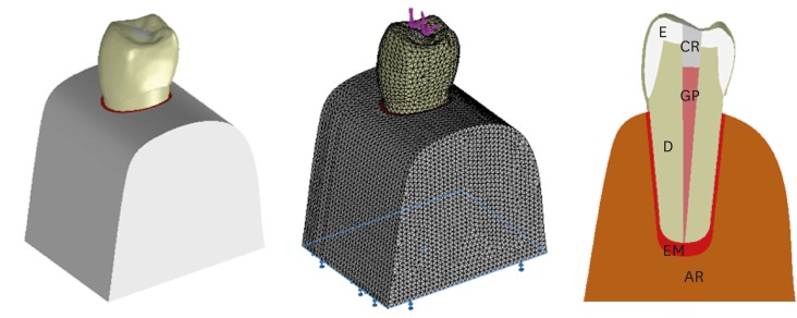
- Critical evaluation of fracture strength testing for endodontically treated teeth: a finite element analysis study
- Emel Uzunoglu-Özyürek, Selen Küçükkaya Eren, Oğuz Eraslan, Sema Belli
- Restor Dent Endod 2019;44(2):e15. Published online April 18, 2019
- DOI: https://doi.org/10.5395/rde.2019.44.e15
-
Abstract
PDFPubReaderePub
Objectives The aim of this study was to investigate whether the diameter and direction of the plunger and simulation of the periodontal ligament (PDL) affected the stress distribution in endodontically treated premolars.
Methods A fracture strength test was simulated via finite element analysis. A base model was set up, and the following parameters were modified: plunger diameter (3 mm vs. 6 mm), plunger direction (vertical vs. 135° angular to the central fossa), and PDL simulation. The analysis was conducted using the CosmosWorks structural analysis program, and the results are presented in terms of von Mises stresses.
Results The smaller plunger increased the stresses at the contact area of the crown, but the plunger diameter had no effect on the stress distribution within the root. An angular plunger direction increased stresses within the root, as well as at the buccal cusp of the crown, compared with the vertical direction. Simulation of the PDL caused higher stress accumulation, especially in the cervical region of the root.
Conclusions The plunger diameter had no effect on the stress distribution in the roots, whereas the plunger direction and PDL simulation did affect the stress distribution. More stringent standards can be established by taking such parameters into account when performing fracture testing in future studies.
-
Citations
Citations to this article as recorded by
- Fracture resistance of endodontically treated teeth restored with polyetheretherketone (PEEK) versus glass fiber posts: A comparative in vitro analysis
Manduwada Vishal, Neha Mehra, Mamta Kaushik, Prabakaran Saravanan
Endodontology.2026; 38(2): 172. CrossRef - Access cavity in endodontics: Balancing precision, preservation, and clinical needs
Dina Abdellatif, Ismail Davut Capar, De Fontaine Sarah, Alfredo Iandolo, Christophe Meyer, Davide Mancino
Journal of Conservative Dentistry and Endodontics.2025; 28(6): 573. CrossRef - Assessment of Stress Distribution with 3 Taper Design Preparation of Root Canal Using Finite Element Analysis
Tejasree Rathod, G. Durgabhavani, Pudu Tirupathi, Nusrath Parveen, Yelloji Paramesh, Prabhakar Dharavattu
Journal of Pharmacy and Bioallied Sciences.2024; 16(Suppl 1): S112. CrossRef - The impact of the filling technique with two sealers in bulk or associated with gutta-percha on the fatigue behavior and failure patterns of endodontically treated teeth
Isabella Marian Lena, Luiza Colpo Chiaratti, Rafaela Oliveira Pilecco, Renan Vaz Machry, João Paulo Mendes Tribst, Cornelis Johannes Kleverlaan, Gabriel Kalil Rocha Pereira, Renata Dornelles Morgental
PeerJ.2024; 12: e18221. CrossRef - Stronger than Ever: Multifilament Fiberglass Posts Boost Maxillary Premolar Fracture Resistance
Naji Kharouf, Eugenio Pedullà, Gianluca Plotino, Hamdi Jmal, Mohammed-El-Habib Alloui, Philippine Simonis, Patrice Laquerriere, Valentina Macaluso, Dina Abdellatif, Raphaël Richert, Youssef Haikel, Davide Mancino
Journal of Clinical Medicine.2023; 12(8): 2975. CrossRef - Neural network approach to evaluate the physical properties of dentin
Mohammad Ali Saghiri, Ali Mohammad Saghiri, Elham Samadi, Devyani Nath, Julia Vakhnovetsky, Steven M. Morgano
Odontology.2023; 111(1): 68. CrossRef - Modelling and evaluating periodontal ligament mechanical behaviour and properties: A scoping review of current approaches and limitations
Enaiyat Ghani Ovy, Dan L. Romanyk, Carlos Flores Mir, Lindsey Westover
Orthodontics & Craniofacial Research.2022; 25(2): 199. CrossRef - FEAr no more! Finite element analysis in orthodontics
Shilpa Chawla, Shailesh Deshmukh
Journal of the International Clinical Dental Research Organization.2022; 14(1): 6. CrossRef - Influence of Methodological Variables on Fracture Strength Tests Results of Premolars with Different Number of Residual Walls. A Systematic Review with Meta-Analysis
Carlo Gaeta, Crystal Marruganti, Emanuele Mignosa, Giovanni Franciosi, Edoardo Ferrari, Simone Grandini
Dentistry Journal.2021; 9(12): 146. CrossRef
- Fracture resistance of endodontically treated teeth restored with polyetheretherketone (PEEK) versus glass fiber posts: A comparative in vitro analysis
- 3,607 View
- 59 Download
- 9 Crossref
- Comparing the effect of a desensitizing material and a self-etch adhesive on dentin sensitivity after periodontal surgery: a randomized clinical trial
- Hila Hajizadeh, Atefeh Nemati-Karimooy, Sara Majidinia, Amir Moeintaghavi, Marjaneh Ghavamnasiri
- Restor Dent Endod 2017;42(3):168-175. Published online July 21, 2017
- DOI: https://doi.org/10.5395/rde.2017.42.3.168
-
Abstract
PDFPubReaderePub
Objectives This double-blind randomized placebo-controlled clinical trial evaluated the ability of a desensitizing agent and a self-etch adhesive on cervical dentin sensitivity (CDS) after periodontal surgery.
Materials and Methods Ninety hypersensitive teeth of 13 subjects were included in the study. After periodontal surgery, the teeth of each posterior sextant treated with one of the following materials: G1: Clearfil S3 Bond (Kuraray Dental), G2: Gluma Desensitizer (Heraeus Kulzer), and G3: placebo (water). The sensitivity was assessed using evaporative stimuli before treatment (baseline, T0), 1 day after treatment (T1), after 1 week (T2), and after 1 month (T3) according to visual analog scale (VAS).
Results Following the treatment, all the 3 groups showed significant reduction of CDS in T1 compared to T0. Reduction of CDS between T1 and T2 was observed only in G1 but there was no significant difference between T2 and T3 in this group. Although we observed a significant difference in T3 compared to T1 and T2 in G2 and G3, comparison of treatment groups in each assessment time showed a significant difference only in T3. According to paired comparison, this was due to the difference between G2 and G3.
Conclusions Dentin sensitivity following periodontal surgery will decrease spontaneously over time, but treating the sensitive teeth with Gluma Desensitizer and Clearfil S3 Bond can have some benefits.
-
Citations
Citations to this article as recorded by- Effect of different material protocols on the control of dentin hypersensitivity: a split-mouth randomized controlled clinical trial
Júlia Marques Martins, Maria Fernanda Ferreira Nogueira, Guilherme José Pimentel Lopes de Oliveira, Alexandre Coelho Machado, Paulo César de Freitas Santos Filho, Hugo Lemes Carlo, Carlos José Soares, Gisele Rodrigues da Silva
Clinical Oral Investigations.2026;[Epub] CrossRef - Biomineralization reaction from nanosized calcium silicate: A new method for reducing dentin hypersensitivity
Mi-Jeong Jeon, Yu-Sung Choi, Jeong-Kil Park, Jin-Soo Ahn, Yu-Chih Chiang, Deog-Gyu Seo
Journal of Dental Sciences.2025; 20(1): 428. CrossRef - Effectiveness of Self-etching Adhesive Only Versus in Combination with Gluma Desensitizer for Preventing Post-composite Sensitivity - A Prospective Study
Hemamalini Rath, Shilpa Mahapatra, Sri Priya Narayanan
Indian Journal of Dental Research.2025; 36(1): 32. CrossRef - Efficacy of seventh generation bonding agents as desensitizers in patients with dentin hypersensitivity: a randomized clinical trial
Sumaiya Shabbir, Shahbaz Ahmed, Syed Jaffar Abbas Zaidi, Sania Riaz, Huma Sarwar, Muhammad Taqi, Zia ur Rahman Khan
BMC Oral Health.2024;[Epub] CrossRef - Investigation of the crystal formation from calcium silicate in human dentinal tubules and the effect of phosphate buffer saline concentration
Mi-Jeong Jeon, Jin-Soo Ahn, Jeong-Kil Park, Deog-Gyu Seo
Journal of Dental Sciences.2024; 19(4): 2278. CrossRef - The effect of fluoride iontophoresis on seal ability of self-etch adhesive in human dentin in vitro
Kanittha Kijsamanmith, Parintorn Wallanon, Chanya Pitchayasatit, Poonnapha Kittiratanaviwat
BMC Oral Health.2022;[Epub] CrossRef - The study of toothpaste desensitizing properties
S. B. Ulitovskiy, O. V. Kalinina, A. A. Leontev, O. V. Khabarova, L. I. Pankrateva, E. S. Soloveva, N. K. Fok
Parodontologiya.2022; 27(1): 81. CrossRef - Effectiveness and cytotoxicity of two desensitizing agents: a dentin permeability measurement and dentin barrier testing in vitro study
Ruodan Jiang, Yongxiang Xu, Feilong Wang, Hong Lin
BMC Oral Health.2022;[Epub] CrossRef - A randomized clinical trial of dentin hypersensitivity reduction over one month after a single topical application of comparable materials
Samar Hatem Abuzinadah, Abdulrahman Jafar Alhaddad
Scientific Reports.2021;[Epub] CrossRef - Comparison between effectiveness of dentine desensitizer and one bottle self-etch adhesive on dentine hypersensitivity
Muhammad Zohaib Younus, Muhammad Adeel Ahmed, Azeem Ul Yaqin Syed, Jiand Malik Baloch, Muhammad Ali, Abubakar Sheikh
Technology and Health Care.2021; 29(6): 1153. CrossRef - A long-term evaluation of experimental potassium oxalate concentrations on dentin hypersensitivity reduction: A triple-blind randomized clinical trial
Alexia da Mata Galvão, Livia Fávaro Zeola, Guilherme Faria Moura, Daniela Navarro Ribeiro Teixeira, Ramon Corrêa de Queiroz Gonzaga, Gisele Rodrigues da Silva, Paulo Vinícius Soares
Journal of Dentistry.2019; 89: 103180. CrossRef
- Effect of different material protocols on the control of dentin hypersensitivity: a split-mouth randomized controlled clinical trial
- 3,643 View
- 11 Download
- 11 Crossref
- White mineral trioxide aggregate mixed with calcium chloride dihydrate: chemical analysis and biological properties
- Hany Mohamed Aly Ahmed, Norhayati Luddin, Thirumulu Ponnuraj Kannan, Khairani Idah Mokhtar, Azlina Ahmad
- Restor Dent Endod 2017;42(3):176-187. Published online April 17, 2017
- DOI: https://doi.org/10.5395/rde.2017.42.3.176
-
Abstract
PDFPubReaderePub
Objectives This study aimed to evaluate the chemical and biological properties of fast-set white mineral trioxide aggregate (FS WMTA), which was WMTA combined with calcium chloride dihydrate (CaCl2·2H2O), compared to that of WMTA.
Materials and Methods Surface morphology, elemental, and phase analysis were examined using scanning electron microscope (SEM), energy dispersive X-ray microanalysis (EDX), and X-ray diffraction (XRD), respectively. The cytotoxicity and cell attachment properties were evaluated on human periodontal ligament fibroblasts (HPLFs) using methyl-thiazol-diphenyltetrazolium (MTT) assay and under SEM after 24 and 72 hours, respectively.
Results Results showed that the addition of CaCl2·2H2O to WMTA affected the surface morphology and chemical composition. Although FS WMTA exhibited a non-cytotoxic profile, the cell viability values of this combination were lesser than WMTA, and the difference was significant in 7 out of 10 concentrations at the 2 time intervals (
p < 0.05). HPLFs adhered over the surface of WMTA and at the interface, after 24 hours of incubation. After 72 hours, there were increased numbers of HPLFs with prominent cytoplasmic processes. Similar findings were observed with FS WMTA, but the cells were not as confluent as with WMTA.Conclusions The addition of CaCl2·2H2O to WMTA affected its chemical properties. The favorable biological profile of FS WMTA towards HPLFs may have a potential impact on its clinical application for repair of perforation defects.
-
Citations
Citations to this article as recorded by- The effect of three additives on properties of mineral trioxide aggregate cements: a systematic review and meta-analysis of in vitro studies
Behnam Bolhari, Faranak Noori, Hadi Assadian, Amir Raee, Sholeh Ghabraei, Ahmad-Reza Shamshiri, Artak Heboyan
BMC Oral Health.2024;[Epub] CrossRef - Evaluation of sorption and solubility of materials based on calcium aluminate
Renata Josipovic, Violeta Petrovic, Marijana Popovic-Bajic, Irena Kuzmanovic-Radman, Mirjana Umicevic-Davidovic, Aleksandra Djeri, Slavoljub Zivkovic
Stomatoloski glasnik Srbije.2023; 70(1): 26. CrossRef - Chitosan-Based Accelerated Portland Cement Promotes Dentinogenic/Osteogenic Differentiation and Mineralization Activity of SHED
Hasan Subhi, Adam Husein, Dasmawati Mohamad, Nik Rozainah Nik Abdul Ghani, Asma-Abdullah Nurul
Polymers.2021; 13(19): 3358. CrossRef - Chemical modification of MTA and CEM cement to decrease setting time and improve bioactivity properties by adding alkaline salts
Faeze Jamali Zavare, Hanieh Nojehdehian, Maryam Moezizadeh, Mehdi Daneshpooya
Journal of Dental Research, Dental Clinics, Dental Prospects.2020; 14(1): 1. CrossRef - Biological effects of acid-eroded MTA Repair HP and ProRoot MTA on human periodontal ligament stem cells
Mar Collado-González, Sergio López-García, David García-Bernal, Ricardo E. Oñate-Sánchez, Christopher J. Tomás-Catalá, Jose M. Moraleda, Adrián Lozano, Leopoldo Forner, Francisco J. Rodríguez-Lozano
Clinical Oral Investigations.2019; 23(10): 3915. CrossRef - Comparative Cytocompatibility and Mineralization Potential of Bio-C Sealer and TotalFill BC Sealer
Sergio López-García, Miguel R. Pecci-Lloret, Julia Guerrero-Gironés, María P. Pecci-Lloret, Adrián Lozano, Carmen Llena, Francisco Javier Rodríguez-Lozano, Leopoldo Forner
Materials.2019; 12(19): 3087. CrossRef - Evaluation of changes in ion release and biological properties of NeoMTA‐Plus and Endocem‐MTA exposed to an acidic environment
F. J. Rodríguez‐Lozano, M. Collado‐González, S. López‐García, D. García‐Bernal, J. M. Moraleda, A. Lozano, L. Forner, L. Murcia, R. E. Oñate‐Sánchez
International Endodontic Journal.2019; 52(8): 1196. CrossRef
- The effect of three additives on properties of mineral trioxide aggregate cements: a systematic review and meta-analysis of in vitro studies
- 2,387 View
- 9 Download
- 7 Crossref
Review Article
- Recognition and management of palatogingival groove for tooth survival: a literature review
- Hee-Jin Kim, Yoorina Choi, Mi-Kyung Yu, Kwang-Won Lee, Kyung-San Min
- Restor Dent Endod 2017;42(2):77-86. Published online April 12, 2017
- DOI: https://doi.org/10.5395/rde.2017.42.2.77
-
Abstract
PDFPubReaderePub
Palatogingival groove (PGG) is an anomaly in the maxillary anterior teeth, often accompanied by the area of bony destruction adjacent to the teeth with no carious or traumatic history. The hidden trap in the tooth can harbor plaque and bacteria, resulting in periodontal destruction with or without pulpal pathologic change. Related diseases can involve periodontal destruction, combined endodontic-periodontal lesions, or separate endodontic and periodontal lesions. Disease severity and prognosis related to PGG depend on several factors, including location, range, depth, and type of the groove. Several materials have been used and recommended for cases of extensive periodontal destruction from PGG to remove and block the inflammatory source and recover the health of surrounding periodontal tissues. Even in cases of severe periodontal destruction, several studies have reported favorable treatment outcomes with proper management. With new options in diagnosis and treatment, clinicians need a detailed understanding of the characteristics, treatment, and prognosis of PGG to successfully manage the condition.
-
Citations
Citations to this article as recorded by- Prevalence of Palatal Grooves on Maxillary Anterior Teeth Using Cone-beam Computed Tomography: A Systematic Review and Meta-Analysis
Oscar Lozano González, Marco Felipe Salas Orozco, Nuria Patiño Marín, Paul V. Abbott, Marc Garcia-Font, Francesc Abella Sans
Journal of Endodontics.2026; 52(1): 14. CrossRef - Endodontic bioceramics: current and futurity aspects
Roma M, Karthik Shetty, Laxmish Mallya, Krishna Prasad Shetty
Frontiers in Oral Health.2026;[Epub] CrossRef - A Unified Deep Learning Framework for Visual Diagnosis of Palatal Radicular Grooves in CBCT Scans: A Multicenter Validation Study
Qikui Zhu, Weitao Fu, Yeyu Lin, Jiaxing Li, Wenhui Tang, Ying Zhang, Rui Zhang, Guanfan Lu, Yao Lin, Jing Shen, Zhuan Bian, Liuyan Meng
Journal of Endodontics.2026; 52(7): 1136. CrossRef - Endodontic and Periodontal Treatment of a Two‐Rooted Maxillary Lateral Incisor With a Type III Palatoradicular Groove: A Case Report With 2‐Year Follow‐Up
Katsuhiro Takeda, Tomoya Naruse, Yohei Takahashi, Reina Kawai, Kimiaki Yuhi, Hideki Shiba, Barbara Lapinska
Case Reports in Dentistry.2026;[Epub] CrossRef - Morphological analysis of palatogingival grooves in an Iraqi population: a retrospective cone-beam computed tomography study
O. B. Taha, N. S. Irhayyim, H. Y. Mohammed, M. Z. AL-Rawas, M.A. A. Naw, J. Y. Abdullah, M. I. Karobari
Endodontics Today.2026; 24(2): 354. CrossRef - Three-year follow-up case report: root canal treatment combined with intentional replantation for treating type III palatogingival groove in a maxillary lateral incisor
Jixu Jia, Miao Cheng, Sumeng Shi, Yanchun Qiao
Frontiers in Oral Health.2025;[Epub] CrossRef - Prevalence of palatogingival groove and its association with periapical lesions and periodontal bone loss: a cone beam computed tomography study
Dilan Pelin Yildirim, Selin Goker Kamali
BMC Oral Health.2025;[Epub] CrossRef - Evaluation of Morphology and Prevalence of Palatoradicular Grooves on Affected Maxillary Anterior Teeth Using Cone-Beam Computed Tomography: An Institutional Retrospective Study
Dilara Baştuğ, Leyla Benan Ayrancı
Applied Sciences.2025; 15(14): 8031. CrossRef - Sulco palato-gengival e suas consequências: Revisão de literatura
Marielli de Paula Gonçalves, Maria Júlia Ribeiro Chalita Vieira, Mikaelly Kawany Martins da Silva, Fabiana Tavares Lunardi Palhari, Maria Isabel Gonçalves Fialho
Research, Society and Development.2025; 14(8): e5014849388. CrossRef - Credibility of Intentional Reimplantation Techniques for Periodontally Compromised Teeth: A Report of Two Cases
Satarupa Suklabaidya, Ilakiya Mathi, Kennedy Babu, Gandhimadhi D, Manoj Margabandhu
Cureus.2025;[Epub] CrossRef - Prevalence of Palatal Radicular Groove in upper Lateral Incisors: A CBCT study at Isfahan Azad dental school
Amirreza Zefreh, Azadeh Torkzadeh, Hajar Shekarchizadeh, Maryam Zare Jahromi, Rojin Ardalani
Contemporary Orofacial Science.2025;[Epub] CrossRef - A classification of radicular grooves from the perspective of periodontology
Huxiao Li, Zhaowei Tai, Jiachen Dong, Zhongchen Song
BMC Oral Health.2025;[Epub] CrossRef - Advancements in Root Canal Therapy: Translational Innovations and the Role of Nanoparticles in Endodontic Treatment
Noha M. Badawi, Mohamed M. Kataia, Hadeel A. Mousa, Mozhgan Afshari
Journal of Nanotechnology.2025;[Epub] CrossRef - Cone-beam computed tomographic evaluation to estimate the prevalence of palatogingival groove in the maxillary anterior teeth and its radiographic characteristics: An institutional retrospective study
Mousumi Biswas, Dibyendu Mazumdar, Binayak Saha, Siddhi Agarwala, Kallol Kumar Saha, Kuntal Chowdhury
Journal of Conservative Dentistry and Endodontics.2024; 27(3): 233. CrossRef - A Three-Dimensional Assessment of a Type I Shallow Palatogingival Groove by Cone Beam Computed Tomography: A Case Report
Ramachandra Reddy Gowda Venkatesha, Karthik Rajaram Mohan, Saramma Mathew Fenn, Sabitha Gokulraj, Kumar Appusamy
Cureus.2024;[Epub] CrossRef - Diagnostic Approaches of Palatogingival Groove: A Systematic Review
Greta Venskutė
Journal of Dental Health and Oral Research.2024; : 1. CrossRef - Palatal groove associated with periodontal lesions: a systematic review illustrated by a decisional tree for management
Yvan Gaudex, Vianney Gandillot, Isabelle Fontanille, Philippe Bouchard, Stephane Kerner, Maria Clotilde Carra
BMC Oral Health.2024;[Epub] CrossRef - Palatogingival Groove: The Known–unknown Devourer
Sumedha Gupta, Sandeep Tandon, Ambika S Rathore, Rinku Mathur, Tripti S Rai, Kanchan Kumari Dhaker
International Journal of Clinical Pediatric Dentistry.2024; 17(S1): S95. CrossRef - Nomogram to predict radicular grooves in maxillary lateral incisors in preoperative orthodontic population
Xiuneng Zhou, Jie Deng, Nianke Liu, Chunhui Yang, Shiyu Li, Yaling Song
Clinical Oral Investigations.2024;[Epub] CrossRef - Management of Palatogingival Groove in Maxillary Lateral Incisor: A Report of a Rare Case With a Brief Review of Literature
Irfan Ansari, Sanjay Miglani, Vijay Yadav, Shamimul Hasan
Cureus.2023;[Epub] CrossRef - Prevalence of palatogingival groove affecting maxillary anterior teeth in Saudi subpopulation: A cone-beam computed tomographic study with literature review
Ali Ibrahim Aljuailan, Roqayah Aljuailan, Rahul N. Gaikwad, Shaul Hameed Kolarkodi, Nasser Rufaydan Alamri
The Saudi Dental Journal.2023; 35(8): 1039. CrossRef - Bioceramics in Endodontics: Updates and Future Perspectives
Xu Dong, Xin Xu
Bioengineering.2023; 10(3): 354. CrossRef - Interdisciplinary approach for diagnosis and management of the tooth with type III palatogingival groove
Harakh Chand Baranwal, Jyoti Yadav
Saudi Endodontic Journal.2023; 13(2): 211. CrossRef - Progress in Diagnosis and Treatment of Palatogingival Groove
倩 郑
Advances in Clinical Medicine.2022; 12(04): 2723. CrossRef - Palatogingival grooves associated with periodontal bone Loss of maxillary incisors in a Chinese population
Rui Zhang, Jie Xiong, Markus Haapasalo, Ya Shen, Liuyan Meng
Australian Endodontic Journal.2022; 48(2): 313. CrossRef - Surgical management of lateral lesions with intentional replantation in single-rooted mandibular first premolars with radicular groove
Ya-Hsin Yu, Minje Kim, Samuel Kratchman, Bekir Karabucak
The Journal of the American Dental Association.2022; 153(4): 371. CrossRef - Management of the palato-radicular groove with a periodontal regenerative procedure and prosthodontic treatment: A case report
Dan-Hua Ling, Wei-Ping Shi, Yan-Hong Wang, Dan-Ping Lai, Yan-Zhen Zhang
World Journal of Clinical Cases.2022; 10(17): 5732. CrossRef - Combined Periodontal and Endodontic Management of Palatal Radicular Groove with Platelet‐Rich Fibrin and Biodentine®
Arjun Hari Rijal, Bhageshwar Dhami, Pratistha Ghimire, Konstantinos Michalakis
Case Reports in Dentistry.2022;[Epub] CrossRef - Intentional replantation combined root resection therapy for the treatment of type III radicular groove with two roots: A case report
Dan Tan, Shi-Ting Li, Hao Feng, Zhong-Chao Wang, Cai Wen, Min-Hai Nie
World Journal of Clinical Cases.2022; 10(20): 6991. CrossRef - DENTAL DEFECTS WITH SUBGINGIVAL EXTENSION: A RESTORATIVE CONUNDRUM
Seema Yadav
INTERNATIONAL JOURNAL OF SCIENTIFIC RESEARCH.2021; : 20. CrossRef - Misdiagnosis or Missed Diagnosis? Cone-Beam Computed Tomography-Aided Multidisciplinary Management of Maxillary Central Incisor with Palatogingival Groove
R. Kurinji Amalavathy, K.M. Vidya, Sonali Nabil Sarooshi, Hrudi Sundar Sahoo
Indian Journal of Dental Sciences.2021; 13(1): 46. CrossRef - Root and Root Canal Morphology: Study Methods and Classifications
Duaa M Shihab , Anas F Mahdee
Journal of Baghdad College of Dentistry.2021; 33(4): 11. CrossRef - Prevalence and radiological characteristics of palatogingival groove: A retrospective cone-beam computed tomography study in an Indian cohort
MS Lekshmi, Sheetal Sharma, ShaliniR Gupta, Sidhartha Sharma, Vijay Kumar, Amrita Chawla, Ajay Logani
Journal of Conservative Dentistry.2021; 24(4): 359. CrossRef - Successful Multidisciplinary Management of an Endodontic‐Periodontal Lesion Associated With a Palato‐Radicular Groove: A Case Report
Diksha Katwal, Jennifer K. Fiorica, Jane Bleuel, Stephen J. Clark
Clinical Advances in Periodontics.2020; 10(2): 88. CrossRef - Anatomical, microbiological, and genetic considerations in treatment of Chinese periodontal patients
Edwin X. J. Goh, Marianne M. A. Ong
Journal of Investigative and Clinical Dentistry.2019;[Epub] CrossRef - A new system for classifying tooth, root and canal anomalies
H. M. A. Ahmed, P. M. H. Dummer
International Endodontic Journal.2018; 51(4): 389. CrossRef
- Prevalence of Palatal Grooves on Maxillary Anterior Teeth Using Cone-beam Computed Tomography: A Systematic Review and Meta-Analysis
- 11,671 View
- 234 Download
- 36 Crossref
Research Article
- Effects of proanthocyanidin, a crosslinking agent, on physical and biological properties of collagen hydrogel scaffold
- Yoorina Choi, Hee-Jin Kim, Kyung-San Min
- Restor Dent Endod 2016;41(4):296-303. Published online October 4, 2016
- DOI: https://doi.org/10.5395/rde.2016.41.4.296
-
Abstract
PDFPubReaderePub
Objectives The purpose of the present study was to evaluate the effects of proanthocyanidin (PAC), a crosslinking agent, on the physical properties of a collagen hydrogel and the behavior of human periodontal ligament cells (hPDLCs) cultured in the scaffold.
Materials and Methods Viability of hPDLCs treated with PAC was measured using the 3-(4,5-dimethylthiazol-2-yl)-2,5-diphenyltetrazolium bromide (MTT) assay. The physical properties of PAC treated collagen hydrogel scaffold were evaluated by the measurement of setting time, surface roughness, and differential scanning calorimetry (DSC). The behavior of the hPDLCs in the collagen scaffold was evaluated by cell morphology observation and cell numbers counting.
Results The setting time of the collagen scaffold was shortened in the presence of PAC (
p < 0.05). The surface roughness of the PAC-treated collagen was higher compared to the untreated control group (p < 0.05). The thermogram of the crosslinked collagen exhibited a higher endothermic peak compared to the uncrosslinked one. Cells in the PAC-treated collagen were observed to attach in closer proximity to one another with more cytoplasmic extensions compared to cells in the untreated control group. The number of cells cultured in the PAC-treated collagen scaffolds was significantly increased compared to the untreated control (p < 0.05).Conclusions Our results showed that PAC enhanced the physical properties of the collagen scaffold. Furthermore, the proliferation of hPDLCs cultured in the collagen scaffold crosslinked with PAC was facilitated. Conclusively, the application of PAC to the collagen scaffold may be beneficial for engineering-based periodontal ligament regeneration in delayed replantation.
-
Citations
Citations to this article as recorded by- Effect of collagen crosslinkers on sodium hypochlorite treated dentin bond strength: a systematic review and meta-analysis
Weiqing Zhou, Shuting Feng, Xiaojun Chu, Shuaimei Xu, Xiongqun Zeng
Frontiers in Bioengineering and Biotechnology.2025;[Epub] CrossRef - Proliferative Effect of Proanthocyanidins on HGF-1 and HPDLF Cells: An In Vitro Study
Evelina Alkimavičienė, Nomeda Basevičienė, Arvydas Strazdauskas, Rasa Banienė, Nijolė Savickienė
Medicina.2025; 61(12): 2098. CrossRef - A highly biocompatible CE-crosslinked collagen implant with exceptional anti-calcification and collagen regeneration capabilities for aging skin rejuvenation
Qi Wang, Huiyu Yan, Linyan Yao, Wenhua Li, Jianxi Xiao
Journal of Materials Chemistry B.2024; 12(18): 4467. CrossRef - Dexamethasone release from hyaluronic acid microparticle and proanthocyanidin-gelatin hydrogel in sciatic tissue regeneration
Kazem Javanmardi, Hamideh Shahbazi, Ava Soltani Hekmat, Mehdi Khanmohammadi, Arash Goodarzi
Journal of Materials Science: Materials in Medicine.2024;[Epub] CrossRef - New Materials Based on Collagen and Taxifolin Derivatives: Production and Properties
Yu. V. Shatalin, M. I. Kobyakova, V. S. Shubina
Биологические мембраны Журнал мембранной и клеточной биологии.2024; 41(1): 82. CrossRef - Modulation of Adhesion and Migration of NIH/3T3 Cells in Collagen Materials by Taxifolin Derivatives
Yu. V. Shatalin, M. I. Kobyakova, V. S. Shubina
Biochemistry (Moscow), Supplement Series A: Membrane and Cell Biology.2023; 17(S1): S85. CrossRef - Development and characterization of crosslinked k-carrageenan/sericin blend with covalent agents or thermal crosslink for indomethacin extended release
Wedja Timóteo Vieira, Meuris Gurgel Carlos da Silva, Laura de Oliveira Nascimento, Melissa Gurgel Adeodato Vieira
International Journal of Biological Macromolecules.2023; 246: 125558. CrossRef - New Challenges and Prospective Applications of Three-Dimensional Bioactive Polymeric Hydrogels in Oral and Craniofacial Tissue Engineering: A Narrative Review
Gamal Abdel Nasser Atia, Hany K. Shalaby, Naema Goda Ali, Shaimaa Mohammed Morsy, Mohamed Mohamady Ghobashy, Hager Abdel Nasser Attia, Paritosh Barai, Norhan Nady, Ahmad S. Kodous, Hasi Rani Barai
Pharmaceuticals.2023; 16(5): 702. CrossRef - Polyphenols: Bioavailability, Microbiome Interactions and Cellular Effects on Health in Humans and Animals
Michael B. Scott, Amy K. Styring, James S. O. McCullagh
Pathogens.2022; 11(7): 770. CrossRef - Advances of Hydrogel Therapy in Periodontal Regeneration—A Materials Perspective Review
Maoxue Li, Jiaxi Lv, Yi Yang, Guoping Cheng, Shujuan Guo, Chengcheng Liu, Yi Ding
Gels.2022; 8(10): 624. CrossRef - Collagen stabilization by natural cross-linkers: A qualitative and quantitative FTIR study on ultra-thin dentin collagen model
Rong WANG, Tyler STANLEY, Xiaomei YAO, Hang LIU, Yong WANG
Dental Materials Journal.2022; 41(3): 440. CrossRef - Cross-Linking Agents for Electrospinning-Based Bone Tissue Engineering
Dong-Jin Lim
International Journal of Molecular Sciences.2022; 23(10): 5444. CrossRef - Dense lamellar scaffold, biomimetically inspired, for reverse cardiac remodeling: Effect of proanthocyanidins and glutaraldehyde
Thais Alves, Juliana Ferreira Souza, Venancio Alves Amaral, Alessandra Candida Rios, Tais Costa, Kessi Crescencio, Fernando Batain, Denise Grotto, Renata Lima, Lindemberg Silveira Filho, Jose Oliveira Junior, Patricia Severino, Norberto Aranha, Marco Chau
Journal of Dispersion Science and Technology.2021; 42(2): 248. CrossRef - The effect of the cross-linker ratio used in gellan gum biomaterial synthesis on biomineralization
Serbülent TÜRK, Burak ÜNLÜ, Mahmut ÖZACAR
Bulletin of Biotechnology.2021; 2(2): 27. CrossRef - The recent advances in scaffolds for integrated periodontal regeneration
Hyun Nyun Woo, Young Joon Cho, Solaiman Tarafder, Chang H. Lee
Bioactive Materials.2021; 6(10): 3328. CrossRef - Plant based cross-linkers for tissue engineering applications
Abhishek Indurkar, Ashish Pandit, Ratnesh Jain, Prajakta Dandekar
Journal of Biomaterials Applications.2021; 36(1): 76. CrossRef - Plant-based biomaterials in tissue engineering
Abhishek Indurkar, Ashish Pandit, Ratnesh Jain, Prajakta Dandekar
Bioprinting.2021; 21: e00127. CrossRef - Traditional Chinese Medicine and orthopedic biomaterials: Host of opportunities from herbal extracts
Huijuan Tang, Andrell Hosein, Monica Mattioli-Belmonte
Materials Science and Engineering: C.2021; 120: 111760. CrossRef - Adsorption of Gold Ions onto Sericin and Alginate Particles Chemically Crosslinked by Proanthocyanidins: a Complete Fixed-Bed Column Study
Nilza Tatiane das Graças Santos, Richard Landers, Meuris Gurgel Carlos da Silva, Melissa Gurgel Adeodato Vieira
Industrial & Engineering Chemistry Research.2020; 59(1): 318. CrossRef - Proanthocyanidin as a crosslinking agent for fibrin, collagen hydrogels and their composites with decellularized Wharton’s-jelly-extract for tissue engineering applications
Elham Hasanzadeh, Narges Mahmoodi, Arefeh Basiri, Faezeh Esmaeili Ranjbar, Zahra Hassannejad, Somayeh Ebrahimi-Barough, Mahmoud Azami, Jafar Ai, Vafa Rahimi-Movaghar
Journal of Bioactive and Compatible Polymers.2020; 35(6): 554. CrossRef - Hydrogels for the Delivery of Plant-Derived (Poly)Phenols
Nicola Micale, Andrea Citarella, Maria Sofia Molonia, Antonio Speciale, Francesco Cimino, Antonella Saija, Mariateresa Cristani
Molecules.2020; 25(14): 3254. CrossRef - Natural biopolymer‐based hydrogels for use in food and agriculture
Miri Klein, Elena Poverenov
Journal of the Science of Food and Agriculture.2020; 100(6): 2337. CrossRef - Grape Seed-Inspired Smart Hydrogel Scaffolds for Melanoma Therapy and Wound Healing
Hongshi Ma, Quan Zhou, Jiang Chang, Chengtie Wu
ACS Nano.2019; 13(4): 4302. CrossRef - Improvement of the Physical Properties of Guided Bone Regeneration Membrane from Porcine Pericardium by Polyphenols-Rich Pomace Extract
Nazario Russo, Clara Cassinelli, Elisa Torre, Marco Morra, Giorgio Iviglia
Materials.2019; 12(16): 2564. CrossRef - Novel Biomedical Applications of Crosslinked Collagen
Lisha Gu, Tiantian Shan, Yu-xuan Ma, Franklin R. Tay, Lina Niu
Trends in Biotechnology.2019; 37(5): 464. CrossRef - The prospects of collagen as a basis for curable and activated osteoplastic materials
N. L. Fatkhudinova, A. V. Vasilyev, T. B. Bukharova, E. O. Osidak, N. V. Starikova, S. P. Domogatsky, D. V. Goldshtein, A. A. Kulakov
Stomatologiya.2018; 97(6): 78. CrossRef
- Effect of collagen crosslinkers on sodium hypochlorite treated dentin bond strength: a systematic review and meta-analysis
- 2,694 View
- 13 Download
- 26 Crossref
Case Reports
- Management of failed periodontal surgical intervention for a furcal lesion with a nonsurgical endodontic approach
- Saeed Asgary, Mahta Fazlyab
- Restor Dent Endod 2014;39(2):115-119. Published online March 21, 2014
- DOI: https://doi.org/10.5395/rde.2014.39.2.115
-
Abstract
PDFPubReaderePub
As long as the prognosis of teeth remains a matter of concern, the endodontic-periodontal relationship will be considered a challenge for the clinician. Many etiologic factors, including bacteria, fungi, and viruses, plus other contributing factors, such as trauma, root resorptions/perforations, and dental malformations, play a role in the co-occurrence of endodontic and periodontal lesions. Whatever the cause, a correct diagnosis on which to base the treatment plan is the key to successful maintenance of the tooth. This article reports the successful endodontic management of a furcation lesion in a mandibular molar that was nonresponsive to a previous periodontal surgical graft. The case had presented a diagnostic challenge for the clinicians, and this article reviews the key points that can lead to a correct diagnosis and treatment planning.
-
Citations
Citations to this article as recorded by- NON-SURGICAL ENDODONTIC TREATMENT OF CHRONIC APICAL PERIODONTITIS WITH EXTENSIVE PERIAPICAL AND FURCATION BONE DESTRUCTION: A CASE REPORT WITH 18-MONTH FOLLOW-UP
Magomedov Ibragim, Supiev Lugman, Omarov Muslim
BULLETIN OF STOMATOLOGY AND MAXILLOFACIAL SURGERY.2026; : 34. CrossRef - Acute periodontal lesions (periodontal abscesses and necrotizing periodontal diseases) and endo‐periodontal lesions
David Herrera, Belén Retamal‐Valdes, Bettina Alonso, Magda Feres
Journal of Clinical Periodontology.2018;[Epub] CrossRef - Acute periodontal lesions (periodontal abscesses and necrotizing periodontal diseases) and endo‐periodontal lesions
David Herrera, Belén Retamal‐Valdes, Bettina Alonso, Magda Feres
Journal of Periodontology.2018;[Epub] CrossRef - The importance of correct diagnosis and treatment in endo-periodontal lesions: a two cases comparison
Sara Bernardi, Christian Frascarelli, Giulia Fantozzi, Silvia Caruso, Robert Gatto, Gianna Maria Nardi, Maria Adelaide Continenza
Dental Update.2016; 43(8): 766. CrossRef
- NON-SURGICAL ENDODONTIC TREATMENT OF CHRONIC APICAL PERIODONTITIS WITH EXTENSIVE PERIAPICAL AND FURCATION BONE DESTRUCTION: A CASE REPORT WITH 18-MONTH FOLLOW-UP
- 4,166 View
- 18 Download
- 4 Crossref
- Diagnosis and treatment of teeth with primary endodontic lesions mimicking periodontal disease: three cases with long-term follow ups
- Jae-Hyung Lim, Ji-Hyun Lee, Su-Jung Shin
- Restor Dent Endod 2014;39(1):56-62. Published online January 20, 2014
- DOI: https://doi.org/10.5395/rde.2014.39.1.56
-
Abstract
PDFPubReaderePub
A tooth with primary endodontic disease that demonstrates a periodontal defect might be extracted because of misdiagnosis as severe periodontal disease or a vertical root fracture. The aim of this case report was to demonstrate the long-term survival of endodontically treated teeth, which had been initially considered unsavable. With meticulous evaluation including the patient's dental history, clinical and radiographic examinations, teeth with primary endodontic lesions could be differentiated and saved after proper root canal treatment. Pain history, vitality test, and radiographic examinations, as well as a general periodontal condition check with periodontal probing on an affected tooth, might be the key methods to differentiate endodontic pathosis from that of periodontal disease.
-
Citations
Citations to this article as recorded by- Variability in diagnostic and therapeutic decision-making for endodontic-periodontal lesions: evidence from a cross-sectional study
Yasir Dilshad Siddiqui, Malik Zain Ul Abideen, Muhammad Rizwan Memon, Farah Tahir, Ammar Ahmed Siddiqui, Muhammad Nadeem Baig, Mohammed Katib Alrowili, Azhar Iqbal, Raha Ahmed, Ahmad Salaar
Frontiers in Public Health.2026;[Epub] CrossRef - From Caries to Periodontal Breakdown: A Biological and Clinical Continuum Linking Cariology, Operative Dentistry, Endodontics, and Periodontology
Yasir Dilshad Siddiqui, Nusrat Sultana, Osama Khattak, Mohammed Zahedul Islam Nizami
Dentistry Journal.2026; 14(6): 380. CrossRef - The morphological and functional relationship between dental pulp and periodontal tissue in the aspect of endo-perio lesions
D. A. Moiseev, S. I. Volkov, A. A. Konov, M. A. Kulyukina
Parodontologiya.2022; 26(4): 289. CrossRef - Evaluation of root morphology of maxillary and mandibular second molars lost due to periodontitis
Akiko Kato, Toshimitsu Hishikawa, Koji Inagaki, Genta Yamamoto, Akio Mitani, Masaki Honda
Journal of Periodontal Research.2020; 55(5): 753. CrossRef - Clinical and Radiographic Characteristics of Endoperiodontitis in Patients with Chronic Generalized Periodontitis
L.N. Dedova, Yu.L. Denisova, N.I. Rossenik
Stomatologist. Minsk.2017; (3(26)): 13. CrossRef - The importance of correct diagnosis and treatment in endo-periodontal lesions: a two cases comparison
Sara Bernardi, Christian Frascarelli, Giulia Fantozzi, Silvia Caruso, Robert Gatto, Gianna Maria Nardi, Maria Adelaide Continenza
Dental Update.2016; 43(8): 766. CrossRef - Surgical management with intentional replantation on a tooth with palato-radicular groove
Jorge Forero-López, Luis Gamboa-Martínez, Laura Pico-Porras, Javier Laureano Niño-Barrera
Restorative Dentistry & Endodontics.2015; 40(2): 166. CrossRef - Subgingival microbiome in smokers and non‐smokers in Korean chronic periodontitis patients
J.‐H. Moon, J.‐H. Lee, J.‐Y. Lee
Molecular Oral Microbiology.2015; 30(3): 227. CrossRef
- Variability in diagnostic and therapeutic decision-making for endodontic-periodontal lesions: evidence from a cross-sectional study
- 4,416 View
- 16 Download
- 8 Crossref
Review Article
- Success and failure of endodontic microsurgery
- Minju Song, Euiseong Kim
- J Korean Acad Conserv Dent 2011;36(6):465-476. Published online November 30, 2011
- DOI: https://doi.org/10.5395/JKACD.2011.36.6.465
-
Abstract
PDFPubReaderePub
In current endodontic practice, introduction of operating microscope, ultrasonic instruments, and microinstruments has induced a big change in the field of surgical retreatment. In this study, we aimed to offer key steps of endodontic microsurgery procedure compared with traditional root-end surgery, and to evaluate factors influencing success and failure based on published articles.
Endodontic microsurgery is a surgical procedure performed with the aid of a microscope, ultrasonic instruments and modern microsurgical instruments. The microscope provides magnification and illumination - essential for identifying minute details of the apical anatomy. Ultrasonic instruments facilitate the precise root-end preparation that is within the anatomical space of the canal. Modern endodontics can therefore be performed with precision and predictability, thus eliminating the disadvantages inherent in traditional periapical surgery such as large osteotomy, beveled apicoectomy, inaccurate root-end preparation and the inability to observe isthmus.
Factors influencing the outcomes of endodontic microsurgery may be diverse, but standardization of procedures can minimize its range. Among patient and tooth-related factors, periodontal status and tooth position are known to be prognostic, but there are only few articles concerning this matter. High-evidence randomized clinical trials or prospective cohort studies are needed to confirm these findings.
-
Citations
Citations to this article as recorded by- Treatment-Related Factors Affecting the Success of Endodontic Microsurgery and the Influence of GTR on Radiographic Healing—A Cone-Beam Computed Tomography Study
Daniel Bieszczad, Jarosław Wichlinski, Tomasz Kaczmarzyk
Journal of Clinical Medicine.2023; 12(19): 6382. CrossRef - Factors Affecting the Success of Endodontic Microsurgery: A Cone-Beam Computed Tomography Study
Daniel Bieszczad, Jaroslaw Wichlinski, Tomasz Kaczmarzyk
Journal of Clinical Medicine.2022; 11(14): 3991. CrossRef - Comparison of Clinical Outcomes of Endodontic Microsurgery: 1 Year versus Long-term Follow-up
Minju Song, Taekjin Nam, Su-Jung Shin, Euiseong Kim
Journal of Endodontics.2014; 40(4): 490. CrossRef - The Influence of Bone Tissue Deficiency on the Outcome of Endodontic Microsurgery: A Prospective Study
Minju Song, Sahng Gyoon Kim, Su-Jung Shin, Hyeon-Cheol Kim, Euiseong Kim
Journal of Endodontics.2013; 39(11): 1341. CrossRef - Prognostic Factors of Clinical Outcomes in Endodontic Microsurgery: A Prospective Study
Minju Song, Sahng Gyoon Kim, Seung-Jong Lee, Baekil Kim, Euiseong Kim
Journal of Endodontics.2013; 39(12): 1491. CrossRef - Is stopping of anticoagulant therapy really required in a minor dental surgery? - How about in an endodontic microsurgery?
Yong-Wook Cho, Euiseong Kim
Restorative Dentistry & Endodontics.2013; 38(3): 113. CrossRef
- Treatment-Related Factors Affecting the Success of Endodontic Microsurgery and the Influence of GTR on Radiographic Healing—A Cone-Beam Computed Tomography Study
- 3,193 View
- 41 Download
- 6 Crossref
Case Report
- Treatment of a lateral incisor anatomically complicated with palatogingival groove
- Moon-Sun Choi, Se-Hee Park, Kyung-Mo Cho, Jin-Woo Kim
- J Korean Acad Conserv Dent 2011;36(3):238-242. Published online May 31, 2011
- DOI: https://doi.org/10.5395/JKACD.2011.36.3.238
-
Abstract
PDFPubReaderePub
Objectives Palatogingival groove is a developmental anomaly that starts near the cingulum of the tooth and runs down the cementoenamel junction in apical direction, terminating at various depths along the roots. While frequently associated with periodontal pockets and bone loss, pulpal necrosis of these teeth may precipitate a combined endodontic-periodontal lesion. This case presents a case of a lateral incisor anatomically complicated with palatogingival groove.
Methods Two patients with lesion associated with the palatogingival groove were chosen for this report. Palatogingival grooves were treated with different restoration materials with endodontic treatment.
Conclusions Maxillary lateral incisor with a palatogingival groove may occur the periodontal disease with pulpal involvement. Elimination of groove may facilitate the periodontal re-attachment and prevent the recurrence.
-
Citations
Citations to this article as recorded by- Endodontic treatment of maxillary lateral incisors with anatomical variations
Moon-Hwan Lee, Jung-Hong Ha, Myoung-Uk Jin, Young-Kyung Kim, Sung-Kyo Kim
Restorative Dentistry & Endodontics.2013; 38(4): 253. CrossRef
- Endodontic treatment of maxillary lateral incisors with anatomical variations
- 1,770 View
- 2 Download
- 1 Crossref
Basic Researchs
- The effect of tumor necrosis factor (TNF)-α to induce matrix metalloproteinase (MMPs) from the human dental pulp, gingival, and periodontal ligament cells
- Eun-Mi Rhim, Sang-Hyuk Park, Duck-Su Kim, Sun-Young Kim, Kyoung-Kyu Choi, Gi-Woon Choi
- J Korean Acad Conserv Dent 2011;36(1):26-36. Published online January 31, 2011
- DOI: https://doi.org/10.5395/JKACD.2011.36.1.26
-
Abstract
PDFPubReaderePub
Objectives In the present study, three kinds of tissues cells (pulp, gingiva, and periodontal ligament) were investigated if those cells express MMP and TIMP when they were stimulated with neuropeptides (substance P, CGRP) or proinflammatory cytokine, TNF-α.
Materials and Methods The cells cultured from human dental pulp (PF), gingiva (GF) and periodontal ligament were (PDLF) stimulated with Mock, SP, TNF-α, and CGRP for 24 hrs and 48 hrs. for an RNase protection assay and Enzyme Linked Immunosorbent Assay.
Cells (PF, GF and PDLF) seeded in 100 mm culture dish were stimulated with SP (10-5, 10-8 M) or only with medium (Mock stimulation) for 4hrs and for 24 hrs for RNase Protection Assay, and they were stimulated with CGRP (10-5 M) and TNF-α (2 ng/mL) for 24 hrs and with various concentraion of TNF-α (2, 10, and 100 ng/mL) for Rnase Protection Assay with a human MMP-1 probe set including MMP 1, 2, 8, 7, 8, 9, 12, and TIMP 2, 3.
In addition, cells (PF, GF and PDLF) were stimulated with Mock and various concentraion of TNF-α (2, 10, and 100 ng/mL) for 24 hrs and with TNF-α (10 ng/mL) for 48 hrs, and the supernatents from the cells were collected for Enzyme Linked Immunosorbent Assay (ELISA) for MMP-1 and MMP-13.
Results The expression of MMPs in PF, GF, PDLF after stimulation with SP and CGRP were not changed compared with Mock stimulation for 4 hrs and 24 hrs. The expression of MMP-1, -12, -13 24 hrs after stimulation with TNF-α were upregulated, however the expression of TIMP-3 in PF, GF, PDLF after stimulation with TNF-α were downregulated. TNF-α (2 ng/mL, 10 ng/mL, 100 ng/mL) increased MMP-1 and MMP-12 expression in PF dose dependently for 24 hrs.
Conclusions TNF-α in the area of inflammation may play an important role in regulating the remodeling of dentin, cementum, and alveolar bone.
-
Citations
Citations to this article as recorded by- Decoding the Ultimate Effects of Dipeptidyl Peptidase‐4 Inhibitors on Angiogenesis: An Updated Comprehensive Review of the Cellular and Molecular Mechanisms
Andrew Z. Zaka, Safwat A. Mangoura, Marwa A. Ahmed, Beshoy Allam
ChemistrySelect.2025;[Epub] CrossRef - Anti‐Inflammatory Effects of Melatonin and 5‐Methoxytryptophol on Lipopolysaccharide‐Induced Acute Pulpitis in Rats
Fatma Kermeoğlu, Umut Aksoy, Abdullah Sebai, Gökçe Savtekin, Hanife Özkayalar, Serkan Sayıner, Ahmet Özer Şehirli, Shuai CHEN
BioMed Research International.2021;[Epub] CrossRef - Cross-Talk between Ciliary Epithelium and Trabecular Meshwork Cells In-Vitro: A New Insight into Glaucoma
Natalie Lerner, Elie Beit-Yannai, Wayne Iwan Lee Davies
PLoS ONE.2014; 9(11): e112259. CrossRef
- Decoding the Ultimate Effects of Dipeptidyl Peptidase‐4 Inhibitors on Angiogenesis: An Updated Comprehensive Review of the Cellular and Molecular Mechanisms
- 2,325 View
- 7 Download
- 3 Crossref
- Biocompatibility of bioaggregate cement on human pulp and periodontal ligament (PDL) derived cells
- Choo-Ryung Chung, Euiseong Kim, Su-Jung Shin
- J Korean Acad Conserv Dent 2010;35(6):473-478. Published online November 30, 2010
- DOI: https://doi.org/10.5395/JKACD.2010.35.6.473
-
Abstract
PDFPubReaderePub
Objectives This study was performed to investigate the biocompatibility of newly introduced Bioaggregate on human pulp and PDL cells.
Materials and Methods Cells were collected from human pulp and PDL tissue of extracted premolars. Cell culture plate was coated either with Bioaggregate or white MTA, then the same number of cells were poured to cell culture dishes. Cell attachment and growth was examined under a phase microscope after 1,3 and 7 days of seeding. Cell viability was measured and the data was analyzed using Student
t -test and one way ANOVA.Results Both types of cells used in this study were well attached and grew healthy on Bioaggregate and MTA coated culture dishes. No cell inhibition zone was observed in Bioaggregate group. There was no statistical difference of viable cells between bioaggreagte and MTA groups.
Conclusions Bioaggregate appeared to be biocompatible compared with white MTA on human pulp and PDL cells.
-
Citations
Citations to this article as recorded by- Evaluation of bioactivity, biocompatibility, and antibacterial properties of tricalcium silicate bone cement modified with wollastonite/ fluorapatite glass and glass-ceramic
H.K. Abd El-Hamid, A.M. Fayad, R.L. Elwan
Ceramics International.2024; 50(14): 25322. CrossRef - Influence of insulin on the healing of exposed dental pulp after pulp capping: An experimental study in a dog model
Mokhtar A. Al‐Anesi, Ashraf M. Abu‐Seida, Salma H. El Ashry, Abeer H. Mahran, Ehab S. Abd‐Elhamid
Special Care in Dentistry.2021; 41(1): 49. CrossRef - ROOT END FILLING MATERIALS – A REVIEW
Bynagari Chandra Shekar, Veerendra Uppin, Madhu Pujar
GLOBAL JOURNAL FOR RESEARCH ANALYSIS.2021; : 5. CrossRef - Effects of two fast-setting calcium-silicate cements on cell viability and angiogenic factor release in human pulp-derived cells
Chooryung J. Chung, Euiseong Kim, Minju Song, Jeong-Won Park, Su-Jung Shin
Odontology.2016; 104(2): 143. CrossRef - Cytotoxicity and physical properties of tricalcium silicate-based endodontic materials
Young-Eun Jang, Bin-Na Lee, Jeong-Tae Koh, Yeong-Joon Park, Nam-Eok Joo, Hoon-Sang Chang, In-Nam Hwang, Won-Mann Oh, Yun-Chan Hwang
Restorative Dentistry & Endodontics.2014; 39(2): 89. CrossRef - Biocompatibility of root-end filling materials: recent update
Payal Saxena, Saurabh Kumar Gupta, Vilas Newaskar
Restorative Dentistry & Endodontics.2013; 38(3): 119. CrossRef
- Evaluation of bioactivity, biocompatibility, and antibacterial properties of tricalcium silicate bone cement modified with wollastonite/ fluorapatite glass and glass-ceramic
- 2,189 View
- 6 Download
- 6 Crossref
Case Report
- Treatment of crown-root fracture with a modified crown fragment reattachment technique
- Chang-Won Song, Min-Ju Song, Su-Jung Shin, Jeong-Won Park
- J Korean Acad Conserv Dent 2010;35(5):395-401. Published online September 30, 2010
- DOI: https://doi.org/10.5395/JKACD.2010.35.5.395
-
Abstract
PDFPubReaderePub
The development of adhesive dentistry has allowed that the crown fragment reattachment can be another option in the treatment of crown fracture. However, additional crown lengthening procedure or extrusion of the tooth may be necessary in the treatment of crown root fracture because subgingival fracture line in close proximity to the alveolar bone leads to challenges for restorative procedure and the violation of the biologic width. This case report presents a modified crown fragment reattachment technique of crown root fracture with pulp exposure, which was done without additional crown lengthening procedures. After the endodontic treatment, the patient was treated using a post insertion and the fragment reattachment technique, which made it possible to preserve the space for the biologic width and maintain a dry surgical field for adequate adhesion through the modification of the fractured coronal fragment. Since a coronal fracture was occurred and reattached afterward, it was observed that the coronal fragment was well maintained without the additional loss of periodontal attachment through 2-year follow up.
- 1,598 View
- 23 Download
Basic Research
- The evaluation of periodontal ligament cells of rat teeth after low-temperature preservation under high pressure
- Jin-Ho Chung, Jin Kim, Seong-Ho Choi, Eui-Seong Kim, Jiyong Park, Seung-Jong Lee
- J Korean Acad Conserv Dent 2010;35(4):285-294. Published online July 31, 2010
- DOI: https://doi.org/10.5395/JKACD.2010.35.4.285
-
Abstract
PDFPubReaderePub
The purpose of this study was to evaluate the viability of periodontal ligament cells of rat teeth after low-temperature preservation under high pressure by means of MTT assay, WST-1 assay. 12 teeth of Sprague-Dawley white female rats of 4 week-old were used for each group.
Both side of the first and second maxillary molars were extracted as atraumatically as possible under tiletamine anesthesia. The experimental groups were group 1 (Immediate extraction), group 2 (Slow freezing under pressure of 3 MPa), group 3 (Slow freezing under pressure of 2 MPa), group 4 (Slow freezing under no additional pressure), group 5 (Rapid freezing in liquid nitrogen under pressure of 2 MPa), group 6 (Rapid freezing in liquid nitrogen under no additional pressure), group 7 (low-temperature preservation at 0℃ under pressure of 2 MPa), group 8 (low-temperature preservation at 0℃ under no additional pressure), group 9 (low-temperature preservation at -5℃ under pressure of 90 MPa). F-medium and 10% DMSO were used as preservation medium and cryo-protectant. For cryo-preservation groups, thawing was performed in 37℃ water bath, then MTT assay, WST-1 assay were processed. One way ANOVA and Tukey HSD method were performed at the 95% level of confidence. The values of optical density obtained by MTT assay and WST-1 were divided by the values of eosin staining for tissue volume standardization.
In both MTT and WST-1 assay, group 7 (0℃/2 MPa) showed higher viability of periodontal ligament cells than other group (2-6, 8) and this was statistically significant (p < 0.05), but showed lower viability than group 1, immediate extraction group (no statistical significance).
By the results of this study, low-temperature preservation at 0℃ under pressure of 2 MPa suggest the possibility for long term preservation of teeth.
-
Citations
Citations to this article as recorded by- Evaluation of the Viability of Rat Periodontal Ligament Cells after Storing at 0℃/2 MPa Condition up to One Week: In Vivo MTT Method
Sun Mi Jang, Sin-Yeon Cho, Eui-Seong Kim, Il-Young Jung, Seung Jong Lee
Journal of Korean Dental Science.2016; 9(1): 1. CrossRef
- Evaluation of the Viability of Rat Periodontal Ligament Cells after Storing at 0℃/2 MPa Condition up to One Week: In Vivo MTT Method
- 1,812 View
- 2 Download
- 1 Crossref
Original Articles
- The comparison of gene expression from human dental pulp cells and periodontal ligament cells
- Hyoun So, Sang-Hyuk Park, Gi-Woon Choi
- J Korean Acad Conserv Dent 2009;34(5):430-441. Published online September 30, 2009
- DOI: https://doi.org/10.5395/JKACD.2009.34.5.430
-
Abstract
PDFPubReaderePub
The purpose of this study was to characterize functional distinction between human dental pulp cells(PC) and periodontal ligament cells(PDLC) using cDNA microarray assay and to confirm the results of the microarray assay using RT-PCR. 3 genes out of 51 genes which were found to be more expressed(>2 fold) in PC were selected, and 3 genes out of 19 genes which were found to be more expressed(>2 fold) in PDLC were selected for RT-PCR as well.
According to this study, the results were as follows:
1. From the microarray assay, 51 genes were more expressed (2 fold) from PC than PDLC.
2. RT-PCR confirmed that ITGA4 and TGF β2 were more expressed in PC than in PDLC.
3. From the microarray assay, 19 genes were more expressed (2 fold) from PDLC than PC.
4. RT-PCR confirmed that LUM, WISP1, and MMP1 were more expressed in PDLC than in PC.
From the present study, different expression of the genes between the PC and PDLC were characterized to show the genes which play an important role in dentinogenesis were more expressed from PC than PDLC, while the genes which were related with collagen synthesis were more expressed from PDLC than PC.
-
Citations
Citations to this article as recorded by- Gene expression profiling in human dental pulp cells treated with mineral trioxide aggregate
Yong-Beom Kim, Won-Jun Shon, WooCheol Lee, Kee-Yeon Kum, Seung-Ho Baek, Kwang-Shik Bae
Journal of Korean Academy of Conservative Dentistry.2010; 35(3): 152. CrossRef
- Gene expression profiling in human dental pulp cells treated with mineral trioxide aggregate
- 1,758 View
- 1 Download
- 1 Crossref
- THE EFFICACY OF PROGRAMMED CRYO-PRESERVATION UNDER PRESSURE IN RAT PERIODONTAL LIGAMENT CELLS
- Young-Eun Lee, Eui-Seong Kim, Jin Kim, Seung-Hoon Han, Seung-Jong Lee
- J Korean Acad Conserv Dent 2009;34(4):356-363. Published online January 14, 2009
- DOI: https://doi.org/10.5395/JKACD.2009.34.4.356
-
Abstract
PDFPubReaderePub
Abstract The purpose of this study was to evaluate the viability of periodontal ligament cells in rat teeth using slow cryo-preservation method under pressure by means of MTT assay and WST-1 assay. Eighteen teeth of Sprague-Dawley white female rats of 4 week-old were used for each group.
Both sides of the first and second maxillary molars were extracted as atraumatically as possible under Tiletamine anesthesia. The experimental groups were group 1 (Immediate control), group 2 (Cold preservation at 4°C for 1 week), group 3 (Slow freezing), group 4 (Slow freezing under pressure of 3 MPa). F-medium and 10% DMSO were used as preservation medium and cryo-protectant. For cryo-preservation groups, thawing was performed in 37°C water bath, then MTT assay and WST-1 assay were processed. One way ANOVA and Tukey method were performed at the 95% level of confidence. The values of optical density obtained by MTT assay and WST-1 were divided by the values of eosin staining for tissue volume standardization.
In both MTT and WST-1 assay, group 4 showed significantly higher viability of periodontal ligament cells than group 2 and 3 (p < 0.05), but showed lower viability than immediate control group.
By the results of this study, slow cryo-preservation method under pressure suggests the possibility for long term cryo-preservation of the teeth.
-
Citations
Citations to this article as recorded by- Effects of Slow Programmable Cryopreservation on Preserving Viability of the Cultured Periodontal Ligament Cells from Human Impacted Third Molar
Jin-Woo Kim, Tae-Yi Kim, Ye-mi Kim, Eun-Kyoung Pang, Sun-Jong Kim
Journal of Korean Dental Science.2015; 8(2): 57. CrossRef - The evaluation of periodontal ligament cells of rat teeth after low-temperature preservation under high pressure
Jin-Ho Chung, Jin Kim, Seong-Ho Choi, Eui-Seong Kim, Jiyong Park, Seung-Jong Lee
Journal of Korean Academy of Conservative Dentistry.2010; 35(4): 285. CrossRef - Comparison of viability of oral epithelial cells stored by different freezing methods
Do-Young Baek, Seung-Jong Lee, Han-Sung Jung, EuiSeong Kim
Journal of Korean Academy of Conservative Dentistry.2009; 34(6): 491. CrossRef
- Effects of Slow Programmable Cryopreservation on Preserving Viability of the Cultured Periodontal Ligament Cells from Human Impacted Third Molar
- 1,798 View
- 1 Download
- 3 Crossref
- Evaluation of the viability of periodontal ligament cell in rat teeth using slow cryopreservation method with magnetic field
- Hyun-Jung Ahn, Eui-Seong Kim, Jin Kim, Duck-Won Kim, Ki-Yeol Kim, Chan-Young Lee, Seung-Jong Lee
- J Korean Acad Conserv Dent 2008;33(4):332-340. Published online July 31, 2008
- DOI: https://doi.org/10.5395/JKACD.2008.33.4.332
-
Abstract
PDFPubReaderePub
The purpose of this study was to evaluate the viability of periodontal ligament cell in rat teeth using slow cryopreservation method with magnetic field through MTT assay and TUNEL test. For each group, 12 teeth of 4 weeks old white female Sprague-Dawley rat were used for MTT assay, and 6 teeth in TUNEL test. The Maxillary left and right, first and second molars were extracted as atraumatically as possible under tiletamine anesthesia. The experimental groups were group1 (immediately extraction), group 2 (cold preservation at 4℃ for 1 week), group 3 (rapid cryopreservation in liquid nitrogen), group 4 (slow cryopreservation with magnetic field of 1 G), and group 5 (slow cryopreservation). F medium was used as preservation medium and 10% DMSO as cryoprotectant. After preservation and thawing, the MTT assay and TUNEL test were processed. One way ANOVA and Scheffe method were performed at the 95% level of confidence. The value of optical density obtained after MTT analysis was divided by the value of eosin staining for tissue volume standardization. In both MTT assay and TUNEL test, it had showed no significant difference among group 3, 4, and 5. And group 3 had showed higher viability of periodontal ligament cell than group 2.
From this study, slow cryopreservation method with magnetic field can be used as one of cryopreservation methods.
-
Citations
Citations to this article as recorded by- The evaluation of periodontal ligament cells of rat teeth after low-temperature preservation under high pressure
Jin-Ho Chung, Jin Kim, Seong-Ho Choi, Eui-Seong Kim, Jiyong Park, Seung-Jong Lee
Journal of Korean Academy of Conservative Dentistry.2010; 35(4): 285. CrossRef - Comparison of viability of oral epithelial cells stored by different freezing methods
Do-Young Baek, Seung-Jong Lee, Han-Sung Jung, EuiSeong Kim
Journal of Korean Academy of Conservative Dentistry.2009; 34(6): 491. CrossRef - The efficacy of programmed cryo-preservation under pressure in rat periodontal ligament cells
Young-Eun Lee, Eui-Seong Kim, Jin Kim, Seung-Hoon Han, Seung-Jong Lee
Journal of Korean Academy of Conservative Dentistry.2009; 34(4): 356. CrossRef
- The evaluation of periodontal ligament cells of rat teeth after low-temperature preservation under high pressure
- 1,911 View
- 1 Download
- 3 Crossref
- Evaluation of the radiopacity and cytotoxicity of resinous root canal sealers
- Chang-Kyu Kim, Hyun-Wook Ryu, Hoon-Sang Chang, Byung-Do Lee, Kyung-San Min, Chan-Ui Hong
- J Korean Acad Conserv Dent 2007;32(5):419-425. Published online September 30, 2007
- DOI: https://doi.org/10.5395/JKACD.2007.32.5.419
-
Abstract
PDFPubReaderePub
The aim of this study was to evaluate the radiopacity and cytotoxicity of three resin-based (AH 26, EZ fill and AD Seal), a zinc oxide-eugenol-based (ZOB Seal), and a calcium hydroxide-based (Sealapex) root canal sealers. Specimens, 10 mm in diameter and 1 mm in thickness, were radiographed simultaneously with an aluminum step wedge using occlusal films, according to ISO 6876/2001 standards. Radiographs were digitized, and the radiopacity of sealers was compared to the different thicknesses of the aluminum step wedge, using the Scion image software. Using the 3-(4,5-dimethylthiazol-2-yl)-2,5-diphenyltetrazolium bromide (MTT) assay, the cytotoxicity of each material was determined in immortalized human periodontal ligament (IPDL) cells.
The results demonstrated that EZ fill was the most radiopaque sealer, while Sealapex was the least radiopaque (p < 0.05). AH 26, AD Seal and ZOB Seal presented intermediate radiopacity values. All the materials evaluated, except for Sealapex, presented the minimum radiopacity required by ISO standards. The cell viabilities of resin-based root canal sealers were statistically higher than that of other type of root canal sealers through the all experimental time. Further, EZ fill showed statistically lower cell viability in 24 and 48 hours compared to AD Seal and in 72 hours compared to all other resin-based root canal sealers. However, there was no correlation between the radiopacity and cytotoxicity of three resin-based root canals sealers (p > 0.05).
These results indicate that resin-based root canal sealer is more biocompatible and has advantage in terms of radiopacity.
-
Citations
Citations to this article as recorded by- A Comparative Evaluation of Two Commonly Used GP Solvents on Different Epoxy Resin-based Sealers: An In Vitro Study
Sakshi Tyagi, Ekta Choudhary, Rajat Chauhan, Ashish Choudhary
International Journal of Clinical Pediatric Dentistry.2020; 13(1): 35. CrossRef - Evaluation of softening ability of Xylene & Endosolv-R on three different epoxy resin based sealers within 1 to 2 minutes - anin vitrostudy
Pratima Ramakrishna Shenoi, Gautam Pyarelal Badole, Rajiv Tarachand Khode
Restorative Dentistry & Endodontics.2014; 39(1): 17. CrossRef - A comparative evaluation of cytotoxicity of root canal sealers: anin vitrostudy
Gautam Pyarelal Badole, Manjusha Madhukar Warhadpande, Ganesh Kothiramji Meshram, Rakesh Namdeoraoji Bahadure, Shubha Gopal Tawani, Gopal Tawani, Shital Gautam Badole
Restorative Dentistry & Endodontics.2013; 38(4): 204. CrossRef - Evaluation of radiopacity and discriminability of various fiber reinforced composite posts
Eun-Hye Lee, Hang-Moon Choi, Se-Hee Park, Jin-Woo Kim, Kyung-Mo Cho
Journal of Korean Academy of Conservative Dentistry.2010; 35(3): 188. CrossRef - A comparative study on radiopacity of root canal sealers
Tae-Min Kim, Seo-Kyoung Kim, In-Nam Hwang, Yun-Chan Hwang, Byung-Cheol Kang, Suk-Ja Yoon, Jae-Seo Lee, Won-Mann Oh
Journal of Korean Academy of Conservative Dentistry.2009; 34(1): 61. CrossRef - A comparative study on radiopacity of canal filling and retrograde root-end filling materials
Yong-Sang Kim, Seo-Kyong Kim, Yun-Chan Hwang, In-Nam Hwang, Won-Mann Oh
Journal of Korean Academy of Conservative Dentistry.2008; 33(2): 107. CrossRef
- A Comparative Evaluation of Two Commonly Used GP Solvents on Different Epoxy Resin-based Sealers: An In Vitro Study
- 2,713 View
- 7 Download
- 6 Crossref
- Evaluation of periodontal ligament cell viability in rat teeth according to various extra-oral dry storage times using MTT assay
- In-Soo Jeon, Eui-Seong Kim, Jin Kim, Seung-Jong Lee
- J Korean Acad Conserv Dent 2006;31(5):398-408. Published online September 30, 2006
- DOI: https://doi.org/10.5395/JKACD.2006.31.5.398
-
Abstract
PDFPubReaderePub
The purpose of this study was to verify the usefulness of MTT analysis as a tool of measurement of the periodontal ligament cell viability from the extracted rat molar.
A total of 80 Sprague-Dawley white female rat of 4 week-old with a body weight of 100 grams were used. The maxillary left and right, first and second molars were extracted under Ketamine anesthesia. Twenty-four teeth of each group (divided as five groups depending upon the time-lapse after extraction such as immediate, 10, 20, 40 and 60 minutes) were immersed in 200 µl of MTT solution (0.5 mg/ml) and processed for optical density measurements . Another 10 teeth of each group were treated as same as above and sectioned at 10 µm for microscopic examination.
All measurements values were divided by the value of hematoxylin-eosin staining which represented the volume of each corresponding samples. Immediate and 10 minute groups showed highest MTT values followed by 20, 40, and 60 minutes consecutively. Statistical significance (p < 0.05) existed between all groups except in immediate versus 10 minute groups and 40 versus 60 minutes. Histological findings also showed similar findings with MTT results in crystal shape and crystal numbers between the experimental groups.
These data indicate that
in vivo MTT analysis may be of value for evaluation of the periodontal ligament cell viability without time- consuming cell culturing processes.-
Citations
Citations to this article as recorded by- Evaluation of the Viability of Rat Periodontal Ligament Cells after Storing at 0℃/2 MPa Condition up to One Week: In Vivo MTT Method
Sun Mi Jang, Sin-Yeon Cho, Eui-Seong Kim, Il-Young Jung, Seung Jong Lee
Journal of Korean Dental Science.2016; 9(1): 1. CrossRef
- Evaluation of the Viability of Rat Periodontal Ligament Cells after Storing at 0℃/2 MPa Condition up to One Week: In Vivo MTT Method
- 1,830 View
- 0 Download
- 1 Crossref
-
Evaluation of periodontal ligament cell viability in rat teeth after frozen preservation using
in-vivo MTT assay - Jae-Wook Kim, Eui-Sung Kim, Jin Kim, Seung-Jong Lee
- J Korean Acad Conserv Dent 2006;31(3):192-202. Published online May 31, 2006
- DOI: https://doi.org/10.5395/JKACD.2006.31.3.192
-
Abstract
PDFPubReaderePub
The purpose of this study was to examine the viability of PDL cells in rat molars by using
in vivo MTT assay, which was used to compare fast cryopreservation group by liquid nitrogen (-196℃) with 4℃ cold preservation group.A total of 74 Sprague-Dawley white female rats of 4 week-old with a body weight of 100 grams were used. The maxillary left and right, first and second molars were extracted as atraumatically as possible under ketamine anesthesia.
Ten teeth of each group were divided as six experimental groups depending upon the preservation. Cryopreservation groups were Group 1 (5% DMSO 6% HES in F medium), Group 2 (10% DMSO in F medium), Group 3 (5% DMSO 6% HES in Viaspan®), Group 4 (10% DMSO in Viaspan®) which were cryopreserved for 1 week and cold preservation groups were Group 5 (F medium), Group 6 (Viaspan®) at 4℃ for 1 week. Immediate extraction group was used as a control. After preservation and thawing, the
in vivo MTT assay was processed. Two way ANOVA and Duncan's Multiple Range Test was performed at the 95% level of confidence. Another 2 teeth of each group were treated as the same manner and frozen sections 10 µm thick for microscopic observation.The value of optical density obtained after
in vivo MTT analysis was divided by the value of eosin staining for tissue volume standardization. Group 1, 2 had significantly higher optical density than Group 3 and 4 which had the lowest OD value. Group 6 had higher OD value than in Group 5 (P < 0.05). Histological findings of periodontal ligament cell, after being stained with MTT solution were consistent with thein vivo MTT assay results.In this study, the groups which were frozen with DMSO as a cryoprotectant and the groups with F medium showed the best results.
-
Citations
Citations to this article as recorded by- Evaluation of the Viability of Rat Periodontal Ligament Cells after Storing at 0℃/2 MPa Condition up to One Week: In Vivo MTT Method
Sun Mi Jang, Sin-Yeon Cho, Eui-Seong Kim, Il-Young Jung, Seung Jong Lee
Journal of Korean Dental Science.2016; 9(1): 1. CrossRef - Comparative study on survival rate of human gingival fibroblasts stored in different storage media
Hee Su Lee, You Sun Lim
Journal of Korean society of Dental Hygiene.2012; 12(4): 733. CrossRef - The evaluation of periodontal ligament cells of rat teeth after low-temperature preservation under high pressure
Jin-Ho Chung, Jin Kim, Seong-Ho Choi, Eui-Seong Kim, Jiyong Park, Seung-Jong Lee
Journal of Korean Academy of Conservative Dentistry.2010; 35(4): 285. CrossRef - Comparison of viability of oral epithelial cells stored by different freezing methods
Do-Young Baek, Seung-Jong Lee, Han-Sung Jung, EuiSeong Kim
Journal of Korean Academy of Conservative Dentistry.2009; 34(6): 491. CrossRef - The efficacy of programmed cryo-preservation under pressure in rat periodontal ligament cells
Young-Eun Lee, Eui-Seong Kim, Jin Kim, Seung-Hoon Han, Seung-Jong Lee
Journal of Korean Academy of Conservative Dentistry.2009; 34(4): 356. CrossRef
- Evaluation of the Viability of Rat Periodontal Ligament Cells after Storing at 0℃/2 MPa Condition up to One Week: In Vivo MTT Method
- 1,840 View
- 0 Download
- 5 Crossref
- The effect of neuropeptides on secretion of Interleukin-8 (IL-8)
- Kyung-Jun Kim, Sang-Hyuk Park, Kyoung-Kyu Choi, Sang-Jin Park
- J Korean Acad Conserv Dent 2006;31(3):153-160. Published online May 31, 2006
- DOI: https://doi.org/10.5395/JKACD.2006.31.3.153
-
Abstract
PDFPubReaderePub
We investigated the secretion of Interleukin-8 (IL-8) from ginviva and periodontal ligament stimulated with Substance P (SP) and Calcitonin Gene-related Peptide (CGRP). Gingiva (GF), periodontal ligament (PDLF) and pulp (PF) tissues were collected from extracted intact 3rd molars.
Cultured cells were stimulated with different concentrations of SP for 4 hrs, and stimulated with SP, CGRP and Tumor Necrosis Factor-α (TNF-α) for 8 hrs. Then RNase Protection Assay was carried out. ELISA was performed using supernatants of stimulated cells for quantitative analysis of IL-8. Results were assessed using student t-test with significance of P < 0.05.
According to this study, the results were as follows:
IL-8 mRNA was detected in all type of cells studied (PF, GF and PDLF).
IL-8 mRNA expression was not increased after stimulating 4 hrs with SP (10-5M) and SP (10-8M) compared with Mock stimulation in all type of cells studied.
IL-8 mRNA expression was not increased after stimulating 8 hrs with SP (10-4M) and CGRP (10-6M) compared with Mock stimulation in all type of cells studied.
TNF-α(2 ng/ml) increased the expression of IL-8 mRNA in all kind of cells studied.
The secretion of IL-8 from GF was increased 8 hrs after the stimulation with CGRP (10-6M) (p < 0.05).
The secretion of IL-8 from PDLF was increased 8 hrs after the stimulation with SP (10-4M) (p < 0.05).
Calcitonin Gene-related Peptide (CGRP) increased Interleukin-8 (IL-8) which plays an important role in chemotaxis of neutrophil in Calcitonin Gene-related Peptide (CGRP) gingival tissue, whereas Substance P increased the secretion of IL-8 from periodontal ligament.
- 1,937 View
- 1 Download
 First
First Prev
Prev

