
Search
- Page Path
- HOME > Search
Research Article
- Calcium silicate-based sealers remnants in isthmuses of mesial roots of mandibular molars: an in vitro evaluation
- David Saldanha de Brito Alencar, Ana Cristina Padilha Janini, Lauter Eston Pelepenko, Brenda Fornazaro Moraes, Francisco Haiter Neto, Marco Antonio Hungaro Duarte, Marina Angélica Marciano
- Restor Dent Endod 2025;50(3):e25. Published online July 15, 2025
- DOI: https://doi.org/10.5395/rde.2025.50.e25
-
 Abstract
 PDF
PDF PubReader
 ePub
ePub - Objectives
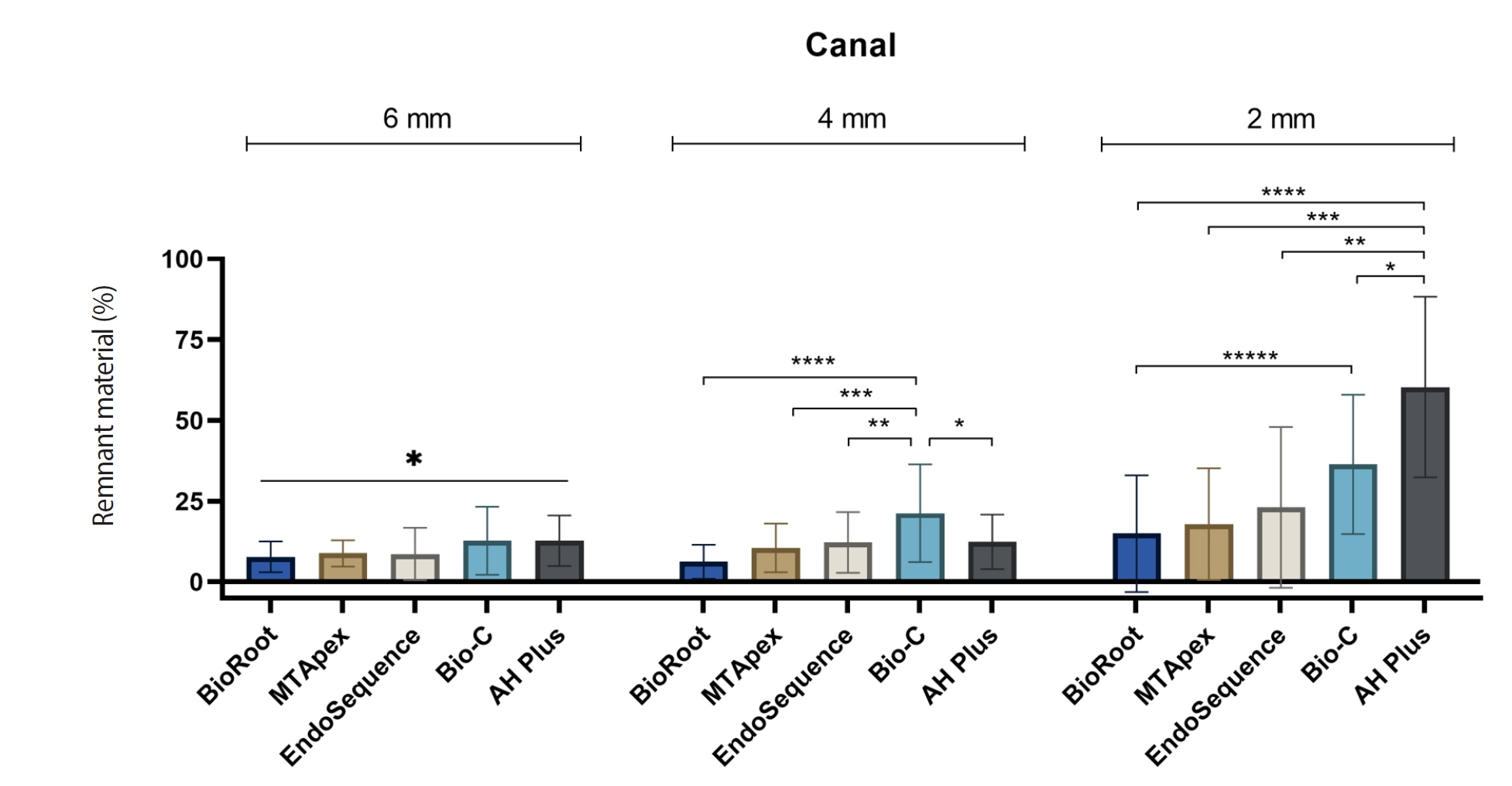
Endodontic retreatment aims to address treatment failure through the removal of root canal filling materials. This in vitro study evaluated the presence of filling material remnants in the mesial root canals, specifically focusing on the isthmuses, of mandibular molars after retreatment.
Methods
One hundred extracted mandibular molar mesial roots with isthmuses were prepared with an R25 file, obturated with one of five calcium silicate-based sealers (BioRoot RCS [Septodont], MTApex [Ultradent Products Inc.], EndoSequence BC Sealer HiFlow [Brasseler USA], Bio-C Sealer [Angelus]) or an epoxy resin-based sealer (AH Plus Jet [Dentsply Maillefer]), all stained with rhodamine B, and stored at 37ºC for 30 days to allow for setting. Retreatment was subsequently performed using R40 and XP-endo Finisher R instruments (FKG Dentaire) with 2.5% sodium hypochlorite irrigation. The presence of remaining filling material was then assessed using confocal microscopy, and setting times were tested per ISO 6876:2012.
Results
AH Plus Jet showed the most remnants at 2 mm and the longest retreatment time. Calcium silicate-based sealers exhibited prolonged setting times under dry conditions, with EndoSequence BC Sealer HiFlow showing a particularly extended setting period.
Conclusions
Despite retreatment, residues remained in all canals and isthmus regions, particularly Bio-C Sealer and AH Plus Jet in apical areas, emphasizing the difficulty of complete removal and the persistence of filling material. -
Citations
Citations to this article as recorded by
- Bonding effects of mechanical removal of bioceramic sealer residues using glycine or glass microparticles abrasion
Jesus Aranda, Julia de Freitas Ceccato, Eduardo Fernández Godoy, João Felipe Besegato, Joissi Ferrari Zaniboni, Regina Guenka Palma-Dibb, Milton Carlos Kuga
International Journal of Adhesion and Adhesives.2026; 148: 104289. CrossRef
- Bonding effects of mechanical removal of bioceramic sealer residues using glycine or glass microparticles abrasion
- 2,841 View
- 130 Download
- 1 Web of Science
- 1 Crossref

Review Article
- Effect of endodontic sealer on postoperative pain: a network meta-analysis
- Cynthia Maria Chaves Monteiro, Ana Cristina Rodrigues Martins, Alessandra Reis, Juliana Larocca de Geus
- Restor Dent Endod 2023;48(1):e5. Published online December 29, 2022
- DOI: https://doi.org/10.5395/rde.2023.48.e5
-
Abstract
PDFPubReaderePub
This systematic review and network meta-analysis aimed to answer the following focused research question: “Does the type of endodontic sealer affect the postoperative pain in patients who received endodontic treatment?” Different databases and grey literature were surveyed. Only one randomized controlled trial were included. The risk of bias in the studies was evaluated by using the Cochrane Collaboration’s tool. A random-effects meta-analysis was conducted to compare the risk and intensity of postoperative pain. The quality of the body of evidence was assessed using the Grading of Recommendations Assessment, Development, and Evaluation approach. Out of 11,601 studies, 15 remained for qualitative analyses and 12 for meta-analysis. Seven studies were classified at high risk of bias, and 8 studies raised some concerns. No significant differences between the endodontic materials were observed in the direct comparisons, both in risk and in intensity of postoperative pain (pairwise comparisons with 2 studies: I2 = 0%;
p > 0.05 and 8 studies: I2 = 23%;p > 0.05, respectively). The certainty of the evidence was graded as low or moderate. There was no difference in the risk and intensity of postoperative pain after filling with different endodontic sealers. Further systematic reviews should be conducted.Trial Registration PROSPERO Identifier:
CRD42020215314 -
Citations
Citations to this article as recorded by- Does the Use of a Bioceramic Sealer Reduce Postoperative Pain Compared With an Epoxy Resin‐Based Sealer After Primary Root Canal Treatment and Retreatment?—An Umbrella Review
Lokhasudhan Govindaraju, Rajeswari Kalaiselvam, Mathan Rajan Rajendran, Aleksandar Jakovljevic, Jelena Jacimovic, Henry F. Duncan, Venkateshbabu Nagendrababu
International Endodontic Journal.2026; 59(3): 341. CrossRef - Evidence synthesis of postoperative pain with bioceramic vs. epoxy resin sealers: umbrella review of randomized trials within existing systematic reviews
Mrunali Dahikar, Ashish Mandwe, Kulvinder Singh Banga, Alexander Maniangat Luke, Suraj Arora, Unmesh Khanvilkar, Ajinkya M. Pawar
Frontiers in Dental Medicine.2026;[Epub] CrossRef - Factors Influencing Apical Extrusion of 2 Types of Endodontic Sealers with Different Delivery Systems
Shumaila Iqbal, Nicholas S. Adams, Josette Camilleri
Journal of Endodontics.2026; 52(5): 806. CrossRef - Evaluation of Postoperative Pain Frequency in Single‐Session Endodontic Treatments With Patency and Foraminal Enlargement
Viviane Barbosa Godoy, Ana Grasiela Limoeiro, Vanessa Sandini, Vini Mehta, Wayne Martins Nascimento, Marilia Fagury Videira Marceliano‐Alves, Marcos Frozoni
Clinical and Experimental Dental Research.2026;[Epub] CrossRef - Silicone vs. Silicon/Silica in Intraoral Healing: A Systematic Review
David Parker, Aditi Bopardikar, Georgios E. Romanos
Materials.2026; 19(7): 1425. CrossRef - Post-Operative Pain After Endodontic Instrumentation, Irrigation and Obturation: An Umbrella Review of Systematic Reviews Published from 2016 to 2025
Fausto Zamparini, Andrea Spinelli, Gioia Quadrini, Maria Giovanna Gandolfi, Carlo Prati
Journal of Clinical Medicine.2026; 15(12): 4775. CrossRef - Comparative Evaluation of Postoperative Pain Following Nonsurgical Endodontic Therapy with Calcium Silicate-Based Sealer and Traditional Sealers: A Systematic Review and Meta-Analysis
Guha Poulomi, Solete Pradeep, Antony Delphine, Arun Nishitha, Surendar Ramamoorthi, Choudhari Sahil, Hima Sandeep Adimulapu
Pesquisa Brasileira em Odontopediatria e Clínica Integrada.2026;[Epub] CrossRef - Effect of occlusal reduction on post-operative pain of symptomatic and asymptomatic molar teeth
Aysenur Kamacı Esen, Fatma Furuncuoğlu, Fatima Betul Basturk, Muhammet Nuri Taşcıoğlu, Masoud Parirokh
Acta Odontologica Scandinavica.2025; 84: 371. CrossRef - An Observational Study on Pain Occurrence After Root Canal Treatment: Role of Operator Experience When Using a Bioceramic Sealer
Mihai Merfea, Ioana Sofia Pop-Ciutrila, Mindra Eugenia Badea, Ada Gabriela Delean, Oana Cimponeriu, Razvan Corneliu Pop, Maria Peter, Iulia Clara Badea, Sanda Ileana Cimpean
Journal of Clinical Medicine.2025; 14(13): 4558. CrossRef - Assessment of Postoperative Pain After Single‐ or Multiple‐Visit Endodontic Therapy and Its Molecular Aspects: A Randomised Controlled Study
Larissa Nunes Rosa Bedene, Denise Piotto Leonardi, Joana Santana Couto, Bruno Marques‐da‐Silva, Marilisa Carneiro Leão Gabardo, João Arnando Brancher, Flávia Sens Fagundes Tomazinho
Australian Endodontic Journal.2025; 51(3): 668. CrossRef - Clinical and Radiographic Outcomes of Root Canal Obturation with Hydraulic Condensation and Tricalcium Silicate Bioceramic Sealer: A 12-Month Observational Study on Periapical Healing
Kostadin Zhekov, Vesela Stefanova
Journal of Functional Biomaterials.2025; 16(11): 412. CrossRef - Comparative evaluation of postoperative pain and periapical healing after root canal treatment using three different endodontic sealers: A randomized controlled clinical trial
Ruchika Pandey, Nitin Kararia, Deepak Kumar Sharma, Vishal Rathod, Anand Vilas Bansod, Dhaval Desai
Journal of Conservative Dentistry and Endodontics.2024; 27(9): 962. CrossRef - Effect of bioceramic-based and resin-based sealers on postoperative discomfort following root canal therapy: a systematic review and meta-analysis
Mansi Supare, Ajinkya M. Pawar, Kashmira Sawant, Dian Agustin Wahjuningrum, Suraj Arora, Firas Elmsmari, Mohmed Isaqali Karobari, Bhagyashree Thakur
PeerJ.2024; 12: e18198. CrossRef - Comparative Evaluation of Incidences of Post Operative Pain in Patient Treated in Single Visit Root Canal Treatment by Using Different Sealers: - An in-Vivo Study
Sadashiv Daokar, Aishwarya Ranjalkar, Kalpana Pawar, Komal Potfode, Dhanashri Padwal, Sana Khan
International Journal of Innovative Science and Research Technology (IJISRT).2024; : 2743. CrossRef
- Does the Use of a Bioceramic Sealer Reduce Postoperative Pain Compared With an Epoxy Resin‐Based Sealer After Primary Root Canal Treatment and Retreatment?—An Umbrella Review
- 8,404 View
- 163 Download
- 12 Web of Science
- 14 Crossref
Research Articles
- Efficacy of reciprocating and rotary retreatment nickel-titanium file systems for removing filling materials with a complementary cleaning method in oval canals
- Said Dhaimy, Hyeon-Cheol Kim, Lamyae Bedida, Imane Benkiran
- Restor Dent Endod 2021;46(1):e13. Published online February 3, 2021
- DOI: https://doi.org/10.5395/rde.2021.46.e13
-
Abstract
PDFPubReaderePub
Objectives This study aimed to evaluate and compare the efficacy of the S1 reciprocating system and the D-Race retreatment rotary system for filling material removal and the apical extrusion of debris.
Materials and Methods Sixty-four freshly extracted maxillary canines were shaped with size 10 and size 15 K-files, instrumented using ProTaper Gold under irrigation with 2.5% sodium hypochlorite (NaOCl), obturated according to the principle of thermo-mechanical condensation with gutta-percha and zinc oxide eugenol sealer, and allowed to set for 3 weeks at 37°C. Subsequently, the teeth were divided into a control group (
n = 4), the D-Race rotary instrument group (n = 30), and the S1 reciprocating instrument group (n = 30). After classical retreatment, the canals were subjected to a complementary approach with the XP-Endo Shaper. Desocclusol was used as a solvent, and irrigation with 2.5% NaOCl was performed. Each group was divided into subgroups according to the timing of radiographic readings. The images were imported into a software program to measure the remaining filling material, the apical extrusion, and the root canal space. The data were statistically analyzed using the Z-test and JASP graphics software.Results No significant differences were found between the D-Race and S1 groups for primary retreatment; however, using a complementary cleaning method increased the removal of remnant filling (
p < 0.05).Conclusions Classical removal of canal filling material may not be sufficient for root canal disinfection, although a complementary finishing approach improved the results. Nevertheless, all systems left some debris and caused apical extrusion.
-
Citations
Citations to this article as recorded by- Effectiveness of different supplementary protocols for remaining filling material removal in endodontic reintervention: an integrative review
Amanda Freitas da Rosa, Bruna Venzke Fischer, Luiz Carlos de Lima Dias-Junior, Anna Victoria Costa Serique, Eduardo Antunes Bortoluzzi, Cleonice da Silveira Teixeira, Lucas da Fonseca Roberti Garcia
Odontology.2024; 112(1): 51. CrossRef - Critical analysis of research methods and experimental models to study removal of root filling materials
Mahdi A. Ajina, Pratik K. Shah, Bun San Chong
International Endodontic Journal.2022; 55(S1): 119. CrossRef - Economic analysis of the different endodontic instrumentation techniques used in the Unified Health System
Laura Paredes Merchan, Livia Fernandes Probst, Ana Clara Correa Duarte Simões, Augusto Cesar Santos Raimundo, Yuri Wanderley Cavalcanti, Denise de Fátima Barros Cavalcante, João Victor Frazão Câmara, Antonio Carlos Pereira
BMC Oral Health.2022;[Epub] CrossRef - Fabrication of a Potential Electrodeposited Nanocomposite for Dental Applications
Chun-Wei Chang, Chen-Han Tsou, Bai-Hung Huang, Kuo-Sheng Hung, Yung-Chieh Cho, Takashi Saito, Chi-Hsun Tsai, Chia-Chien Hsieh, Chung-Ming Liu, Wen-Chien Lan
Inorganics.2022; 10(10): 165. CrossRef - Influence of Filling Material Remnants on the Diffusion of Hydroxyl Ions in Endodontically Retreated Teeth: An Ex Vivo Study
Vania Portela Ditzel Westphalen, Marilisa Carneiro Leao Gabardo, Natanael Henrique Ribeiro Mattos, Camila Paiva Perin, Liliane Roskamp, Cristiano Miranda de Araújo, Luiz Fernando Fariniuk, Flares Baratto–Filho
The Journal of Contemporary Dental Practice.2022; 23(8): 768. CrossRef - Efficacy of Removing Thermafil and GuttaCore from Straight Root Canal Systems Using a Novel Non-Surgical Root Canal Re-Treatment System: A Micro-Computed Tomography Analysis
Vicente Faus-Llácer, Rubén Linero Pérez, Ignacio Faus-Matoses, Celia Ruiz-Sánchez, Álvaro Zubizarreta-Macho, Salvatore Sauro, Vicente Faus-Matoses
Journal of Clinical Medicine.2021; 10(6): 1266. CrossRef
- Effectiveness of different supplementary protocols for remaining filling material removal in endodontic reintervention: an integrative review
- 2,854 View
- 46 Download
- 7 Web of Science
- 6 Crossref
-
The effect of individualization of fiberglass posts using bulk-fill resin-based composites on cementation: an
in vitro study - Rodrigo Barros Esteves Lins, Jairo Matozinho Cordeiro, Carolina Perez Rangel, Thiago Bessa Marconato Antunes, Luís Roberto Marcondes Martins
- Restor Dent Endod 2019;44(4):e37. Published online October 18, 2019
- DOI: https://doi.org/10.5395/rde.2019.44.e37
-
Abstract
PDFPubReaderePub
Objectives This study evaluated the bond strength of various fiberglass post cementation techniques using different resin-based composites.
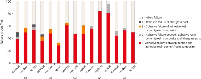
Materials and Methods The roots from a total of 100 bovine incisors were randomly assigned to 5 treatment groups: G1, post + Scotchbond Multi-Purpose (SBMP) + RelyX ARC luting agent; G2, relined post (Filtek Z250) + SBMP + RelyX ARC; G3, individualized post (Filtek Z250) + SBMP; G4, individualized post (Filtek Bulk-Fill) + SBMP; G5, individualized post (Filtek Bulk-Fill Flow) + SBMP. The samples were subjected to the push-out (
n = 10) and pull-out (n = 10) bond strength tests. Data from the push-out bond strength test were analyzed using 2-way analysis of variance (ANOVA) with the Bonferronipost hoc test, and data from the pull-out bond strength test were analyzed using 1-way ANOVA.Results The data for push-out bond strength presented higher values for G2 and G5, mainly in the cervical and middle thirds, and the data from the apical third showed a lower mean push-out bond strength in all groups. No significant difference was noted for pull-out bond strength among all groups. The most frequent failure modes observed were adhesive failure between dentine and resin and mixed failure.
Conclusions Fiberglass post cementation using restorative and flowable bulk-fill composites with the individualization technique may be a promising alternative to existing methods of post cementation.
-
Citations
Citations to this article as recorded by- EVALUATION OF PUSH-OUT BOND STRENGTH OF GLASS FIBER POSTS USING DIFFERENT LUTING CEMENTS
Jannah Mohammed, Maha Agha
BULLETIN OF STOMATOLOGY AND MAXILLOFACIAL SURGERY.2025; : 274. CrossRef - EVALUATION OF PUSH-OUT BOND STRENGTH OF GLASS FIBER POSTS USING DIFFERENT LUTING CEMENTS
Jannah Mohammed, Jannah Mohammed
BULLETIN OF STOMATOLOGY AND MAXILLOFACIAL SURGERY.2025; : 274. CrossRef - Effects of a relined fiberglass post with conventional and self-adhesive resin cement
Wilton Lima dos Santos Junior, Marina Rodrigues Santi, Rodrigo Barros Esteves Lins, Luís Roberto Marcondes Martins
Restorative Dentistry & Endodontics.2024;[Epub] CrossRef - Fracture resistance of weakened roots restored with relined or milled CAD-CAM glass fiber posts
Belizane das Graças Oliveira MAIA, Thais da Silva Alves SANTOS, Cláudio Antonio Talge CARVALHO, Francielle Silvestre VERNER, Rafael Binato JUNQUEIRA
Dental Materials Journal.2023; 42(1): 92. CrossRef - Evaluation of pretreatments on intra‐radicular dentin bond strength of self‐adhesive resin cements
Marina Rodrigues Santi, Rodrigo Barros Esteves Lins, Beatriz Ometto Sahadi, Luís Roberto Marcondes Martins, Jorge Rodrigo Soto‐Montero
Journal of Esthetic and Restorative Dentistry.2022; 34(7): 1051. CrossRef - Comparison of the Mechanical Properties and Push-out Bond Strength of Self-adhesive and Conventional Resin Cements on Fiber Post Cementation
MR Santi, RBE Lins, BO Sahadi, JR Soto-Montero, LRM Martins
Operative Dentistry.2022; 47(3): 346. CrossRef - Glass fiber posts
Renata Pereira, Rodrigo Barros Esteves Lins, Victória Castelan Rodrigues, Débora Alves Nunes Leite Lima, Luís Roberto Marcondes Martins, Flávio Henrique Baggio Aguiar
Brazilian Journal of Oral Sciences.2020; 19: e207508. CrossRef
- EVALUATION OF PUSH-OUT BOND STRENGTH OF GLASS FIBER POSTS USING DIFFERENT LUTING CEMENTS
- 2,147 View
- 14 Download
- 7 Crossref
- A micro-computed tomographic study of remaining filling materials of two bioceramic sealers and epoxy resin sealer after retreatment
- KyungJae Kim, Da Vin Kim, Sin-Young Kim, SungEun Yang
- Restor Dent Endod 2019;44(2):e18. Published online April 26, 2019
- DOI: https://doi.org/10.5395/rde.2019.44.e18
-
Abstract
PDFPubReaderePub
Objective This study evaluated the presence of residual root canal filling material after retreatment using micro-computed tomography (micro-CT).
Materials and Methods Extracted human teeth (single- and double-rooted,
n = 21/each; C-shaped,n = 15) were prepared with ProFile and randomly assigned to three subgroups for obturation with gutta-percha and three different sealers (EndoSeal MTA, EndoSequence BC sealer, and AH Plus). After 10 days, the filling material was removed and the root canals were instrumented one size up from the previous master apical file size. The teeth were scanned using micro-CT before and after retreatment. The percentage of remaining filling material after retreatment was calculated at the coronal, middle, and apical thirds. Data were analyzed using the Kruskal-Wallis test and Mann-WhitneyU test with Bonferronipost hoc correction.Results The tested sealers showed no significant differences in the percentage of remaining filling material in single- and double-rooted teeth, although EndoSeal MTA showed the highest value in C-shaped roots (
p < 0.05). The percentage of remaining filling material of AH Plus and EndoSeal MTA was significantly higher in C-shaped roots than in single- or double-roots (p < 0.05), while that of BC sealer was similar across all root types. EndoSeal MTA showed the highest values at the apical thirds of single- and double-roots (p < 0.05); otherwise, no significant differences were observed among the coronal, middle, and apical thirds.Conclusions Within the limitations of this study, a large amount of EndoSeal MTA remained after retreatment, especially in C-shaped root canals.
-
Citations
Citations to this article as recorded by- A laboratory study comparing two methods for removing plastic carrier obturators from severely curved root canals
Tania Gancedo-Gancedo, Patricia Pereira-Lores, Venkateshbabu Nagendrababu, Paul MH Dummer, Jenifer Martín-González, Alba Bello-Castro, Inmaculada Tomás, Benjamín Martín-Biedma, Pablo Castelo-Baz
BMC Oral Health.2026;[Epub] CrossRef - Comparative evaluation of the retreatability of bioceramic-based root canal sealers: an in vitro study
Sinem Esen Mutlu, Tamer Taşdemir, Tugba Kosar
Journal of the Australian Ceramic Society.2026;[Epub] CrossRef - Development of a deep neural network and empirical model for predicting local gas holdup profiles in bubble columns
Sebastián Uribe, Ahmed Alalou, Mario E. Cordero, Muthanna Al‐Dahhan
The Canadian Journal of Chemical Engineering.2025; 103(6): 2918. CrossRef - An In Vitro Comparison of Epoxy Resin Sealer Removal During Endodontic Retreatment
Prashant A Bondarde, Aditi S Patkar, Aishwarya R Pawar, Rukmini Pande, Akshata Deshpande, Rachana S Agrawal, Seema Gupta
Cureus.2025;[Epub] CrossRef - Calcium silicate-based sealers remnants in isthmuses of mesial roots of mandibular molars: an in vitro evaluation
David Saldanha de Brito Alencar, Ana Cristina Padilha Janini, Lauter Eston Pelepenko, Brenda Fornazaro Moraes, Francisco Haiter Neto, Marco Antonio Hungaro Duarte, Marina Angélica Marciano
Restorative Dentistry & Endodontics.2025; 50(3): e25. CrossRef - Push-out bond strength of two endodontic sealers in retreated canals using different solvents
Sara Gamal Ghanem, Walaa M. Ghoneim, Ahmed H. Labib
Tanta Dental Journal.2025; 22(3): 504. CrossRef - Assessing Volume of Two Sealers’ Remnants after Reinstrumentation Using 3D Imaging Technology: An In Vitro Comparative Study
Khalel Mutaz Dawod, Raghad Abdulrazzaq Al-Hashimi
The Journal of Contemporary Dental Practice.2025; 26(8): 743. CrossRef - Removal efficacy of two different root canal sealers in retrograde cavities: a micro-CT study
Özge Başar, Ahter Şanal Çıkman, Cangül Keskin
BMC Oral Health.2025;[Epub] CrossRef - Evaluation of the retreatability of bioceramic root canal sealers with various formulations in simulated grooves
Meltem Sümbüllü, Afzal Ali, Abdulaziz Bakhsh, Hakan Arslan
PeerJ.2025; 13: e20398. CrossRef - Correlation of Bond Strength and Dentinal Tubule Penetration Evaluation of Four Different Endodontic Sealers: AH Plus, MTA Fillapex, Endoseal MTA, and Endoseal TCS (Maruchi): An In Vitro Study
Arezoo Mirzaei Sadeghloo, Seyedali Seyedmajidi, Akam Saeidi, Elham Mahmoudi, Murilo Baena Lopes
International Journal of Dentistry.2025;[Epub] CrossRef - Root canal cleanliness and debris extrusion following retreatment of thermoplastic injection technique and bioceramic-based root canal sealer
Deniz Bender, Mert Ocak, Emel Uzunoğlu Özyürek
Clinical Oral Investigations.2024;[Epub] CrossRef - The Effect of Different Obturation Techniques Using Different Root Canal Sealers on the Residual Filling Material After Retreatment Procedures
M Sarı, K Yılmaz
Nigerian Journal of Clinical Practice.2024; 27(2): 174. CrossRef - Effect of Different Obturation Techniques on the Amount of Debris Extrusion During Endodontic Retreatment Using XP Endo Retreatment Set Files (In vitro Study)
Pawan Mohamad Amin, Hawzhen Mohammed Saeed
Sulaimani Dental Journal.2023; 10: 49. CrossRef - The efficiency of different irrigation activation techniques in the removal of calcium silicate‐based endodontic sealer from artificially created groove
Meltem Sümbüllü, Afzal Ali, Mine Büker, Hakan Arslan
Australian Endodontic Journal.2023; 49(S1): 238. CrossRef - Efficiency of diode laser and ultrasonic‐activated irrigation in retreatment of gutta percha and bioceramic sealer: An in vitro study
Rahaf A. Almohareb, Reem M. Barakat, Noor Aljarallah, Halah Mudhish, Amjaad Almutairi, Fahda N. Algahtani
Australian Endodontic Journal.2023; 49(2): 318. CrossRef - Efficiency of the new reciprocating and rotary systems with or without ultrasonics in removing root-canals filling with calcium silicate-based sealer (MTA)
Ahmad A. Madarati, Aya M. N. Sammani, Ahmad A. Alnazzawi, Ali Alrahlah
BMC Oral Health.2023;[Epub] CrossRef - Retreatability of calcium silicate‐based root canal sealer using reciprocating instrumentation with different irrigation activation techniques in single‐rooted canals
Daniele Angerame, Matteo De Biasi, Davide Porrelli, Lorenzo Bevilacqua, Riccardo Zanin, Matteo Olivi, Vassilios Kaitsas, Giovanni Olivi
Australian Endodontic Journal.2022; 48(3): 415. CrossRef - Critical analysis of research methods and experimental models to study removal of root filling materials
Mahdi A. Ajina, Pratik K. Shah, Bun San Chong
International Endodontic Journal.2022; 55(S1): 119. CrossRef - An Updated Review on Properties and Indications of Calcium Silicate‐Based Cements in Endodontic Therapy
Fateme Eskandari, Alireza Razavian, Rozhina Hamidi, Khadije Yousefi, Susan Borzou, Zohaib Khurshid
International Journal of Dentistry.2022;[Epub] CrossRef - How do imaging protocols affect the assessment of root-end fillings?
Fernanda Ferrari Esteves Torres, Reinhilde Jacobs, Mostafa EzEldeen, Karla de Faria-Vasconcelos, Juliane Maria Guerreiro-Tanomaru, Bernardo Camargo dos Santos, Mário Tanomaru-Filho
Restorative Dentistry & Endodontics.2022;[Epub] CrossRef - The Efficacy of Er:YAG Laser-Activated Shock Wave-Enhanced Emission Photoacoustic Streaming Compared to Ultrasonically Activated Irrigation and Needle Irrigation in the Removal of Bioceramic Filling Remnants from Oval Root Canals—An Ex Vivo Study
Gabrijela Kapetanović Petričević, Marko Katić, Valentina Brzović Rajić, Ivica Anić, Ivona Bago
Bioengineering.2022; 9(12): 820. CrossRef - An in vitro comparative evaluation of retreatability of a bioceramic and resin sealer using cone-beam computed tomography analysis
Sumit Sharma, Ramya Raghu, Ashish Shetty, Subhashini Rajasekhara, Harika Lakshmisetty, G. Bharath
Endodontology.2022; 34(3): 173. CrossRef - Positive and negative properties of four endodontic sealant groups: a systematic review
E. V. Chestnyh, I. O. Larichkin, M. V. Iusufova, D. I. Oreshkina, E. I. Oreshkina, V. S. Minakova, S. V. Plekhanova
Kuban Scientific Medical Bulletin.2021; 28(3): 130. CrossRef - Retrievability of bioceramic-based sealers in comparison with epoxy resin-based sealer assessed using microcomputed tomography: A systematic review of laboratory-based studies
Buvaneshwari Arul, Aswathi Varghese, Anisha Mishra, Subashini Elango, Sairathna Padmanaban, Velmurugan Natanasabapathy
Journal of Conservative Dentistry.2021; 24(5): 421. CrossRef - Micro CT pilot evaluation of removability of two endodontic sealers
David Colmenar, Tenzin Tamula, Qiang Zhu, Chul Ahn, Carolyn Primus, Takashi Komabayashi
Journal of Oral Science.2021; 63(4): 306. CrossRef - Comparison of Obturation Quality between Calcium Silicate-Based Sealers and Resin-Based Sealers for Endodontic Re-treatment
Hye-Ryeon Jin, Young-Eun Jang, Yemi Kim
Materials.2021; 15(1): 72. CrossRef - Micro-computed tomographic evaluation of a new system for root canal filling using calcium silicate-based root canal sealers
Mario Tanomaru-Filho, Fernanda Ferrari Esteves Torres, Jader Camilo Pinto, Airton Oliveira Santos-Junior, Karina Ines Medina Carita Tavares, Juliane Maria Guerreiro-Tanomaru
Restorative Dentistry & Endodontics.2020;[Epub] CrossRef - Micro-computed tomographic evaluation of the flow and filling ability of endodontic materials using different test models
Fernanda Ferrari Esteves Torres, Juliane Maria Guerreiro-Tanomaru, Gisselle Moraima Chavez-Andrade, Jader Camilo Pinto, Fábio Luiz Camargo Villela Berbert, Mario Tanomaru-Filho
Restorative Dentistry & Endodontics.2020;[Epub] CrossRef - Retreatment efficacy of hydraulic calcium silicate sealers used in single cone obturation
M. Garrib, J. Camilleri
Journal of Dentistry.2020; 98: 103370. CrossRef
- A laboratory study comparing two methods for removing plastic carrier obturators from severely curved root canals
- 3,242 View
- 38 Download
- 29 Crossref
Review Article
- Unwanted effects due to interactions between dental materials and magnetic resonance imaging: a review of the literature
- Sherin Jose Chockattu, Deepak Byathnal Suryakant, Sophia Thakur
- Restor Dent Endod 2018;43(4):e39. Published online August 30, 2018
- DOI: https://doi.org/10.5395/rde.2018.43.e39
-
Abstract
PDFPubReaderePub
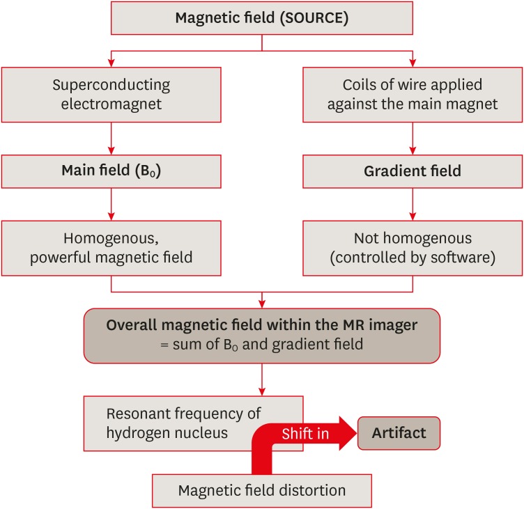
Magnetic resonance imaging (MRI) is an advanced diagnostic tool used in both medicine and dentistry. Since it functions based on a strong uniform static magnetic field and radiofrequency pulses, it is advantageous over imaging techniques that rely on ionizing radiation. Unfortunately, the magnetic field and radiofrequency pulses generated within the magnetic resonance imager interact unfavorably with dental materials that have magnetic properties. This leads to unwanted effects such as artifact formation, heat generation, and mechanical displacement. These are a potential source of damage to the oral tissue surrounding the affected dental materials. This review aims to compile, based on the current available evidence, recommendations for dentists and radiologists regarding the safety and appropriate management of dental materials during MRI in patients with orthodontic appliances, maxillofacial prostheses, dental implants, direct and indirect restorative materials, and endodontic materials.
-
Citations
Citations to this article as recorded by- Postoperative MRI in cranio-maxillofacial and oral reconstruction: A prospective comparative pilot study on artifact reduction
Adib Al-Haj Husain, Sameena Sandhu, Maximilian Eberhard Hermann Wagner, Suen An Nynke Lie, Egon Burian, Daniel Zedler, Bernd Stadlinger, Peter Kessler, Harald Essig
Journal of Cranio-Maxillofacial Surgery.2026; 54(6): 104522. CrossRef - Artifacts in magnetic resonance imaging of the head and neck: Unwanted effects caused by implant-supported restorations fabricated with different alloys
Lauren Bohner, Dieter Dirksen, Marcel Hanisch, Newton Sesma, Johannes Kleinheinz, Norbert Meier
The Journal of Prosthetic Dentistry.2025; 133(6): 1574. CrossRef - The influence of preformed metal crowns versus zirconia crowns on the diagnostic quality of magnetic resonance images
O. Dalzell, P. Haghighi, J. Ho, T. Rayner, L. Vidarsson, G. A. Garisto
European Archives of Paediatric Dentistry.2025; 26(1): 109. CrossRef - Interference of titanium and zirconia implants on dental-dedicated MR image quality: ex vivo and in vivo assessment
Katrine M Johannsen, Jennifer Christensen, Louise Hauge Matzen, Brian Hansen, Rubens Spin-Neto
Dentomaxillofacial Radiology.2025; 54(2): 132. CrossRef - Accuracy of Ionizing‐Radiation‐Based and Non‐Ionizing Imaging Assessments for the Diagnosis of Periodontitis: Systematic Review and Meta‐Analysis
Nicola Discepoli, Isabella De Rubertis, Cecile Wasielewski, Giuseppe Troiano, Maria Clotilde Carra
Journal of Clinical Periodontology.2025; 52(S29): 74. CrossRef - The Effect of MRI Exposure on the Shear Bond Strength and Adhesive Remnant Index of Different Bracket Types
Luka Šimunović, Jakov Stojanović, Katarina Tečić, Dijana Zadravec, Senka Meštrović
Dentistry Journal.2025; 13(3): 108. CrossRef - Impact of Artifacts Caused by Intraoral Dental Materials in Magnetic Resonance Imaging
Divya Josephraj, Ravindranath Vineetha, Priya Pattath Sankaran, Prakashini Koteshwara, Mathangi Kumar, Kalyana Chakravarthy Pentapati
Pesquisa Brasileira em Odontopediatria e Clínica Integrada.2025;[Epub] CrossRef - Orthodontic appliances and their diagnostic impact to brain MRI
Lisa Latzko, Anna Schmit, Bernhard Glodny, Astrid E. Grams, Christoph Birkl, Adriano G. Crismani
Clinical Oral Investigations.2025;[Epub] CrossRef - Impact of Intra-Oral Dental Materials on Magnetic Resonance Imaging: A Perspective Survey from Dental Professionals
Sejal Gupta, Mathangi Kumar, Kalyana C Pentapati, Ravindranath Vineetha, Vinu Thomas George, Nidambur Vasudev Ballal, Priya Pattath Sankaran
Journal of Pharmacy and Bioallied Sciences.2025; 17(Suppl 1): S551. CrossRef - Beyond radiation: Emerging applications of MRI in dental diagnostics and clinical practice
Gerta Halilaj, Nebi Cemeta
Journal of Dentistry and Multidisciplinary Sciences.2025; 1(1): 31. CrossRef - Nonionizing diagnostic imaging modalities for visualizing health and pathology of periodontal and peri‐implant tissues
Andy Wai Kan Yeung, Abeer AlHadidi, Rutvi Vyas, Michael M. Bornstein, Hiroshi Watanabe, Ray Tanaka
Periodontology 2000.2024; 95(1): 87. CrossRef - Cortical thickness and grey-matter volume anomaly detection in individual MRI scans: Comparison of two methods
David Romascano, Michael Rebsamen, Piotr Radojewski, Timo Blattner, Richard McKinley, Roland Wiest, Christian Rummel
NeuroImage: Clinical.2024; 43: 103624. CrossRef - Association between dental restorations and artefacts on head magnetic resonance images in paediatric patients
Pitchaya Tunlayadechanont, Padcha Tunlayadechanont, Nantana Sriudomporn, Ploy Wisetsathon, Duangporn Duangthip, Varangkanar Jirarattanasopha
International Journal of Paediatric Dentistry.2024; 34(5): 546. CrossRef - Commercially Pure Titanium Implants With Selenium and Hyaluronic Acid Coating for Dental Applications
Soorya Ganesh, Gheena S, Kalaiyarasan Madhu
Cureus.2024;[Epub] CrossRef - Multibraided Fixed Retainers with Different Diameters after Magnetic Resonance Imaging (MRI): In Vitro Study Investigating Temperature Changes and Bonding Efficacy
Maria Francesca Sfondrini, Maurizio Pascadopoli, Paola Gandini, Lorenzo Preda, Domenico Sfondrini, Karin Bertino, Cinzia Rizzi, Andrea Scribante
Dentistry Journal.2024; 12(8): 255. CrossRef - Chronic non-bacterial osteomyelitis of the mandible – orthodontic considerations and management: A case report
Saskia Andrea Schwabe, Sean Booth, Susi Caldwell
Journal of Orthodontics.2024; 51(4): 415. CrossRef - Magnetic resonance imaging in the diagnosis of periodontal and periapical disease
Katrine Mølgaard Johannsen, João Marcus de Carvalho E Silva Fuglsig, Louise Hauge Matzen, Jennifer Christensen, Rubens Spin-Neto
Dentomaxillofacial Radiology.2023;[Epub] CrossRef - Surveillance of head neck cancer: Case for personalized and standardized surveillance
Shrikant B. Mali
Oral Oncology.2023; 139: 106354. CrossRef - Effect of Magnetic Resonance Imaging at 1.5 T and 3 T on Temperature and Bond Strength of Orthodontic Bands with Welded Tubes: An In Vitro Study
Maria Francesca Sfondrini, Simone Gallo, Maurizio Pascadopoli, Cinzia Rizzi, Andrea Boldrini, Simone Santagostini, Luca Anemoni, Maria Sole Prevedoni Gorone, Lorenzo Preda, Paola Gandini, Andrea Scribante
Materials.2023; 16(2): 651. CrossRef - Magnetic resonance imaging artefacts caused by orthodontic appliances and/or implant-supported prosthesis: a systematic review
Katrine Mølgaard Johannsen, João Marcus de Carvalho E Silva Fuglsig, Brian Hansen, Ann Wenzel, Rubens Spin-Neto
Oral Radiology.2023; 39(2): 394. CrossRef - Magnetic resonance imaging investigations in patients with metallic dental prosthesis: “The associated dilemma for medical fraternity and the dentist's role”
Ritika Bhambhani, SantanuSen Roy, Shubha Joshi
The Journal of Indian Prosthodontic Society.2023; 23(2): 203. CrossRef - Recent advances in the application and biological mechanism of silicon nitride osteogenic properties: a review
Ziyi Liu, Ruijie Wang, Wenjing Liu, Yushan Liu, Xiaoli Feng, Fujian Zhao, Pei Chen, Longquan Shao, Mingdeng Rong
Biomaterials Science.2023; 11(21): 7003. CrossRef - Techniques, Tricks, and Stratagems of Oral Cavity Computed Tomography and Magnetic Resonance Imaging
Davide Maraghelli, Michele Pietragalla, Linda Calistri, Luigi Barbato, Luca Giovanni Locatello, Martina Orlandi, Nicholas Landini, Antonio Lo Casto, Cosimo Nardi
Applied Sciences.2022; 12(3): 1473. CrossRef - GEÇICI VE DAIMI SIMANLARIN DENTINE OLAN BAĞLANMA DAYANIMI ÜZERINE MANYETIK REZONANS GÖRÜNTÜLEME İŞLEMININ ETKISININ ARAŞTIRILMASI
Melih ÜLGEY, Oğuzhan GÖRLER, İsmail ŞALK, Derya ÖZDEMİR DOĞAN
Atatürk Üniversitesi Diş Hekimliği Fakültesi Dergisi.2022; : 1. CrossRef - Performance of PROPELLER FSE T2WI in reducing metal artifacts of material porcelain fused to metal crown: a clinical preliminary study
Wenjin Li, Jing Shi, Wenjin Bian, Jianting Li, Xiaoqing Chen, Juan Feng, Jiali Yu, Jun Wang, Jinliang Niu
Scientific Reports.2022;[Epub] CrossRef - Tracking the Molecular Fingerprint of Head and Neck Cancer for Recurrence Detection in Liquid Biopsies
Araceli Diez-Fraile, Joke De Ceulaer, Charlotte Derpoorter, Christophe Spaas, Tom De Backer, Philippe Lamoral, Johan Abeloos, Tim Lammens
International Journal of Molecular Sciences.2022; 23(5): 2403. CrossRef - Review on Biocompatibility and Prospect Biomedical Applications of Novel Functional Metallic Glasses
Michał Biały, Mariusz Hasiak, Amadeusz Łaszcz
Journal of Functional Biomaterials.2022; 13(4): 245. CrossRef - MRI compatibility of orthodontic brackets and wires: systematic review article
Adrienn Dobai, Fanni Dembrovszky, Tamás Vízkelety, Péter Barsi, Fanni Juhász, Csaba Dobó-Nagy
BMC Oral Health.2022;[Epub] CrossRef - The interaction and interference of preformed metal crowns on magnetic resonance imaging: a scoping review with a systematic methodology
O. Sumner, R. Goldsmith, N. Heath, G. D. Taylor
European Archives of Paediatric Dentistry.2021; 22(6): 1023. CrossRef - An Evidence-based Protocol for the Management of Orthodontic Patients Undergoing MRI Scans
Rachael Shivam, Sheelagh Rogers, Nicholas Drage
Orthodontic Update.2021; 14(1): 32. CrossRef - Reversal of Osseointegration as a Novel Perspective for the Removal of Failed Dental Implants: A Review of Five Patented Methods
Rolf G. Winnen, Kristian Kniha, Ali Modabber, Faruk Al-Sibai, Andreas Braun, Reinhold Kneer, Frank Hölzle
Materials.2021; 14(24): 7829. CrossRef - Magnetic resonance imaging as a diagnostic tool for periodontal disease: A prospective study with correlation to standard clinical findings—Is there added value?
Monika Probst, Egon Burian, Teresa Robl, Dominik Weidlich, Dimitrios Karampinos, Teresa Brunner, Claus Zimmer, Florian Andreas Probst, Matthias Folwaczny
Journal of Clinical Periodontology.2021; 48(7): 929. CrossRef - An Update of the Possible Applications of Magnetic Resonance Imaging (MRI) in Dentistry: A Literature Review
Rodolfo Reda, Alessio Zanza, Alessandro Mazzoni, Andrea Cicconetti, Luca Testarelli, Dario Di Nardo
Journal of Imaging.2021; 7(5): 75. CrossRef - Implant-supported overdentures: part 1
David Gray, Jaymit Patel
British Dental Journal.2021; 231(2): 94. CrossRef - Oral and dental considerations in pediatric cancers
Priyanshi Ritwik, Tammuella E. Chrisentery-Singleton
Cancer and Metastasis Reviews.2020; 39(1): 43. CrossRef - Recent advances in bioelectronics chemistry
Yin Fang, Lingyuan Meng, Aleksander Prominski, Erik N. Schaumann, Matthew Seebald, Bozhi Tian
Chemical Society Reviews.2020; 49(22): 7978. CrossRef - Imaging of root canal treatment using ultra high field 9.4T UTE-MRI – a preliminary study
Maximilian Timme, Max Masthoff, Nina Nagelmann, Malte Masthoff, Cornelius Faber, Sebastian Bürklein
Dentomaxillofacial Radiology.2020; 49(1): 20190183. CrossRef - Magnetic resonance imaging based computer‐guided dental implant surgery—A clinical pilot study
Florian Andreas Probst, Josef Schweiger, Maria Juliane Stumbaum, Dimitrios Karampinos, Egon Burian, Monika Probst
Clinical Implant Dentistry and Related Research.2020; 22(5): 612. CrossRef - Magnetic resonance imaging artifacts produced by dental implants with different geometries
Lauren Bohner, Norbert Meier, Felix Gremse, Pedro Tortamano, Johannes Kleinheinz, Marcel Hanisch
Dentomaxillofacial Radiology.2020; 49(8): 20200121. CrossRef - Implications and Considerations of Dental Materials in MRI: A Case Report and Literature Review
Brenton J. Wilson, Phoebe E. O’hare, John Zacariah, Wen Lin Chai
Case Reports in Dentistry.2020;[Epub] CrossRef
- Postoperative MRI in cranio-maxillofacial and oral reconstruction: A prospective comparative pilot study on artifact reduction
- 11,039 View
- 79 Download
- 40 Crossref
Research Articles
- Push-out bond strength of a self-adhesive resin cement used as endodontic sealer
- Eduardo Diogo Gurgel-Filho, Felipe Coelho Lima, Vicente de Paula Aragão Saboia, Tauby de Souza Coutinho-Filho, Aline de Almeida Neves, Emmanuel João Nogueira Leal da Silva
- Restor Dent Endod 2014;39(4):282-287. Published online August 20, 2014
- DOI: https://doi.org/10.5395/rde.2014.39.4.282
-
Abstract
PDFPubReaderePub
Objectives The aim of the present study was to investigate the bond strength of RelyX Unicem (3M) to root canal dentin when used as an endodontic sealer.
Materials and Methods Samples of 24 single-rooted teeth were prepared with Gates Glidden drills and K3 files. After that, the roots were randomly assigned to three experimental groups (
n = 8) according to the filling material, (1) AH Plus (Dentsply De Trey GmbH)/Gutta-Percha cone; (2) Epiphany SE (Pentron)/Resilon cone; (3) RelyX Unicem/Gutta-Percha cone. All roots were filled using a single cone technique associated to vertical condensation. After the filling procedures, each tooth was prepared for a push-out bond strenght test by cutting 1 mm-thick root slices. Loading was performed on a universal testing machine at a speed of 0.5 mm/min. One-way analysis of variance and Tukey test for multiple comparisons were used to compare the results among the experimental groups.Results Epiphany SE/Resilon showed significantly lower push-out bond strength than both AH Plus/Gutta-Percha and RelyX Unicem/Gutta-Percha (
p < 0.05). There was no significant difference in bond strength between AH Plus/Gutta-Percha and RelyX Unicem/Gutta-Percha (p > 0.05).Conclusions Under the present
in vitro conditions, bond strength to root dentin promoted by RelyX Unicem was similar to AH Plus. Epiphany SE/Resilon resulted in lower bond strength values when compared to both materials.-
Citations
Citations to this article as recorded by- In vitro comparative evaluation of physicochemical and mechanical properties, cytocompatibility, and antimicrobial efficacy of various bioceramic root canal sealers
Fushi Wang, Jiaxing Li, Jingjing Wan, Siyuan Li, Shijia Tang, Li Wang, Liuyan Meng
Ceramics International.2026; 52(7): 9561. CrossRef - In-Vitro Comparative Adhesion Evaluation of Bioceramic and Dual-Cure Resin Endodontic Sealers Using SEM, AFM, Push-Out and FTIR
Radu Marcel Chisnoiu, Marioara Moldovan, Doina Prodan, Andrea Maria Chisnoiu, Dana Hrab, Ada Gabriela Delean, Alexandrina Muntean, Doina Iulia Rotaru, Ovidiu Pastrav, Mihaela Pastrav
Applied Sciences.2021; 11(10): 4454. CrossRef - Push-out Bond Strength of Fiber Posts Cemented Using New Universal Adhesives on Etched and Nonetched Intraradicular Dentin
Hani F Ounsi, Simone Grandini, Marco Ferrari, Valentina Spicciarelli, Giacomo Corsentino, Crystal Marruganti
The Journal of Contemporary Dental Practice.2020; 21(1): 91. CrossRef - Comparison of push-out bond strength of three different obturating systems to intraradicular dentin: An In vitro study
MohammedKhwaja Moinuddin, LKarthik Prasad, Nimeshika Ramachandruni, Shekar Kamishetty, RaviChandra Cherkupalli
Contemporary Clinical Dentistry.2019; 10(4): 631. CrossRef - The influence of methodological variables on the push‐out resistance to dislodgement of root filling materials: a meta‐regression analysis
F. M. Collares, F. F. Portella, S. B. Rodrigues, R. K. Celeste, V. C. B. Leitune, S. M. W. Samuel
International Endodontic Journal.2016; 49(9): 836. CrossRef - Effect of photon induced photoacoustic streaming (PIPS) on bond strength to dentine of two root canal filling materials
Ivana Miletić, Nicoletta Chieffi, Carlo Rengo, Marco Ferrari, Dan Nathanson, Anja Baraba
Lasers in Surgery and Medicine.2016; 48(10): 951. CrossRef
- In vitro comparative evaluation of physicochemical and mechanical properties, cytocompatibility, and antimicrobial efficacy of various bioceramic root canal sealers
- 2,451 View
- 6 Download
- 6 Crossref
- Comparative analysis of physicochemical properties of root perforation sealer materials
- Maura Cristiane Gonçales Orçati Dorileo, Fábio Luis Miranda Pedro, Matheus Coelho Bandeca, Orlando Aguirre Guedes, Ricardo Dalla Villa, Alvaro Henrique Borges
- Restor Dent Endod 2014;39(3):201-209. Published online June 30, 2014
- DOI: https://doi.org/10.5395/rde.2014.39.3.201
-
Abstract
PDFPubReaderePub
Objectives This study evaluated the solubility, dimensional alteration, pH, electrical conductivity, and radiopacity of root perforation sealer materials.
Materials and Methods For the pH test, the samples were immersed in distilled water for different periods of time. Then, the samples were retained in plastic recipients, and the electrical conductivity of the solution was measured. The solubility, dimensional alteration, and radiopacity properties were evaluated according to Specification No. 57 of the American National Standards Institute/American Dental Association (ANSI/ADA). Statistical analyses were carried out using analysis of variance (ANOVA) and Tukey's test at a significance level of 5%. When the sample distribution was not normal, a nonparametric ANOVA was performed with a Kruskal-Wallis test (α = 0.05).
Results The results showed that white structural Portland cement (PC) had the highest solubility, while mineral trioxide aggregate (MTA)-based cements, ProRoot MTA (Dentsply-Tulsa Dental) and MTA BIO (Ângelus Ind. Prod.), had the lowest values. MTA BIO showed the lowest dimensional alteration values and white PC presented the highest values. No differences among the tested materials were observed in the the pH and electrical conductivity analyses. Only the MTA-based cements met the ANSI/ADA recommendations regarding radiopacity, overcoming the three steps of the aluminum step wedge.
Conclusions On the basis of these results, we concluded that the values of solubility and dimensional alteration of the materials were in accordance with the ANSI/ADA specifications. PCs did not fulfill the ANSI/ADA requirements regarding radiopacity. No differences were observed among the materials with respect to the pH and electrical conductivity analyses.
-
Citations
Citations to this article as recorded by- Chemical and in vivo analyses of calcium silicate‐based materials in bone and connective tissues
Ana Cristina Padilha Janini, Lauter Eston Pelepenko, Brenda Fornazaro Moraes, Victor Augusto Benedicto dos Santos, Matheus Barros‐Costa, Isabela Alvarenga Maciel dos Santos, Fábio Roberto de Souza Batista, Juliana de Aguiar Silveira Meira, Mariza Akemi Ma
International Endodontic Journal.2025; 58(3): 484. CrossRef - A comparative study on solubility of root end filling materials - An in vitro study
Supriya Makam, Geeta Hiremath, Balaram Naik
Endodontology.2025; 37(2): 205. CrossRef - Physicochemical and antibacterial properties of ZnO/chitosan-modified mineral trioxide aggregate composites
Mariyam Mariyam, Siti Sunarintyas, Leny Yuliatun, Dyah Irnawati, Adhi Dwi Hatmanto, Nuryono Nuryono
Case Studies in Chemical and Environmental Engineering.2024; 9: 100749. CrossRef - Portland Cement: An Overview as a Root Repair Material
Shahriar Shahi, Elaheh Fakhri, Hamidreza Yavari, Solmaz Maleki Dizaj, Sara Salatin, Khadijeh Khezri, Victor Feitosa
BioMed Research International.2022;[Epub] CrossRef - A micro-computed tomographic study using a novel test model to assess the filling ability and volumetric changes of bioceramic root repair materials
Fernanda Ferrari Esteves Torres, Jader Camilo Pinto, Gabriella Oliveira Figueira, Juliane Maria Guerreiro-Tanomaru, Mario Tanomaru-Filho
Restorative Dentistry & Endodontics.2021;[Epub] CrossRef - COMPARISON OF MINERAL TRIOXIDE AGGREGATE, ENDOSEQUENCE ROOT REPAIR MATERIAL, AND BIODENTINE USED FOR REPAIRING ROOT PERFORATIONS: A SYSTEMATIC REVIEW
Faisal ALGHAMDİ, Esraa ALJAHDALİ
Cumhuriyet Dental Journal.2019; 22(4): 469. CrossRef - Effects of the addition of nanoparticulate calcium carbonate on setting time, dimensional change, compressive strength, solubility and pH of MTA
A. Bernardi, E. A. Bortoluzzi, W. T. Felippe, M. C. S. Felippe, W. S. Wan, C. S. Teixeira
International Endodontic Journal.2017; 50(1): 97. CrossRef - The use of a biocompatible cement in endodontic surgery. A randomized clinical trial 1
Sérgio Ribeiro da Silva, José Dias da Silva Neto, Taylor Brandão Schnaider, Daniela Francescato Veiga, Neil Ferreira Novo, Marcos Mesquita Filho, Lydia Masako Ferreira
Acta Cirurgica Brasileira.2016; 31(6): 422. CrossRef - Evaluation of Sealing Effect and Working Time of Root Canal Filling MTA Materials
Hyojin Kim, Youngjin Kim, Soonhyeun Nam, Kwon Taeyub, Hyunjung Kim
THE JOURNAL OF THE KOREAN ACADEMY OF PEDTATRIC DENTISTRY.2016; 43(2): 129. CrossRef - Portland cement versus MTA as a root-end filling material. A pilot study
Sérgio Ribeiro da Silva, José Dias da Silva Neto, Daniela Francescato Veiga, Taylor Brandão Schnaider, Lydia Masako Ferreira
Acta Cirurgica Brasileira.2015; 30(2): 160. CrossRef
- Chemical and in vivo analyses of calcium silicate‐based materials in bone and connective tissues
- 2,654 View
- 7 Download
- 10 Crossref
- Cytotoxicity of newly developed pozzolan cement and other root-end filling materials on human periodontal ligament cell
- Minju Song, Tae-Sun Yoon, Sue-Youn Kim, Euiseong Kim
- Restor Dent Endod 2014;39(1):39-44. Published online January 20, 2014
- DOI: https://doi.org/10.5395/rde.2014.39.1.39
-
Abstract
PDFPubReaderePub
Objectives The purpose of this study was to evaluate
in vitro cytotoxicity of the pozzolan cement and other root-end filling materials using human periodontal ligament cell.Materials and Methods Endocem (Maruchi), white ProRoot MTA (Dentsply), white Angelus MTA (Angelus), and Super EBA (Bosworth Co.) were tested after set completely in an incubator at 37℃ for 7 days, Endocem was tested in two ways: 1) immediately after mixing (fresh specimens) and 2) after setting completely like other experimental materials. The methods for assessment included light microscopic examination, cell counting and WST-1 assay on human periodontal ligament cell.
Results In the results of microscopic examination and cell counting, Super EBA showed significantly lower viable cell than any other groups (
p < 0.05). As the results of WST-1 assay, compared with untreated control group, there was no significant cell viability of the Endocem group. However, the fresh mixed Endocem group had significantly less cell viability. The cells exposed to ProRoot MTA and Angelus MTA showed the highest viability, whereas the cells exposed to Super EBA displayed the lowest viability (p < 0.05).Conclusions The cytotoxicity of the pozzolan cement (Endocem) was comparable with ProRoot MTA and Angelus MTA. Considering the difficult manipulation and long setting time of ProRoot MTA and Angelus MTA, Endocem can be used as the alternative of retrofilling material.
-
Citations
Citations to this article as recorded by- In Vitro Biocompatibility of Calcium Silicate-Based Materials for Retrograde Endodontic Treatment Under Different Setting Conditions
Kremena Markova, Neshka Manchorova-Veleva, Veselina Todorova, Lyubomir Vangelov, Desislava Petkova
Journal of Functional Biomaterials.2026; 17(3): 124. CrossRef - Effects of Three Retrograde Filling Materials on Production of Inflammatory Cytokines and Resorbing Mediators
Samaneh Arab, Marjan Bahraminasab, Masoumeh Motamedi, Jamshid Hadjati, Alaviye Vahid
Journal of Microbiota.2024;[Epub] CrossRef - Physicochemical Properties, Cytocompatibility, and Biocompatibility of a Bioactive Glass Based Retrograde Filling Material
Kazumasa Murata, Ayako Washio, Takahiko Morotomi, Thira Rojasawasthien, Shoichiro Kokabu, Chiaki Kitamura
Nanomaterials.2021; 11(7): 1828. CrossRef - Cell migration and osteo/odontogenesis stimulation of iRoot FS as a potential apical barrier material in apexification
Y. Liu, X. M. Liu, J. Bi, S. Yu, N. Yang, B. Song, X. Chen
International Endodontic Journal.2020; 53(4): 467. CrossRef - Biocompatibility of Biodentine™ ® with Periodontal Ligament Stem Cells: In Vitro Study
Duaa Abuarqoub, Nazneen Aslam, Hanan Jafar, Zakariya Abu Harfil, Abdalla Awidi
Dentistry Journal.2020; 8(1): 17. CrossRef - A micro-computed tomographic study of remaining filling materials of two bioceramic sealers and epoxy resin sealer after retreatment
KyungJae Kim, Da Vin Kim, Sin-Young Kim, SungEun Yang
Restorative Dentistry & Endodontics.2019;[Epub] CrossRef - Comparison of Gap Volume after Retrofilling Using 4 Different Filling Materials: Evaluation by Micro–computed Tomography
Sue Youn Kim, Hyeon-Cheol Kim, Su-Jung Shin, Euiseong Kim
Journal of Endodontics.2018; 44(4): 635. CrossRef - Anti-inflammatory and Mineralization Effects of ProRoot MTA and Endocem MTA in Studies of Human and Rat Dental Pulps In Vitro and In Vivo
Do-Hee Kim, Ji-Hyun Jang, Bin-Na Lee, Hoon-Sang Chang, In-Nam Hwang, Won-Mann Oh, Sun-Hun Kim, Kyung-San Min, Jeong-Tae Koh, Yun-Chan Hwang
Journal of Endodontics.2018; 44(10): 1534. CrossRef - Effects of Three Calcium Silicate Cements on Inflammatory Response and Mineralization-Inducing Potentials in a Dog Pulpotomy Model
Chung-Min Kang, Jiwon Hwang, Je Seon Song, Jae-Ho Lee, Hyung-Jun Choi, Yooseok Shin
Materials.2018; 11(6): 899. CrossRef - Cytocompatibility of Biodentine and iRoot FS with human periodontal ligament cells: an in vitro study
T. Luo, J. Liu, Y. Sun, Y. Shen, L. Zou
International Endodontic Journal.2018; 51(7): 779. CrossRef - Biological response of commercially available different tricalcium silicate‐based cements and pozzolan cement
Serhat Köseoğlu, Tuğba Pekbağr?yan?k, Ebru Kucukyilmaz, Mehmet Sağlam, Sukru Enhos, Ayşe Akgün
Microscopy Research and Technique.2017; 80(9): 994. CrossRef - Biological efficacy of two mineral trioxide aggregate (MTA)-based materials in a canine model of pulpotomy
Myeongyeon LEE, Chung-Min KANG, Je Seon SONG, Yooseok SHIN, Seunghye KIM, Seong-Oh KIM, Hyung-Jun CHOI
Dental Materials Journal.2017; 36(1): 41. CrossRef - Cytotoxicities and genotoxicities of cements based on calcium silicate and of dental formocresol
Hyunjung Ko, Youngdan Jeong, Miri Kim
Mutation Research/Genetic Toxicology and Environmental Mutagenesis.2017; 815: 28. CrossRef - Bioactive-glass in Endodontic Therapy and Associated Microsurgery
Andrea Corrado Profeta, Gian Marco Prucher
The Open Dentistry Journal.2017; 11(1): 164. CrossRef - A Randomized Controlled Study of Mineral Trioxide Aggregate and Super Ethoxybenzoic Acid as Root-end Filling Materials in Endodontic Microsurgery: Long-term Outcomes
Sunil Kim, Minju Song, Su-Jung Shin, Euiseong Kim
Journal of Endodontics.2016; 42(7): 997. CrossRef - Effects of two fast-setting calcium-silicate cements on cell viability and angiogenic factor release in human pulp-derived cells
Chooryung J. Chung, Euiseong Kim, Minju Song, Jeong-Won Park, Su-Jung Shin
Odontology.2016; 104(2): 143. CrossRef - Cytotoxicity and Initial Biocompatibility of Endodontic Biomaterials (MTA and Biodentine™) Used as Root-End Filling Materials
Diana María Escobar-García, Eva Aguirre-López, Verónica Méndez-González, Amaury Pozos-Guillén
BioMed Research International.2016; 2016: 1. CrossRef - In Vitro Cytotoxicity Evaluation of Three Root-End Filling Materials in Human Periodontal Ligament Fibroblasts
Hernán Coaguila-Llerena, Abraham Vaisberg, Zulema Velásquez-Huamán
Brazilian Dental Journal.2016; 27(2): 187. CrossRef - Dynamic intratubular biomineralization following root canal obturation with pozzolan‐based mineral trioxide aggregate sealer cement
Yeon‐Jee Yoo, Seung‐Ho Baek, Kee‐Yeon Kum, Won‐Jun Shon, Kyung‐Mi Woo, WooCheol Lee
Scanning.2016; 38(1): 50. CrossRef - Management of Idiopathic External Root Resorption: A case report
Yoorina Choi, Hyeon-Ha Kim, Su-Jung Park
The Korean Journal of Oral and Maxillofacial Pathology.2016; 40(5): 865. CrossRef - A Randomized Controlled Study of the Use of ProRoot Mineral Trioxide Aggregate and Endocem as Direct Pulp Capping Materials
Minju Song, Minji Kang, Hyeon-Cheol Kim, Euiseong Kim
Journal of Endodontics.2015; 41(1): 11. CrossRef - Comparative Analysis of Selected Physicochemical Properties of Pozzolan Portland and MTA-Based Cements
Maura Cristiane Gonçales Orçati Dorileo, Ricardo Dalla Villa, Orlando Aguirre Guedes, Andreza Maria Fábio Aranha, Alex Semenoff-Segundo, Matheus Coelho Bandeca, Alvaro Henrique Borges
International Scholarly Research Notices.2014; 2014: 1. CrossRef - Surgical endodontics: past, present, and future
James L. Gutmann
Endodontic Topics.2014; 30(1): 29. CrossRef - Comparative analysis of physicochemical properties of root perforation sealer materials
Maura Cristiane Gonçales Orçati Dorileo, Fábio Luis Miranda Pedro, Matheus Coelho Bandeca, Orlando Aguirre Guedes, Ricardo Dalla Villa, Alvaro Henrique Borges
Restorative Dentistry & Endodontics.2014; 39(3): 201. CrossRef
- In Vitro Biocompatibility of Calcium Silicate-Based Materials for Retrograde Endodontic Treatment Under Different Setting Conditions
- 2,400 View
- 1 Download
- 24 Crossref
Case Report
- Use of temporary filling material for index fabrication in Class IV resin composite restoration
- Kun-Young Kim, Sun-Young Kim, Duck-Su Kim, Kyoung-Kyu Choi
- Restor Dent Endod 2013;38(2):85-89. Published online May 28, 2013
- DOI: https://doi.org/10.5395/rde.2013.38.2.85
-
Abstract
PDFPubReaderePub
When a patient with a fractured anterior tooth visits the clinic, clinician has to restore the tooth esthetically and quickly. For esthetic resin restoration, clinician can use 'Natural Layering technique' and an index for palatal wall may be needed. In this case report, we introduce pre-restoration index technique on a Class IV defect, in which a temporary filling material is used for easy restoration. Chair-side index fabrication for Class IV restoration is convenient and makes a single-visit treatment possible.
-
Citations
Citations to this article as recorded by- Combining a CAD-CAM composite resin palatal wall with a direct composite resin layering technique for the restoration of a large Class IV fracture: A clinical report
Jingjin Liu, Junling Zhang, Weicai Liu, Shanshan Liang
The Journal of Prosthetic Dentistry.2025; 134(5): 1359. CrossRef - A digital workflow for layering composite resin restorations by using 3-dimensionally printed templates to replicate the contralateral tooth accurately and rapidly
Junjing Zhang, Lin Fan, Chenyang Xie, Junying Li, Yuqiang Zhang, Haiyang Yu
The Journal of Prosthetic Dentistry.2024; 131(5): 774. CrossRef - Direct composite resin restoration of a class IV fracture by using 3D printing technology: A clinical report
Yi Gao, Jiyao Li, Bo Dong, Min Zhang
The Journal of Prosthetic Dentistry.2021; 125(4): 555. CrossRef - Esthetic rehabilitation of single anterior edentulous space using fiber-reinforced composite
Hyeon Kim, Min-Ju Song, Su-Jung Shin, Yoon Lee, Jeong-Won Park
Restorative Dentistry & Endodontics.2014; 39(3): 220. CrossRef
- Combining a CAD-CAM composite resin palatal wall with a direct composite resin layering technique for the restoration of a large Class IV fracture: A clinical report
- 2,497 View
- 11 Download
- 4 Crossref
Basic Research
- The effect of several root-end filling materials on MG63 osteoblast-like cells
- Jeong-Ho Lee, Won-Jun Shon, WooCheol Lee, Seung-Ho Baek
- J Korean Acad Conserv Dent 2010;35(3):222-228. Published online May 31, 2010
- DOI: https://doi.org/10.5395/JKACD.2010.35.3.222
-
Abstract
PDFPubReaderePub
The purpose of this study was to compare mineral trioxide aggregate (MTA; Dentsply, Tulsa Dental, Tulsa, OK, USA), which is widely used as root-end filling material, with DiaRoot BioAggregate (DB; Innovative BioCaramix Inc, Vancouver, BC, Canada), newly developed product, by using MG63 osteoblast-like cells. MTA, DB, and Intermediate Restorative Material (IRM; Dentsply Caulk, Milford, DE, USA) were used for root-end filling material while tissue culture plastic was used for control group. Each material was mixed and, the mixtures were left to set for 24 hours. MG63 cells were seeded to each group and then they were cultured for attachment for 4 hours. Following the attachment of cells to the root-end filling material, early cellular response was observed. After another 12 hours'culture, the level of attachment between cells and material was observed and in order to identify the effect of each material to bone formation, transforming growth factor beta1 (TGFβ1) and osteocalin (OC) were estimated by using enzyme-linked immunosorbent assay (ELISA), and the amount of alkaline phosphatase (ALP) was also measured. The data were analyzed using one-way ANOVA. As a result, only at OC and the number of cells which were attached to materials, there was no statistical difference between MTA and DB. At other items, there was statistically significant difference in all groups. Although DB has not shown exactly the same cellular response like that of MTA, the number of attached cells shows that biocompatibility of the material and OC indicates bone formation rate. Therefore, if DB is used for root end filling material, it is expected to lead to similar results to MTA.
-
Citations
Citations to this article as recorded by- Comparative analysis of physicochemical properties of root perforation sealer materials
Maura Cristiane Gonçales Orçati Dorileo, Fábio Luis Miranda Pedro, Matheus Coelho Bandeca, Orlando Aguirre Guedes, Ricardo Dalla Villa, Alvaro Henrique Borges
Restorative Dentistry & Endodontics.2014; 39(3): 201. CrossRef - Biocompatibility of root-end filling materials: recent update
Payal Saxena, Saurabh Kumar Gupta, Vilas Newaskar
Restorative Dentistry & Endodontics.2013; 38(3): 119. CrossRef - Bone regeneration in a periodontally challenged hopeless tooth
Jammula Surya Prasanna, Parupalli Karunakar, Dasari Rajashree, Raji V. Solomon
Journal of Dr. NTR University of Health Sciences.2013; 2(4): 296. CrossRef
- Comparative analysis of physicochemical properties of root perforation sealer materials
- 2,127 View
- 3 Download
- 3 Crossref
Original Articles
- Apical microleakage of MTA with 4-META/MMA & TBB resin as a root-end filling material
- Jin-Cheol Kim, Mi-Ri Kim, Hyun-Jung Ko, Won-Kyung Yang
- J Korean Acad Conserv Dent 2009;34(4):371-376. Published online July 31, 2009
- DOI: https://doi.org/10.5395/JKACD.2009.34.4.371
-
Abstract
PDFPubReaderePub
We evaluated
in vitro microleakage of Mineral Trioxide Aggregate (MTA) powder with 4-methacryloxyethyl trimellitate anhydride (4-META) / methyl methacrylate (MMA) & tri-n-butylborane (TBB) resin as a retrograde filling material by using methylene blue dye method.Fifty-two single rooted, extracted teeth were instrumented and obturated with gutta percha and AH plus sealer. The apical 3mm of each root was resected and 3mm deep ultrasonic root end preparation was done. External surface of roots was coated with nail varnish. Prepared teeth were randomly divided into five groups; Negative control: completely covered with nail varnish; Positive control: coated with nail varnish except for apical foramen; Group 1 (retrofilled with Portland cement); Group 2 (retrofilled with MTA); Group 3 (retrofilled with MTA powder mixed with 4-META/MMA & TBB resin). Immediately after completion of root-end filling, all specimens were submerged in methylene blue dye for 72 hours in 37℃ incubator. The roots were longitudinally sectioned and measured for extent of dye penetration by three different examiners under microscope (×10). The results were statistically analyzed using one way ANOVA and Turkey's HSD test. No leakage was evident in negative control and complete leakage in positive control group. Group 3 showed significantly less leakage than group 1 and 2 (p < 0.01). There was no significant difference between group 1 and 2 (p > 0.01).
It was concluded that MTA powder with 4-META/MMA & TBB resin was excellent in reducing initial apical microleakage.
-
Citations
Citations to this article as recorded by- Characteristics of novel root-end filling material using epoxy resin and Portland cement
Sang-Jin Lee, Jin Chung, Hee-Sam Na, Eun-Joo Park, Hyo-Jin Jeon, Hyeon-Cheol Kim
Clinical Oral Investigations.2013; 17(3): 1009. CrossRef - Sealing Ability of Three Different Materials Used as Retrograde Filling
Ji-Hoon Park, Seung-Bok Kang, Yong-Hoon Choi, Ji-Hyun Bae
Journal of Korean Dental Science.2012; 5(2): 60. CrossRef - Physical properties of novel composite using Portland cement for retro-filling material
Sang-Jin Lee, Ok-In Cho, Jiwan Yum, Jeong-Kil Park, Bock Hur, Hyeon-Cheol Kim
Journal of Korean Academy of Conservative Dentistry.2010; 35(6): 445. CrossRef
- Characteristics of novel root-end filling material using epoxy resin and Portland cement
- 1,786 View
- 1 Download
- 3 Crossref
- Polymerization shrinkage, hygroscopic expansion and microleakage of resin-based temporary filling materials
- Nak Yeon Cho, In-Bog Lee
- J Korean Acad Conserv Dent 2008;33(2):115-124. Published online March 31, 2008
- DOI: https://doi.org/10.5395/JKACD.2008.33.2.115
-
Abstract
PDFPubReaderePub
The purpose of this study was to measure the polymerization shrinkage and hygroscopic expansion of resin-based temporary filling materials and to evaluate microleakage at the interface between the materials and cavity wall.
Five resin-based temporary filing materials were investigated: Fermit (Vivadent), Quicks (Dentkist), Provifil (Promedica), Spacer (Vericom), Clip (Voco). Caviton (GC) was also included for comparison. Polymerization shrinkage of five resin-based temporary filling materials was measured using the bonded disc method. For the measurement of hygroscopic expansion, the discs of six cured temporary filling materials were immersed in saline and a LVDT displacement sensor was used to measure the expansion for 7 days. For estimating of microleakage, Class I cavities were prepared on 120 extracted human molars and randomly assigned to 6 groups of 20 each. The cavities in each group were filled with six temporary filling materials. All specimens were submitted to 1000 thermo-cycles, with temperature varying from 5℃/55℃. Microleakage was determined using a dye penetration test.
The results were as follows:
Fermit had significantly less polymerization shrinkage than the other resin-based temporary filling materials. Fermit (0.22 %) < Spacer (0.38 %) < Quicks (0.64 %), Provifil (0.67 %), Clip (0.67 %)
Resin-based temporary filling materials showed 0.43 - 1.1 % expansion in 7 days.
Fermit showed the greatest leakage, while Quicks exhibited the least leakage.
There are no correlation between polymerization shrinkage or hygroscopic expansion and microleakage of resin-based temporary filling materials.
-
Citations
Citations to this article as recorded by- Comparison of color stability, gloss, mechanical and physical properties according to dental temporary filling materials type
Ji-Won Choi, You-Young Shin, Song-Yi Yang
Korean Journal of Dental Materials.2022; 49(3): 97. CrossRef - Comparative analysis of strain according to two wavelengths of light source and constant temperature bath deposition in ultraviolet-curing resin for dental three-dimensional printing
Dong-Yeon Kim, Gwang-Young Lee, Hoo-Won Kang, Cheon-Seung Yang
Journal of Korean Acedemy of Dental Technology.2020; 42(3): 208. CrossRef - Effect of cavity disinfectants on antibacterial activity and microtensile bond strength in class I cavity
Bo-Ram KIM, Man-Hwan OH, Dong-Hoon SHIN
Dental Materials Journal.2017; 36(3): 368. CrossRef - Shear bond strength of a self-adhesive resin cement to resin-coated dentin
Jee-Youn Hong, Cheol-Woo Park, Jeong-Uk Heo, Min-Ki Bang, Jae-Jun Ryu
The Journal of Korean Academy of Prosthodontics.2013; 51(1): 27. CrossRef - Coronal microleakage of four temporary restorative materials in Class II-type endodontic access preparations
Sang-Mi Yun, Lorena Karanxha, Hee-Jin Kim, Sung-Ho Jung, Su-Jung Park, Kyung-San Min
Restorative Dentistry & Endodontics.2012; 37(1): 29. CrossRef - Microtensile bond strength of resin inlay bonded to dentin treated with various temporary filling materials
Tae-Woo Kim, Bin-Na Lee, Young-Jung Choi, So-Young Yang, Hoon-Sang Chang, Yun-Chan Hwang, In-Nam Hwang, Won-Mann Oh
Journal of Korean Academy of Conservative Dentistry.2011; 36(5): 419. CrossRef - The Effect of Temporary Filling Materials on The Adhesion between Dentin Adhesive-coated Surface and Resin Inlay
Tae-Gun Kim, Kwang-Won Lee, Mi-Kyung Yu
Journal of Korean Academy of Conservative Dentistry.2008; 33(6): 553. CrossRef
- Comparison of color stability, gloss, mechanical and physical properties according to dental temporary filling materials type
- 2,065 View
- 13 Download
- 7 Crossref
- A comparative study on radiopacity of canal filling and retrograde root-end filling materials
- Yong-Sang Kim, Seo-Kyong Kim, Yun-Chan Hwang, In-Nam Hwang, Won-Mann Oh
- J Korean Acad Conserv Dent 2008;33(2):107-114. Published online March 31, 2008
- DOI: https://doi.org/10.5395/JKACD.2008.33.2.107
-
Abstract
PDFPubReaderePub
This study was performed to assess the radiopacity of a variety of canal filling and retrograde root-end filling materials according to the specification concerning root canal obturation materials.
Ten materials including Gutta-percha pellets, amalgam, Fuji II LC, Dyract® AP, Super EBA®, IRM®, AH 26®, Sealapex™, Tubli-Seal™ and dentin were evaluated in this study. In the first part, densitometric reading of an each step of aluminum step wedge on occlusal film were performed at 60 kVp (0.2, 0.3, 0.4 s), 70 kVp (0.2, 0.3, 0.33 s) to decide appropriate voltage and exposure time. In the second part, ten specimens which are 5 mm in diameter and 0.5, 1.0, 1.5, 2.0, 2.5, 3.0 mm in thickness, were fabricated from each material studied. The specimens were radiographed simultaneously with an aluminum step wedge under decided condition (60 kVp, 0.2 s). The mean radiographic density values of the materials were transformed into radiopacity expressed equivalent thickness of aluminum (mm Al).
The following results were obtained.
Among the various conditions including 0.2 s, 0.3 s, 0.4 s at 60 kVp and 0.2 s, 0.3 s, 0.33 s at 70 kVp, the appropriate voltage and exposure time that meet the requirement of density from 0.5 to 2.0 was 0.2 s at 60 kVp.
All of the materials in this study had greater radiopacity than the minimun level recommended by ISO No. 4049 standards.
Most of the materials had greater radiopacity than 3 mm Al requirement of ANSI/ADA specification No. 57 (2000) and ISO No. 6876 (2001) standards except for Fuji II LC and Dyract.
It suggests that all experimental canal filling and retrograde root-end filling materials have a sufficient radiopacity that meet the requirement concerning root canal obturation materials except for Fuji II LC and Dyract.
-
Citations
Citations to this article as recorded by- Evaluation of prognosis related to compliance with supportive periodontal treatment in patients with chronic periodontitis: a clinical retrospective study
Jong-Bin Lee, Hye-Jung Shin, Dae-Yeob Kim, Eun-Kyoung Pang
Journal of Periodontal & Implant Science.2019; 49(2): 76. CrossRef
- Evaluation of prognosis related to compliance with supportive periodontal treatment in patients with chronic periodontitis: a clinical retrospective study
- 1,852 View
- 0 Download
- 1 Crossref
- An electrochemical study of the sealing ability of three retrofilling materials
- Dong-Sung Park, Suh-Jin Sohn, Tae-Seok Oh, Hyun-Mi Yoo, Chan-Je Park, Soon-Ho Yim, Young-Kyoo Lee, Seung-Bum Kye
- J Korean Acad Conserv Dent 2004;29(4):365-369. Published online July 31, 2004
- DOI: https://doi.org/10.5395/JKACD.2004.29.4.365
-
Abstract
PDFPubReaderePub
The purpose of this study was to evaluate the apical sealing ability of Super-EBA, MTA and Dyract-flow as retrofilling materials. Forty-eight extracted human teeth with straight and single root canal were used in this study. The root canals were prepared to a #40 apical canal size and obturated with gutter-percha. Apicoectomies were performed and root end cavities were prepared to a depth of 3mm using an ultrasonic device. The root end cavities were filled with Super-EBA, MTA or Dyract-flow. Leakage was measured using an electrochemical technique for 4 weeks.
According to this study, the results were as follows.
1. Increasing leakage with time was observed in all groups.
2. No significant difference was noted among the 3 groups with time (p = 0.216).
3. No significant difference was noted among the 3 groups when measured within the same time interval (p = 0.814).
The results of this study suggest that the sealing ability of Dyract-flow is equal to that of Super-EBA and MTA, and Dyract-flow may be an alternative to other materials for root-end filling.
-
Citations
Citations to this article as recorded by- Evaluation of Sealing Effect and Working Time of Root Canal Filling MTA Materials
Hyojin Kim, Youngjin Kim, Soonhyeun Nam, Kwon Taeyub, Hyunjung Kim
THE JOURNAL OF THE KOREAN ACADEMY OF PEDTATRIC DENTISTRY.2016; 43(2): 129. CrossRef - Comparative analysis of physicochemical properties of root perforation sealer materials
Maura Cristiane Gonçales Orçati Dorileo, Fábio Luis Miranda Pedro, Matheus Coelho Bandeca, Orlando Aguirre Guedes, Ricardo Dalla Villa, Alvaro Henrique Borges
Restorative Dentistry & Endodontics.2014; 39(3): 201. CrossRef
- Evaluation of Sealing Effect and Working Time of Root Canal Filling MTA Materials
- 2,168 View
- 1 Download
- 2 Crossref
- Spectrophotometric evaluation of sealing effects of several root-end filling materials
- Jin-Gyu Yi, Sang-Jin Park, Kyung-Kyu Choi, Gi-Woon Choi
- J Korean Acad Conserv Dent 2003;28(6):449-456. Published online November 30, 2003
- DOI: https://doi.org/10.5395/JKACD.2003.28.6.449
-
Abstract
PDFPubReaderePub
The purpose of this study is to evaluate the sealing effect of several root-end filling materials using spectrophotometric analysis. 180 single root teeth with one canal were instrumented and canal filled. Root resected and root end preparation was made. Teeth were randomly classified to 5 experimental group(MTA, EBA, IRM, TCP, ZOE) and 1 control group according to root-end filling material MTA group used PRO ROOT MTA, EBA group used Super EBA, TCP group used NEW APATITE LINER TYPE II main component of which is α-tricalcium phosphate(TCP). According to manufacture's instruction experimental material was mixed and retrfilled. After 2% methylene blue solution penetration absorbance for each test sample was measured with spectrophotometer (JASCO UV-530, Japan).
The mean absorbance of control and experimental group was as follows;
MTA : 0.092, IRM : 0.226, Super EBA : 0.255, ZOE : 0.374, Control : 0.425, TCP : 0.501 and the result analyzed by Turkey test at P=0.05 level.
Conclusions of this study are as follows;
The absorbance increase in follwing sequence MTA, IRM, Super EBA, ZOE, Control, TCP.
MTA showed the least leakage but was not significant with IRM or Super EBA and was significant with control or TCP(p<0.05).
TCP had the most leakage and was not significant with control group.
- 1,340 View
- 4 Download
 First
First Prev
Prev

