
Search
- Page Path
- HOME > Search
Research Articles
- Effect of high irradiance and short exposure curing time on the fracture toughness of bulk-fill resin-based composite: an in vitro study
- Beatriz Ometto Sahadi, Tainah Oliveira Rifane, Carolina Bosso André, Vitaliano Gomes Araújo-Neto, Richard Thomas Bengt Price, Marcelo Giannini
- Restor Dent Endod 2026;51(2):e23. Published online April 20, 2026
- DOI: https://doi.org/10.5395/rde.2026.51.e23
-
 Abstract
 PDF
PDF PubReader
 ePub
ePub - Objectives
This study aimed to determine the effect of high irradiance and short exposure time on the fracture toughness of bulk-fill resin-based composites (RBCs).
Methods
Three RBCs were tested: Tetric PowerFill (TPF; Ivoclar Vivadent), Opus Bulk Fill APS (OBF; FGM Dental Group), and Filtek One Bulk Fill (FOB; Solventum). Sixty single-edge-notched disc specimens were prepared using a fracture toughness mold. Each group consisted of 20 samples, divided into two subgroups (n = 10). The RBCs were lightcured either for 3 seconds in high-irradiance mode (‘3s cure’) or for the manufacturer-recommended times (TPF, 10 seconds; OBF, 30 seconds; FOB, 20 seconds) in ‘high power’ mode using the Bluephase PowerCure (Ivoclar Vivadent). The peak spectral wavelength was measured using a spectrophotometer. Specimens were tested on a universal testing machine, and data were analyzed by two-way analysis of variance and Bonferroni test (α = 0.05).
Results
Radiant exposure values (J/cm²) were 9.5 for the 3-second mode and 12.4, 24.8, and 37.1 for 10, 20, and 30 seconds (high power mode), respectively. FOB (4.22 and 3.79 MPa∙m0.5 for 20 and 3 seconds) had the highest mean fracture toughness, while OBF showed the lowest (2.01 and 2.10 MPa∙m0.5 for 30 and 3 seconds). TPF produced intermediate results (2.72 and 2.70 MPa∙m0.5 for 10 and 3 seconds). Exposure time did not affect TPF and OBF, while the 3-second exposure significantly reduced the fracture toughness for FOB.
Conclusions
The RBCs tested had different fracture toughness values regardless of exposure time. High irradiance and short exposure can reduce fracture toughness depending on the RBC tested.
- 812 View
- 70 Download

- Enhancing antimicrobial properties of a resin-based material via incorporation of a powdered phytotherapeutic extract: an in vitro experimental study
- Rodolfo Xavier de Sousa-Lima, Maria Eduarda Lima do Nascimento Marinho, Janielly Cristina Costa da Silva, Moan Jéfter Fernandes Costa, Pedro Henrique Sette-de-Souza, Giana da Silveira Lima, Boniek Castillo Dutra Borges
- Restor Dent Endod 2026;51(1):e2. Published online January 20, 2026
- DOI: https://doi.org/10.5395/rde.2026.51.e2
-
Abstract
PDFPubReaderePub
- Objectives
This study aimed to evaluate the degree of conversion (DC), immediate enamel bond strength (IEBS), antimicrobial activity, and release of the active principle of a resin-based material (RBM) enriched with the powdered Schinopsis brasiliensis (Braúna) stem antibacterial extract.
Methods
The RBM was enriched with 0, 1.25, 2.5, 5, 10, and 20 wt% powdered Braúna extract. The DC (n = 7) was assessed using micro-Raman spectroscopy. The IEBS (n = 7) was determined through the microshear test until failure, and failure modes were examined under a stereomicroscope. The antimicrobial activity (n = 15) was assessed by quantifying colony-forming units, and the release of the active principle was determined using ultra-high-performance liquid chromatography. One-way analysis of variance/Tukey and Kruskal-Wallis/Dunn tests were utilized to analyze the data (p < 0.05).
Results
Materials with 10 wt% and 20 wt% extract showed the lowest DC statistically. However, for IEBS, there were no statistically significant differences among the different groups. All materials released the active principle, but only those with 20 wt% and 10 wt% extract could inhibit biofilm formation similarly to 0.12% chlorhexidine.
Conclusions
Adding powdered Braúna extract between 10 wt% and 20 wt% is a promising alternative to provide an antimicrobial function to RBMs.
- 2,292 View
- 233 Download
- Ex vivo comparative analysis of retrievability among four calcium silicate-based sealers for regaining apical patency
- Darian Shomali, Timothy Kirkpatrick, Sang Won Kwak, Hyeon-Cheol Kim, Ji Wook Jeong
- Restor Dent Endod 2026;51(1):e3. Published online January 14, 2026
- DOI: https://doi.org/10.5395/rde.2026.51.e3
-
Abstract
PDFPubReaderePub
- Objectives
Efficient retrievability is a key requirement for endodontic sealers. This study evaluated the retrievability of four different calcium silicate-based sealers (CSS).
Methods
A total of 153 single-rooted human teeth with straight canals were decoronated to a standardized working length of 12 mm. The canals were negotiated to working length using K files up to size 15/.02, followed by rotary instrumentation up to 35/.04, 2 mm short of working length. The teeth were randomly assigned to five groups: NeoSEALER Flo (NEO; Avalon Biomed), Ceraseal (CS; Meta Biomed), Endosequence BC Sealer (BC; Brasseler USA), AH Plus Bioceramic Sealer (AHB; Dentsply Sirona), and a negative control group. Sealer application and obturation with a 35/.04 gutta-percha cone were performed. After incubation at 37°C in 100% humidity for 7 days, retreatment was performed until apical patency was obtained, with retrievability assessed by regaining apical patency. One-way analysis of variance and Tukey contrast test were used to determine whether there was a significant difference among the four different CSS (p < 0.05).
Results
Success rates in regaining apical patency were NEO (79.4%), CS (37.0%), BC (50.0%), and AHB (69.7%). NEO demonstrated the highest retrievability, while CS had the lowest (p < 0.01).
Conclusions
The type of CSS used has a considerable impact on retreatment difficulty. Among the tested sealers, Neo- SEALER Flo showed the highest retrievability, making it the most retrievable CSS in terms of retreatment efficacy.
- 2,185 View
- 180 Download
- Nanoleakage of apical sealing using a calcium silicate-based sealer according to canal drying methods
- Yoon-Joo Lee, Kyung-Mo Cho, Se-Hee Park, Yoon Lee, Jin-Woo Kim
- Restor Dent Endod 2024;49(2):e20. Published online April 19, 2024
- DOI: https://doi.org/10.5395/rde.2024.49.e20
-
Abstract
PDFPubReaderePub
Objectives This study investigated the nanoleakage of root canal obturations using calcium silicate-based sealer according to different drying methods.
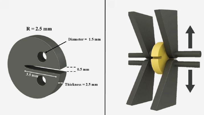
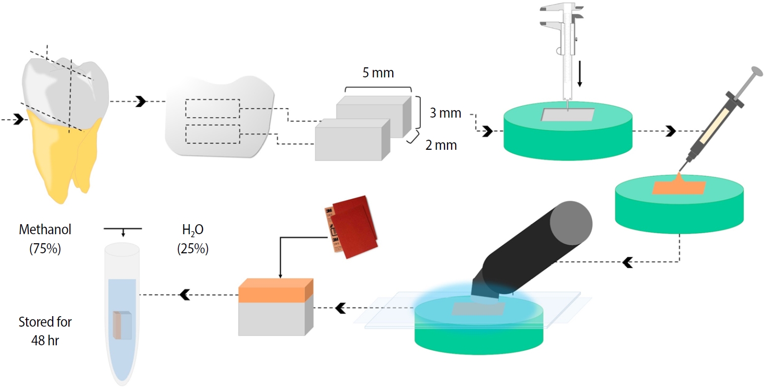
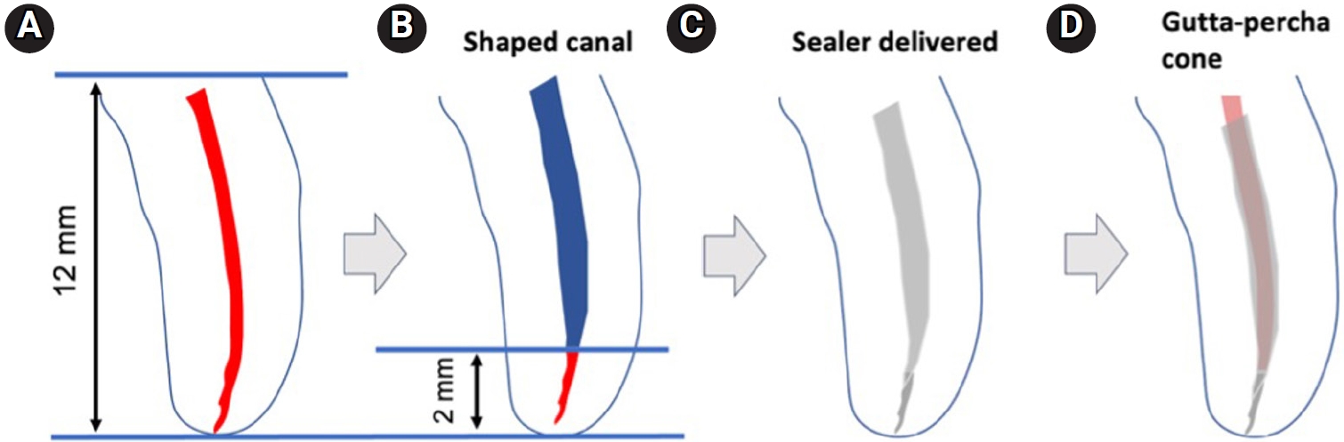
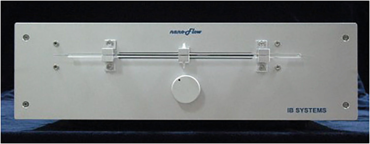
Materials and Methods Fifty-two extracted mandibular premolars with a single root canal and straight root were selected for this study. After canal preparation with a nickel-titanium rotary file system, the specimens were randomly divided into 4 groups according to canal drying methods (1: complete drying, 2: blot drying/distilled water, 3: blot drying/NaOCl, 4: aspiration only). The root canals were obturated using a single-cone filling technique with a calcium silicate–based sealer. Nanoleakage was evaluated using a nanoflow device after 24 hours, 1 week, and 1 month. Data were collected twice per second at the nanoscale and measured in nanoliters per second. Data were statistically analyzed using the Kruskal-Wallis and Mann–Whitney
U -tests (p < 0.05).Results The mean flow rate measured after 24 hours showed the highest value among the time periods in all groups. However, the difference in the flow rate between 1 week and 1 month was not significant. The mean flow rate of the complete drying group was the highest at all time points. After 1 month, the mean flow rate in the blot drying group and the aspiration group was not significantly different.
Conclusions Within the limitations of this study, the canal drying method had a significant effect on leakage and sealing ability in root canal obturations using a calcium silicate-based sealer. Thus, a proper drying procedure is critical in endodontic treatment.
-
Citations
Citations to this article as recorded by
- Effects of interactions between sealers and irrigants on the physicochemical and surface characteristics of endodontic sealers
Hye-In Kim, Yeon-Ju You, Yemi Kim, Jin-Woo Kim, Young-Eun Jang
Clinical Oral Investigations.2026;[Epub] CrossRef - Assessment of Shear Bond Strength of MTA and ApaCal ART Liners to a Universal Bonding Agent: An In Vitro Study
Angel Kurian, Banibrata Lahiri, Archana A Thomas, Gautham Anilkumar, Fawaz Pullishery, Reshma Raju
The Journal of Contemporary Dental Practice.2026; 27(3): 254. CrossRef
- Effects of interactions between sealers and irrigants on the physicochemical and surface characteristics of endodontic sealers
- 3,571 View
- 134 Download
- 1 Web of Science
- 2 Crossref
- Comparison between a bulk-fill resin-based composite and three luting materials on the cementation of fiberglass-reinforced posts
- Carlos Alberto Kenji Shimokawa, Paula Mendes Acatauassú Carneiro, Tamile Rocha da Silva Lobo, Roberto Ruggiero Braga, Míriam Lacalle Turbino, Adriana Bona Matos
- Restor Dent Endod 2023;48(3):e30. Published online August 8, 2023
- DOI: https://doi.org/10.5395/rde.2023.48.e30
-
Abstract
PDFPubReaderePub
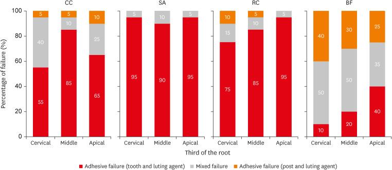
Objectives This study verified the possibility of cementing fiberglass-reinforced posts using a flowable bulk-fill composite (BF), comparing its push-out bond strength and microhardness with these properties of 3 luting materials.
Materials and Methods Sixty endodontically treated bovine roots were used. Posts were cemented using conventional dual-cured cement (CC); self-adhesive cement (SA); dual-cured composite (RC); and BF. Push-out bond strength (
n = 10) and microhardness (n = 5) tests were performed after 1 week and 4 months of storage. Two-way repeated measures analysis of variance (ANOVA), 1-way ANOVA,t -test, and Tukeypost-hoc tests were applied for the push-out bond strength and microhardness results; and Pearson correlation test was applied to verify the correlation between push-out bond strength and microhardness results (α = 0.05).Results BF presented higher push-out bond strength than CC and SA in the cervical third before aging (
p < 0.01). No differences were found between push-out bond strength before and after aging for all the luting materials (p = 0.84). Regarding hardness, only SA presented higher values measured before than after aging (p < 0.01). RC and BF did not present 80% of the maximum hardness at the apical regions. A strong positive correlation was found between the luting materials' push-out bond strength and microhardness (p < 0.01, R2 = 0.7912).Conclusions The BF presented comparable or higher push-out bond strength and microhardness than the luting materials, which indicates that it could be used for cementing resin posts in situations where adequate light curing is possible.
-
Citations
Citations to this article as recorded by- Effects of a relined fiberglass post with conventional and self-adhesive resin cement
Wilton Lima dos Santos Junior, Marina Rodrigues Santi, Rodrigo Barros Esteves Lins, Luís Roberto Marcondes Martins
Restorative Dentistry & Endodontics.2024;[Epub] CrossRef
- Effects of a relined fiberglass post with conventional and self-adhesive resin cement
- 2,946 View
- 53 Download
- 1 Web of Science
- 1 Crossref
Review Article
- Calcium silicate-based root canal sealers: a literature review
- Miyoung Lim, Chanyong Jung, Dong-Hoon Shin, Yong-bum Cho, Minju Song
- Restor Dent Endod 2020;45(3):e35. Published online June 9, 2020
- DOI: https://doi.org/10.5395/rde.2020.45.e35
-
Abstract
PDFPubReaderePub
Epoxy resin-based sealers are currently widely used, and several studies have considered AH Plus to be the gold-standard sealer. However, it still has limitations, including possible mutagenicity, cytotoxicity, inflammatory response, and hydrophobicity. Drawing upon the advantages of mineral trioxide aggregate, calcium silicate-based sealers were introduced with high levels of biocompatibility and hydrophilicity. Because of the hydrophilic environment in root canals, water resorption and solubility of root canal sealers are important factors contributing to their stability. Sealers displaying lower microleakage and stronger push-out bond strength are also needed to endure the dynamic tooth environment. Although the physical properties of calcium silicate-based sealers meet International Organization for Standardization recommendations, and they have consistently reported to be biocompatible, they have not overcome conventional resin-based sealers in actual practice. Therefore, further studies aiming to improve the physical properties of calcium silicate-based sealers are needed.
-
Citations
Citations to this article as recorded by- Does the Use of a Bioceramic Sealer Reduce Postoperative Pain Compared With an Epoxy Resin‐Based Sealer After Primary Root Canal Treatment and Retreatment?—An Umbrella Review
Lokhasudhan Govindaraju, Rajeswari Kalaiselvam, Mathan Rajan Rajendran, Aleksandar Jakovljevic, Jelena Jacimovic, Henry F. Duncan, Venkateshbabu Nagendrababu
International Endodontic Journal.2026; 59(3): 341. CrossRef - Evidence synthesis of postoperative pain with bioceramic vs. epoxy resin sealers: umbrella review of randomized trials within existing systematic reviews
Mrunali Dahikar, Ashish Mandwe, Kulvinder Singh Banga, Alexander Maniangat Luke, Suraj Arora, Unmesh Khanvilkar, Ajinkya M. Pawar
Frontiers in Dental Medicine.2026;[Epub] CrossRef - Effects of interactions between sealers and irrigants on the physicochemical and surface characteristics of endodontic sealers
Hye-In Kim, Yeon-Ju You, Yemi Kim, Jin-Woo Kim, Young-Eun Jang
Clinical Oral Investigations.2026;[Epub] CrossRef - Physical and mechanical properties of a strontium silicate-based sealer
Shannon Wong, Xiaofei Zhu, Tun-Yi Hsu, Sami Chogle, Russell A. Giordano, Yuwei Fan
Odontology.2026;[Epub] CrossRef - Assessment of Release of Calcium Ions from Apical Plugs of Different Bioceramic Sealers in Regenerative Endodontics for Immature Permanent Teeth
Alok Dubey, Nischitha Naik, Bhavana Sujanamulk, Mushir Mulla, Munaz Mulla, Alkananda Sahoo, Mohamed T Salama, Murali Patla S Bhat
World Journal of Dentistry.2026; 17(1): 69. CrossRef - Push-out bond strength of bioceramic-based sealers following different irrigation activation techniques
Fatma Begüm Peker, Hümeyra Çapkın, Ahsen Narbay
Journal of Applied Biomaterials & Functional Materials.2026;[Epub] CrossRef - In Vitro Assessment of Osteogenic Modulation and Molecular Responses Induced by Contemporary Endodontic Sealers in MC3T3-E1 Pre-Osteoblasts
Yuka Miyamoto, Yuka Kato, Ryan Needle, Julie Yongsook Kim, Jin Koo Kim, Paul H. Krebsbach, Insoon Chang
Dentistry Journal.2026; 14(3): 160. CrossRef - The Versatile Role of Rare Earth Elements in Biomaterials
Alfred Ngowi, Thywill Cephas Dzogbewu, Mohammad Rezwan Habib
Advances in Materials Science and Engineering.2026;[Epub] CrossRef - Thermal stability and setting dynamics of nanozinc oxide and AH Plus sealers: An in vitro study
Maryam Gharechahi, Amirreza Panjtanian, Pooya Ahmadi, Arsalan Shahri
Saudi Endodontic Journal.2026; 16(2): 167. CrossRef - Evaluación in vitro de la solubilidad, radiopacidad y pH de un sellador endodóntico biocerámico experimental preparado con diferentes proporciones de silicato de calcio y radiopacificador // In vitro evaluation of solubility, radiopacity and pH of an exp
Alejandro Leonhardt, Osvaldo Zmener, Nicolás Paduli, Roberto Della Porta, Miguel Chantiri
Revista de la Asociación Odontológica Argentina.2026;[Epub] CrossRef - Physiochemical properties of premixed bioceramic-based root canal sealer reinforced with copper oxide nanoparticles
Prasanti Kumari Pradhan, Neelanjana Majee, Gaurav Patri, Prahlad Saraf, Debkant Jena, Akansha Tilokani, Pratik Agrawal
Journal of Conservative Dentistry and Endodontics.2026; 29(6): 672. CrossRef - Biocompatibility and Elemental Characterization of Bioceramic Root Canal Sealers vs Resin Sealer AH Plus
Eduardo Adrián Sánchez Barajas, Alejandra Rodríguez Hidalgo, Marine Ortiz Magdaleno , Rafael Álvarez-Chimal, Amaury Pozos-Guillén , Febe Carolina Vázquez Vázquez
Odovtos - International Journal of Dental Sciences.2026;[Epub] CrossRef - Comparative Evaluation of Postoperative Pain Following Nonsurgical Endodontic Therapy with Calcium Silicate-Based Sealer and Traditional Sealers: A Systematic Review and Meta-Analysis
Guha Poulomi, Solete Pradeep, Antony Delphine, Arun Nishitha, Surendar Ramamoorthi, Choudhari Sahil, Hima Sandeep Adimulapu
Pesquisa Brasileira em Odontopediatria e Clínica Integrada.2026;[Epub] CrossRef - Impact of Thermal Cycling on Volumetric Stability of Endodontic Filling Materials
Petra Dijanic, Marko Katic, Ivan Tomasic, Ana Ivanisevic, Jurica Matijevic
Clinical and Experimental Dental Research.2026;[Epub] CrossRef - Comparative Evaluation of Sealer Voids Using Single-Cone Obturation With Different Root Canal Sealers: An In Vitro Scanning Electron Microscopic Study
Rakhi Singh, Arwa Hussain Chaiwala, Mohan Kumar Reddy Sirigiri, Devna Sharma, Aby Kuruvilla, Rupak Kumar Dasarraju
Cureus.2026;[Epub] CrossRef - Influence of calcium hydroxide/nano-chitosan as intracanal medication on push-out bond strength of epoxy resin and bioceramic sealers: an ex-vivo study
Khaled A. Ghozi, Mohamed A. Gomaa, Amany E. Badr
BMC Oral Health.2026;[Epub] CrossRef - Effect of Different Tapered Gutta-Percha Points on Push-Out Bond Strength of Two Root Canal Sealers
Warattama Suksaphar, Pakit Tungsawat, Ninnita Wongwatanasanti, Siripat Lertnantapanya, Prattana Yodmanothum
European Journal of General Dentistry.2025; 14(03): 285. CrossRef - Effect of Electrical Heat Carrier Temperature on Bacterial Leakage of Endodontically Treated Teeth Using a Bioceramic Sealer
Mir Ahmad Nabavi, Mahmood Reza Kalantar Motamedi, Pedram Fattahi, Saber Khazaei
Clinical and Experimental Dental Research.2025;[Epub] CrossRef - Nanoparticles modified bioceramic sealers on solubility, antimicrobial efficacy, pushout bond strength and marginal adaptation at apical-third of canal dentin
Basil Almutairi, Fahad Alkhudhairy
PeerJ.2025; 13: e18840. CrossRef - Assessing the antimicrobial properties of bioceramic sealers enhanced with herbal extracts against E. faecalis
KS Sachin, K Shibani Shetty, KB Jeyalakshmi, S Harishma, S Harshini
Folia Medica.2025;[Epub] CrossRef - Estudio comparativo de la solubilidad de dos selladores endodónticos biocerámicos y un sellador a base de resinas
//Comparative study of the solubility of two bioceramic endodontic sealers and one epoxi-resin based sealer
Alejandro Leonhardt, Nicolás Paduli, Osvaldo Zmener, Miguel Chantiri
Revista de la Asociación Odontológica Argentina.2025; : 1. CrossRef - Enhancing root canal sealing: Exploring the sealing potential of epoxy and calcium silicate-based sealers with chitosan nanoparticle enhancement
S. Harishma, Srilekha Jayakumar, K Shibani Shetty, Barkavi Panchatcharam, Jwaalaa Rajkumar, S. Harshini
Endodontology.2025; 37(3): 306. CrossRef - Evaluation of the Genotoxicity and Cytotoxicity of Bioceramic Endodontic Sealers in HepG2 and V79 Cell Lines: An In Vitro Study Using the Comet and Micronucleus Assays
Antonija Tadin, Marija Badrov, Danijela Juric Kacunic, Nada Galic, Matea Macan, Ivan Kovacic, Davor Zeljezic
Journal of Functional Biomaterials.2025; 16(5): 169. CrossRef - In Vitro Apatite-Forming Ability of Different Root Canal Sealers (A Comparative Study)
Raghad A Al-Askary, Wiaam M. O. Al-Ashou, Sawsan H. Al-Jubori
Journal of International Society of Preventive and Community Dentistry.2025; 15(2): 173. CrossRef - Microstructural and elemental characterization of novel bioactive glass bioceramic sealer using Fourier transform infrared and X-ray diffraction analysis
Poulomi Guha, Pradeep Solete, Delphine Antony, Nishitha Arun, Mohmed Isaqali Karobari, Surendar Ramamoorthi
Journal of Conservative Dentistry and Endodontics.2025; 28(5): 412. CrossRef - Microstructural and Elemental Characterization of Calcium Silicate-Based Sealers
Mateusz Radwanski, Ireneusz Piwonski, Tomasz Szmechtyk, Salvatore Sauro, Monika Lukomska-Szymanska
Nanomaterials.2025; 15(10): 756. CrossRef - Apical negative pressure-enhanced sealer infiltration for obturating long oval-shaped root canals with the single-cone technique
Yaxu Feng, Brian E. Bergeron, Shijin Zhang, Danyang Sun, Kole Fisher, Franklin R. Tay, Bing Fan
Journal of Dentistry.2025; 160: 105909. CrossRef - Effects of different apical preparation sizes and root canal sealers on the fracture resistance of roots aged for 12 months in endodontically retreated mandibular premolars
Dilek Hancerliogullari, Sevda Durust Baris, Ali Turkyilmaz, Ali Erdemir
British Dental Journal.2025;[Epub] CrossRef - Influence of different endodontic treatment protocols on tooth survival: A retrospective cohort study with multistate analysis and group balancing
Ahmed Elmaasarawi, Mohamed Mekhemar, Andreas Bartols
International Endodontic Journal.2025; 58(10): 1529. CrossRef - Evaluation of 2,6-xylidine precipitate on sealer penetration of calcium silicate-based sealer and resin-based sealer: An in vitro study
M. B. Kalpana, Divya Shetty, Rajaram Naik
Endodontology.2025; 37(2): 183. CrossRef - Translational Advances in Regenerative Dentistry: Functional Biomaterials and Emerging Technologies
Seher Yaylacı, Hacer Eberliköse, Hakan Ceylan
Current Oral Health Reports.2025;[Epub] CrossRef - Marginal adaptation of heat and non-heat compatible bioceramic sealers in warm obturation: an in vitro SEM study
Thanomsuk Jearanaiphaisarn, Thanida Leelayuttakarn, Panisara Amatamahuthana, Pinmanus Chenpairojsakul, Keskanya Subbalekha, Pavena Chivatxaranukul
Scientific Reports.2025;[Epub] CrossRef - Influence of irrigating solutions on the hydration of calcium silicate-based dental biomaterials: An in vitro study
Pradeep M. Divya, Amit Jena, Saumyakanta Mohanty, Govind Shashirekha, Rashmi Rekha Mallick, Priyanka Sarangi
Journal of Conservative Dentistry and Endodontics.2025; 28(8): 758. CrossRef - Multispecies Biofilms Treated With Endodontic Sealers or Calcium Hydroxide: Antimicrobial Activity and Changes in Community Composition
Steven K. Uttech, Ronald Ordinola‐Zapata, W. Craig Noblett, Maria Martell, Bruno Lima, Christopher Staley
International Endodontic Journal.2025; 58(11): 1764. CrossRef - A comparative analysis of adhesion abilities between AH Plus® Bioceramic, Ceraseal® and AH Plus® on root canal dentine surfaces
Ike Dwi Maharti, Indira Larasputri, Nendar Herdianto, Anggraini Margono, Riesma Tasomara, Romilda Rosseti
Journal of Conservative Dentistry and Endodontics.2025; 28(9): 881. CrossRef - Clinical and radiographic success of single-cone bioceramic obturation versus traditional techniques: a systematic review and meta-analysis of randomized controlled trials
Firas Elmsmari, Yousef Elsayed, Abdelrahman Aboubakr, Mahdi Kaafarani, Osama Nour, Ajinkya M. Pawar
Journal of Oral Biology and Craniofacial Research.2025; 15(6): 1422. CrossRef - The Effect of Irrigation Solutions on the Setting Time, Solubility, and pH of Three Types of Premixed Bioceramic‐Based Root Canal Sealers
Kitichai Singharat, Ninnita Wongwatanasanti, Warattama Suksaphar, Pakit Tungsawat, Zhengrui Li
International Journal of Dentistry.2025;[Epub] CrossRef - Endodontie – State of the Art von A bis Z
Will Qian, Andreas Bartols
Zahnmedizin up2date.2025; 19(04): 281. CrossRef - Assessing Volume of Two Sealers’ Remnants after Reinstrumentation Using 3D Imaging Technology: An In Vitro Comparative Study
Khalel Mutaz Dawod, Raghad Abdulrazzaq Al-Hashimi
The Journal of Contemporary Dental Practice.2025; 26(8): 743. CrossRef - Functional and Bioactive Performance of Premixed Bioceramic Sealers with Warm Obturation: A Scoping Review
Patryk Wiśniewski, Stanisław Krokosz, Małgorzata Pietruska, Anna Zalewska
Gels.2025; 11(11): 932. CrossRef - Correlation of Bond Strength and Dentinal Tubule Penetration Evaluation of Four Different Endodontic Sealers: AH Plus, MTA Fillapex, Endoseal MTA, and Endoseal TCS (Maruchi): An In Vitro Study
Arezoo Mirzaei Sadeghloo, Seyedali Seyedmajidi, Akam Saeidi, Elham Mahmoudi, Murilo Baena Lopes
International Journal of Dentistry.2025;[Epub] CrossRef - Osteogenic Potential of Various Premixed Hydraulic Calcium Silicate-Based Sealers on Human Bone Marrow Stem Cells
Na-Hyun You, Donghee Lee, Yemi Kim, Sieun Nam, Sin-Young Kim
Materials.2025; 18(23): 5326. CrossRef - Polydopamine‐Functionalized Zinc Oxide Nanoparticles as a Root Canal Sealer: Characterization, Biological, and Physicochemical Properties
Arul Nayagi Raj, Aditya Shetty, Lakshmi Nidhi Rao, Giuseppe Ciccarella
Bioinorganic Chemistry and Applications.2025;[Epub] CrossRef - Management of rarely seen internal tunnelling root resorption associated with a maxillary permanent incisor
Kirsty A. Carney, Thibault N. E. Colloc, Julie K. Kilgariff
British Dental Journal.2024; 236(12): 955. CrossRef - Top tips for treatment planning: tooth-by-tooth prognosis - Part 3: endodontic prognosis
Prashanti Eachempati, Andrew Harris, Guy Lambourn, Tony Francis, Ewen McColl
British Dental Journal.2024; 237(9): 686. CrossRef - Retreatability of calcium silicate-based sealers based on micro-computed tomographic evaluation − A systematic review
Sundus Mohammed Bukhary
The Saudi Dental Journal.2024; 36(10): 1278. CrossRef - Evaluation of Setting Time, Flowability, Film Thickness, and Radiopacity of Experimental Monocalcium Silicate‐Based Root Canal Sealers
Sukanya Juntha, Pakit Tungsawat, Ninnita Wongwatanasanti, Warattama Suksaphar, Siripat Lertnantapanya, Carlos M. Ardila
International Journal of Dentistry.2024;[Epub] CrossRef - Root Canal Treatment and Demand for Continuing Education among Thai Dental Practitioners
Ninnita Wongwatanasanti, Pakit Tungsawat, Warattama Suksaphar, Siripat Lertnantapanya, Prattana Yodmanotham
The Open Dentistry Journal.2024;[Epub] CrossRef - Clinical outcome of non-surgical root canal treatment using different sealers and techniques of obturation in 237 patients: A retrospective study
Mateusz Radwanski, Krystyna Pietrzycka, Tan Fırat Eyüboğlu, Mutlu Özcan, Monika Lukomska-Szymanska
Clinical Oral Investigations.2024;[Epub] CrossRef - Endodontic sealers after exposure to chlorhexidine digluconate: An assessment of physicochemical properties
Vasileios Kapralos, Josette Camilleri, Andreas Koutroulis, Håkon Valen, Dag Ørstavik, Pia Titterud Sunde
Dental Materials.2024; 40(3): 420. CrossRef - Assessment the bioactivity of zinc oxid eugenol sealer after the addition of different concentrations of nano hydroxyapatite-tyrosine amino acid
Rasha M. Al-Shamaa, Raghad A. Al-Askary
Brazilian Journal of Oral Sciences.2024; 23: e243733. CrossRef - Interfacial adaptation of newly prepared nano-tricalcium silicate-58s bioactive glass-based endodontic sealer
Nawal A. Al-Sabawi, Sawsan Hameed Al-Jubori
Journal of Dental Research, Dental Clinics, Dental Prospects.2024; 18(2): 115. CrossRef - Marginal adaptation of customized gutta percha cone with calcium silicate based sealer versus MTA and biodentine apical plugs in simulated immature permanent teeth (an in vitro study)
Mary M. Mina, Sybel M. Moussa, Mahmoud R. Aboelseoud
BMC Oral Health.2024;[Epub] CrossRef - Solubility of Endoseal and AH26 Root Canal Sealers
Nooshin Fakhari, Ali Reza Mirjani, Abbas Bagheri, Jalil Modaresi
Journal of Research in Dental and Maxillofacial Sciences.2024; 9(1): 1. CrossRef - Novel bioactive nanospheres show effective antibacterial effect against multiple endodontic pathogens
Jin Liu, Haoze Wu, Jun Qiu, Sirui Yang, Doudou Xiang, Xinhua Zhang, Jinxin Kuang, Min Xiao, Qing Yu, Xiaogang Cheng
Heliyon.2024; 10(7): e28266. CrossRef - Evaluation of canal patency and cleanliness following retreatment of bioceramic sealer‐obturated root canals using three different irrigant activation protocols
Daiasharailang Lyngdoh, Sharique Alam, Huma Iftekhar, Surendra Kumar Mishra
Australian Endodontic Journal.2024; 50(3): 475. CrossRef - Antibiofilm Efficacy of Calcium Silicate-Based Endodontic Sealers
Matilde Ruiz-Linares, Vsevolod Fedoseev, Carmen Solana, Cecilia Muñoz-Sandoval, Carmen María Ferrer-Luque
Materials.2024; 17(16): 3937. CrossRef - Enhancing the Biological Properties of Organic–Inorganic Hybrid Calcium Silicate Cements: An In Vitro Study
Minji Choi, Jiyoung Kwon, Ji-Hyun Jang, Duck-Su Kim, Hyun-Jung Kim
Journal of Functional Biomaterials.2024; 15(11): 337. CrossRef - Cytotoxicity and cell migration evaluation of a strontium silicate-based root canal sealer on stem cells from rat apical papilla: an in vitro study
Guanglei Zhou, Yu Zhao, Liangjing Cai, Liwei Liu, Xu Li, Lu Sun, Jiayin Deng
BMC Oral Health.2024;[Epub] CrossRef - An In Vitro Comparative Analysis of Physico–Mechanical Properties of Commercial and Experimental Bioactive Endodontic Sealers
Abdulmajeed Kashaf, Faisal Alonaizan, Khalid S. Almulhim, Dana Almohazey, Deemah Abdullah Alotaibi, Sultan Akhtar, Ashwin C. Shetty, Abdul Samad Khan
Bioengineering.2024; 11(11): 1079. CrossRef - Chemical, Antibacterial, and Cytotoxic Properties of Four Different Endodontic Sealer Leachates Over Time
Jo-Hsun Chen, Veksina Raman, Sarah A. Kuehne, Josette Camilleri, Josefine Hirschfeld
Journal of Endodontics.2024; 50(11): 1612. CrossRef - Comparative Analysis of Fracture Resistance of Endodontic Sealer Types and Filling Methods
Yun Song, Kee-Deog Kim, Bock-Young Jung, Wonse Park, Nan-Sim Pang
Materials.2024; 18(1): 40. CrossRef - Comparative Evaluation of Removal of Bioceramic Sealers Using Rotary Retreatment Files Supplemented with Passive Ultrasonic Activation: An In Vitro Study
Anuradha B Patil, Amrut Bambawale, Pooja R Barghare, Sumanthini V Margasahayam, Divya Naik, Jayeeta S Verma
World Journal of Dentistry.2024; 15(4): 292. CrossRef - Nonsurgical Endodontic Management of Nonperforating Internal Root Resorption in a Maxillary Central Incisor: A Case Report with a 4-Year Follow-Up
Paras M. Gehlot, Divya S. Rajkumar, Annapoorna B. Mariswamy, Upendra Natha N. Reddy, Chaitanya Chappidi
Journal of Pharmacy and Bioallied Sciences.2024; 16(Suppl 3): S3005. CrossRef - Evaluating the Sealing Performance of Endodontic Sealers: Insights Into Achieving Complete Sealing
Ajay Chhabra, Ramya K P., Saravana Prathap, Priyanka Yadav, Himani Mehra, Sona J Parvathy
Cureus.2024;[Epub] CrossRef - Effects of vehicles on the physical properties and biocompatibility of premixed calcium silicate cements
Gitae SON, Gyeung Mi SEON, Sang Hoon CHOI, Hyeong-Cheol YANG
Dental Materials Journal.2024; 43(2): 276. CrossRef - Comparative cytotoxicity study of putty- and powder-type calcium silicate cements
Sora Park, Dohyun Cho, Ji Hyeon Yoon, Yeonjoo Kang, Quang Canh Vo, Gitae Son, Hongjoo Park, Hyeong-Cheol Yang
Korean Journal of Dental Materials.2024; 51(4): 259. CrossRef - Physical-chemical properties and acellular bioactivity of newly prepared nano-tricalcium silicate-58s bioactive glass-based endodontic sealer
Nawal A. Al-Sabawi, Sawsan Hameed Al-Jubori
Journal of Oral Biosciences.2023; 65(4): 305. CrossRef - Dentinal Tubule Penetrability and Bond Strength of Two Novel Calcium Silicate-Based Root Canal Sealers
Karissa Shieh, Jack Yang, Elsa Heng Zhu, Ove Andreas Peters, Sepanta Hosseinpour
Materials.2023; 16(9): 3309. CrossRef - Cytotoxicity and Mineralization Activity of Calcium Silicate-Based Root Canal Sealers Compared to Conventional Resin-Based Sealer in Human Gingival Fibroblast Cells
Mohammad Shokrzadeh, Farzaneh Sadat Motafeghi, Anahita Lotfizadeh, Mohammad Ghorbani, Azam Haddadi Kohsar, Cesar Rogério Pucci
International Journal of Dentistry.2023; 2023: 1. CrossRef - Effect of three different photosensitizers in photodynamic therapy on bond strength of a calcium silicate‐based sealer to radicular dentin
Cihan Küden, Seda Nur Karakaş
Australian Endodontic Journal.2023; 49(S1): 265. CrossRef - Effect of endodontic sealer on postoperative pain: a network meta-analysis
Cynthia Maria Chaves Monteiro, Ana Cristina Rodrigues Martins, Alessandra Reis, Juliana Larocca de Geus
Restorative Dentistry & Endodontics.2023;[Epub] CrossRef - Antimicrobial Activity of Five Calcium Silicate Based Root Canal Sealers against a Multispecies Engineered Biofilm: An In Vitro Study
Carla Zogheib, Issam Khalil, Wajih Hage, Dolla Karam Sarkis, Mireille Kallasy, Germain Sfeir, May Mallah, Roula El Hachem
The Journal of Contemporary Dental Practice.2023; 24(9): 707. CrossRef - Calcium silicate sealers in endodontics
Archana Chavan, Nidambur Ballal
Acta stomatologica Naissi.2023; 39(87): 2624. CrossRef - Assessing the Sealing Performance and Clinical Outcomes of Endodontic Treatment in Patients with Chronic Apical Periodontitis Using Epoxy Resin and Calcium Salicylate Seals
Razvan Mihai Horhat, Bogdan Andrei Bumbu, Laura Orel, Oana Velea-Barta, Laura Cirligeriu, Gratiana Nicoleta Chicin, Marius Pricop, Mircea Rivis, Stefania Dinu, Delia Ioana Horhat, Felix Bratosin, Roxana Manuela Fericean, Rodica Anamaria Negrean, Luminita
Medicina.2023; 59(6): 1137. CrossRef -
In Vitro Cytotoxicity and Mineralization Potential of an Endodontic Bioceramic Material
Soumya Sheela, Mohannad Nassar, Fatma M. AlGhalban, Mehmet O. Gorduysus
European Journal of Dentistry.2023; 17(02): 548. CrossRef - Dislodgment Resistance, Adhesive Pattern, and Dentinal Tubule Penetration of a Novel Experimental Algin Biopolymer-Incorporated Bioceramic-Based Root Canal Sealer
Galvin Sim Siang Lin, Norhayati Luddin, Huwaina Abd Ghani, Josephine Chang Hui Lai, Tahir Yusuf Noorani
Polymers.2023; 15(5): 1317. CrossRef - Impact of Final Irrigation Protocol on the Push-Out Bond Strength of Two Types of Endodontic Sealers
Germain Sfeir, Frédéric Bukiet, Wajih Hage, Roula El Hachem, Carla Zogheib
Materials.2023; 16(5): 1761. CrossRef - Clinical Approaches to the Three-Dimensional Endodontic Obturation Protocol for Teeth with Periapical Bone Lesions
Angela Gusiyska, Elena Dyulgerova
Applied Sciences.2023; 13(17): 9755. CrossRef - Evaluating the bioactivity of endodontic sealers with respect to their thermo-nanomechanical properties
Andreea Marica, Luminita Fritea, Florin Banica, Iosif Hulka, Gerlinde Rusu, Cosmin Sinescu, Traian Octavian Costea, Simona Cavalu
Materials Science-Poland.2023; 41(3): 126. CrossRef - Advances and challenges in regenerative dentistry: A systematic review of calcium phosphate and silicate-based materials on human dental pulp stem cells
B. Christie, N. Musri, N. Djustiana, V. Takarini, N. Tuygunov, M.N. Zakaria, A. Cahyanto
Materials Today Bio.2023; 23: 100815. CrossRef - Radiographic Evaluation of Periapical Healing Rates Between Bio-Ceramic Sealer and AH+ Sealer: A Retrospective Study
Dalia Nayil Alharith, Iman T. Mansi, YoumnaElsaid Abdulmotalib, HebaFuad Amous, TagreedSuliman Aljulban, Haifa Mohammed Al Aiban, Sali Mohamad Haffar
Annals of Dental Specialty.2023; 11(2): 124. CrossRef - Obturation canalaire
N. Linas, M.-L. Munoz-Sanchez, N. Decerle, P.-Y. Cousson
EMC - Médecine buccale.2023; 16(5): 1. CrossRef - Biodentine Inhibits the Initial Microbial Adhesion of Oral Microbiota In Vivo
Ali Al-Ahmad, Michael Haendel, Markus Altenburger, Lamprini Karygianni, Elmar Hellwig, Karl Wrbas, Kirstin Vach, Christian Tennert
Antibiotics.2022; 12(1): 4. CrossRef - Pilot Evaluation of Sealer-Based Root Canal Obturation Using Epoxy-Resin-Based and Calcium-Silicate-Based Sealers: A Randomized Clinical Trial
Minju Song, Min-Gyu Park, Sang-Won Kwak, Ruben H. Kim, Jung-Hong Ha, Hyeon-Cheol Kim
Materials.2022; 15(15): 5146. CrossRef - The antibacterial activity of mineral trioxide aggregate containing calcium fluoride
Miyoung Lim, Seunghoon Yoo
Journal of Dental Sciences.2022; 17(2): 836. CrossRef - Physicochemical and Mechanical Properties of Premixed Calcium Silicate and Resin Sealers
Naji Kharouf, Salvatore Sauro, Ammar Eid, Jihed Zghal, Hamdi Jmal, Anta Seck, Valentina Macaluso, Frédéric Addiego, Francesco Inchingolo, Christine Affolter-Zbaraszczuk, Florent Meyer, Youssef Haikel, Davide Mancino
Journal of Functional Biomaterials.2022; 14(1): 9. CrossRef - Comparison of Fracture Resistance between Single-cone and Warm Vertical Compaction Technique Using Bio-C Sealer® in Mandibular Incisors: An In Vitro Study
Raphael Lichaa, George Deeb, Rami Mhanna, Carla Zogheib
The Journal of Contemporary Dental Practice.2022; 23(2): 143. CrossRef - In vitro physicochemical characterization of five root canal sealers and their influence on an ex vivo oral multi‐species biofilm community
Flavia M. Saavedra, Lauter E. Pelepenko, William S. Boyle, Anqi Zhang, Christopher Staley, Mark C. Herzberg, Marina A. Marciano, Bruno P. Lima
International Endodontic Journal.2022; 55(7): 772. CrossRef - Premixed Calcium Silicate-Based Root Canal Sealer Reinforced with Bioactive Glass Nanoparticles to Improve Biological Properties
Min-Kyung Jung, So-Chung Park, Yu-Jin Kim, Jong-Tae Park, Jonathan C. Knowles, Jeong-Hui Park, Khandmaa Dashnyam, Soo-Kyung Jun, Hae-Hyoung Lee, Jung-Hwan Lee
Pharmaceutics.2022; 14(9): 1903. CrossRef - A critical analysis of research methods and experimental models to study root canal fillings
Gustavo De‐Deus, Erick Miranda Souza, Emmanuel João Nogueira Leal Silva, Felipe Gonçalves Belladonna, Marco Simões‐Carvalho, Daniele Moreira Cavalcante, Marco Aurélio Versiani
International Endodontic Journal.2022; 55(S2): 384. CrossRef - Bioactivity Potential of Bioceramic-Based Root Canal Sealers: A Scoping Review
Mauro Schmitz Estivalet, Lucas Peixoto de Araújo, Felipe Immich, Adriana Fernandes da Silva, Nadia de Souza Ferreira, Wellington Luiz de Oliveira da Rosa, Evandro Piva
Life.2022; 12(11): 1853. CrossRef - The influence of humidity on bond strength of AH Plus, BioRoot RCS, and Nanoseal-S sealers
Sunanda Laxman Gaddalay, Damini Vilas Patil, Ramchandra Kabir
Endodontology.2022; 34(3): 202. CrossRef - The Effect of Bioceramic HiFlow and EndoSequence Bioceramic Sealers on Increasing the Fracture Resistance of Endodontically Treated Teeth: An In Vitro Study
Mohamad Khir Abdulsamad Alskaf, Hassan Achour, Hasan Alzoubi
Cureus.2022;[Epub] CrossRef - Unravelling the effects of ibuprofen-acetaminophen infused copper-bioglass towards the creation of root canal sealant
Chitra S, Riju Chandran, Ramya R, Durgalakshmi D, Balakumar S
Biomedical Materials.2022; 17(3): 035001. CrossRef - A Micro-CT Analysis of Initial and Long-Term Pores Volume and Porosity of Bioactive Endodontic Sealers
Mateusz Radwanski, Michal Leski, Adam K. Puszkarz, Jerzy Sokolowski, Louis Hardan, Rim Bourgi, Salvatore Sauro, Monika Lukomska-Szymanska
Biomedicines.2022; 10(10): 2403. CrossRef - A comprehensive in vitro comparison of the biological and physicochemical properties of bioactive root canal sealers
Sabina Noreen Wuersching, Christian Diegritz, Reinhard Hickel, Karin Christine Huth, Maximilian Kollmuss
Clinical Oral Investigations.2022; 26(10): 6209. CrossRef - Stability and solubility test of endodontic materials
Ivan Matovic, Jelena Vucetic
Stomatoloski glasnik Srbije.2022; 69(4): 169. CrossRef - Antimicrobial effectiveness of root canal sealers againstEnterococcus faecalis
Paola Castillo-Villagomez, Elizabeth Madla-Cruz, Fanny Lopez-Martinez, Idalia Rodriguez-Delgado, Jorge Jaime Flores-Treviño, Guadalupe Ismael Malagon-Santiago, Myriam Angelica de La Garza-Ramos
Biomaterial Investigations in Dentistry.2022; 9(1): 47. CrossRef - Tricalcium silicate cement sealers
Anita Aminoshariae, Carolyn Primus, James C. Kulild
The Journal of the American Dental Association.2022; 153(8): 750. CrossRef - Influence of variations in the environmental pH on the solubility and water sorption of a calcium silicate‐based root canal sealer
E. J. N. L. Silva, C. M. Ferreira, K. P. Pinto, A. F. A. Barbosa, M. V. Colaço, L. M. Sassone
International Endodontic Journal.2021; 54(8): 1394. CrossRef - Calcium Silicate-Based Root Canal Sealers: A Narrative Review and Clinical Perspectives
Germain Sfeir, Carla Zogheib, Shanon Patel, Thomas Giraud, Venkateshbabu Nagendrababu, Frédéric Bukiet
Materials.2021; 14(14): 3965. CrossRef - Development of A Nano-Apatite Based Composite Sealer for Endodontic Root Canal Filling
Angelica Bertacci, Daniele Moro, Gianfranco Ulian, Giovanni Valdrè
Journal of Composites Science.2021; 5(1): 30. CrossRef - Bone repair in defects filled with AH Plus sealer and different concentrations of MTA: a study in rat tibiae
Jessica Emanuella Rocha Paz, Priscila Oliveira Costa, Albert Alexandre Costa Souza, Ingrid Macedo de Oliveira, Lucas Fernandes Falcão, Carlos Alberto Monteiro Falcão, Maria Ângela Area Leão Ferraz, Lucielma Salmito Soares Pinto
Restorative Dentistry & Endodontics.2021;[Epub] CrossRef - Characterization, Antimicrobial Effects, and Cytocompatibility of a Root Canal Sealer Produced by Pozzolan Reaction between Calcium Hydroxide and Silica
Mi-Ah Kim, Vinicius Rosa, Prasanna Neelakantan, Yun-Chan Hwang, Kyung-San Min
Materials.2021; 14(11): 2863. CrossRef - Synthesis and Characterization of Novel Calcium-Silicate Nanobioceramics with Magnesium: Effect of Heat Treatment on Biological, Physical and Chemical Properties
Konstantina Kazeli, Ioannis Tsamesidis, Anna Theocharidou, Lamprini Malletzidou, Jonathan Rhoades, Georgia K. Pouroutzidou, Eleni Likotrafiti, Konstantinos Chrissafis, Theodoros Lialiaris, Lambrini Papadopoulou, Eleana Kontonasaki, Evgenia Lymperaki
Ceramics.2021; 4(4): 628. CrossRef - Calcium Silicate Cements vs. Epoxy Resin Based Cements: Narrative Review
Mario Dioguardi, Cristian Quarta, Diego Sovereto, Giuseppe Troiano, Khrystyna Zhurakivska, Maria Bizzoca, Lorenzo Lo Muzio, Lucio Lo Russo
Oral.2021; 1(1): 23. CrossRef - In Vitro Microleakage Evaluation of Bioceramic and Zinc-Eugenol Sealers with Two Obturation Techniques
Francesco De Angelis, Camillo D’Arcangelo, Matteo Buonvivere, Rachele Argentino, Mirco Vadini
Coatings.2021; 11(6): 727. CrossRef - Efficacy Of Calcium Silicate-Based Sealers In Root Canal Treatment: A Systematic Review
Hattan Mohammed Omar Baismail, Mohammed Ghazi Moiser Albalawi, Alaa Mofareh Thoilek Alanazi, Muhannad Atallah Saleem Alatawi, Badr Soliman Alhussain
Annals of Dental Specialty.2021; 9(1): 87. CrossRef - Apical Sealing Ability of Two Calcium Silicate-Based Sealers Using a Radioactive Isotope Method: An In Vitro Apexification Model
Inês Raquel Pereira, Catarina Carvalho, Siri Paulo, José Pedro Martinho, Ana Sofia Coelho, Anabela Baptista Paula, Carlos Miguel Marto, Eunice Carrilho, Maria Filomena Botelho, Ana Margarida Abrantes, Manuel Marques Ferreira
Materials.2021; 14(21): 6456. CrossRef
- Does the Use of a Bioceramic Sealer Reduce Postoperative Pain Compared With an Epoxy Resin‐Based Sealer After Primary Root Canal Treatment and Retreatment?—An Umbrella Review
- 16,928 View
- 330 Download
- 110 Crossref
Research Articles
- Effect of hydrofluoric acid-based etchant at an elevated temperature on the bond strength and surface topography of Y-TZP ceramics
- Mi-Kyung Yu, Myung-Jin Lim, Noo-Ri Na, Kwang-Won Lee
- Restor Dent Endod 2020;45(1):e6. Published online December 3, 2019
- DOI: https://doi.org/10.5395/rde.2020.45.e6
-
Abstract
PDFPubReaderePub
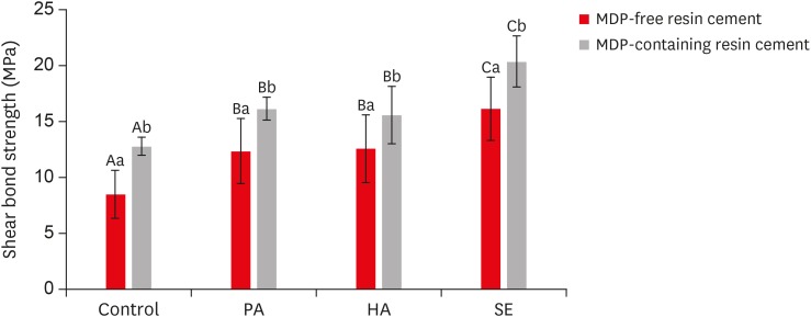
Objectives This study investigated the effects of a hydrofluoric acid (HA; solution of hydrogen fluoride [HF] in water)-based smart etching (SE) solution at an elevated temperature on yttria-stabilized tetragonal zirconia polycrystal (Y-TZP) ceramics in terms of bond strength and morphological changes.
Materials and Methods Eighty sintered Y-TZP specimens were prepared for shear bond strength (SBS) testing. The bonding surface of the Y-TZP specimens was treated with 37% phosphoric acid etching at 20°C–25°C, 4% HA etching at 20°C–25°C, or HA-based SE at 70°C–80°C. In all groups, zirconia primers were applied to the bonding surface of Y-TZP. For each group, 2 types of resin cement (with or without methacryloyloxydecyl dihydrogen phosphate [MDP]) were used. SBS testing was performed. Topographic changes of the etched Y-TZP surface were analyzed using scanning electron microscopy and atomic force microscopy. The results were analyzed and compared using 2-way analysis of variance.
Results Regardless of the type of resin cement, the highest bond strength was measured in the SE group, with significant differences compared to the other groups (
p < 0.05). In all groups, MDP-containing resin cement yielded significantly higher bond strength values than MDP-free resin cement (p < 0.05). It was also shown that the Y-TZP surface was etched by the SE solution, causing a large change in the surface topography.Conclusions Bond strength significantly improved when a heated HA-based SE solution was applied to the Y-TZP surface, and the etched Y-TZP surface was more irregular and had higher surface roughness.
-
Citations
Citations to this article as recorded by- Etchability of zirconia ceramics and its effect on adhesion: A systematic review and meta-analysis
Anina Sieber, Luiza Freitas Brum Souza, Tan Fırat Eyüboğlu, Mutlu Özcan
International Journal of Adhesion and Adhesives.2026; 148: 104303. CrossRef - Evaluation of Different Surface Roughening Techniques on Clear Aligner Attachments Bonded to Monolithic Zirconia: In Vitro Study
Nehal F Albelasy, Ahmad M Hafez, Abdullah S Alhunayni
The Journal of Contemporary Dental Practice.2025; 25(12): 1104. CrossRef - Effect of Acid Surface Treatments on the Shear Bond Strength of Metal Bracket to Zirconia Ceramics
Punchanit Wongrachit, Bancha Samruajbenjakun, Boonlert Kukiattrakoon, Tanapat Jearanai, Supontep Teerakanok, Pannapat Chanmanee
Ceramics.2024; 7(2): 689. CrossRef - Exploring Zirconia Adhesion: Pre and Postsintering Physical Surface Treatment, Chemical Treatment, and Cement Interactions
Flávia Gonçalves, Mirko Dennys Ayala-Perez, Francisco Carlos dos Santos Reis, Walter Gomes Miranda-Júnior, Letícia Cristina Cidreira Boaro, Heng Bo Jiang
BioMed Research International.2024;[Epub] CrossRef - Evaluation of zirconia surfaces and shear bond strength after acid–etching with ultrasonic vibration
Xiaozhen Zhang, Hepeng Nie, Jiaxin Lv, Shanshan Yuan, Juan Wang, Kunzhan Cai, Jin Wu, Qingqing Zhang, Chunbo Tang
Materials Research Express.2024; 11(2): 025401. CrossRef - Effects of Surface-Etching Systems on the Shear Bond Strength of Dual-Polymerized Resin Cement and Zirconia
Sang-Hyun Kim, Kyung Chul Oh, Hong-Seok Moon
Materials.2024; 17(13): 3096. CrossRef - Zirconia bond strength durability following artificial aging: A systematic review and meta-analysis of in vitro studies
Athanasios E. Rigos, Katia Sarafidou, Eleana Kontonasaki
Japanese Dental Science Review.2023; 59: 138. CrossRef - Y-TZP Physicochemical Properties Conditioned with ZrO2 and SiO2 Nanofilms and Bond Strength to Dual Resin Cement
Ricardo Faria Ribeiro, Danilo Flamini Oliveira, Camila Bussola Tovani, Ana Paula Ramos, Ana Flavia Sanches Borges, Adriana Claudia Lapria Faria, Rossana Pereira de Almeida, Renata Cristina Silveira Rodrigues
Materials.2022; 15(22): 7905. CrossRef - Effect of the nanofilm-coated zirconia ceramic on resin cement bond strength
Viviane Maria Gonçalves de Figueiredo, Alecsandro de Moura Silva, Marcos Massi, Argemiro Soares da Silva Sobrinho, José Renato Cavalcanti de Queiroz, João Paulo Barros Machado, Renata Falchete do Prado, Lafayette Nogueira Junior
Journal of Dental Research, Dental Clinics, Dental Prospects.2022; 16(3): 170. CrossRef - Change of phase transformation and bond strength of Y-TZP with various hydrofluoric acid etching
Mi-Kyung Yu, Eun-Jin Oh, Myung-Jin Lim, Kwang-Won Lee
Restorative Dentistry & Endodontics.2021;[Epub] CrossRef - Changes in Bond Strength and Topography for Y-TZP Etched with Hydrofluoric Acid Depending on Concentration and Temperature Conditions
Hyo-Eun Kim, Myung-Jin Lim, Mi-Kyung Yu, Kwang-Won Lee
Medicina.2020; 56(11): 568. CrossRef - Do different sintering conditions influence bond strength between the resin cements and a currently used esthetic zirconia?
Fatma Ayse Sanal, Hamiyet Kilinc
Journal of Adhesion Science and Technology.2020; 34(16): 1809. CrossRef
- Etchability of zirconia ceramics and its effect on adhesion: A systematic review and meta-analysis
- 2,999 View
- 15 Download
- 12 Crossref
- Bacterial leakage and micro-computed tomography evaluation in round-shaped canals obturated with bioceramic cone and sealer using matched single cone technique
- Kallaya Yanpiset, Danuchit Banomyong, Kanet Chotvorrarak, Ratchapin Laovanitch Srisatjaluk
- Restor Dent Endod 2018;43(3):e30. Published online July 5, 2018
- DOI: https://doi.org/10.5395/rde.2018.43.e30
-
Abstract
PDFPubReaderePub
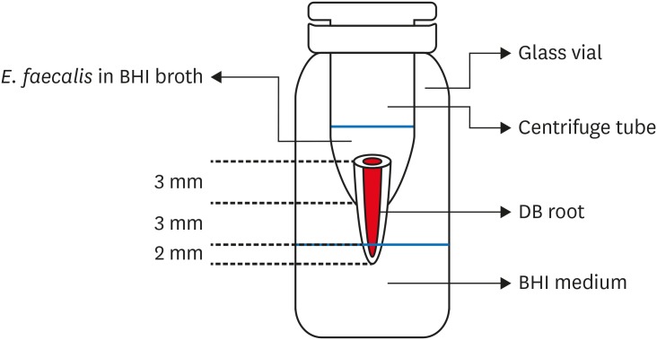
Objectives To evaluate sealing ability of root canals obturated with bioceramic-impregnated gutta percha cone (BCC) or gutta percha (GP), with bioceramic sealer (BCS) or AH Plus (AH; Dentsply-Maillefer), in roundly-prepared canals using matched single-cone technique, based on bacterial leakage test, and to analyze obturation quality using micro-computed tomography (CT) analysis.
Materials and Methods Ninety-two distobuccal roots of maxillary molars were prepared using nickel-titanium files to apical size 40/0.06. The roots were divided into 4 groups (
n = 20) that were obturated with a master cone and sealer: GP/AH, BCC/AH, GP/BCS, and BCC/BCS. Bacterial leakage model usingEnterococcus faecalis was used to evaluate sealing ability for 60-day period. Obturated samples from each group (n = 4) were analyzed using micro-CT.Results All groups showed bacterial leakage at 20%–45% of samples with mean leakage times of 42–52 days. There were no significant differences in bacterial leakage among the groups. Micro-CT showed minimal gaps and voids in all groups at less than 1%.
Conclusions In roundly-prepared canals, the single cone obturation with BCC/BCS was comparable to GP/AH for bacterial leakage at 60 days.
-
Citations
Citations to this article as recorded by- Time-Dependent Volumetric and Porosity Changes of Bioceramic, Silicone Bioactive Glass-Based, and Epoxy Resin-Based Root Canal Sealers: A Micro-CT Analysis
Thanh Quang Nguyen, Chantida Pawaputanon Na Mahasarakham, Pinpana Thaweesit, Kanet Chotvorrarak, Angsana Jainaen
European Journal of Dentistry.2026;[Epub] CrossRef - Assessment of Sealing Ability and Degradation Resistance of a Hydrogel‐Based Root Canal Filling Material Using a Bacterial Leakage Model and SEM Analysis
Else Ellermann, Daniel Richter, Ignasi Belda Punzano, Andreas Schmocker, Mark Bispinghoff, Tan Fırat Eyüboğlu, Mutlu Özcan, Yoel A. Klug
BioMed Research International.2026;[Epub] CrossRef - Marginal Adaptation of GuttaFlow 2, GuttaFlow Bioseal, and AH Plus in the Apical Third of Human Teeth Obturated by the Single-Cone Technique
Sayeh Abbasnejad, Faezeh Abbasi, Solmaz Araghi, Sohrab Tour Savadkouhi
Journal of Research in Dental and Maxillofacial Sciences.2026; 11(2): 107. CrossRef - Influence of Delivery Method of Hydraulic Calcium Silicate Cement‐Based Sealer on Void Formation: A Micro‐Computed Tomography Study
Yasmina Andreani, Ryan David Butler, Diana Patalwala, Paul V. Abbott, Mostafa M. A. Elkholy
Australian Endodontic Journal.2026;[Epub] CrossRef - Impact of Thermal Cycling on Volumetric Stability of Endodontic Filling Materials
Petra Dijanic, Marko Katic, Ivan Tomasic, Ana Ivanisevic, Jurica Matijevic
Clinical and Experimental Dental Research.2026;[Epub] CrossRef - Effect of Root Dentin Moisture on the Apical Sealing Ability of Root Canal Sealers: In vitro Study
Zahraa Khalil Alani, Manal Hussain Abd-alla
Al-Rafidain Journal of Medical Sciences ( ISSN 2789-3219 ).2025; 8(2): 122. CrossRef - Synthesis, physical properties, and root canal sealing of experimental MTA- and salicylate-based root canal sealers
Rafael Pino Vitti, Kusai Baroudi, Tarun Walia, Raghavandra M. Shetty, Flávia Goulart da Rosa Cardoso, Flávia de Moura Pereira, Evandro Piva, Cesar Henrique Zanchi, Gabriel Flores Abuna, Carolina Oliveira de Lima, Emmanuel João Nogueira Leal Silva, Flávio
PLOS One.2025; 20(7): e0329476. CrossRef - Impact of cone system compatibility on single cone bioceramic obturation in canals prepared with variable taper NiTi rotary files
Reem M. Barakat, Rahaf A. Almohareb, Njoom Aleid, Hoor Almowais, Aljawhara Alharbi, Meshal Al-Sharafa, Ali Alrahlah
Scientific Reports.2025;[Epub] CrossRef - Estudio de la obturación con selladores biocerámicos de conductos radiculares de premolares inferiores
Alicia Beatriz Bonafé, Cecilia Inés Rourera, Carla Pedraza, Yamila Victoria Zanoni, Soledad Salduna, Cecilia Noemi De Caso, Gabriela Martín
Methodo Investigación Aplicada a las Ciencias Biológicas.2025; 10(3): 31. CrossRef - Sealing ability of mineral trioxide aggregate: A scoping review of laboratory assessment methods
Kenta Tsuchiya, Salvatore Sauro, Jukka P. Matinlinna, Hidehiko Sano, Monica Yamauti, Deepak Mehta, Kyung‐San Min, Atsushi Tomokiyo
European Journal of Oral Sciences.2025;[Epub] CrossRef - Bacterial Leakage Testing in Dentistry: A Comprehensive Review on Methods, Models, and Clinical Relevance
Niher Tabassum Snigdha, Mohmed Isaqali Karobari, Sukhamoy Gorai
Scientifica.2025;[Epub] CrossRef - In vitro comparative evaluation of apical leakage using a bioceramic sealer with three different obturating techniques: A glucose leakage model
Tanvi S Agrawal, Shishir Singh, Rajesh S Podar, Gaurav Kulkarni, Anuprita Gadkari, Navin Agarwal
Journal of Conservative Dentistry and Endodontics.2024; 27(1): 76. CrossRef - In Vitro Microscopical and Microbiological Assessment of the Sealing Ability of Calcium Silicate-Based Root Canal Sealers
Karin Christine Huth, Sabina Noreen Wuersching, Leander Benz, Stefan Kist, Maximilian Kollmuss
Journal of Functional Biomaterials.2024; 15(11): 341. CrossRef - Comparison between AH plus sealer and total fill bioceramic sealer performance in previously untreated and retreatment cases of maxillary incisors with large-sized periapical lesion: a randomized controlled trial
Eisa Wahbi, Hassan Achour, Yasser Alsayed Tolibah
BDJ Open.2024;[Epub] CrossRef - Bacterial sealing ability of calcium silicate-based sealer for endodontic surgery: an in-vitro study
Mai M. Mansour, Sybel M. Moussa, Marwa A. Meheissen, Mahmoud R. Aboelseoud
BMC Oral Health.2024;[Epub] CrossRef - Assessment the bioactivity of zinc oxid eugenol sealer after the addition of different concentrations of nano hydroxyapatite-tyrosine amino acid
Rasha M. Al-Shamaa, Raghad A. Al-Askary
Brazilian Journal of Oral Sciences.2024; 23: e243733. CrossRef - Assessment of Bacterial Sealing Ability of Two Different Bio-Ceramic Sealers in Single-Rooted Teeth Using Single Cone Obturation Technique: An In Vitro Study
Doaa M. AlEraky, Ahmed M. Rahoma, Hatem M. Abuohashish, Abdullh AlQasser, Abbas AlHamali, Hussain M. AlHussain, Hussain M. AlShoalah, Zakrya AlSaghah, Abdulrahman Khattar, Shimaa Rifaat
Applied Sciences.2023; 13(5): 2906. CrossRef - How do imaging protocols affect the assessment of root-end fillings?
Fernanda Ferrari Esteves Torres, Reinhilde Jacobs, Mostafa EzEldeen, Karla de Faria-Vasconcelos, Juliane Maria Guerreiro-Tanomaru, Bernardo Camargo dos Santos, Mário Tanomaru-Filho
Restorative Dentistry & Endodontics.2022;[Epub] CrossRef - The impact of Morse taper implant design on microleakage at implant-healing abutment interface
Soyeon KIM, Joo Won LEE, Jae-Heon KIM, Van Mai TRUONG, Young-Seok PARK
Dental Materials Journal.2022; 41(5): 767. CrossRef - A critical analysis of research methods and experimental models to study root canal fillings
Gustavo De‐Deus, Erick Miranda Souza, Emmanuel João Nogueira Leal Silva, Felipe Gonçalves Belladonna, Marco Simões‐Carvalho, Daniele Moreira Cavalcante, Marco Aurélio Versiani
International Endodontic Journal.2022; 55(S2): 384. CrossRef - Micro‐CT assessment of gap‐containing areas along the gutta‐percha‐sealer interface in oval‐shaped canals
Gustavo De‐Deus, Gustavo O. Santos, Iara Zamboni Monteiro, Daniele M. Cavalcante, Marco Simões‐Carvalho, Felipe G. Belladonna, Emmanuel J. N. L. Silva, Erick M. Souza, Raphael Licha, Carla Zogheib, Marco A. Versiani
International Endodontic Journal.2022; 55(7): 795. CrossRef - Comparison of Sealing Ability of Bioceramic Sealer, AH Plus, and GuttaFlow in Conservatively Prepared Curved Root Canals Obturated with Single-Cone Technique: An In vitro Study
Shalan Kaul, Ajay Kumar, Bhumika Kamal Badiyani, Laxmi Sukhtankar, M. Madhumitha, Amit Kumar
Journal of Pharmacy and Bioallied Sciences.2021; 13(Suppl 1): S857. CrossRef - Micro-CT Evaluation of Four Root Canal Obturation Techniques
Mahmood Reza Kalantar Motamedi, Amin Mortaheb, Maryam Zare Jahromi, Brett E. Gilbert, Marilena Vivona
Scanning.2021; 2021: 1. CrossRef - Effects of Both Fiber Post/Core Resin Construction System and Root Canal Sealer on the Material Interface in Deep Areas of Root Canal
Hiroki Miura, Shinji Yoshii, Masataka Fujimoto, Ayako Washio, Takahiko Morotomi, Hiroshi Ikeda, Chiaki Kitamura
Materials.2021; 14(4): 982. CrossRef - Sealing ability and microbial leakage of root-end filling materials: MTA versus epoxy resin: A systematic review and meta-analysis
Mario Dioguardi, Mario Alovisi, Diego Sovereto, Giuseppe Troiano, Giancarlo Malagnino, Michele Di Cosola, Angela Pia Cazzolla, Luigi Laino, Lorenzo Lo Muzio
Heliyon.2021; 7(7): e07494. CrossRef - Development of A Nano-Apatite Based Composite Sealer for Endodontic Root Canal Filling
Angelica Bertacci, Daniele Moro, Gianfranco Ulian, Giovanni Valdrè
Journal of Composites Science.2021; 5(1): 30. CrossRef - BIOCERAMIC-BASED ROOT CANAL SEALERS
L Somolová, Z Zapletalová, M Rosa, B Novotná, I Voborná, Y Morozova
Česká stomatologie a praktické zubní lékařství.2021; 121(4): 116. CrossRef - Calcium Silicate-Based Root Canal Sealers: A Narrative Review and Clinical Perspectives
Germain Sfeir, Carla Zogheib, Shanon Patel, Thomas Giraud, Venkateshbabu Nagendrababu, Frédéric Bukiet
Materials.2021; 14(14): 3965. CrossRef - Physico-Chemical Properties of Calcium-Silicate vs. Resin Based Sealers—A Systematic Review and Meta-Analysis of Laboratory-Based Studies
Viresh Chopra, Graham Davis, Aylin Baysan
Materials.2021; 15(1): 229. CrossRef - Comparison of apical sealing ability of bioceramic sealer and epoxy resin-based sealer using the fluid filtration technique and scanning electron microscopy
Widcha Asawaworarit, Thitapa Pinyosopon, Kanittha Kijsamanmith
Journal of Dental Sciences.2020; 15(2): 186. CrossRef - Micro-computed tomographic evaluation of a new system for root canal filling using calcium silicate-based root canal sealers
Mario Tanomaru-Filho, Fernanda Ferrari Esteves Torres, Jader Camilo Pinto, Airton Oliveira Santos-Junior, Karina Ines Medina Carita Tavares, Juliane Maria Guerreiro-Tanomaru
Restorative Dentistry & Endodontics.2020;[Epub] CrossRef - A micro-computed tomographic evaluation of root canal filling with a single gutta-percha cone and calcium silicate sealer
Jong Cheon Kim, Maung Maung Kyaw Moe, Sung Kyo Kim
Restorative Dentistry & Endodontics.2020;[Epub] CrossRef - Comparative evaluation of sealing ability of gutta percha and resilon as root canal filling materials- a systematic review
Pragya Pandey, Himanshi Aggarwal, A.P. Tikku, Arpit Singh, Rhythm Bains, Shambhavi Mishra
Journal of Oral Biology and Craniofacial Research.2020; 10(2): 220. CrossRef - Micro-computed tomographic evaluation of the flow and filling ability of endodontic materials using different test models
Fernanda Ferrari Esteves Torres, Juliane Maria Guerreiro-Tanomaru, Gisselle Moraima Chavez-Andrade, Jader Camilo Pinto, Fábio Luiz Camargo Villela Berbert, Mario Tanomaru-Filho
Restorative Dentistry & Endodontics.2020;[Epub] CrossRef - Root fillings with a matched-taper single cone and two calcium silicate–based sealers: an analysis of voids using micro-computed tomography
Eugenio Pedullà, Roula S. Abiad, Gianluca Conte, Giusy R. M. La Rosa, Ernesto Rapisarda, Prasanna Neelakantan
Clinical Oral Investigations.2020; 24(12): 4487. CrossRef - Influence of different disinfection protocols on gutta-percha cones surface roughness assessed by two different methods
A.M. Nunes, J.P. Gouvea, L. da Silva
Journal of Materials Research and Technology.2019; 8(6): 5464. CrossRef - Endodontic sealers based on calcium silicates: a systematic review
David Donnermeyer, Sebastian Bürklein, Till Dammaschke, Edgar Schäfer
Odontology.2019; 107(4): 421. CrossRef
- Time-Dependent Volumetric and Porosity Changes of Bioceramic, Silicone Bioactive Glass-Based, and Epoxy Resin-Based Root Canal Sealers: A Micro-CT Analysis
- 3,740 View
- 57 Download
- 37 Crossref
- Color stability of bulk-fill and incremental-fill resin-based composites polished with aluminum-oxide impregnated disks
- Uzay Koc-Vural, Ismail Baltacioglu, Pinar Altinci
- Restor Dent Endod 2017;42(2):118-124. Published online March 6, 2017
- DOI: https://doi.org/10.5395/rde.2017.42.2.118
-
Abstract
PDFPubReaderePub
Objectives This study aimed to evaluate the color stability of bulk-fill and nanohybrid resin-based composites polished with 3 different, multistep, aluminum-oxide impregnated finishing and polishing disks.
Materials and Methods Disk-shaped specimens (8 mm in diameter and 4 mm in thickness) were light-cured between two glass slabs using one nanohybid bulk-fill (Tetric EvoCeram, Ivoclar Vivadent), one micro-hybrid bulk-fill (Quixfil, Dentsply), and two nanohybrid incremental-fill (Filtek Ultimate, 3M ESPE; Herculite XRV Ultra, Kerr) resin-based composites, and aged by thermocycling (between 5 - 55℃, 3,000 cycles). Then, they were divided into subgroups according to the polishing procedure as SwissFlex (Coltène/Whaledent), Optidisc (Kerr), and Praxis TDV (TDV Dental) (
n = 12 per subgroup). One surface of each specimen was left unpolished. All specimens were immersed in coffee solution at 37℃. The color differences (ΔE) were measured after 1 and 7 days of storage using a colorimeter based on CIE Lab system. The data were analyzed by univariate ANOVA, Mann-Whitney U test, and Friedmann tests (α = 0.05).Results Univariate ANOVA detected significant interactions between polishing procedure and composite resin and polishing procedure and storage time (
p < 0.05). Significant color changes were detected after 1 day storage in coffee solution (p < 0.05), except Quixfil/Optidisc which was color-stable after 7 days (p > 0.05). Polishing reduced the discoloration resistance of Tetric EvoCeram/SwissFlex, Tetric EvoCeram/Praxis TDV, Quixfil-SwissFlex, and all Herculite XRV Ultra groups after 7 days storage (p < 0.05).Conclusions Discoloration resistance of bulk-fill resin-based composites can be significantly affected by the polishing procedures.
-
Citations
Citations to this article as recorded by- Effect of polishing systems on the roughness, color, and staining of conventional and bulk-fill resin composites with and without S-PRG filler
Eliane Noriko Takahashi Moreira, Waldemir Francisco Vieira-Junior, Cecilia Pedroso Turssi, Fabiana Mantovani Gomes França, Roberta Tarkany Basting
Clinical Oral Investigations.2025;[Epub] CrossRef - Evaluation of color stability and surface roughness of smart monochromatic resin composite in comparison to universal resin composites after immersion in staining solutions
Ammar Shawkat Abdul kareem, Wegdan Mohamed Abdel-Fattah, Marihan Ibrahim Lotfy El Gayar
BMC Oral Health.2025;[Epub] CrossRef - Properties of Composite Resins Early After Curing: Color Stability, Degree of Conversion, and Dimensional Stability
Sahar Amirinejad, Narges Panahandeh, Hassan Torabzadeh, Hannah Wesley
International Journal of Dentistry.2025;[Epub] CrossRef - Surface roughness and color change of methacrylate and ormocer-based direct composite versus indirect CAD/CAM composite blocks
Somaya Ali Saleh, Danya Hashem
The Saudi Dental Journal.2024; 36(12): 1559. CrossRef - Comparative Evaluation of the Color Stability and Clinical performance of bulk-filled composites: A Split-mouth Randomized Controlled Trial
Karuna YM, Srikant N, Kundabala M, Anupama Nayak P, Ashwin Rao, Maimoona TM
Research Journal of Pharmacy and Technology.2023; : 5091. CrossRef - Color stability of bulk‐fill compared to conventional resin‐based composites: A scoping review
Gaetano Paolone, Mauro Mandurino, Nicola Scotti, Giuseppe Cantatore, Markus B. Blatz
Journal of Esthetic and Restorative Dentistry.2023; 35(4): 657. CrossRef - Color stability of resin‐based composites: Staining procedures with liquids—A narrative review
Gaetano Paolone, Sara Formiga, Francesca De Palma, Luca Abbruzzese, Luca Chirico, Salvatore Scolavino, Cecilia Goracci, Giuseppe Cantatore, Alessandro Vichi
Journal of Esthetic and Restorative Dentistry.2022; 34(6): 865. CrossRef - Comparison of mechanical and optical properties of a newly marketed universal composite resin with contemporary universal composite resins: An in vitro study
Sevil Gurgan, Uzay Koc Vural, Ivana Miletic
Microscopy Research and Technique.2022; 85(3): 1171. CrossRef - Color stability and surface roughness of resin based direct and indirect restorative materials
Bilge ERSÖZ, Serpil Karaoğlanoğlu, Elif Aybala Oktay, Numan Aydın
European Annals of Dental Sciences.2021;[Epub] CrossRef - Longevity of direct diastema closure and recontouring restorations with resin composites in maxillary anterior teeth: A 4‐year clinical evaluation
Bora Korkut, Cafer Türkmen
Journal of Esthetic and Restorative Dentistry.2021; 33(4): 590. CrossRef - Assessment of microhardness and color stability of micro-hybrid and nano-filled composite resins
D Barve, P Dave, M Gulve, S Saquib, G Das, M Sibghatullah, S Chaturvedi
Nigerian Journal of Clinical Practice.2021; 24(10): 1499. CrossRef - Effect of Modeling Resins on Microhardness of Resin Composites
Ezgi T. Bayraktar, Pinar Y. Atali, Bora Korkut, Ezgi G. Kesimli, Bilge Tarcin, Cafer Turkmen
European Journal of Dentistry.2021; 15(03): 481. CrossRef - One-Year Clinical Performance of the Fast-Modelling Bulk Technique and Composite-Up Layering Technique in Class I Cavities
Louis Hardan, Layla Sidawi, Murad Akhundov, Rim Bourgi, Maroun Ghaleb, Sarah Dabbagh, Krzysztof Sokolowski, Carlos Enrique Cuevas-Suárez, Monika Lukomska-Szymanska
Polymers.2021; 13(11): 1873. CrossRef - Color stability of bulk‐fill and universal composite restorations with dissimilar dentin replacement materials
Vesna Miletic, Jovana Marjanovic, Djordje N. Veljovic, Jovana N. Stasic, Violeta Petrovic
Journal of Esthetic and Restorative Dentistry.2019; 31(5): 520. CrossRef - Color Stability of Bulk-Fill Resin Composites after Immersion in Different Media
Sungkyoon Kang, Jihyun Song
THE JOURNAL OF THE KOREAN ACADEMY OF PEDTATRIC DENTISTRY.2019; 46(4): 353. CrossRef - Co-Blend Application Mode of Bulk Fill Composite Resin
Mohammad Al-Nabulsi, Alaa Daud, Cynthia Yiu, Hanan Omar, Salvatore Sauro, Amr Fawzy, Umer Daood
Materials.2019; 12(16): 2504. CrossRef - Color of bulk‐fill composite resin restorative materials
Çağatay Barutcigil, Kubilay Barutcigil, Mehmet Mustafa Özarslan, Ayşe Dündar, Burak Yilmaz
Journal of Esthetic and Restorative Dentistry.2018;[Epub] CrossRef - The effect of a bleaching agent on the pigment removal and the bond strength of bulk-fill resin composites: an in vitro study
Caroline de Farias CHARAMBA, Renally Bezerra Wanderley LIMA, Sônia Saeger MEIRELES, Rosângela Marques DUARTE, Ana Karina Maciel ANDRADE
Revista de Odontologia da UNESP.2018; 47(5): 298. CrossRef - Effects of Fibers on Color and Translucency Changes of Bulk-Fill and Anterior Composites after Accelerated Aging
Ali Riza Tuncdemir, Mehmet Esad Güven
BioMed Research International.2018; 2018: 1. CrossRef - A Novel Technique for Bulk‐Fill Resin‐Based Restorations: Achieving Function and Esthetics in Posterior Teeth
Gerardo Durán Ojeda, Ismael Henríquez Gutiérrez, José Pablo Tisi, Abelardo Báez Rosales, Michelle A. Chinelatti
Case Reports in Dentistry.2017;[Epub] CrossRef
- Effect of polishing systems on the roughness, color, and staining of conventional and bulk-fill resin composites with and without S-PRG filler
- 2,564 View
- 11 Download
- 20 Crossref
- Effect of organic acids in dental biofilm on microhardness of a silorane-based composite
- Sedighe Sadat Hashemikamangar, Seyed Jalal Pourhashemi, Mohammad Talebi, Nazanin Kiomarsi, Mohammad Javad Kharazifard
- Restor Dent Endod 2015;40(3):188-194. Published online June 2, 2015
- DOI: https://doi.org/10.5395/rde.2015.40.3.188
-
Abstract
PDFPubReaderePub
Objectives This study evaluated the effect of lactic acid and acetic acid on the microhardness of a silorane-based composite compared to two methacrylate-based composite resins.
Materials and Methods Thirty disc-shaped specimens each were fabricated of Filtek P90, Filtek Z250 and Filtek Z350XT. After measuring of Vickers microhardness, they were randomly divided into 3 subgroups (
n = 10) and immersed in lactic acid, acetic acid or distilled water. Microhardness was measured after 48 hr and 7 day of immersion. Data were analyzed using repeated measures ANOVA (p < 0.05). The surfaces of two additional specimens were evaluated using a scanning electron microscope (SEM) before and after immersion.Results All groups showed a reduction in microhardness after 7 day of immersion (
p < 0.001). At baseline and 7 day, the microhardness of Z250 was the greatest, followed by Z350 and P90 (p < 0.001). At 48 hr, the microhardness values of Z250 and Z350 were greater than P90 (p < 0.001 for both), but those of Z250 and Z350 were not significantly different (p = 0.095). Also, the effect of storage media on microhardness was not significant at baseline, but significant at 48 hr and after 7 day (p = 0.001 andp < 0.001, respectively). Lactic acid had the greatest effect.Conclusions The microhardness of composites decreased after 7 day of immersion. The microhardness of P90 was lower than that of other composites. Lactic acid caused a greater reduction in microhardness compared to other solutions.
-
Citations
Citations to this article as recorded by- Effect of hydroelectrolytic beverages on the roughness and microhardness of bulk fill resin composites
Renata Siqueira Scatolin, Caio Castro Grigoletto, Laura Nobre Ferraz, Rafael Pino Vitti
Brazilian Journal of Oral Sciences.2025; 24: e254003. CrossRef - Investigating the effect of three carbonated drinks on tooth enamel roughness and microhybrid composite
Sara Akbari Fard, Saeed Nemati Anaraki, Haleh Kazemi -Yazdi, Mahsa Qenaat
journal of research in dental sciences.2024; 21(3): 174. CrossRef - Evaluating the effect of natural, industrial juices and beverage on orthodontic bonding composite (in-vitro study)
Rusal S Ahmed, Alan I Saleem
Journal of Baghdad College of Dentistry.2023; 35(3): 10. CrossRef - Stoichiometric models of sucrose and glucose fermentation by oral streptococci: Implications for free acid formation and enamel demineralization
Marzieh Mansouri, Evan P. O'Brien, Karabi Mondal, Chien-Chia Chen, James L. Drummond, Luke Hanley, Karl J. Rockne
Dental Materials.2023; 39(4): 351. CrossRef - Effect of mouthwashes on the microhardness of aesthetic composite restorative materials
Noura Abdulaziz Alessa
Anales del Sistema Sanitario de Navarra.2023;[Epub] CrossRef - Evaluation of the Effect of Natural and Industrial Orange Juices and Beverage on Surface Roughness of Orthodontic Bonding Composite
Rusal Saad Ahmed, Alan Issa Saleem
Dental Hypotheses.2022; 13(3): 107. CrossRef - Effects of particle distribution and calculation method on results of nano-indentation technique in heterogeneous nanocomposites-experimental and numerical approaches
M. Heidari, A. Karimzadeh, M.R. Ayatollahi, M.Y. Yahya
International Journal of Solids and Structures.2021; 225: 111054. CrossRef - New Resin-Based Bulk-Fill Composites: in vitro Evaluation of Micro-Hardness and Depth of Cure as Infection Risk Indexes
Marco Colombo, Simone Gallo, Claudio Poggio, Vittorio Ricaldone, Carla Renata Arciola, Andrea Scribante
Materials.2020; 13(6): 1308. CrossRef - Tribological Behavior of Restorative Dental Microcomposites After Exposure to Mouth Acids
A. C. Branco, J. Brito, M. Codorniz, M. Steinhausen, F. Martins, J. Reis, P. Maurício, R. Colaço, A. P. Serro
Tribology Letters.2019;[Epub] CrossRef - Vickers Micro-Hardness of New Restorative CAD/CAM Dental Materials: Evaluation and Comparison after Exposure to Acidic Drink
Marco Colombo, Claudio Poggio, Alessandro Lasagna, Marco Chiesa, Andrea Scribante
Materials.2019; 12(8): 1246. CrossRef - 30 Months Clinical Evaluation of Posterior Composite Resin Restorations
Serdar Akarsu, Hüseyin Özgür Özdemir
The Journal of Dentists.2018; 6: 6. CrossRef - Survival and Associated Risk Factors of Selective Caries Removal Treatments in Primary Teeth: A Retrospective Study in a High Caries Risk Population
Ximena C. Melgar, Niek J.M. Opdam, Marcos Britto Correa, Renata Franzon, Flávio Fernando Demarco, Fernando B. Araujo, Luciano Casagrande
Caries Research.2017; 51(5): 466. CrossRef
- Effect of hydroelectrolytic beverages on the roughness and microhardness of bulk fill resin composites
- 2,318 View
- 9 Download
- 12 Crossref
- The effect of resin thickness on polymerization characteristics of silorane-based composite resin
- Sung-Ae Son, Hyoung-Mee Roh, Bock Hur, Yong-Hoon Kwon, Jeong-Kil Park
- Restor Dent Endod 2014;39(4):310-318. Published online September 5, 2014
- DOI: https://doi.org/10.5395/rde.2014.39.4.310
-
Abstract
PDFPubReaderePub
Objectives This study examined the influence of the resin thickness on the polymerization of silorane- and methacrylate-based composites.
Materials and Methods One silorane-based (Filtek P90, 3M ESPE) and two methacrylate-based (Filtek Z250 and Z350, 3M ESPE) composite resins were used. The number of photons were detected using a photodiode detector at the different thicknesses (thickness, 1, 2 and 3 mm) specimens. The microhardness of the top and bottom surfaces was measured (
n = 15) using a Vickers hardness with 200 gf load and 15 sec dwell time conditions. The degree of conversion (DC) of the specimens was determined using Fourier transform infrared spectroscopy (FTIR). Scratched powder of each top and bottom surface of the specimen dissolved in ethanol for transmission FTIR spectroscopy. The refractive index was measured using a Abbe-type refractometer. To measure the polymerization shrinkage, a linometer was used. The results were analyzed using two-way ANOVA and Tukey's test atp < 0.05 level.Results The silorane-based resin composite showed the lowest filler content and light attenuation among the specimens. P90 showed the highest values in the DC and the lowest microhardness at all depth. In the polymerization shrinkage, P90 showed a significantly lower shrinkage than the rest two resin products (
p < 0.05). P90 showed a significantly lower refractive index than the remaining two resin products (p < 0.05).Conclusions DC, microhardness, polymerization rate and refractive index linearly decreased as specimen thickness linearly increased. P90 showed much less polymerization shrinkage compared to other specimens. P90, even though achieved the highest DC, showed the lowest microhardness and refractive index.
-
Citations
Citations to this article as recorded by- Effect of Different Coring Thicknesses on Tooth Movement During Processing of Complete Dentures: An In Vitro Study
Akshaya Kadunganari, Nandakumar Karamannattil, Aysha Mohamed Ali KP, Neethu Niduvote Poyil, Priyanka K, Shahnaz Shahnawaz
Cureus.2026;[Epub] CrossRef - A Year-Long Comparison of Dentin Bond Strength Using the Co-Curing Technique and Conventional Adhesive Application
Josipa Vukelja Bosnić, Eva Klarić, Ivan Sever, Zrinka Tarle
Journal of Composites Science.2025; 9(3): 131. CrossRef - Chameleon Effect of Universal Shade Composite Polymers in Repairing CAD/CAM Lithium Disilicate
Gaetano Paolone, Giacomo Collivasone, Niccolò De Masi, Alicia Heinichen, Katia Greco, Enrico Gherlone, Giuseppe Cantatore
Materials.2025; 18(13): 3020. CrossRef - The influence of inorganic fillers on the light transmission through resin-matrix composites during the light-curing procedure: an integrative review
Rita Fidalgo-Pereira, Daniela Carpio, Orlanda Torres, Oscar Carvalho, Filipe Silva, Bruno Henriques, Mutlu Özcan, Júlio C. M. Souza
Clinical Oral Investigations.2022; 26(9): 5575. CrossRef - Conversion, Polymerization Shrinkage, Heat Generation, and Depth of Cure of Novel Dental Composites
Saad Liaqat, Humaira Jabeen
Pakistan BioMedical Journal.2022;[Epub] CrossRef - Effect of Polymerization on the Color of Resin Composites
B Korkut, G Dokumacigil, N Murat, PY Atali, B Tarcin, GB Gocmen
Operative Dentistry.2022; 47(5): 514. CrossRef - Shrinkage Stress and Temperature Variation in Resin Composites Cured via Different Photoactivation Methods: Insights for Standardisation of the Photopolymerisation
Guilherme dos Santos Sousa, Gabriel Felipe Guimarães, Edilmar Marcelino, José Eduardo Petit Rodokas, Arilson José de Oliveira Júnior, Ivana Cesarino, Alcides Lopes Leão, Carla dos Santos Riccardi, Mohammad Arjmand, Rafael Plana Simões
Polymers.2021; 13(13): 2065. CrossRef - Effect of the incorporation of silica blow spun nanofibers containing silver nanoparticles (SiO2/Ag) on the mechanical, physicochemical, and biological properties of a low-viscosity bulk-fill composite resin
Soraya Salmanzadeh Ardestani, Roberta Ferreti Bonan, Mariaugusta Ferreira Mota, Rosiane Maria da Costa Farias, Romualdo Rodrigues Menezes, Paulo Rogério Ferreti Bonan, Panmella Pereira Maciel, Flávia Maria de Moraes Ramos-Perez, André Ulisses Dantas Batis
Dental Materials.2021; 37(10): 1615. CrossRef - Light-Curing Units, Photoinitiators System, and Monomers on Physico-Mechanical Properties of Experimental Composite Resins
Gustavo Furlan da Silva Prezotto, Weverteon Soares de Lima, Rafael Pino Vitti, Ariel Farias da Silva, Mário Alexandre Coelho Sinhoreti, William Cunha Brandt
Matéria (Rio de Janeiro).2020;[Epub] CrossRef - Influence of Different Cordless Light-emitting-diode Units and Battery Levels on Chemical, Mechanical, and Physical Properties of Composite Resin
IO Cardoso, AC Machado, DNR Teixeira, FC Basílio, A Marletta, PV Soares
Operative Dentistry.2020; 45(4): 377. CrossRef - Shrinkage in composites: An enigma
Dhakshinamoorthy Malarvizhi, Arumugam Karthick, NewBegin Selvakumar Gold Pearlin Mary, Alagarsamy Venkatesh
Journal of International Oral Health.2019; 11(5): 244. CrossRef - Development and status of resin composite as dental restorative materials
Xinxuan Zhou, Xiaoyu Huang, Mingyun Li, Xian Peng, Suping Wang, Xuedong Zhou, Lei Cheng
Journal of Applied Polymer Science.2019;[Epub] CrossRef - Effect of the Time of Salivary Contamination during Light Curing on Degree of Conversion and Microhardness of a Restorative Composite Resin
Rasoul Sahebalam, Alireza Boruziniat, Fahimeh Mohammadzadeh, Abdolrasoul Rangrazi
Biomimetics.2018; 3(3): 23. CrossRef - LIGHT POLYMERIZATION OF PHOTO-CURED COMPOSITE MATERIALS: MODERN APPROACHES AND APPLICATION PECULIARITIES
O. A. Udod, V. H. Tsentilo, O. M. Adamenko
Bulletin of Problems Biology and Medicine.2018; 2(4): 72. CrossRef - Resistencia a la compresión del ionómero de vidrio y de la resina compuesta. Estudio in vitro
Sara Blanco Lerech, Sebastián Frías Tarón, Arnulfo Tarón Dunoyer, José María Bustillo Arrieta, Antonio Díaz Caballero
Revista Odontológica Mexicana.2017; 21(2): 109. CrossRef - Compressive strength of glass ionomer and composite resin. In vitro study
Sara Blanco Lerech, Sebastián Frías Tarón, Arnulfo Tarón Dunoyer, José María Bustillo Arrieta, Antonio Díaz Caballero
Revista Odontológica Mexicana.2017; 21(2): e107. CrossRef - Influência de três modos de fotopolimerização sobre a microdureza de três resinas compostas
Andréa Cristina Schneider, Márcio José Mendonça, Roberta Bento Rodrigues, Priscilla do Monte Ribeiro Busato, Veridiana Camilotti
Polímeros.2016; 26(spe): 37. CrossRef - Vickers microhardness comparison of 4 composite resins with different types of filler.
Rene García-Contreras, Rogelio Scougall-Vilchis, Laura Acosta-Torres, Concepción Arenas-Arrocena, Rigoberto García-Garduño, Javier de la Fuente-Hernández
Journal Oral Of Research.2015; 4(5): 313. CrossRef
- Effect of Different Coring Thicknesses on Tooth Movement During Processing of Complete Dentures: An In Vitro Study
- 2,644 View
- 11 Download
- 18 Crossref
- Microtensile bond strength of silorane-based composite specific adhesive system using different bonding strategies
- Laura Alves Bastos, Ana Beatriz Silva Sousa, Brahim Drubi-Filho, Fernanda de Carvalho Panzeri Pires-de-Souza, Lucas da Fonseca Roberti Garcia
- Restor Dent Endod 2015;40(1):23-29. Published online August 25, 2014
- DOI: https://doi.org/10.5395/rde.2015.40.1.23
-
Abstract
PDFPubReaderePub
Objectives The aim of this study was to evaluate the effect of pre-etching on the bond strength of silorane-based composite specific adhesive system to dentin.
Materials and Methods Thirty human molars were randomly divided into 5 groups according to the different bonding strategies. For teeth restored with silorane-based composite (Filtek Silorane, 3M ESPE), the specific self-etching adhesive system (Adhesive System P90, 3M ESPE) was used with and without pre-etching (Pre-etching/Silorane and Silorane groups). Teeth restored with methacrylate based-composite (Filtek Z250, 3M ESPE) were hybridized with the two-step self-etching system (Clearfil SE Bond, Kuraray), with and without pre-etching (Pre-etching/Methacrylate and Methacrylate groups), or three-step adhesive system (Adper Scotchbond Multi-Purpose, 3M ESPE) (Three-step/Methacrylate group) (
n = 6). The restored teeth were sectioned into stick-shaped test specimens (1.0 × 1.0 mm), and coupled to a universal test machine (0.5 mm/min) to perform microtensile testing.Results Pre-etching/Methacrylate group presented the highest bond strength values, with significant difference from Silorane and Three-step/Methacrylate groups (
p < 0.05). However, it was not significantly different from Preetching/Silorane and Methacrylate groups.Conclusions Pre-etching increased bond strength of silorane-based composite specific adhesive system to dentin.
-
Citations
Citations to this article as recorded by- Is the presence of 10-MDP associated to higher bonding performance for self-etching adhesive systems? A meta-analysis of in vitro studies
Julia Fehrenbach, Cristina Pereira Isolan, Eliseu Aldrighi Münchow
Dental Materials.2021; 37(10): 1463. CrossRef
- Is the presence of 10-MDP associated to higher bonding performance for self-etching adhesive systems? A meta-analysis of in vitro studies
- 2,129 View
- 5 Download
- 1 Crossref
- Push-out bond strength of a self-adhesive resin cement used as endodontic sealer
- Eduardo Diogo Gurgel-Filho, Felipe Coelho Lima, Vicente de Paula Aragão Saboia, Tauby de Souza Coutinho-Filho, Aline de Almeida Neves, Emmanuel João Nogueira Leal da Silva
- Restor Dent Endod 2014;39(4):282-287. Published online August 20, 2014
- DOI: https://doi.org/10.5395/rde.2014.39.4.282
-
Abstract
PDFPubReaderePub
Objectives The aim of the present study was to investigate the bond strength of RelyX Unicem (3M) to root canal dentin when used as an endodontic sealer.
Materials and Methods Samples of 24 single-rooted teeth were prepared with Gates Glidden drills and K3 files. After that, the roots were randomly assigned to three experimental groups (
n = 8) according to the filling material, (1) AH Plus (Dentsply De Trey GmbH)/Gutta-Percha cone; (2) Epiphany SE (Pentron)/Resilon cone; (3) RelyX Unicem/Gutta-Percha cone. All roots were filled using a single cone technique associated to vertical condensation. After the filling procedures, each tooth was prepared for a push-out bond strenght test by cutting 1 mm-thick root slices. Loading was performed on a universal testing machine at a speed of 0.5 mm/min. One-way analysis of variance and Tukey test for multiple comparisons were used to compare the results among the experimental groups.Results Epiphany SE/Resilon showed significantly lower push-out bond strength than both AH Plus/Gutta-Percha and RelyX Unicem/Gutta-Percha (
p < 0.05). There was no significant difference in bond strength between AH Plus/Gutta-Percha and RelyX Unicem/Gutta-Percha (p > 0.05).Conclusions Under the present
in vitro conditions, bond strength to root dentin promoted by RelyX Unicem was similar to AH Plus. Epiphany SE/Resilon resulted in lower bond strength values when compared to both materials.-
Citations
Citations to this article as recorded by- In vitro comparative evaluation of physicochemical and mechanical properties, cytocompatibility, and antimicrobial efficacy of various bioceramic root canal sealers
Fushi Wang, Jiaxing Li, Jingjing Wan, Siyuan Li, Shijia Tang, Li Wang, Liuyan Meng
Ceramics International.2026; 52(7): 9561. CrossRef - In-Vitro Comparative Adhesion Evaluation of Bioceramic and Dual-Cure Resin Endodontic Sealers Using SEM, AFM, Push-Out and FTIR
Radu Marcel Chisnoiu, Marioara Moldovan, Doina Prodan, Andrea Maria Chisnoiu, Dana Hrab, Ada Gabriela Delean, Alexandrina Muntean, Doina Iulia Rotaru, Ovidiu Pastrav, Mihaela Pastrav
Applied Sciences.2021; 11(10): 4454. CrossRef - Push-out Bond Strength of Fiber Posts Cemented Using New Universal Adhesives on Etched and Nonetched Intraradicular Dentin
Hani F Ounsi, Simone Grandini, Marco Ferrari, Valentina Spicciarelli, Giacomo Corsentino, Crystal Marruganti
The Journal of Contemporary Dental Practice.2020; 21(1): 91. CrossRef - Comparison of push-out bond strength of three different obturating systems to intraradicular dentin: An In vitro study
MohammedKhwaja Moinuddin, LKarthik Prasad, Nimeshika Ramachandruni, Shekar Kamishetty, RaviChandra Cherkupalli
Contemporary Clinical Dentistry.2019; 10(4): 631. CrossRef - The influence of methodological variables on the push‐out resistance to dislodgement of root filling materials: a meta‐regression analysis
F. M. Collares, F. F. Portella, S. B. Rodrigues, R. K. Celeste, V. C. B. Leitune, S. M. W. Samuel
International Endodontic Journal.2016; 49(9): 836. CrossRef - Effect of photon induced photoacoustic streaming (PIPS) on bond strength to dentine of two root canal filling materials
Ivana Miletić, Nicoletta Chieffi, Carlo Rengo, Marco Ferrari, Dan Nathanson, Anja Baraba
Lasers in Surgery and Medicine.2016; 48(10): 951. CrossRef
- In vitro comparative evaluation of physicochemical and mechanical properties, cytocompatibility, and antimicrobial efficacy of various bioceramic root canal sealers
- 2,451 View
- 6 Download
- 6 Crossref
- Evaluation of internal adaptation of dental adhesive restorations using micro-CT
- Oh-Hyun Kwon, Sung-Ho Park
- Restor Dent Endod 2012;37(1):41-49. Published online March 2, 2012
- DOI: https://doi.org/10.5395/rde.2012.37.1.41
-
Abstract
PDFPubReaderePub
Objectives The internal adaptation of composite restorations with or without resin modified glass ionomer cement (RMGIC) was analyzed non-destructively using Microcomputed tomography (micro-CT).
Materials and Methods Thirty intact human teeth were used. The specimens were divided into 3 groups. In the control group, the cavities were etched with 10% phosphoric acid for 15 sec. Composite resin was filled into the cavity without adhesive. In group 1, light cured glass ionomer cement (GIC, Fuji II LC, GC) was applied as a base. The cavities were then etched, bonded, light cured and filled with composites. In group 2, the cavities were then etched, bonded, light cured and filled with composites without base application. They were immersed in a 25% silver nitrate solution. Micro-CT was performed before and after mechanical loading. One-way ANOVA with Duncan analysis was used to compare the internal adaptation between the groups before or after loading. A paired
t -test was used to compare internal adaptation before and after mechanical loading. All statistical inferences were made within the 95% confidence interval.Results The silver nitrate solution successfully penetrated into the dentinal tubules from the pulp spaces, and infiltrated into the gap between restoration and pulpal floor. Group 2 showed a lower adaptation than the control group and group 1 (
p < 0.05). There was no significant difference between the control group and group 1. For all groups, there was a significant difference between before and after mechanical loading (p < 0.05).Conclusions The internal adaptation before and after loading was better when composites were bonded to tooth using adhesive than composites based with RMGIC.
-
Citations
Citations to this article as recorded by- Micro-computed tomography in preventive and restorative dental research: A review
Mehrsima Ghavami-Lahiji, Reza Tayefeh Davalloo, Gelareh Tajziehchi, Paria Shams
Imaging Science in Dentistry.2021; 51(4): 341. CrossRef - Gaps at the interface between dentine and self‐adhesive resin cement in post‐endodontic restorations quantified in 3D by phase contrast‐enhanced micro‐CT
A. P. Soares, K. Bitter, A. Lagrange, A. Rack, H. Shemesh, P. Zaslansky
International Endodontic Journal.2020; 53(3): 392. CrossRef - Hard X-ray phase-contrast-enhanced micro-CT for quantifying interfaces within brittle dense root-filling-restored human teeth
Ana Prates Soares, Uwe Blunck, Kerstin Bitter, Sebastian Paris, Alexander Rack, Paul Zaslansky
Journal of Synchrotron Radiation.2020; 27(4): 1015. CrossRef - Comparison of micro-CT and conventional dye penetration for microleakage assessment after different aging conditions
Rayssa Ferreira Zanatta, Annette Wiegand, Christian Dullin, Alessandra Bühler Borges, Carlos Rocha Gomes Torres, Marta Rizk
International Journal of Adhesion and Adhesives.2019; 89: 161. CrossRef - Comparison of Internal Adaptation of Bulk-fill and Increment-fill Resin Composite Materials
FS Alqudaihi, NB Cook, KE Diefenderfer, MC Bottino, JA Platt
Operative Dentistry.2019; 44(1): E32. CrossRef - Polymerization shrinkage assessment of dental resin composites: a literature review
Dalia Kaisarly, Moataz El Gezawi
Odontology.2016; 104(3): 257. CrossRef - Non-destructive evaluation of an internal adaptation of resin composite restoration with swept-source optical coherence tomography and micro-CT
Seung-Hoon Han, Alireza Sadr, Junji Tagami, Sung-Ho Park
Dental Materials.2016; 32(1): e1. CrossRef - Micro-CT evaluation of internal adaptation in resin fillings with different dentin adhesives
Seung-Hoon Han, Sung-Ho Park
Restorative Dentistry & Endodontics.2014; 39(1): 24. CrossRef
- Micro-computed tomography in preventive and restorative dental research: A review
- 2,492 View
- 9 Download
- 8 Crossref
Basic Research
- The effects of total-etch, wet-bonding, and light-curing of adhesive on the apical seal of a resin-based root canal filling system
- Won-Il Ryu, Won-Jun Shon, Seung-Ho Baek, In-Han Lee, Byeong-Hoon Cho
- J Korean Acad Conserv Dent 2011;36(5):385-396. Published online September 30, 2011
- DOI: https://doi.org/10.5395/JKACD.2011.36.5.385
-
Abstract
PDFPubReaderePub
Objectives This study evaluated the effects of adhesion variables such as the priming concepts of canal wall and the curing modes of adhesives on the sealing ability of a resin-based root canal filling system.
Materials and Methods Apical microleakage of the Resilon-RealSeal systems filled with 3 different combinations of adhesion variables was compared with the conventional gutta-percha filling using a dye penetration method. Experimental groups were SEDC, Resilon (Resilon Research LLC) filling with self-etch RealSeal (SybronEndo) primer and dual-cure RealSeal sealer; NELC, Resilon filling with no etching, Scotchbond Multi-Purpose (3M ESPE) primer application and light-curing adhesive; and TELC, Resilon filling with Scotchbond Multi-Purpose primer and adhesive used under total etch / wet bonding and light-cure protocols. GPCS, gutta-percha filling with conventional AH26 plus sealer, was the control group.
Results The median longitudinal dye penetration length of TELC was significantly shorter than those of GPCS and SEDC (Kruskal-Wallis test,
p < 0.05). In the cross-sectional microleakage scores, TELC showed significant differences from other groups at 2 to 5 mm from the apical foramen (Kruskal-Wallis test,p < 0.05).Conclusions When a resin-based root canal filling material was used, compared to the self-etching primer and the dual-cure sealer, the total etch/wet-bonding with primer and light-curing of adhesive showed improved apical sealing and was highly recommended.
- 1,844 View
- 1 Download
Original Articles
- Clinical study of shade improvement and safety of polymer-based pen type BlancTic Forte whitening agent containing 8.3% Carbamide peroxide
- Jin-Kyung Lee, Sun-Hong Min, Sung-Tae Hong, So-Ram Oh, Shin-Hye Chung, Young-Hye Hwang, Sung-Yeop You, Kwang-Shik Bae, Seung-Ho Baek, Woo-Cheol Lee, Won-Jun Son, Kee-Yeon Kum
- J Korean Acad Conserv Dent 2009;34(2):154-161. Published online March 31, 2009
- DOI: https://doi.org/10.5395/JKACD.2009.34.2.154
-
Abstract
PDFPubReaderePub
This clinical study evaluated the whitening effect and safety of polymer based-pen type BlancTis Forte (NIBEC) containing 8.3% carbamide peroxide. Twenty volunteers used the BlancTis Forte whitening agent for 2 hours twice a day for 4 weeks. As a control, Whitening Effect Pen (LG) containing 3% hydrogen peroxide was used by 20 volunteers using the same protocol. The change in shade (ΔE*, color difference) was measured using Shadepilot™ (DeguDent) before, during, and after bleaching (2 weeks, 4 weeks, and post-bleaching 4 weeks). A clinical examination for any side effects (tooth hypersensitivity or soft tissue complications) was also performed at each check-up. The following results were obtained.
1. Both the experimental and control groups displayed a noticeable change in shade (ΔE) of over 2. No significant differences were found between the two groups (p > 0.05), implying that the two agents have a similar whitening effect.
2. The whitening effect was mainly due to changes in a and b values rather than in L value (brightness). The experimental group showed a significantly higher change in b value, thus yellow shade, than the control (p < 0.05).
3. None of the participants complained of tooth hypersensitivity or soft tissue complications, confirming the safety of both whitening agents.
-
Citations
Citations to this article as recorded by- Surface Damage and Bleaching Effect according to the Application Type of Home Tooth Bleaching Applicants
Na-Yeoun Tak, Do-Seon Lim, Hee-Jung Lim, Im-Hee Jung
Journal of Dental Hygiene Science.2020; 20(4): 252. CrossRef - Efficacy of a self - applied paint - on whitening gel combined with wrap
Soo-Yeon Kim, Jae-Hyun Ahn, Ji-Young Kim, Jin-Woo Kim, Se-Hee Park, Kyung-Mo Cho
Journal of Dental Rehabilitation and Applied Science.2018; 34(3): 175. CrossRef
- Surface Damage and Bleaching Effect according to the Application Type of Home Tooth Bleaching Applicants
- 2,116 View
- 2 Download
- 2 Crossref
- Stress analysis of maxillary premolars with composite resin restoration of notch-shaped class V cavity and access cavity; Three-dimensional finite element study
- Seon-Hwa Lee, Hyeon-Cheol Kim, Bock Hur, Kwang-Hoon Kim, Kwon Son, Jeong-Kil Park
- J Korean Acad Conserv Dent 2008;33(6):570-579. Published online November 30, 2008
- DOI: https://doi.org/10.5395/JKACD.2008.33.6.570
-
Abstract
PDFPubReaderePub
The purpose of this study was to investigate the distribution of tensile stress of canal obturated maxillary second premolar with access cavity and notch-shaped class V cavity restored with composite resin using a 3D finite element analysis.
The tested groups were classified as 8 situations by only access cavity or access cavity with notch-shaped class VS cavity (S or N), loading condition (L1 or L2), and with or without glass ionomer cement base (R1 or R2). A static load of 500 N was applied at buccal and palatal cusps. Notch-shaped cavity and access cavity were filled microhybrid composite resin (Z100) with or without GIC base (Fuji II LC). The tensile stresses presented in the buccal cervical area, palatal cervical area and occlusal surface were analyzed using ANSYS.
Tensile stress distributions were similar regardless of base. When the load was applied on the buccal cusp, excessive high tensile stress was concentrated around the loading point and along the central groove of occlusal surface. The tensile stress values of the tooth with class V cavity were slightly higher than that of the tooth without class V cavity. When the load was applied the palatal cusp, excessive high tensile stress was concentrated around the loading point and along the central groove of occlusal surface. The tensile stress values of the tooth without class V cavity were slightly higher than that of the tooth with class V cavity.
- 1,648 View
- 0 Download
- Microleakage of resilon by methacrylate-based sealer and self-adhesive resin cement
- Sun-Young Ham, Jin-Woo Kim, Hye-Jin Shin, Kyung-Mo Cho, Se-Hee Park
- J Korean Acad Conserv Dent 2008;33(3):204-212. Published online May 31, 2008
- DOI: https://doi.org/10.5395/JKACD.2008.33.3.204
-
Abstract
PDFPubReaderePub
The purpose of this study was to compare the apical microleakage in root canal filled with Resilon by methacrylate-based root canal sealer or 2 different self-adhesive resin cements. Seventy single-rooted extracted human teeth were sectioned at the CEJ perpendicular to the long axis of the roots with diamond disk. Canal preparation was performed with crown-down technique using Profile NiTi rotary instruments and GG drill. Each canal was prepared to ISO size 40, .04 taper and 1 mm short from the apex. The prepared roots were randomly divided into 4 experimental groups of 15 roots each and 5 roots each for positive and negative control group. The root canals were filled by lateral condensation as follows. Group 1: Guttapercha with AH-26, Group 2: Resilon with RealSeal primer & sealer, Group 3: Resilon with Rely-X Unicem, Group 4: Resilon with BisCem. After stored in 37℃, 100% humidity chamber for 7 days, the roots were coated with 2 layers of nail varnish except apical 3 mm. The roots were then immersed in 1% methylene blue dye for 7 days. Apical microleakage was measured by a maximum length of linear dye penetration after roots were separated longitudinally. One way ANOVA and Scheffe's post-hoc test were performed for statistical analysis. Group 1 showed the least apical leakage and there was no statistical significance between Group 2, 3, 4. According to the results, the self adhesive resin cement is possible to use as sealer instead of primer & sealant when root canal filled by Resilon.
- 1,283 View
- 1 Download
- Microleakage of resilon: Effects of several self-etching primer
- Jong-Hyeon O, Se-Hee Park, Hye-Jin Shin, Kyung-Mo Cho, Jin-Woo Kim
- J Korean Acad Conserv Dent 2008;33(2):133-140. Published online March 31, 2008
- DOI: https://doi.org/10.5395/JKACD.2008.33.2.133
-
Abstract
PDFPubReaderePub
The purpose of this study was to compare the apical microleakage in root canal filled with Resilon by several self-etching primers and methacrylate-based root canal sealer. Seventy single-rooted human teeth were used in this study. The canals were instrumented by a crown-down manner with Gate-Glidden drills and .04 Taper Profile to ISO #40. The teeth were randomly divided into four experimental groups of 15 teeth each according to root canal filling material and self-etching primers and two control groups (positive and negative) of 5 teeth each as follows: group 1 - gutta percha and AH26® sealer; group 2 - Resilon, RealSeal™ primer and RealSeal™ sealer; group 3 - Resilon, Clearfil SE Bond® primer and RealSeal™ sealer group 4 - Resilon, AdheSe® primer and RealSeal™ sealer. Apical leakage was measured by a maximum length of linear dye penetration of roots sectioned longitudinally by diamond disk. Statistical analysis was performed using the One-way ANOVA followed by Scheffe's test. There were no statistical differences in the mean apical dye penetration among the groups 2, 3 and 4 of self-etching primers. And group 1, 2 and 3 had also no statistical difference in apical dye penetration. But, there was statistical difference between group 1 and 4 (p < 0.05). The group 1 showed the least dye penetration. According to the results of this study, Resilon with self-etching primer was not sealed root canal better than gutta precha with AH26® at sealing root canals. And there was no significant difference in apical leakage among the three self-etching primers.
- 1,237 View
- 1 Download
- Polymerization shrinkage, hygroscopic expansion and microleakage of resin-based temporary filling materials
- Nak Yeon Cho, In-Bog Lee
- J Korean Acad Conserv Dent 2008;33(2):115-124. Published online March 31, 2008
- DOI: https://doi.org/10.5395/JKACD.2008.33.2.115
-
Abstract
PDFPubReaderePub
The purpose of this study was to measure the polymerization shrinkage and hygroscopic expansion of resin-based temporary filling materials and to evaluate microleakage at the interface between the materials and cavity wall.
Five resin-based temporary filing materials were investigated: Fermit (Vivadent), Quicks (Dentkist), Provifil (Promedica), Spacer (Vericom), Clip (Voco). Caviton (GC) was also included for comparison. Polymerization shrinkage of five resin-based temporary filling materials was measured using the bonded disc method. For the measurement of hygroscopic expansion, the discs of six cured temporary filling materials were immersed in saline and a LVDT displacement sensor was used to measure the expansion for 7 days. For estimating of microleakage, Class I cavities were prepared on 120 extracted human molars and randomly assigned to 6 groups of 20 each. The cavities in each group were filled with six temporary filling materials. All specimens were submitted to 1000 thermo-cycles, with temperature varying from 5℃/55℃. Microleakage was determined using a dye penetration test.
The results were as follows:
Fermit had significantly less polymerization shrinkage than the other resin-based temporary filling materials. Fermit (0.22 %) < Spacer (0.38 %) < Quicks (0.64 %), Provifil (0.67 %), Clip (0.67 %)
Resin-based temporary filling materials showed 0.43 - 1.1 % expansion in 7 days.
Fermit showed the greatest leakage, while Quicks exhibited the least leakage.
There are no correlation between polymerization shrinkage or hygroscopic expansion and microleakage of resin-based temporary filling materials.
-
Citations
Citations to this article as recorded by- Comparison of color stability, gloss, mechanical and physical properties according to dental temporary filling materials type
Ji-Won Choi, You-Young Shin, Song-Yi Yang
Korean Journal of Dental Materials.2022; 49(3): 97. CrossRef - Comparative analysis of strain according to two wavelengths of light source and constant temperature bath deposition in ultraviolet-curing resin for dental three-dimensional printing
Dong-Yeon Kim, Gwang-Young Lee, Hoo-Won Kang, Cheon-Seung Yang
Journal of Korean Acedemy of Dental Technology.2020; 42(3): 208. CrossRef - Effect of cavity disinfectants on antibacterial activity and microtensile bond strength in class I cavity
Bo-Ram KIM, Man-Hwan OH, Dong-Hoon SHIN
Dental Materials Journal.2017; 36(3): 368. CrossRef - Shear bond strength of a self-adhesive resin cement to resin-coated dentin
Jee-Youn Hong, Cheol-Woo Park, Jeong-Uk Heo, Min-Ki Bang, Jae-Jun Ryu
The Journal of Korean Academy of Prosthodontics.2013; 51(1): 27. CrossRef - Coronal microleakage of four temporary restorative materials in Class II-type endodontic access preparations
Sang-Mi Yun, Lorena Karanxha, Hee-Jin Kim, Sung-Ho Jung, Su-Jung Park, Kyung-San Min
Restorative Dentistry & Endodontics.2012; 37(1): 29. CrossRef - Microtensile bond strength of resin inlay bonded to dentin treated with various temporary filling materials
Tae-Woo Kim, Bin-Na Lee, Young-Jung Choi, So-Young Yang, Hoon-Sang Chang, Yun-Chan Hwang, In-Nam Hwang, Won-Mann Oh
Journal of Korean Academy of Conservative Dentistry.2011; 36(5): 419. CrossRef - The Effect of Temporary Filling Materials on The Adhesion between Dentin Adhesive-coated Surface and Resin Inlay
Tae-Gun Kim, Kwang-Won Lee, Mi-Kyung Yu
Journal of Korean Academy of Conservative Dentistry.2008; 33(6): 553. CrossRef
- Comparison of color stability, gloss, mechanical and physical properties according to dental temporary filling materials type
- 2,065 View
- 13 Download
- 7 Crossref
- The bonding durability of total etching adhesives on dentin
- Mi-Ra Jung, Gi-Woon Choi, Sang-Hyuk Park, Sang-Jin Park
- J Korean Acad Conserv Dent 2007;32(4):365-376. Published online July 31, 2007
- DOI: https://doi.org/10.5395/JKACD.2007.32.4.365
-
Abstract
PDFPubReaderePub
The purpose of this study was to evaluate the effect of different etching times on microtensile bond strength (µTBS) to dentin both initial and after thermocycling with 3 different types of total-etching adhesives.
Fifty four teeth were divided into 18 groups by etching times (5, 15, 25 sec), adhesives types (Scotchbond Multipurpose (SM), Single Bond (SB), One-Step (OS)), and number of thermocycling (0, 2,000 cycles).
Flat dentin surfaces were prepared on mid-coronal dentin of extracted third molars. After exposed fresh dentin surfaces were polished with 600-grit SiC papers, each specimen was acid-etched with 35% phosphoric acid (5, 15, 25 sec) and bonded with 3 different types of total etching adhesives respectively. Then, hybrid composite Z-250 was built up. Half of them were not thermocycled (control group) and the others were subjected to 2,000 thermocycle (experimental group). They were sectioned occluso-gingivally into 1.0 × 1.0 mm2 composite-dentin beams and tested with universal testing machine at a crosshead speed of 1.0 mm/min.
Within limited data of this study, the results were as follows
1. There was no statistically significant difference in µTBS between the thermocycled and non-thermocycled groups, except for both SM and SB etched for 25 sec.
2. In thermocycled SM and SB groups, bond strength decreased by extended etching time.
In total etching systems, adhesive durability for dentin could be affected by type of solvents in adhesive and etching time. Especially, extended etching time may cause deteriorate effects on bond strength when ethanol-based adhesive was used.
-
Citations
Citations to this article as recorded by- Difference in bond strength according to filling techniques and cavity walls in box-type occlusal composite resin restoration
Eun-Joo Ko, Dong-Hoon Shin
Journal of Korean Academy of Conservative Dentistry.2009; 34(4): 350. CrossRef
- Difference in bond strength according to filling techniques and cavity walls in box-type occlusal composite resin restoration
- 1,778 View
- 6 Download
- 1 Crossref
- Study on the interface between light-cured glass ionomer base and indirect composite resin inlay and dentin
- Song-Hee Lee, Dong-Jun Kim, Yun-Chan Hwang, Won-Mann Oh, In-Nam Hwang
- J Korean Acad Conserv Dent 2005;30(3):158-169. Published online May 31, 2005
- DOI: https://doi.org/10.5395/JKACD.2005.30.3.158
-
Abstract
PDFPubReaderePub
This study was done to evaluate the shear bond strength between light-cured glass ionomer cement (GIC) base and resin cement for luting indirect resin inlay and to observe bonding aspects which is produced at the interface between them by SEM.
Two types of light cured GIC (Fuji II LC Improved, GC Co. Tokyo, Japan and Vitrebond™, 3M, Paul, Minnesota, U.S.A) were used in this study. For shear bond test, GIC specimens were made and immersed in 37℃ distilled water for 1 hour, 24 hours, 1 week and 2 weeks. Eighty resin inlays were prepared with Artglass® (Heraeus Kultzer, Germany) and luted with Variolink® II (Ivoclar Vivadent, Liechtenstein).
Shear bond strength of each specimen was measured and fractured surface were examined. Statistical analysis was done with one-way ANOVA.
Twenty four extracted human third molars were selected and Class II cavities were prepared and GIC based at axiopulpal lineangle. The specimens were immersed in 37℃ distilled water for 1 hour, 24 hours, 1 week and 2 weeks. And then the resin inlays were luted to prepared teeth. The specimens were sectioned vertically with low speed saw. The bonding aspect of the specimens were observed by SEM (JSM-5400®, Jeol, Tokyo, Japan). There was no significant difference between the shear bond strength according to storage periods of light cured GIC base. And cohesive failure was mostly appeared in GIC. On scanning electron micrograph, about 30 - 120 µm of the gaps were observed on the interface between GIC base and dentin. No gaps were observed on the interface between GIC and resin inlay.
-
Citations
Citations to this article as recorded by- Comparative analysis of strain according to two wavelengths of light source and constant temperature bath deposition in ultraviolet-curing resin for dental three-dimensional printing
Dong-Yeon Kim, Gwang-Young Lee, Hoo-Won Kang, Cheon-Seung Yang
Journal of Korean Acedemy of Dental Technology.2020; 42(3): 208. CrossRef
- Comparative analysis of strain according to two wavelengths of light source and constant temperature bath deposition in ultraviolet-curing resin for dental three-dimensional printing
- 1,802 View
- 1 Download
- 1 Crossref
- Rewetting effect of water-based primer on the air-dried dentin
- Ki-Young Kim, Jeong-Kil Park, Bock Hur
- J Korean Acad Conserv Dent 2003;28(6):498-503. Published online November 30, 2003
- DOI: https://doi.org/10.5395/JKACD.2003.28.6.498
-
Abstract
PDFPubReaderePub
The purpose of this study was to evaluate the rewetting effect of water-based primer on the air-dried dentin. In this
in vitro study, freshly extracted non-caries human molars and three-step adhesive system(SBMP) were used.Freshly extracted non-caries human molars and three-step adhesive system(SBMP) were used. Flat occlusal dentin surface were prepared using low-speed diamond saw. Prepared teeth were randomly divided into three groups. Group 1.(W); etched(35% phosphoric acid for 15s) and blot-dried, Group 2.(5D); 5s air-dried, Group 3.(30D); 30s air-dried. To obtain color contrast in CLSM observation, primer was mixed with rhodamine B and bonding resin was mixed with fluorescein. Microscopic sample of each group were obtained after longitudinal section. Morphological investigation of resin-dentin interface and thickness of hybrid layer measurement using CLSM were done. Microtensile bond strength for each specimen was measured. Specimen were observed under microscope to examine the failure patterns of interface between resin and dentin.
The results of this study were as follows:
The results(mean) of Thickness of hybrid layer were W;19.67, 5D;20.9, 30D;10µm. Only 30D had statistically significant differences to W and 5D(P<0.05).
The results(mean) of Microtensile bond strength were W;16.02, 5D;14.69, 30D;11.14MPa. Only 30D had statistically significant differences to W and 5D(P<0.05).
There were positive correlation between Thickness of hybrid layer and microtensile bond strength(P<0.05).
- 1,420 View
- 4 Download
- Micro-shear bond strength of resin-modified glass ionomer and resin-based adhesives to dentin
- Hyun-Kyung Hong, Kyoung-Kyu Choi, Sang-Hyuk Park, Sang-Jin Park
- J Korean Acad Conserv Dent 2003;28(4):314-325. Published online July 31, 2003
- DOI: https://doi.org/10.5395/JKACD.2003.28.4.314
- 1,642 View
- 1 Download
 First
First Prev
Prev

