
Previous issues
- Page Path
- HOME > Browse articles > Previous issues
- Volume 47 (4); November 2022
-


Review Article
- Influence of the root canal filling technique on the success rate of primary endodontic treatments: a systematic review
- Daniel Feijolo Marconi, Giovana Siocheta da Silva, Theodoro Weissheimer, Isadora Ames Silva, Gabriel Barcelos Só, Leonardo Thomasi Jahnke, Jovito Adiel Skupien, Marcus Vinicius Reis Só, Ricardo Abreu da Rosa
- Restor Dent Endod 2022;47(4):e40. Published online October 11, 2022
- DOI: https://doi.org/10.5395/rde.2022.47.e40
-
 Abstract
 PDF
PDF Supplementary Material
 PubReader
 ePub
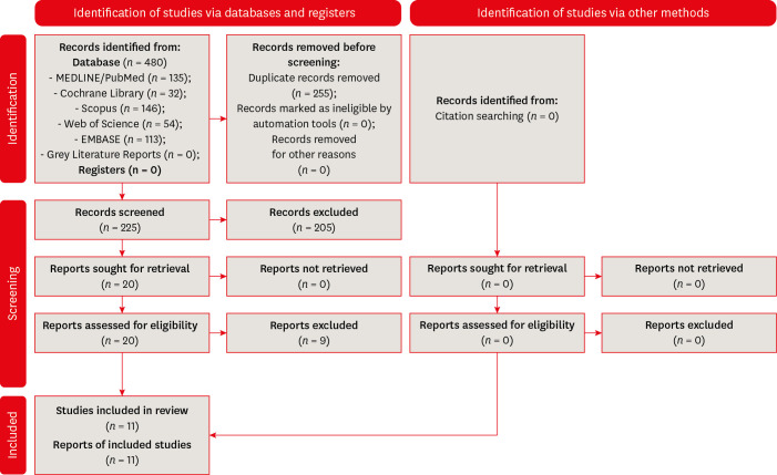
ePub Objectives This study aimed to investigate the influence of different obturation techniques compared to cold lateral compaction on the success rate of primary non-surgical endodontic treatments.
Materials and Methods Systematic searches were performed for studies published up to May 17th, 2022 in MEDLINE/PubMed, Cochrane Library, Web of Science, Scopus, EMBASE, and Grey Literature Reports. Randomized clinical trials and nonrandomized (nonrandomized clinical trials, prospective or retrospective) studies that evaluated the success rate of primary non-surgical endodontic treatments obturated with the cold lateral compaction (control) and other obturation techniques were included. The revised Cochrane risk of bias tools for randomized trials (RoB 2) and nonrandomized studies of interventions (ROBINS-I) were used to evaluate the risk of bias. The Grading of Recommendations Assessment, Development, and Evaluation (GRADE) tool was used to evaluate the certainty of evidence.
Results Eleven studies (4 randomized clinical trials (RCTs), 4 prospective, and 3 retrospectives) were included. Two RCTs were classified as having some concerns risk of bias and 2 as a low risk of bias. Two nonrandomized studies were classified as having a critical risk of bias and 5 as having a moderate risk of bias. The GRADE analysis demonstrated a very low to moderate certainty of evidence.
Conclusions This systematic review generally evidenced no differences in the success rate of primary non-surgical endodontic treatments when the cold lateral compaction technique and other obturation techniques are performed. Further well-designed studies are still necessary.
-
Citations
Citations to this article as recorded by
- Assessing Sealing Ability of C-Root SP Strontium Silicate Sealer With Different Obturation Techniques: An in vitro Study
Suixin Hu, Jianshe Li, Meng Xu, Laiqing Xu, Yangming Yin, Peng Xue, Liping Dong, Lin Wang, Huixia He, Ying Liu, Qiang Luo, Fei Chen
International Dental Journal.2026; 76(1): 109283. CrossRef - Effect of root canal filling techniques and materials on endodontic treatment outcomes: a systematic review and meta-analysis
Ahmed Mushtaq, Sura Alsanafi, Firas Elmsmari, José A. González, Marc Garcia-Font, Francesc Abella Sans, Kelvin I. Afrashtehfar, Paul V. Abbott
Scientific Reports.2026;[Epub] CrossRef - Dimensional and cross-sectional compatibility of contemporary nickel–titanium files and their corresponding gutta-percha cones: A micro-computed tomography study
Cem Sarkan, Faruk Haznedaroglu
The Saudi Dental Journal.2026;[Epub] CrossRef - Comparative Analysis Of Obturation Techniques In Endodontics: Lateral Vs. Thermoplasticized. Thermoplasticized
Juan Esteban Díaz Pacheco , Rómulo Guillermo López Torres , Verónica Alejandra Salame Ortíz
Salud, Ciencia y Tecnología.2025; 5: 1626. CrossRef - Effect of ultrasonic activation of endodontic sealers on root canal filling quality during the single-cone obturation procedure: a systematic review and meta-analysis of laboratory-based studies
Shuting Feng, Weiqing Zhou, Xiaojun Chu, Shuaimei Xu, Xiongqun Zeng
Odontology.2025; 113(4): 1380. CrossRef - In Vitro and In Vivo Evaluation of a New Experimental Polydimethylsiloxane-Based Endodontic Sealer
Fabiola Cardoso Maldonado, Cesar Gaitan Fonseca, Carlos Bermudez Jimenez, Luis Alejandro Aguilera Galaviz, Margarita L. Martinez-Fierro, Lorena Troncoso Vazquez, Martha Eugenia Reyes Ortiz
Journal of Functional Biomaterials.2025; 16(11): 402. CrossRef - Evaluation of three obturation techniques in 3D-printed models of oval canals with standardized prepared morphology: a micro-CT study
Wenjun Xia, Qisheng Gu, Yingshuang Song, Yunjia Liu, Xuetao Deng, Wenhao Qian
BMC Oral Health.2025;[Epub] CrossRef - Clinical and Radiographic Failure of Nonsurgical Endodontic Treatment and Retreatment Using Single-cone Technique With Calcium Silicate-based Sealers: A Systematic Review and Meta-analysis
Mohammad A. Sabeti, Negah Karimpourtalebi, Arash Shahravan, Omid Dianat
Journal of Endodontics.2024; 50(6): 735. CrossRef - Method of microbial decontamination of endodontic absorbent paper points: a randomised experimental study
O. A. Pavlovskaya, O. A. Kachanova, V. V. Volobuev, M. N. Mitropanova, A. R. Gazarova, V. Y. Zobenko, A. G. Uvarova
Pediatric dentistry and dental prophylaxis.2024; 24(2): 157. CrossRef - The Push-Out Bond Strength, Surface Roughness, and Antimicrobial Properties of Endodontic Bioceramic Sealers Supplemented with Silver Nanoparticles
Karla Navarrete-Olvera, Nereyda Niño-Martínez, Idania De Alba-Montero, Nuria Patiño-Marín, Facundo Ruiz, Horacio Bach, Gabriel-Alejandro Martínez-Castañón
Molecules.2024; 29(18): 4422. CrossRef - Clinical outcome of non-surgical root canal treatment using different sealers and techniques of obturation in 237 patients: A retrospective study
Mateusz Radwanski, Krystyna Pietrzycka, Tan Fırat Eyüboğlu, Mutlu Özcan, Monika Lukomska-Szymanska
Clinical Oral Investigations.2024;[Epub] CrossRef
- Assessing Sealing Ability of C-Root SP Strontium Silicate Sealer With Different Obturation Techniques: An in vitro Study
- 8,990 View
- 145 Download
- 10 Web of Science
- 11 Crossref

Research Articles
- Correlation between different methodologies used to evaluate the marginal adaptation of proximal dentin gingival margins elevated using a glass hybrid
- Hoda S. Ismail, Brian R. Morrow, Ashraf I. Ali, Rabab El. Mehesen, Franklin Garcia-Godoy, Salah H. Mahmoud
- Restor Dent Endod 2022;47(4):e36. Published online September 3, 2022
- DOI: https://doi.org/10.5395/rde.2022.47.e36
-
Abstract
PDFPubReaderePub
Objectives This study aimed to evaluate the effect of aging on the marginal quality of glass hybrid (GH) material used to elevate dentin gingival margins, and to analyze the consistency of the results obtained by 3
in vitro methods.Materials and Methods Ten teeth received compound class II cavities with subgingival margins. The dentin gingival margins were elevated with GH, followed by resin composite. The GH/gingival dentin interfaces were examined through digital microscopy, scanning electron microscopy (SEM) using resin replicas, and according to the World Dental Federation (FDI) criteria. After initial evaluations, all teeth were subjected to 10,000 thermal cycles, followed by repeating the same marginal evaluations and energy dispersive spectroscopy (EDS) analysis for the interfacial zone of 2 specimens. Marginal quality was expressed as the percentage of continuous margin at ×200 for microscopic techniques and as the frequency of each score for FDI ranking. Data were analyzed using the paired sample
t -test, Wilcoxon signed-rank test, and Pearson and Spearmen correlation coefficients.Results None of the testing techniques proved the significance of the aging factor. Moderate and strong significant correlations were found between the testing techniques. The EDS results suggested the presence of an ion-exchange layer along the GH/gingival dentin interface of aged specimens.
Conclusions The marginal quality of the GH/dentin gingival interface defied aging by thermocycling. The replica SEM and FDI ranking results had stronger correlations with each other than either showed with the digital microscopy results.
-
Citations
Citations to this article as recorded by- Influence of Cavity Design, Bonding‐Banding Sequence, and Thermomechanical Loading on the Marginal Adaptation of Class II Resin Composite Restorations
Hoda Saleh Ismail, Ashraf Ibrahim Ali, Hanan Ahmed Nabil Soliman
Journal of Esthetic and Restorative Dentistry.2026;[Epub] CrossRef - Elemental and micromorphological analysis of ion releasing restoration/carious dentin interface
Alaa Esmat Abdelsalam, Hoda Saleh Ismail, Hamdi Hosni Hamama
Scientific Reports.2025;[Epub] CrossRef - Marginal Seating of Zirconium Monolithic Crown Restorations Utilising Different Types of Luting Agents - A Comparative Study
Zahraa M. Al-Hawwaz, Farah Adnan Taha, Adel F. Ibraheem, Lena S. Abdullah
Indian Journal of Dental Research.2025; 36(3): 303. CrossRef - Comparison of the biomechanical effects of the post-core crown, endocrown and inlay crown after deep margin elevation and its clinical significance
Feng Wu, Xiaomin Su, Yue Shi, Juan Bai, Jing Feng, Xilin Sun, Xuanqi Wang, Hongyan Wang, Jiayu Wen, Jie Kang
BMC Oral Health.2024;[Epub] CrossRef - Effect of different restorative systems and aging on marginal adaptation of resin composites to deep proximal margins
Hoda S. Ismail, Ashraf I. Ali
Journal of Esthetic and Restorative Dentistry.2024; 36(2): 346. CrossRef - Elevación de margen profundo, Revisión sistemática
Doris Stefania Abad Cordero, Silvia Alexandra Reinoso Ortiz, Dolores Aracely Cedeño Zambrano, Manuel León Velastegui
Anatomía Digital.2023; 6(4.3): 401. CrossRef
- Influence of Cavity Design, Bonding‐Banding Sequence, and Thermomechanical Loading on the Marginal Adaptation of Class II Resin Composite Restorations
- 3,624 View
- 56 Download
- 3 Web of Science
- 6 Crossref
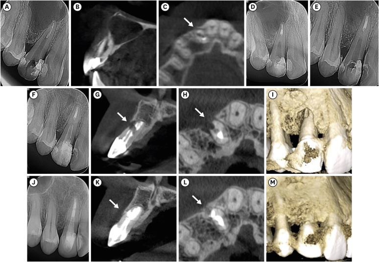
- Apical periodontitis in mesiobuccal roots of maxillary molars: influence of anatomy and quality of root canal treatment, a CBCT study
- Samantha Jannone Carrion, Marcelo Santos Coelho, Adriana de Jesus Soares, Marcos Frozoni
- Restor Dent Endod 2022;47(4):e37. Published online September 19, 2022
- DOI: https://doi.org/10.5395/rde.2022.47.e37
-
Abstract
PDFPubReaderePub
Objectives This study aimed to evaluate the prevalence of apical periodontitis (AP) in the mesiobuccal roots of root canal-treated maxillary molars.
Materials and Methods One thousand cone-beam computed tomography images of the teeth were examined by 2 dental specialists in oral radiology and endodontics. The internal anatomy of the roots, Vertucci’s classification, quality of root canal treatment, and presence of missed canals were evaluated; additionally, the correlation between these variables and AP was ascertained.
Results A total of 1,000 roots (692 first molars and 308 second molars) encompassing 1,549 canals were assessed, and the quality of the root canal filling in the majority (56.9%) of the canals was satisfactory. AP was observed in 54.4% of the teeth. A mesiolingual canal in the mesiobuccal root (MB2 canal) was observed in 54.9% of the images, and the majority (83.5%) of these canals were not filled. Significant associations were observed between the presence of an MB2 canal and the quality of the root canal filling and the presence of AP.
Conclusions AP was detected in more than half of the images. The MB2 canals were frequently missed or poorly filled.
-
Citations
Citations to this article as recorded by- Prevalence of Unfilled MB2 Canals and Their Association with Apical Periodontitis: A CBCT-Based Cross-Sectional Study in a German Population
Maythem Al Fartousi, Christian Ralf Gernhardt
Diagnostics.2026; 16(5): 796. CrossRef - When to perform cone beam computed tomography (CBCT) in primary root canal treatment? A CBCT-based cross-sectional study on the prevalence of MB2 canal in maxillary first molars
Bledar Lilaj, Elias Salzmann, Gernot Paul Hönigl, Rinet Dauti, Anton Dobsak, Sophie Pock, Barbara Cvikl
Dentomaxillofacial Radiology.2026; 55(4): 381. CrossRef - Deep learning–based detection of the second mesiobuccal canal in maxillary first molars using cone-beam computed tomography
Jiao Lin, Jialing Liu, Yuxin Jiang, Yang Liu, Bixin Wen, Shihao Li, Chenglong Li
BMC Oral Health.2026;[Epub] CrossRef - Anatomical Configuration of the MB2 Canal Using High-Resolution Cone-Beam Computed Tomography
Luciana Magrin Blank-Gonçalves, Emmanuel João Nogueira Leal da Silva, Monikelly do Carmo Chagas Nascimento, Ana Grasiela Limoeiro, Luiz Roberto Coutinho Manhães-Jr
Journal of Endodontics.2025; 51(5): 609. CrossRef - The Effect of Age and Gender on the Distance Between the Maxillary Sinus Cortical Bone and Maxillary Molars: A Cone-Beam Tomography Analysis
Thaysa Menezes Constantino, Marília Fagury Videira Marceliano-Alves, Vivian Ronquete, Ana Grasiela da Silva Limoeiro, Pablo Andres Amoroso-Silva, Mariano Simon Pedano, Tchilalo Boukpessi, Fábio Vidal, Thais Machado de Carvalho Coutinho
Sinusitis.2025; 9(1): 9. CrossRef - Retrospective study of the morphology of third maxillary molars among the population of Lower Silesia based on analysis of cone beam computed tomography
Anna Olczyk, Barbara Malicka, Katarzyna Skośkiewicz-Malinowska, Mohmed Isaqali Karobari
PLOS ONE.2024; 19(2): e0299123. CrossRef - Relationship between apical periodontitis and missed canals in mesio-buccal roots of maxillary molars: CBCT study
Badi B. Alotaibi, Kiran I. Khan, Muhammad Q. Javed, Smita D. Dutta, Safia S. Shaikh, Nawaf M. Almutairi
Journal of Taibah University Medical Sciences.2024; 19(1): 18. CrossRef - APICAL PERIODONTITIS IN MAXILLARY MOLARS WITH MISSED SECOND MESIO-BUCCAL ROOT CANAL: A CBCT STUDY
Cristina Coralia Nistor, Ioana Suciu , Ecaterina Ionescu , Anca Dragomirescu , Elena Coculescu , Andreea Baluta
Romanian Journal of Oral Rehabilitation.2024; 16(3): 100. CrossRef - Anatomic Comparison of Contralateral Maxillary Second Molars Using High-Resolution Micro-CT
Ghassan Dandache, Umut Aksoy, Mehmet Birol Ozel, Kaan Orhan
Symmetry.2023; 15(2): 420. CrossRef
- Prevalence of Unfilled MB2 Canals and Their Association with Apical Periodontitis: A CBCT-Based Cross-Sectional Study in a German Population
- 4,553 View
- 72 Download
- 8 Web of Science
- 9 Crossref
- Calcium-doped zinc oxide nanocrystals as an innovative intracanal medicament: a pilot study
- Gabriela Leite de Souza, Thamara Eduarda Alves Magalhães, Gabrielle Alves Nunes Freitas, Nelly Xiomara Alvarado Lemus, Gabriella Lopes de Rezende Barbosa, Anielle Christine Almeida Silva, Camilla Christian Gomes Moura
- Restor Dent Endod 2022;47(4):e38. Published online October 4, 2022
- DOI: https://doi.org/10.5395/rde.2022.47.e38
-
Abstract
PDFPubReaderePub
Objectives This study investigated the cytotoxicity, radiopacity, pH, and dentinal tubule penetration of a paste of 1.0% calcium-doped zinc oxide nanocrystals (ZnO:1.0Ca) combined with propylene glycol (PRG) or polyethylene glycol and propylene glycol (PEG-PRG).
Materials and Methods The pastes were prepared by mixing calcium hydroxide [Ca(OH)2] or ZnO:1.0Ca with PRG or a PEG-PRG mixture. The pH was evaluated after 24 and 96 hours of storage in deionized water. Digital radiographs were acquired for radiopacity analysis and bubble counting of each material. The materials were labeled with 0.1% fluorescein and applied to root canals, and images of their dentinal tubule penetration were obtained using confocal laser scanning microscopy. RAW264.7 macrophages were placed in different dilutions of culture media previously exposed to the materials for 24 and 96 hours and tested for cell viability using the MTT assay. Analysis of variance and the Tukey test (
α = 0.05) were performed.Results ZnO:1.0Ca materials showed lower viability at 1:1 and 1:2 dilutions than Ca(OH)2 materials (
p < 0.0001). Ca(OH)2 had higher pH values than ZnO:1.0Ca at 24 and 96 hours, regardless of the vehicle (p < 0.05). ZnO:1.0Ca pastes showed higher radiopacity than Ca(OH)2 pastes (p < 0.01). No between-material differences were found in bubble counting (p = 0.0902). The ZnO:1.0Ca pastes had a greater penetration depth than Ca(OH)2 in the apical third (p < 0.0001).Conclusions ZnO:1.0Ca medicaments presented higher penetrability, cell viability, and radiopacity than Ca(OH)2. Higher values of cell viability and pH were present in Ca(OH)2 than in ZnO:1.0Ca.
-
Citations
Citations to this article as recorded by-
Nano calcium zincate-assisted synthesis of benzo[
d
]thiazol-2-yl phenylisoxazoles: quantum computational,
in silico
molecular docking simulations and DNA interaction
A. K. Smitha, V. Srinivasa Murthy, B. Vinay Kumar, M. Sennappan, A. H. Shridhar, Lohit Naik, K. Yogendra, N. Madhusudhana
Nucleosides, Nucleotides & Nucleic Acids.2026; 45(3): 261. CrossRef - Nanomaterial-Enhanced Dentistry: A Clinical Perspective
Selvam Manoj, Radhakrishnan Sreena, Rajkumar Divya, Starlin Ebinesh, Shenbagaraman Akshaya, Srikumar Sugantha Angel, Arputharaj Joseph Nathanael
ACS Biomaterials Science & Engineering.2025; 11(8): 4671. CrossRef
-
Nano calcium zincate-assisted synthesis of benzo[
d
]thiazol-2-yl phenylisoxazoles: quantum computational,
in silico
molecular docking simulations and DNA interaction
- 2,812 View
- 37 Download
- 2 Web of Science
- 2 Crossref
- The prevalence and characteristics of external cervical resorption based on cone-beam computed tomographic imaging: a cross-sectional study
- Matheus Diniz Ferreira, Matheus Barros-Costa, Felipe Ferreira Costa, Deborah Queiroz Freitas
- Restor Dent Endod 2022;47(4):e39. Published online October 11, 2022
- DOI: https://doi.org/10.5395/rde.2022.47.e39
-
Abstract
PDFPubReaderePub
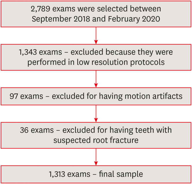
Objectives This study investigated the prevalence and characteristics of external cervical resorption (ECR) regarding sex, age, tooth, stages of progression, and portal of entry, using cone-beam computed tomography (CBCT) scans.
Materials and Methods CBCT scans of 1,313 patients from a Brazilian subpopulation comprising 883 female and 430 male patients (mean age, 55.2 years), acquired using a PreXion 3D CBCT unit, were evaluated. All permanent teeth included in the scans were evaluated for the presence of ECR according to the 3-dimensional classification and the portal of entry. The association between the presence of ECR and the factors studied was assessed using the χ2 test. Intra-observer agreement was analyzed with the kappa test (
α = 0.05).Results In total, 6,240 teeth were analyzed, of which 84 (1.35%) were affected by ECR. A significant association was found between the presence of ECR and sex, with a higher prevalence in male patients (
p = 0.002). The most frequently affected teeth were the mandibular and maxillary central incisors. The most common height was the mid-third of the root. For the portal of entry, 44% of cases were on the proximal surfaces, 40.5% on the lingual/palatal surface and 15.5% on the buccal surface. Intra-observer agreement was excellent.Conclusions The prevalence of ECR was 1.35%, with a higher prevalence in male patients and a wide age distribution. The mandibular and maxillary central incisors were the most commonly affected teeth, and cases of ECR most frequently showed a height into the mid-third of the root and proximal entry.
-
Citations
Citations to this article as recorded by- Features of external root resorption as predictors of disease progression: A CBCT cross-sectional study
Tânia Maria Soares Reis, Daniella Ribeiro Ferrari, Rafael Binato Junqueira, Priscila Dias Peyneau, Eduardo Murad Villoria, Maria Augusta Visconti, Francielle Silvestre Verner
Odontology.2026; 114(2): 720. CrossRef - External Cervical Resorption Treatment: A Single‐Center Retrospective Cohort Study of Cases Treated Over a 20‐Year Period
Terrell F. Pannkuk
Dental Traumatology.2026; 42(4): 446. CrossRef - Conservative Management of Class III Invasive Cervical Resorption Using the Heithersay Technique: A Case Report
Kalyana Ponangi
Cureus.2026;[Epub] CrossRef - Non‐Interventional Resolution of External Cervical Root Resorption: A Case Report
Farnam Pourreza-Jorshari, Georgios Patouris, Faraz Pourreza-Jorshari, Lee Feinberg, Shalini Kanagasingam, Hannah Wesley
Case Reports in Dentistry.2026;[Epub] CrossRef - Survival of single-tooth implants in sites with and without history of external cervical root resorption: a register-based cohort study
Fernando José Mota de Almeida, Jonas Norberg Cedering, Mattias Pettersson
BMC Oral Health.2026;[Epub] CrossRef - Reabsorción cervical externa. Informe de un caso // External cervical resorption: A case report
Osvaldo Zmener
Revista de la Asociación Odontológica Argentina.2026;[Epub] CrossRef - Prise en charge des lésions cervicales
C. Mocquot, L. Detzen, I. Fontanille, B. Orlik, F. Decup
EMC - Médecine buccale.2025; 18(3): 1. CrossRef - Prevalence and Characterization of External Cervical Resorption Using Cone Beam Computed Tomography
Isadora Carneiro Pereira Machado, Marilia Oliveira Morais, Adriana Lustosa Pereira Bicalho, Patricia Helena Pereira Ferrari, Juliano Martins Bueno, José Luiz Cintra Junqueira, Mariana Quirino Silveira Soares
Journal of Endodontics.2024; 50(2): 164. CrossRef - Influence of tube current and metal artifact reduction on the diagnosis of external cervical resorption in teeth adjacent to a dental implant in CBCT: an ex-vivo study
Thamiles Gonzalez-Passos, Matheus Barros-Costa, Matheus L Oliveira, Deborah Queiroz Freitas
Clinical Oral Investigations.2024;[Epub] CrossRef - Maxillary anterior teeth with extensive root resorption treated with multidisciplinary approach: A case report
Thais Machado de Carvalho Coutinho, Carollyne Souza Campello, Juliana Pires Abdelnur, Vivian Ronquete, Carlos Henrique Sardenberg Pereira, Marilia F Marceliano-Alves
International Journal of Case Reports and Images.2023; 14(1): 8. CrossRef - Clinical and radiographic features of external cervical resorption – An observational study
Shanon Patel, Francesc Abella, Kreena Patel, Paul Lambrechts, Nassr Al‐Nuaimi
International Endodontic Journal.2023; 56(12): 1475. CrossRef
- Features of external root resorption as predictors of disease progression: A CBCT cross-sectional study
- 5,200 View
- 67 Download
- 9 Web of Science
- 11 Crossref
- Evaluation of blood clot, platelet-rich plasma, and platelet-rich fibrin–mediated regenerative endodontic procedures in teeth with periapical pathology: a CBCT study
- Swati Markandey, Haridas Das Adhikari
- Restor Dent Endod 2022;47(4):e41. Published online October 21, 2022
- DOI: https://doi.org/10.5395/rde.2022.47.e41
-
Abstract
PDFSupplementary MaterialPubReaderePub
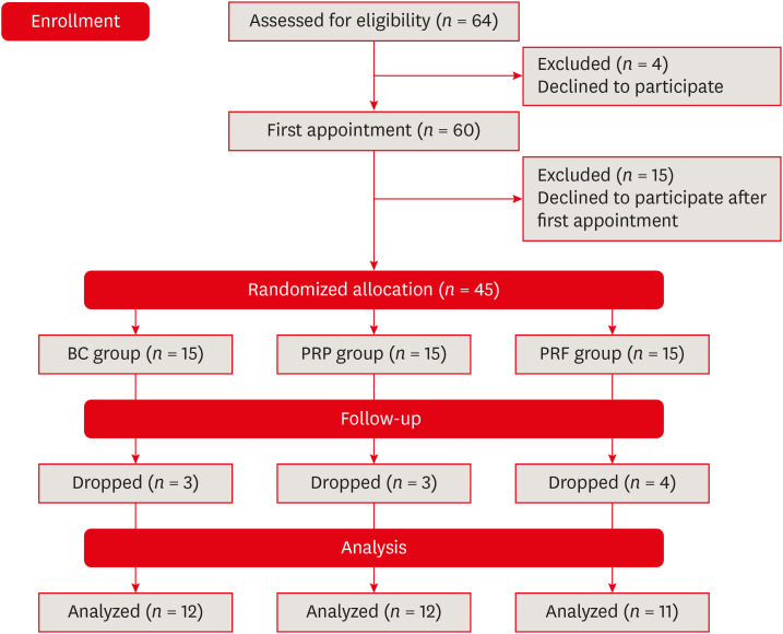
Objectives This study compared the clinical and radiological outcomes of regenerative endodontic procedures (REPs) using blood clots (BCs), platelet-rich plasma (PRP), and platelet-rich fibrin (PRF) through intraoral periapical radiography (IOPAR) and cone-beam computed tomography (CBCT).
Materials and Methods Forty-five single-rooted necrotic teeth with periapical pathology were randomly allocated to receive BC, PRP, or PRF as an individual scaffold. Outcomes were evaluated in 35 teeth in 23 patients with a follow-up period of 12–24 months through qualitative IOPAR scoring and quantitative CBCT measurements. Healing of periapical lesions and in immature teeth, changes in the apical foramen diameter (AFD), root wall thickness (RWT), and root length (RL) were assessed. A
p value less than 0.05 was considered to indicate statistical significance.Results All teeth were asymptomatic except 1 in the PRP group. Periapical lesion healing was seen in all except 2 teeth in the BC group and 3 in the PRP group. Both IOPAR and CBCT revealed no significant differences in bone healing or changes in AFD, RWT, and RL among the 3 groups. A positive pulp sensibility response to the cold test was seen in 2 teeth in the BC group, but none to the electric pulp test. Intracanal calcification (ICC) was evident in more teeth in the BC group than in the PRP and PRF groups, and was also significantly higher in immature teeth.
Conclusions Our results revealed that BC, PRP, and PRF have similar potential as scaffolds in REPs, and ICC may be a concern for long-term outcomes.
-
Citations
Citations to this article as recorded by- Regenerative Endodontics and Stem Cell-Based Therapies – A Systematic Review
Wjoud Ahmed Alshamrani, Sarah Sulaiman Alzarea, Joud Khalid Alabbas, Ayah Khalid Alabbas, Mawiyah Ibrahim Aljaddua, Osama Khattak, Rakhi Issrani
Journal of Pharmacy and Bioallied Sciences.2026; 18(Suppl 1): S29. CrossRef - Collagen Scaffolds in Regenerative Endodontic Procedures: Current Evidence, Limitations, and Future Perspectives
Qiong-Ling Shi, Xiao Zhu, Chen Chen, Jing-Yi Chen, Dan-Yang Lu, Ying Shi, Yan-Qi Chen, Zhi-Fang Wu
Polymers.2026; 18(7): 894. CrossRef - Clinical and Radiographic Evaluation of Platelet-rich Fibrin as an Adjunct to Conventional Endodontic Therapy in Periapical Lesions
Neelam D Chandwani, Anjali Gayke, Ranjana Deshmukh, FNU Reetika, Anusi Chikro, Shweta Habbu
Journal of Modern Dentistry Insights.2026; 2(1): 4. CrossRef - Efficacy of combined scaffolds and sodium hypochlorite in regenerative endodontics of immature teeth (in-vivo study)
Hanan Arb, Abeer Darrag, Neveen Shaheen, Dina Attia
Tanta Dental Journal.2026; 23(2): 240. CrossRef - Regenerative potential of concentrated growth factor compared to platelet-rich fibrin in treatment of necrotic mature teeth: a randomized clinical trial
Taghreed Salah, Wael Hussein, Heba Abdelkafy
BDJ Open.2025;[Epub] CrossRef - Clinical and radiographic outcomes of non-surgical retreatment of mature maxillary incisors using two regenerative endodontic techniques in adolescents: a 24-month randomized clinical trial
Ahmad Abdel Hamid Elheeny, Sherif Shafik EL Bahnasy, Yassmin Mohamed ElMakawi, Mohammed Turky, Eman Farouk Ahmed, Norhan Khaled Omar Wahba
BDJ Open.2025;[Epub] CrossRef - Clinical and Radiographic Outcomes of Autologous Platelet-Rich Products in Regenerative Endodontics: A Systematic Review and Meta-Analysis
Raewyn Huang, Wei Chen, Matthew Fang, Ove A. Peters, Sepanta Hosseinpour
Dentistry Journal.2025; 13(6): 236. CrossRef - Regenerative Endodontics in the Treatment of Periapical Lesions of Endodontic Origin: A Systematic Review of Randomized Controlled Trials
BelHaj Salah Kawthar, Berrima Fatma, Boukhris Hanen, Gnaba Imen, Ben Youssef Souha
The Journal of Contemporary Dental Practice.2025; 26(6): 623. CrossRef - Advanced Platelet-Rich Fibrin Plus Sealed Exclusively with Glass Ionomer Cement: Setting a New Standard for Healing, Aesthetics and Predictive Modelling in Regenerative Endodontics
Dubravka Turjanski, Dragutin Lisjak, Petra Bučević Sojčić, Jelena Valpotić, Tea Borojević Renić, Kristina Goršeta, Domagoj Glavina
Materials.2025; 18(18): 4421. CrossRef - Evaluation of different scaffolds and intracanal medications in revascularization of nonvital immature permanent teeth
Mona M. A. Sameia, Abeer M. Darrag, Neveen A. Shaheen, Dina A. Attia
Tanta Dental Journal.2025; 22(4): 693. CrossRef - Biomimetic pulp scaffolds prepared from extracellular matrix derived from stem cells from human exfoliated deciduous teeth promote pulp–dentine complex regeneration
Ning Yang, Rou Shen, Wenxiao Yang, Shengcai Zhang, Tianxing Gong, Yao Liu
International Endodontic Journal.2024; 57(9): 1279. CrossRef - Arrest and Repair of Inflammatory Root Resorption After an Endodontic Regeneration Procedure – A Hypothesis and Case Report
Arieh Y. Kaufman, Bill Kahler
Journal of Endodontics.2024; 50(12): 1743. CrossRef - Effect of Platelet Rich Plasma in Regenerative Endodontic Treatment: A Review of Clinical Trials
Hojat Rezazadeh, Mehrnaz Okhovatfard
Research Journal of Pharmacy and Technology.2023; : 5562. CrossRef
- Regenerative Endodontics and Stem Cell-Based Therapies – A Systematic Review
- 6,532 View
- 169 Download
- 7 Web of Science
- 13 Crossref
-
The influence of sodium hypochlorite concentration on the fibrin structure of human blood clots and transforming growth factor-beta 1 release: an
ex vivo study - Anisha Mishra, Velmurugan Natanasabapathy, Nandini Suresh
- Restor Dent Endod 2022;47(4):e42. Published online October 31, 2022
- DOI: https://doi.org/10.5395/rde.2022.47.e42
-
Abstract
PDFSupplementary MaterialPubReaderePub
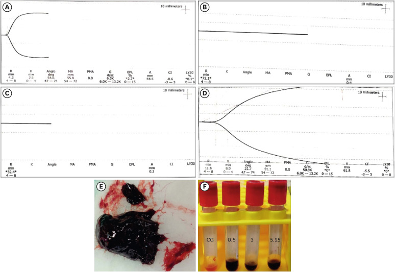
Objective This study investigated the effects of various concentrations of sodium hypochlorite (NaOCl) on human whole-blood clotting kinetics, the structure of the blood clots formed, and transforming growth factor (TGF)-β1 release.
Materials and Methods Human whole blood was collected from 5 healthy volunteers and divided into 4 groups: CG (control, 0.5 mL of blood), BN0.5 (0.5 mL of blood with 0.5 mL of 0.5% NaOCl), BN3 (0.5 mL of blood with 0.5 mL of 3% NaOCl), and BN5.25 (0.5 mL of blood with 0.5 mL of 5.25% NaOCl). The effects of NaOCl on clotting kinetics, structure of fibrin and cells, and release of TGF-β1 were assessed using thromboelastography (TEG), scanning electron microscopy (SEM), and enzyme-linked immunosobent assay, respectively. Statistical analysis was conducted using the Kruskal Wallis and Mann-Whitney
U tests, followed by thepost hoc Dunn test. Ap value < 0.05 indicated statistical significance.Results The blood samples in BN0.5 and BN3 did not clot, whereas the TEG of BN5.25 showed altered clot formation. Samples from the CG and BN3 groups could only be processed with SEM, which showed that the latter lacked fibrin formation and branching of fibers, as well as clumping of red blood cells with surface roughening and distortion. TGF-β1 release was significantly highest in BN3 when all groups were compared to CG (
p < 0.05).Conclusions Each concentration of NaOCl affected the release of TGF-β1 from blood clots and altered the clotting mechanism of blood by affecting clotting kinetics and cell structure.
-
Citations
Citations to this article as recorded by- Evaluation of the cytotoxicity of a broad-spectrum antiseptic using a model of erythrocyte hemolysis in an in vitro experiment
S. P. Rubnikovich, O. E. Bekjanova, L. E. Khasanova, Sh. F. Shamsieva, S. X. Alimova, M. M. Astanakulova, N. T. Babadjanova, X. Sh. Mirzaev
Proceedings of the National Academy of Sciences of Belarus, Medical series.2026; 23(2): 95. CrossRef - Effect of Adjunctive Ozone Application Protocols on Dentin-Derived Growth Factor Release: An In Vitro Study
Sude Göbüt, Melis Oya Ateş, Ali Keleş, Fatma Avcıoğlu
Journal of Clinical Medicine.2026; 15(11): 4277. CrossRef - Cytotoxic Effects of Synthetic and Herbal Endodontic Irrigants on Human Red Blood Cells: An In Vitro Study
Panna Mangat, Bhaviya Chandel, Mampi Biswas, Sara Trivedy, Akshata Gupta, Nayan Shree, Seema Gupta
Cureus.2025;[Epub] CrossRef
- Evaluation of the cytotoxicity of a broad-spectrum antiseptic using a model of erythrocyte hemolysis in an in vitro experiment
- 3,079 View
- 39 Download
- 1 Web of Science
- 3 Crossref
- Surface gloss, gloss retention, and color stability of 2 nano-filled universal resin composites
- Gustavo Fabián Molina, Ricardo Juan Cabral, Ignacio Mazzola, Michael Burrow
- Restor Dent Endod 2022;47(4):e43. Published online October 31, 2022
- DOI: https://doi.org/10.5395/rde.2022.47.e43
-
Abstract
PDFPubReaderePub
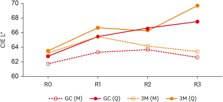
Objectives This study compared the surface gloss (SG), gloss retention (GR), and color stability (CS) of 2 universal resin composites after chemical (CA) and mechanical (MA) aging.
Materials and Methods Twenty disc-shaped samples of G-ænial A´Chord (GC-Europe) and Filtek Universal (3M-ESPE) were polished with sequential abrasive papers. For CA, specimens were stored in 1 mL of 75% ethanol for 15 days at 37°C, and readings (SG, GR, and CS) were obtained at baseline and 5, 10, and 15 days. For MA, specimens were subjected to 10,750 simulated brushing cycles. SG and CS were evaluated after every 3,583 cycles. SG was measured with a glossmeter (geometrical configuration: 60°), and values were expressed in gloss units. Color was measured with a spectrophotometer using the CIE-L*a*b* color system. The Student’s
t -test, 1-way analysis of variance, and Scheffé test were used for statistical analysis (α = 0.05).Results G-ænial presented significantly higher SG values than Filtek (
p = 0.02), with GR reductions of 5.2% (CA) and 5.3% (MA) for G-ænial and 7.6% (CA) and 7.2% (MA) for Filtek. The aging protocol had no statistically significant effect on SG or GR (p = 0.25) from baseline to the final readings. G-ænial–MA presented the lowest color difference(∆E = 1.8), and G-ænial–CA and Filtek–CA had the largest changes (∆E = 8.6 and∆E = 11.8, respectively).Conclusion G-ænial presented higher SG values and better CS. Both restorative materials demonstrated acceptable GR and CS. Aging protocols impacted these properties negatively.
-
Citations
Citations to this article as recorded by- Color stability, surface roughness, and surface morphology of universal composites
Mohammad Meniawi, Nazlı Şirinsükan, Esra Can
Odontology.2026; 114(1): 149. CrossRef - RETRACTED: Surface gloss and micro‐CT analysis of additively and subtractively manufactured resin composites and zirconia after simulated tooth brushing with different bristle types and toothpaste formulations: An in vitro study
Ahmet Faruk Ertürk, Rafat Sasany, Seyed Ali Mosaddad, Merve Yelken Kendirci
Journal of Prosthodontics.2026; 35(5): 1. CrossRef - Surface roughness of composite resins subjected to brushing with whitening toothpastes: an in vitro study
Nicolle Madruga Ramos FERREIRA, Vinicius Funghetto LIPPERT, Amanda Baptista da Silva HECK, Ana Maria SPOHR, Marcel Ferreira KUNRATH, Carlos Alberto FELDENS, Paulo Floriani KRAMER
Brazilian Oral Research.2025;[Epub] CrossRef - Evaluation of Color Stability and Surface Abrasion of Nano-modified Glass Ionomer Cement with Dentifrices: An In Vitro Study
Jessy Paulraj, Subhabrata Maiti, Harini Palani
International Journal of Prosthodontics and Restorative Dentistry.2025; 15(1): 10. CrossRef - Security inks with silanized zinc oxide quantum dots and cellulose ethers for the safeguarding of cultural heritage objects
Andrea Louise Matulac, Themis Krasoudaki, Francesca Battaglia, Carlo Spadoni, Martina Piletti, Daniela Iacopino, Rodorico Giorgi
Applied Materials Today.2025; 44: 102718. CrossRef - Gastric acid challenge: Mechanical proficiency and surface gloss of tooth-colored restorative materials
Ozge Gizem Yenidunya, Tugba Misilli, Ebru Yilmaz
BMC Oral Health.2025;[Epub] CrossRef - Impact of in-office bleaching agents on the optical properties of universal resin composites: an in vitro analysis
Esra Özyurt, Merve Nezir, Hanife Altınışık, Mediha Büyükgöze Dindar
BMC Oral Health.2025;[Epub] CrossRef - Aging and Staining Effects on Optical Properties of Flowable Composites
M. M. Sly, Y. Korkmaz‐Ceyhan, F. Dini, R. L. Ocampo Escobedo, E. Abram, R. D. Paravina
Journal of Biomedical Materials Research Part B: Applied Biomaterials.2025;[Epub] CrossRef - Evaluation of The Effect of In-Office Bleaching Agent on Mechanical Properties of Different Single-Shade Resin Composites: An In-Vitro Study
Merve Nezir, Hanife Altınışık, Esra Özyurt
ADO Klinik Bilimler Dergisi.2025; 14(3): 197. CrossRef - Surface gloss changes in 3D-printed resin materials following different polishing procedures and aging protocols
Ilayda Yumak, Hayal Boyacioglu, Lezize Sebnem Turkun
BMC Oral Health.2025;[Epub] CrossRef - The Gloss Retention of Esthetic Restorations Following Simulated Brushing with Charcoal Oral Products: An In-Vitro Study
Fadia Awadalkreem, Nancy S Farghal, Nadin A Abouelhonoud, Raiyan I Khan
The Journal of Contemporary Dental Practice.2024; 25(5): 473. CrossRef - Effect of different finishing and polishing systems on surface properties of universal single shade resin-based composites
Ghada Alharbi, Hend NA Al Nahedh, Loulwa M. Al-Saud, Nourah Shono, Ahmed Maawadh
BMC Oral Health.2024;[Epub] CrossRef - The Effect of Chemical Degradation and Polishing on the Gloss of Composite Dental Materials
Ružica Zovko, Stipo Cvitanović, Mirela Mabić, Zdenko Šarac, Anka Ćorić, Domagoj Glavina, Kristina Goršeta
Materials.2023; 16(10): 3727. CrossRef
- Color stability, surface roughness, and surface morphology of universal composites
- 4,329 View
- 65 Download
- 10 Web of Science
- 13 Crossref
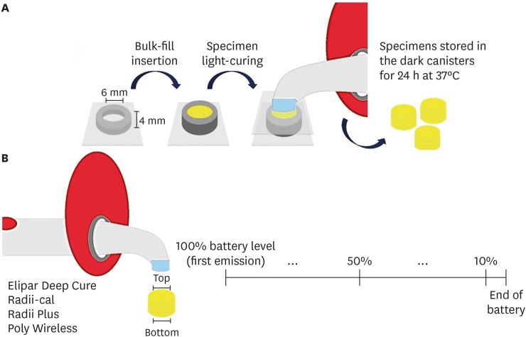
- Relationship between battery level and irradiance of light-curing units and their effects on the hardness of a bulk-fill composite resin
- Fernanda Harumi Oku Prochnow, Patricia Valéria Manozzo Kunz, Gisele Maria Correr, Marina da Rosa Kaizer, Carla Castiglia Gonzaga
- Restor Dent Endod 2022;47(4):e45. Published online November 3, 2022
- DOI: https://doi.org/10.5395/rde.2022.47.e45
-
Abstract
PDFPubReaderePub
Objectives This study evaluated the relationship between the battery charge level and irradiance of light-emitting diode (LED) light-curing units (LCUs) and how these variables influence the Vickers hardness number (VHN) of a bulk-fill resin.
Materials and Methods Four LCUs were evaluated: Radii Plus (SDI), Radii-cal (SDI), Elipar Deep Cure (Filtek Bulk Fill, 3M Oral Care), and Poly Wireless (Kavo Kerr). Irradiance was measured using a radiometer every ten 20-second activations until the battery was discharged. Disks (4 mm thick) of a bulk-fill resin (Filtek Bulk Fill, 3M Oral Care) were prepared, and the VHN was determined on the top and bottom surfaces when light-cured with the LCUs with battery levels at 100%, 50% and 10%. Data were analyzed by 2-way analysis of variance, the Tukey’s test, and Pearson correlations (α = 5%).
Results Elipar Deep Cure and Poly Wireless showed significant differences between the irradiance when the battery was fully charged versus discharged (10% battery level). Significant differences in irradiance were detected among all LCUs, within each battery condition tested. Hardness ratios below 80% were obtained for Radii-cal (10% battery level) and for Poly Wireless (50% and 10% battery levels). The battery level showed moderate and strong, but non-significant, positive correlations with the VHN and irradiance.
Conclusions Although the irradiance was different among LCUs, it decreased in half of the devices along with a reduction in battery level. In addition, the composite resin effectiveness of curing, measured by the hardness ratio, was reduced when the LCUs’ battery was discharged.
-
Citations
Citations to this article as recorded by- Effect of erosive solutions and thermal cycling on the surface properties of universal injectable and regular consistency resin composites
Ahmed Abbas Rhaif, Hoda Saleh Ismail, Tawakol Ahmed Ahmed Enab, Nadia Mohamed Zaghloul
BMC Oral Health.2025;[Epub] CrossRef - Effect of Battery Level During Successive Charging Cycles on the Performance of Certified and Low-cost Uncertified Light-curing Units Available on E-commerce
TS Peres, G Oliveira, SP da Silva Sakamoto, M da Silva Faria, HL Carlo, CJ Soares
Operative Dentistry.2024; 49(6): 673. CrossRef - Influence of Exposure Distance on Light Irradiance of Dental Curing Lamps in Various Operating Modes
Anna Lehmann, Kacper Nijakowski, Marta Mroczyk, Filip Podgórski, Beata Czarnecka, Anna Surdacka
Applied Sciences.2024; 14(21): 9999. CrossRef - ESTADO DA INTENSIDADE LUMINOSA DAS LÂMPADAS DE FOTOPOLIMERIZAÇÃO DAS CLÍNICAS ODONTOLÓGICAS DOS CENTROS DE SAÚDE DA CIDADE DE CUENCA
Milton Alexis Quinchiguano Caraguay, David Ismael Bravo Achundia , Esteban Eduardo Amoroso Calle, Manuel Estuardo Bravo Calderon
RECISATEC - REVISTA CIENTÍFICA SAÚDE E TECNOLOGIA - ISSN 2763-8405.2023; 3(6): e36296. CrossRef
- Effect of erosive solutions and thermal cycling on the surface properties of universal injectable and regular consistency resin composites
- 2,890 View
- 42 Download
- 5 Web of Science
- 4 Crossref
Case Report
- Endodontic micro-resurgery and guided tissue regeneration of a periapical cyst associated to recurrent root perforation: a case report
- Fernando Córdova-Malca, Hernán Coaguila-Llerena, Lucía Garré-Arnillas, Jorge Rayo-Iparraguirre, Gisele Faria
- Restor Dent Endod 2022;47(4):e35. Published online September 3, 2022
- DOI: https://doi.org/10.5395/rde.2022.47.e35
-
Abstract
PDFPubReaderePub
Although the success rates of microsurgery and micro-resurgery are very high, the influence of a recurrent perforation combined with radicular cyst remains unclear. A 21-year-old white female patient had a history of root perforation in a previously treated right maxillary lateral incisor. Analysis using cone-beam computed tomography (CBCT) revealed an extensive and well-defined periapical radiolucency, involving the buccal and palatal bone plate. The perforation was sealed with bioceramic material (Biodentine) in the pre-surgical phase. In the surgical phase, guided tissue regeneration (GTR) was performed by combining xenograft (lyophilized bovine bone) and autologous platelet-rich fibrin applied to the bone defect. The root-end preparation was done using an ultrasonic tip. The retrograde filling was performed using a bioceramic material (Biodentine). Histopathological analysis confirmed a radicular cyst. The patient returned to her referring practitioner to continue the restorative procedures. CBCT analysis after 1-year recall revealed another perforation in the same place as the first intervention, ultimately treated by micro-resurgery using the same protocol with GTR, and a bioceramic material (MTA Angelus). The 2-year recall showed healing and bone neoformation. In conclusion, endodontic micro-resurgery with GTR showed long-term favorable results when a radicular cyst and a recurrent perforation compromised the success.
-
Citations
Citations to this article as recorded by- Outcome of endodontic micro-resurgery: A systematic review
Faisal Alnassar, Riyadh Alroomy, Qamar Hashem, Abdullah Alqedairi, Nabeel Almotairy
Saudi Endodontic Journal.2025; 15(2): 112. CrossRef - Platelet-Rich Plasma and Platelet-Rich Fibrin in Endodontics: A Scoping Review
Simão Rebimbas Guerreiro, Carlos Miguel Marto, Anabela Paula, Joana Rita de Azevedo Pereira, Eunice Carrilho, Manuel Marques-Ferreira, Siri Vicente Paulo
International Journal of Molecular Sciences.2025; 26(12): 5479. CrossRef - Non-surgical Approach to a Maxillary Cyst-Like Lesion: Orthograde Endodontic Treatment With Neodymium-Doped Yttrium Aluminum Garnet (Nd:YAG) Decontamination of the Canal System
Beatrice Spaggiari, Paolo Vescovi, Silvia Pizzi, Roberta Iaria, Ilaria Giovannacci
Cureus.2025;[Epub] CrossRef - Persistent Periradicular Lesion Associated With Concurrent Root Fracture and Odontogenic Keratocyst: A Case Report
Mehdi Vatanpour, Fatemeh Rezaei
Clinical Case Reports.2025;[Epub] CrossRef - Management of Apico-marginal Defects With Endodontic Microsurgery and Guided Tissue Regeneration: A Report of Thirteen Cases
Abayomi O. Baruwa, Jorge N.R. Martins, Mariana D. Pires, Beatriz Pereira, Pedro May Cruz, António Ginjeira
Journal of Endodontics.2023; 49(9): 1207. CrossRef
- Outcome of endodontic micro-resurgery: A systematic review
- 4,403 View
- 82 Download
- 4 Web of Science
- 5 Crossref
Erratum
- Erratum: Correction of missing funding information. A new phantom to evaluate the tissue dissolution ability of endodontic irrigants and activating devices
- Kimia Khoshroo, Brinda Shah, Alexander Johnson, John Baeten, Katherine Barry, Mohammadreza Tahriri, Mohamed S. Ibrahim, Lobat Tayebi
- Restor Dent Endod 2022;47(4):e44. Published online November 2, 2022
- DOI: https://doi.org/10.5395/rde.2022.47.e44
- 1,459 View
- 19 Download
 First
First Prev
Prev

