
Previous issues
- Page Path
- HOME > Browse articles > Previous issues
- Volume 46 (4); November 2021
-


Research Articles
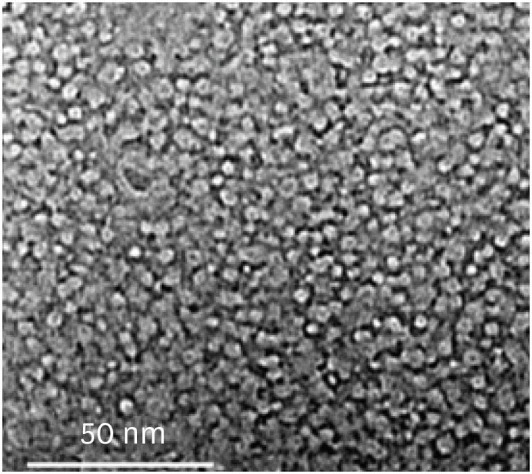
- Laboratory model to evaluate efficacy of an experimental titanium oxide nanofibers bleaching agent
- Clayton Tran, Ellin Choi, Brittany Watu, Udochukwu Oyoyo, Christopher Perry, So Ran Kwon
- Restor Dent Endod 2021;46(4):e47. Published online September 2, 2021
- DOI: https://doi.org/10.5395/rde.2021.46.e47
-
 Abstract
 PDF
PDF PubReader
 ePub
ePub Objectives This study aimed to use a laboratory model to evaluate the efficacy of an experimental bleaching agent.
Materials and Methods The model used human extracted molars that were treated and measured for bleaching efficacy. Teeth (
n = 50) were distributed into 5 groups: Negative control (NC): immersion in water for 8 hours; Nanofibers (NFs): Experimental titanium dioxide nanofibers with stirring and light activation for 8 hours; Whitestrips (WS): Crest 3D White Glamorous White Whitestrips, 2 applications daily for 30 minutes, 14 days; 1% hydrogen peroxide (HP) standard: 1% hydrogen peroxide for 8 hours; and 30% HP standard: 30% hydrogen peroxide for 8 hours. Instrumental measurements were performed using a spectrophotometer. Results were recorded at baseline, 1-day post-bleaching, and 1-week post-bleaching. Kruskal-Wallis procedure was used to determine differences in color change. Pearson correlation was used to evaluate the relationship between visual and instrumental measurements. Tests of hypotheses were 2-sided with alpha = 0.05.Results There was no significant difference in color parameters (L1, a1, b1, and shade guide units [SGU]) at baseline (
p > 0.05). There was a significant difference among the groups for overall color change (ΔE*ab) and change in shade guide units (ΔSGU) at 1-day and 1-week post-bleaching (p < 0.05). The higher the HP concentration, the higher the color change as expressed in ΔSGU and ΔE*ab. The negative control exceeded the perceptibility threshold of ΔE* = 1.2 regardless of time point. NFs showed a decrease in chroma, but were not statistically different compared to the negative control.Conclusions The laboratory model was successful in screening an experimental bleaching agent.
-
Citations
Citations to this article as recorded by
- Evaluating the Efficacy of Titanium Dioxide Nanoparticles in Combination with Commonly Used Bleaching Agents: An In Vitro Study
Rajasekhar Vemareddy, Sudhakar Naidu, Bala Raju Korrai, Shanmukha Nagadevara, Someshwar Battu, Jyotsnanjali Thati, Sivaji Kavuri
World Journal of Dentistry.2024; 15(5): 377. CrossRef
- Evaluating the Efficacy of Titanium Dioxide Nanoparticles in Combination with Commonly Used Bleaching Agents: An In Vitro Study
- 2,700 View
- 17 Download
- 1 Crossref

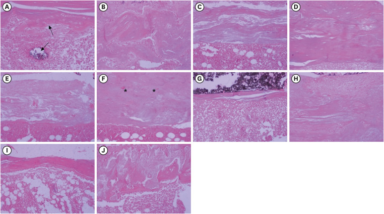
- Bone repair in defects filled with AH Plus sealer and different concentrations of MTA: a study in rat tibiae
- Jessica Emanuella Rocha Paz, Priscila Oliveira Costa, Albert Alexandre Costa Souza, Ingrid Macedo de Oliveira, Lucas Fernandes Falcão, Carlos Alberto Monteiro Falcão, Maria Ângela Area Leão Ferraz, Lucielma Salmito Soares Pinto
- Restor Dent Endod 2021;46(4):e48. Published online September 2, 2021
- DOI: https://doi.org/10.5395/rde.2021.46.e48
-
Abstract
PDFPubReaderePub
Objectives This study aimed to evaluate the effects on bone repair of different concentrations of mineral trioxide aggregate (MTA) added to AH Plus.
Materials and Methods Bone tissue reactions were evaluated in 30 rats (
Rattus norvegicus ) after 7 and 30 days. In the AH + MTA10, AH + MTA20, and AH + MTA30 groups, defects in the tibiae were filled with AH Plus with MTA in proportions of 10%, 20% and 30%, respectively; in the MTA-FILL group, MTA Fillapex was used; and in the control group, no sealer was used. The samples were histologically analyzed to assess bone union and maturation. The Kruskal-Wallis and Mann-Whitney tests were performed for multiple pairwise comparisons (p ≤ 0.05).Results At the 7-day time point, AH + MTA10 was superior to MTA-FILL with respect to bone union, and AH + MTA20 was superior to MTA-FILL with respect to bone maturity (
p < 0.05). At the 30-day time point, both the AH + MTA10 and AH + MTA20 experimental sealers were superior not only to MTA-FILL, but also to AH + MTA30 with respect to both parameters (p < 0.05). The results of the AH + MTA10 and AH + MTA20 groups were superior to those of the control group for both parameters and experimental time points (p < 0.05).Conclusions The results suggest the potential benefit of using a combination of these materials in situations requiring bone repair.
-
Citations
Citations to this article as recorded by- Analysis of the cytotoxicity and bioactivity of CeraSeal, BioRoot™ and AH Plus® sealers in pre-osteoblast lineage cells
Luciano Aparecido de Almeida-Junior, Giuliana de Campos Chaves Lamarque, Henry Herrera, Maya Fernanda Manfrin Arnez, Francine Lorencetti-Silva, Raquel Assed Bezerra Silva, Léa Assed Bezerra Silva, Francisco Wanderley Garcia Paula-Silva
BMC Oral Health.2024;[Epub] CrossRef - A Review of the research methods and progress of biocompatibility evaluation of root canal sealers
Xiliang Yang, Tianxia Zheng, Nuoya Yang, Zihan Yin, Wuliang Wang, Yuhong Bai
Australian Endodontic Journal.2023; 49(S1): 508. CrossRef - Effect of Vitapex Combined with AH-Plus Paste on Inflammation in Middle-Aged and Elderly Patients with Periodontal-Endodontic Disease
Rong Hu, Fulan Zhang, Xiangyu Guo, Youren Jing, Xiaowan Lin, Liping Tian, Min Tang
Computational and Mathematical Methods in Medicine.2022; 2022: 1. CrossRef
- Analysis of the cytotoxicity and bioactivity of CeraSeal, BioRoot™ and AH Plus® sealers in pre-osteoblast lineage cells
- 2,789 View
- 17 Download
- 4 Web of Science
- 3 Crossref
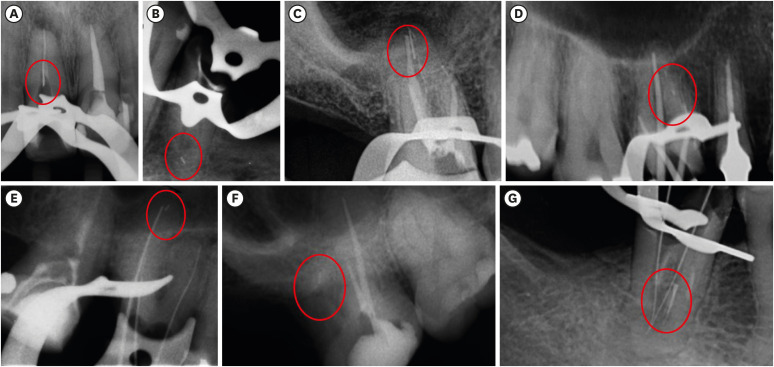
- Fracture incidence of Reciproc instruments during root canal retreatment performed by postgraduate students: a cross-sectional retrospective clinical study
- Liliana Machado Ruivo, Marcos de Azevedo Rios, Alexandre Mascarenhas Villela, Alexandre Sigrist de Martin, Augusto Shoji Kato, Rina Andrea Pelegrine, Ana Flávia Almeida Barbosa, Emmanuel João Nogueira Leal Silva, Carlos Eduardo da Silveira Bueno
- Restor Dent Endod 2021;46(4):e49. Published online September 9, 2021
- DOI: https://doi.org/10.5395/rde.2021.46.e49
-
Abstract
PDFPubReaderePub
Objectives To evaluate the fracture incidence of Reciproc R25 instruments (VDW) used during non-surgical root canal retreatments performed by students in a postgraduate endodontic program.
Materials and Methods From the analysis of clinical record cards and periapical radiographs of root canal retreatments performed by postgraduate students using the Reciproc R25, a total of 1,016 teeth (2,544 root canals) were selected. The instruments were discarded after a single use. The general incidence of instrument fractures and its frequency was analyzed considering the group of teeth and the root thirds where the fractures occurred. Statistical analysis was performed using the χ2 test (
p < 0.01).Results Seven instruments were separated during the procedures. The percentage of fracture in relation to the number of instrumented canals was 0.27% and 0.68% in relation to the number of instrumented teeth. Four fractures occurred in maxillary molars, 1 in a mandibular molar, 1 in a mandibular premolar and 1 in a maxillary incisor. A greater number of fractures was observed in molars when compared with the number of fractures observed in the other dental groups (
p < 0.01). Considering all of the instrument fractures, 71.43% were located in the apical third and 28.57% in the middle third (p < 0.01). One instrument fragment was removed, one bypassed, while in 5 cases, the instrument fragment remained inside the root canal.Conclusions The use of Reciproc R25 instruments in root canal retreatments carried out by postgraduate students was associated with a low incidence of fractures.
-
Citations
Citations to this article as recorded by- Reciprocating Kinematics in Endodontics: 18 Years of Scientific Evolution and Global Research Trends
Luiz Fernando Monteiro Czornobay, Aurélio de Oliveira Rocha, Lucas Menezes dos Anjos, Christiane Cabral Leite, Bruno Alexandre Pacheco de Castro Henriques, Cleonice da Silveira Teixeira, Emmanuel João Nogueira Leal da Silva, Lucas da Fonseca Roberti Garci
International Endodontic Journal.2026;[Epub] CrossRef - Reciprocating Torsional Fatigue and Mechanical Tests of Thermal-Treated Nickel Titanium Instruments
Victor Talarico Leal Vieira, Alejandro Jaime, Carlos Garcia Puente, Giuliana Soimu, Emmanuel João Nogueira Leal Silva, Carlos Nelson Elias, Gustavo de Deus
Journal of Endodontics.2025; 51(3): 359. CrossRef - Neodymium-Doped Yttrium Aluminum Perovskite (Nd:YAP) Laser in the Elimination of Endodontic Nickel-Titanium Files Fractured in Rooted Canals (Part 2: Teeth With Significant Root Curvature)
Amaury Namour, Marwan El Mobadder, Clément Cerfontaine, Patrick Matamba, Lucia Misoaga, Delphine Magnin , Praveen Arany, Samir Nammour
Cureus.2025;[Epub] CrossRef - Temperature-Dependent Effects on Cyclic Fatigue Resistance in Three Reciprocating Endodontic Systems: An In Vitro Study
Marcela Salamanca Ramos, José Aranguren, Giulia Malvicini, Cesar De Gregorio, Carmen Bonilla, Alejandro R. Perez
Materials.2025; 18(5): 952. CrossRef - The Cost of Instrument Retrieval on the Root Integrity
Marco A. Versiani, Hugo Sousa Dias, Emmanuel J. N. L. Silva, Felipe G. Belladonna, Jorge N. R. Martins, Gustavo De‐Deus
International Endodontic Journal.2025; 58(12): 1948. CrossRef - Multimethod analysis of large‐ and low‐tapered single file reciprocating instruments: Design, metallurgy, mechanical performance, and irrigation flow
Emmanuel João Nogueira Leal Silva, Fernando Peña‐Bengoa, Natasha C. Ajuz, Victor T. L. Vieira, Jorge N. R. Martins, Duarte Marques, Ricardo Pinto, Mario Rito Pereira, Francisco Manuel Braz‐Fernandes, Marco A. Versiani
International Endodontic Journal.2024; 57(5): 601. CrossRef - Nd: YAP Laser in the Elimination of Endodontic Nickel-Titanium Files Fractured in Rooted Canals (Part 1: Teeth With Minimal Root Curvature)
Amaury Namour, Marwan El Mobadder, Patrick Matamba, Lucia Misoaga, Delphine Magnin , Praveen Arany, Samir Nammour
Cureus.2024;[Epub] CrossRef - Cyclic Fatigue of Different Reciprocating Endodontic Instruments Using Matching Artificial Root Canals at Body Temperature In Vitro
Sebastian Bürklein, Paul Maßmann, Edgar Schäfer, David Donnermeyer
Materials.2024; 17(4): 827. CrossRef - Endodontic Orthograde Retreatments: Challenges and Solutions
Alessio Zanza, Rodolfo Reda, Luca Testarelli
Clinical, Cosmetic and Investigational Dentistry.2023; Volume 15: 245. CrossRef - Design, metallurgy, mechanical properties, and shaping ability of 3 heat-treated reciprocating systems: a multimethod investigation
Emmanuel J. N. L. Silva, Jorge N. R. Martins, Natasha C. Ajuz, Henrique dos Santos Antunes, Victor Talarico Leal Vieira, Francisco Manuel Braz-Fernandes, Felipe Gonçalves Belladonna, Marco Aurélio Versiani
Clinical Oral Investigations.2023; 27(5): 2427. CrossRef - Noncontact 3D evaluation of surface topography of reciprocating instruments after retreatment procedures
Miriam Fatima Zaccaro-Scelza, Renato Lenoir Cardoso Henrique Martinez, Sandro Oliveira Tavares, Fabiano Palmeira Gonçalves, Marcelo Montagnana, Emmanuel João Nogueira Leal da Silva, Pantaleo Scelza
Brazilian Dental Journal.2022; 33(3): 38. CrossRef
- Reciprocating Kinematics in Endodontics: 18 Years of Scientific Evolution and Global Research Trends
- 3,618 View
- 34 Download
- 8 Web of Science
- 11 Crossref
- Errors in light-emitting diodes positioning when curing bulk fill and incremental composites: impact on properties after aging
- Abdulrahman A. Balhaddad, Isadora M. Garcia, Haifa Maktabi, Maria Salem Ibrahim, Qoot Alkhubaizi, Howard Strassler, Fabrício M. Collares, Mary Anne S. Melo
- Restor Dent Endod 2021;46(4):e51. Published online September 24, 2021
- DOI: https://doi.org/10.5395/rde.2021.46.e51

-
Abstract
PDFPubReaderePub
Objectives This study aimed to evaluate the effect of improper positioning single-peak and multi-peak lights on color change, microhardness of bottom and top, and surface topography of bulk fill and incremental composites after artificial aging for 1 year.
Materials and Methods Bulk fill and incremental composites were cured using multi-peak and single-peak light-emitting diode (LED) following 4 clinical conditions: (1) optimal condition (no angulation or tip displacement), (2) tip-displacement (2 mm), (3) slight tip angulation (α = 20°) and (4) moderate tip angulation (α = 35°). After 1-year of water aging, the specimens were analyzed for color changes (ΔE), Vickers hardness, surface topography (Ra, Rt, and Rv), and scanning electron microscopy.
Results For samples cured by single-peak LED, the improper positioning significantly increases the color change compared to the optimal position regardless of the type of composite (
p < 0.001). For multi-peak LED, the type of resin composite and the curing condition displayed a significant effect on ΔE (p < 0.001). For both LEDs, the Vickers hardness and bottom/top ratio of Vickers hardness were affected by the type of composite and the curing condition (p < 0.01).Conclusions The bulk fill composite presented greater resistance to wear, higher color stability, and better microhardness than the incremental composite when subjected to improper curing. The multi-peak LED improves curing under improper conditions compared to single-peak LED. Prevention of errors when curing composites requires the attention of all personnel involved in the patient's care once the clinical relevance of the appropriate polymerization reflects on reliable long-term outcomes.
-
Citations
Citations to this article as recorded by- A clinical survey of the output intensity of 50 light-curing units in dental clinics across Davangere and Mangalore region using a spectrometer system
Elizbeth Christy Jose, Sakshi Jha, Prema Shantagouda Biradar, J Arun, TN Nandini, Thushara Mohanan
International Journal of Oral Health Sciences.2025; 15(1): 41. CrossRef - The demineralization resistance and mechanical assessments of different bioactive restorative materials for primary and permanent teeth: an in vitro study
Maria Salem Ibrahim, Fahad Rakad Aldhafeeri, Abdullah Sami Banaemah, Mana S. Alhaider, Yousif A. Al-Dulaijan, Abdulrahman A. Balhaddad
BDJ Open.2024;[Epub] CrossRef - Inorganic Compounds as Remineralizing Fillers in Dental Restorative Materials: Narrative Review
Leena Ibraheem Bin-Jardan, Dalal Ibrahim Almadani, Leen Saleh Almutairi, Hadi A. Almoabid, Mohammed A. Alessa, Khalid S. Almulhim, Rasha N. AlSheikh, Yousif A. Al-Dulaijan, Maria S. Ibrahim, Afnan O. Al-Zain, Abdulrahman A. Balhaddad
International Journal of Molecular Sciences.2023; 24(9): 8295. CrossRef
- A clinical survey of the output intensity of 50 light-curing units in dental clinics across Davangere and Mangalore region using a spectrometer system
- 2,545 View
- 22 Download
- 2 Web of Science
- 3 Crossref
- Effect of hydrogel-based antibiotic intracanal medicaments on crown discoloration
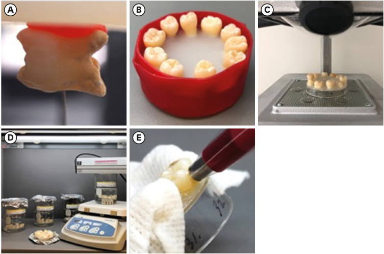
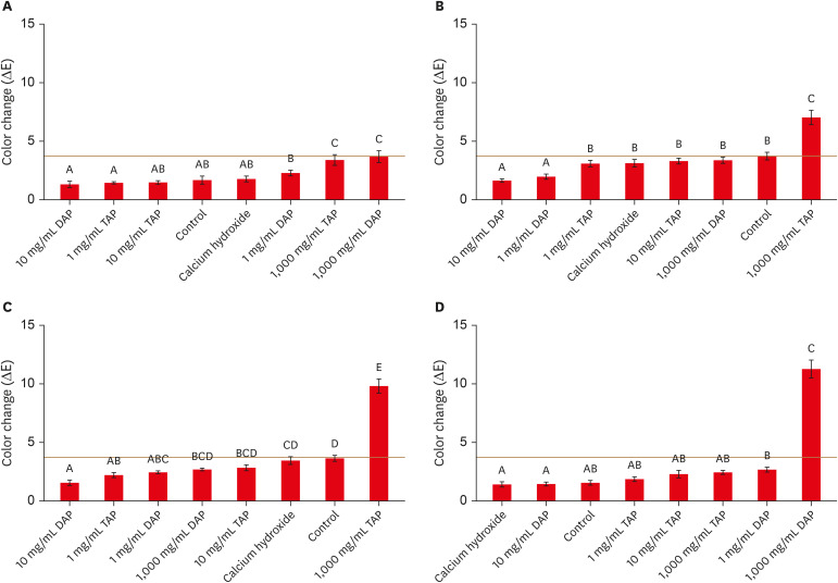
- Rayan B. Yaghmoor, Jeffrey A. Platt, Kenneth J. Spolnik, Tien Min Gabriel Chu, Ghaeth H. Yassen
- Restor Dent Endod 2021;46(4):e52. Published online October 5, 2021
- DOI: https://doi.org/10.5395/rde.2021.46.e52
-
Abstract
PDFPubReaderePub
Objectives This study evaluated the effects of low and moderate concentrations of triple antibiotic paste (TAP) and double antibiotic paste (DAP) loaded into a hydrogel system on crown discoloration and explored whether application of an adhesive bonding agent prevented crown discoloration.
Materials and Methods Intact human molars (
n = 160) were horizontally sectioned 1 mm apical to the cementoenamel junction. The crowns were randomized into 8 experimental groups (calcium hydroxide, Ca[OH]2; 1, 10, and 1,000 mg/mL TAP and DAP; and no medicament. The pulp chambers in half of the samples were coated with an adhesive bonding agent before receiving the intracanal medicament. Color changes (ΔE) were detected by spectrophotometry after 1 day, 1 week, and 4 weeks, and after 5,000 thermal cycles, with ΔE = 3.7 as a perceptible threshold. The 1-samplet -test was used to determine the significance of color changes relative to 3.7. Analysis of variance was used to evaluate the effects of treatment, adhesive, and time on color change, and the level of significance wasp < 0.05.Results Ca(OH)2 and 1 and 10 mg/mL DAP did not cause clinically perceivable tooth discoloration. Adhesive agent use significantly decreased tooth discoloration in the 1,000 mg/mL TAP group up to 4 weeks. However, adhesive use did not significantly improve coronal discoloration after thermocycling when 1,000 mg/mL TAP was used.
Conclusions Ca(OH)2 and 1 and 10 mg/mL DAP showed no clinical discoloration. Using an adhesive significantly improved coronal discoloration up to 4 weeks with 1,000 mg/mL TAP.
-
Citations
Citations to this article as recorded by- Comparative analysis of tooth discoloration induced by different intracanal medicaments in regenerative endodontics: A systematic review and network meta-analysis
Ashlesha Nageshwar Madankar, Sulabha Radke, Shanmuga Priya, Darshan Dakshindas
Endodontology.2026; 38(1): 8. CrossRef - Tooth discoloration caused by nanographene oxide as an irrigant and intracanal medicament in the endodontic treatment of extracted single-rooted teeth: An ex-vivo study
Abbas Abbaszadegan, Zeinab Rafiee, Bahar Asheghi, Ahmad Gholami, Mohmed Isaqali Karobari
PLOS One.2025; 20(6): e0325430. CrossRef - Root development of immature necrotic permanent teeth following regenerative endodontic process: Case series
Abbasali Khademi, Pedram Iranmanesh, Ali Akhavan, Movahed Ghassem Yeganeh, Samira Khalifezade Esfahani
Dental Research Journal.2025;[Epub] CrossRef - Root Canal Dentin Microhardness after Contact with Antibiotic Medications: An In Vitro Study
Amanda Palmeira Arruda Nogueira, Renata Grazziotin-Soares, Adriana Marques Mesquita Leal, Sérgio Alves Guida Freitas Júnior, Bruna Laís Lins Gonçalves, José Bauer, Meire Coelho Ferreira, Ceci Nunes Carvalho
Dentistry Journal.2024; 12(7): 201. CrossRef - Potential Crown Discoloration Induced by the Combination of Various Intracanal Medicaments and Scaffolds Applied in Regenerative Endodontic Therapy
NB Altun, A Turkyilmaz
Nigerian Journal of Clinical Practice.2024; 27(7): 897. CrossRef
- Comparative analysis of tooth discoloration induced by different intracanal medicaments in regenerative endodontics: A systematic review and network meta-analysis
- 3,157 View
- 43 Download
- 4 Web of Science
- 5 Crossref
- Push-out bond strength and marginal adaptation of apical plugs with bioactive endodontic cements in simulated immature teeth
- Maria Aparecida Barbosa de Sá, Eduardo Nunes, Alberto Nogueira da Gama Antunes, Manoel Brito Júnior, Martinho Campolina Rebello Horta, Rodrigo Rodrigues Amaral, Stephen Cohen, Frank Ferreira Silveira
- Restor Dent Endod 2021;46(4):e53. Published online October 20, 2021
- DOI: https://doi.org/10.5395/rde.2021.46.e53
-
Abstract
PDFPubReaderePub
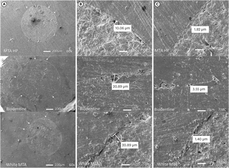
Objectives This study evaluates the bond strength and marginal adaptation of mineral trioxide aggregate (MTA) Repair HP and Biodentine used as apical plugs; MTA was used as reference material for comparison.
Materials and Methods A total of 30 single-rooted teeth with standardized, artificially created open apices were randomly divided into 3 groups (
n = 10 per group), according to the material used to form 6-mm-thick apical plugs: group 1 (MTA Repair HP); group 2 (Biodentine); and group 3 (white MTA). Subsequently, the specimens were transversely sectioned to obtain 2 (cervical and apical) 2.5-mm-thick slices per root. Epoxy resin replicas were observed under a scanning electron microscope to measure the gap size at the material/dentin interface (the largest and smaller gaps were recorded for each replica). The bond strength of the investigated materials to dentin was determined using the push-out test. The variable bond strengths and gap sizes were evaluated independently at the apical and cervical root dentin slices. Data were analyzed using descriptive and analytic statistics.Results The comparison between the groups regarding the variables' bond strengths and gap sizes showed no statistical difference (
p > 0.05) except for a single difference in the smallest gap at the cervical root dentin slice, which was higher in group 3 than in group 1 (p < 0.05).Conclusions The bond strength and marginal adaptation to root canal walls of MTA HP and Biodentine cement were comparable to white MTA.
-
Citations
Citations to this article as recorded by- Evaluation of marginal adaptation and bond strength of apical root canal plugs using different bioceramic cements
Michel Sena Fernandes Faria Lima, Alberto Nogueira da Gama Antunes, Kênia Maria Pereira Soares de Toubes, Fábio Fernandes Borém Bruzinga, Camila de Sousa Caneschi, Luís Fernando dos Santos Alves Morgan, Frank Ferreira Silveira
BMC Oral Health.2026;[Epub] CrossRef - Application of Biodentine for Apexification of Immature Teeth of Children: A Scoping Review
Liz M Gerard, Sumit Gaur
International Journal of Clinical Pediatric Dentistry.2025; 18(5): 573. CrossRef - Evaluation of the root dentin bond strength and intratubular biomineralization of a premixed calcium aluminate-based hydraulic bioceramic endodontic sealer
Yu-Na Lee, Min-Kyeong Kim, Hee-Jin Kim, Mi-Kyung Yu, Kwang-Won Lee, Kyung-San Min
Journal of Oral Science.2024; 66(2): 96. CrossRef - Managing Cracked Teeth with Root Extension: A Prospective Preliminary Study Using Biodentine™ Material
Kênia Maria Soares de Toubes, Isabella Sousa Corrêa, Regina Célia Lopes Valadares, Stephanie Quadros Tonelli, Fábio Fernandes Borém Bruzinga, Frank Ferreira Silveira, Dr Karthikeyan Ramalingam
International Journal of Dentistry.2024;[Epub] CrossRef - Marginal adaptation of customized gutta percha cone with calcium silicate based sealer versus MTA and biodentine apical plugs in simulated immature permanent teeth (an in vitro study)
Mary M. Mina, Sybel M. Moussa, Mahmoud R. Aboelseoud
BMC Oral Health.2024;[Epub] CrossRef - Comparative Evaluation of Push-Out Bond Strength of Conventional Mineral Trioxide Aggregate, Biodentine, a Modified Mineral Trioxide Aggregate, and Two Novel Antibacterial-Enhanced Mineral Trioxide Aggregates
Arokia Rajkumar Shancy Merlin, Vignesh Ravindran, Ganesh Jeevanandan, Rajalakshmanan Eswaramoorthy, Abirami Arthanari
Cureus.2024;[Epub] CrossRef - Push out bond strength of hydraulic cements used at different thicknesses
C. Ruiz Durán, Dra L. Gancedo-Caravia, V. Vera González, C. González Losada
BMC Oral Health.2023;[Epub] CrossRef - Effects of different calcium-silicate based materials on fracture resistance of immature permanent teeth with replacement root resorption and osteoclastogenesis
Gabriela Leite de Souza, Gabrielle Alves Nunes Freitas, Maria Tereza Hordones Ribeiro, Nelly Xiomara Alvarado Lemus, Carlos José Soares, Camilla Christian Gomes Moura
Restorative Dentistry & Endodontics.2023;[Epub] CrossRef
- Evaluation of marginal adaptation and bond strength of apical root canal plugs using different bioceramic cements
- 3,496 View
- 43 Download
- 9 Web of Science
- 8 Crossref
- Change of phase transformation and bond strength of Y-TZP with various hydrofluoric acid etching
- Mi-Kyung Yu, Eun-Jin Oh, Myung-Jin Lim, Kwang-Won Lee
- Restor Dent Endod 2021;46(4):e54. Published online October 20, 2021
- DOI: https://doi.org/10.5395/rde.2021.46.e54
-
Abstract
PDFPubReaderePub
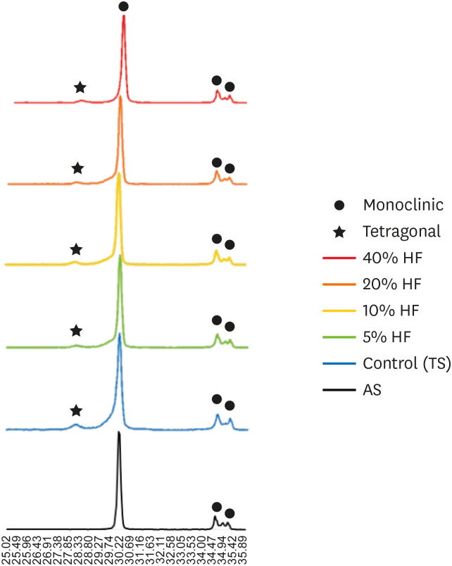
Objectives The purpose of this study was to quantify phase transformation after hydrofluoric acid (HF) etching at various concentrations on the surface of yttria-stabilized tetragonal zirconia polycrystal (Y-TZP), and to evaluate changes in bonding strength before and after thermal cycling.
Materials and Methods A group whose Y-TZP surface was treated with tribochemical silica abrasion (TS) was used as the control. Y-TZP specimens from each experimental group were etched with 5%, 10%, 20%, and 40% HF solutions at room temperature for 10 minutes. First, to quantify the phase transformation, Y-TZP specimens (
n = 5) treated with TS, 5%, 10%, 20% and 40% HF solutions were subjected to X-ray diffraction. Second, to evaluate the change in bond strength before and after thermal cycling, zirconia primer and MDP-containing resin cement were sequentially applied to the Y-TZP specimen. After 5,000 thermal cycles for half of the Y-TZP specimens, shear bond strength was measured for all experimental groups (n = 10).Results The monoclinic phase content in the 40% HF-treated group was higher than that of the 5%, 10%, and 20% HF-treated groups, but lower than that of TS-treated group (
p < 0.05). The 40% HF-treated group showed significantly higher bonding strength than the TS, 5%, and 10% HF-treated groups, even after thermal cycling (p < 0.05).Conclusions Through this experiment, the group treated with SiO2 containing air-borne abrasion on the Y-TZP surface showed higher phase transformation and higher reduction in bonding strength after thermal cycling compared to the group treated with high concentration HF.
-
Citations
Citations to this article as recorded by- Phase transition regulation and enhancement of optical properties in YPO4:Eu3+ through the influence of alkali metal ions
Junwei Zhan, Liusai Yang, Yaoxian Zhu, Yifan Zhu, Jianlei Liu, Siyan Peng, Jianping Zou
Journal of Molecular Structure.2026; 1352: 144420. CrossRef - Etchability of zirconia ceramics and its effect on adhesion: A systematic review and meta-analysis
Anina Sieber, Luiza Freitas Brum Souza, Tan Fırat Eyüboğlu, Mutlu Özcan
International Journal of Adhesion and Adhesives.2026; 148: 104303. CrossRef - Effect of BaTiO3 particle size and distribution on the BaTiO3-3Y-TZP interface reactivity of bioactive ceramic composites
João Pinto, Michael Gasik, Óscar Carvalho, Filipe S. Silva
Ceramics International.2026; 52(12): 18587. CrossRef - Improving the Clinical Performance of Dental Implants Through Advanced Surface Treatments: The Case of Ti and ZrO2 Coatings
Mohamed Aissi, Qanita Tayyaba, Azzedine Er-Ramly, Hendra Hermawan, Nadia Merzouk
Metals.2025; 15(3): 320. CrossRef - Enhancing the bonding of zirconia to resin by constructing a graded zirconia-glass composite surface
Zhiqi Yan, Jiale Li, Jing Chen, Zhe Zhao, Fan Li, Ling Zhang, Jihua Chen, Fu Wang
Surfaces and Interfaces.2025; 64: 106374. CrossRef - Surface property changes observed in zirconia during etching with high-concentration hydrofluoric acid over various immersion times
Ga-Eul YOU, Myung-Jin LIM, Kyung-San MIN, Mi-Kyung YU, Kwang-Won LEE
Dental Materials Journal.2024; 43(1): 52. CrossRef - Effect of surface treatments on the bond strength for different generation of zirconia CAD/CAM blocks
Man-Jong Cho, Sunwoong Song, Shin Hye Chung, Young-Seok Park, Bum-Soon Lim
Korean Journal of Dental Materials.2024; 51(3): 157. CrossRef - Is zirconia surface etching a viable alternative to airborne particle abrasion? A systematic review and meta-analysis of in vitro studies
Carlo D'Alessandro, Uros Josic, Claudia Mazzitelli, Tatjana Maravic, Laurel Graham, Carlo Barausse, Annalisa Mazzoni, Lorenzo Breschi, Markus B. Blatz
Journal of Dentistry.2024; 151: 105394. CrossRef - Exploring Zirconia Adhesion: Pre and Postsintering Physical Surface Treatment, Chemical Treatment, and Cement Interactions
Flávia Gonçalves, Mirko Dennys Ayala-Perez, Francisco Carlos dos Santos Reis, Walter Gomes Miranda-Júnior, Letícia Cristina Cidreira Boaro, Heng Bo Jiang
BioMed Research International.2024;[Epub] CrossRef - 3Y-TZP electrostatic painting to increase bond strength to dentin and dental prostheses
Alessandro Brito Thomaz, Carlos Nelson Elias, Heraldo Elias Salomão dos Santos, Celso Renato de Souza Resende, Claudinei dos Santos
Journal of Materials Research and Technology.2023; 26: 9063. CrossRef - Effect of surface topography and wettability on shear bond strength of Y-TZP ceramic
Suriyakul Wongsue, Ornnicha Thanatvarakorn, Taweesak Prasansuttiporn, Piyarat Nimmanpipug, Thanapat Sastraruji, Keiichi Hosaka, Richard M. Foxton, Masatoshi Nakajima
Scientific Reports.2023;[Epub] CrossRef - Adhesive Cementation of Zirconia Based Ceramics-Surface Modification Methods Literature Review
Magdalena Szawioła-Kirejczyk, Karolina Chmura, Krzysztof Gronkiewicz, Andrzej Gala, Jolanta E. Loster, Wojciech Ryniewicz
Coatings.2022; 12(8): 1067. CrossRef - Y-TZP Physicochemical Properties Conditioned with ZrO2 and SiO2 Nanofilms and Bond Strength to Dual Resin Cement
Ricardo Faria Ribeiro, Danilo Flamini Oliveira, Camila Bussola Tovani, Ana Paula Ramos, Ana Flavia Sanches Borges, Adriana Claudia Lapria Faria, Rossana Pereira de Almeida, Renata Cristina Silveira Rodrigues
Materials.2022; 15(22): 7905. CrossRef - Enhanced osteogenic activity of titania-modified zirconia implant by ultraviolet irradiation
Shuang Tang, Yan Wang, Zhenyu Zong, Ning Ding, Zutai Zhang
Frontiers in Bioengineering and Biotechnology.2022;[Epub] CrossRef
- Phase transition regulation and enhancement of optical properties in YPO4:Eu3+ through the influence of alkali metal ions
- 3,078 View
- 29 Download
- 15 Web of Science
- 14 Crossref
- Adhesive systems applied to dentin substrate under electric current: systematic review
- Carolina Menezes Maciel, Tatiane Cristina Vieira Souto, Bárbara de Almeida Pinto, Laís Regiane Silva-Concilio, Kusai Baroudi, Rafael Pino Vitti
- Restor Dent Endod 2021;46(4):e55. Published online November 5, 2021
- DOI: https://doi.org/10.5395/rde.2021.46.e55
-
Abstract
PDFPubReaderePub
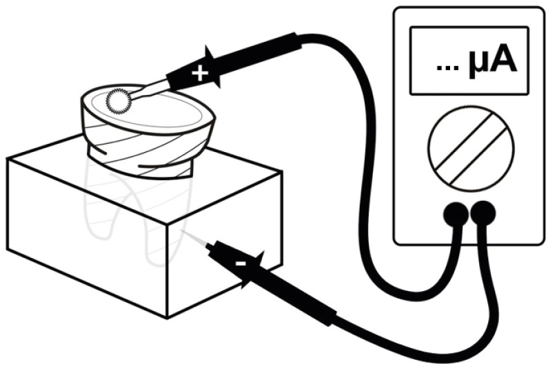
Objectives The purpose of this systematic review was to collect and discuss the technique of adhesive systems application on dentin substrate under electric current.
Materials and Methods The first search strategy was based on data available at PubMed, LILACS, Scielo, Scopus, and Cochrane Library, using a combination of descriptors such as “dentin bond agents OR adhesive system AND electric current OR electrobond” or “dentin bonding agents OR dentin bonding agent application OR adhesive system AND electric current OR electrobond”, with no limit regarding the publication year. The second search strategy was based on the articles' references found previously. An additional search strategy was applied that concerned the proposed theme in the SBU-UNICAMP (Unicamp's Library System Institutional Repository).
Results Twelve studies published between 2006 and 2020 were found. The analyses of the selected studies showed that the use of electric current during adhesive systems application on dentin, whether conventional or self-conditioning, increases resinous monomer infiltration in the dentin substrate, which improves the hybridization processes and the bond strength of the restorative material to dentin.
Conclusions Despite the favorable results related to the use of this technique, there is still no specific protocol for the application of adhesive systems under electric current.
-
Citations
Citations to this article as recorded by- Dental pulp stem cell responses to electric field stimulation: A pilot in vitro study with potential implications for dentin-pulp regeneration
Xiaolin Li, Yu Gu, Qiuling Zhong, Qian Liu, Vsevolod Telezhkin, Alastair J Sloan, Lihong Qiu, Bing Song
Journal of Dentistry.2026; : 106924. CrossRef - Advances in Resin-Dentin Bonding: Evaluating Pre-Treatment Techniques for Improved Adhesion
Rim Bourgi
Journal of Dental Health and Oral Research.2025; : 1. CrossRef - Iontophoresis effects of two-step self-etch and total-etch systems on dentin permeability and sealing of composite restoration under simulated pulpal pressure
Orapin Ajcharanukul, Peeraya Santikulluk, Palat Sasingha, Sirithorn Sabpawat, Kanokporn Sukyanan
BMC Oral Health.2022;[Epub] CrossRef
- Dental pulp stem cell responses to electric field stimulation: A pilot in vitro study with potential implications for dentin-pulp regeneration
- 2,788 View
- 21 Download
- 1 Web of Science
- 3 Crossref
- Combination of a new ultrasonic tip with rotary systems for the preparation of flattened root canals
- Karina Ines Medina Carita Tavares, Jáder Camilo Pinto, Airton Oliveira Santos-Junior, Fernanda Ferrari Esteves Torres, Juliane Maria Guerreiro-Tanomaru, Mario Tanomaru-Filho
- Restor Dent Endod 2021;46(4):e56. Published online October 27, 2021
- DOI: https://doi.org/10.5395/rde.2021.46.e56

-
Abstract
PDFPubReaderePub
Objectives This study evaluated 2 nickel-titanium rotary systems and a complementary protocol with an ultrasonic tip and a small-diameter instrument in flattened root canals.
Materials and Methods Thirty-two human maxillary second premolars with flattened canals (buccolingual diameter ≥4 times larger than the mesiodistal diameter) at 9 mm from the radiographic apex were selected. The root canals were prepared by ProDesign Logic (PDL) 30/0.01 and 30/0.05 or Hyflex EDM (HEDM) 10/0.05 and 25/0.08 (
n = 16), followed by application of the Flatsonic ultrasonic tip in the cervical and middle thirds and a PDL 25/0.03 file in the apical third (FPDL). The teeth were scanned using micro-computed tomography before and after the procedures. The percentage of volume increase, debris, and uninstrumented surface area were analyzed using the Kruskal-Wallis, Dunn, Wilcoxon, analysis of variance/Tukey, and paired and unpairedt -tests (α = 0.05).Results No significant difference was found in the volume increase and uninstrumented surface area between PDL and HEDM (
p > 0.05). PDL had a higher percentage of debris than HEDM in the middle and apical thirds (p < 0.05). The FPDL protocol resulted in less debris and uninstrumented surface area for PDL and HEDM (p < 0.05). This protocol, with HEDM, reduced debris in the middle and apical thirds and uninstrumented surface area in the apical third (p < 0.05).Conclusions High percentages of debris and uninstrumented surface area were observed after preparation of flattened root canals. The HEDM, Flatsonic tip, and 25/0.03 instrument protocol enhanced cleaning in flattened root canals.
-
Citations
Citations to this article as recorded by- Kök Kanal Tedavisi Yenilemelerinde Ultrasonik Uç Kullanımı
Ayşenur Kızıltaş Gül, Turan Mert Hisar, Seniha Miçooğulları
Selcuk Dental Journal.2025; 12(1): 157. CrossRef - Flatsonic Ultrasonic Tip Optimizes the Removal of Remaining Filling Material in Flattened Root Canals: A Micro–computed Tomographic Analysis
Airton Oliveira Santos-Junior, Karina Ines Medina Carita Tavares, Jáder Camilo Pinto, Fernanda Ferrari Esteves Torres, Juliane Maria Guerreiro-Tanomaru, Mário Tanomaru-Filho
Journal of Endodontics.2024; 50(5): 612. CrossRef - The Effect of Combined Ultrasonic Tip and Mechanized Instrumentation on the Reduction of the Percentage of Non-Instrumented Surfaces in Oval/Flat Root Canals: A Systematic Review and Meta-Analysis
Marcella Dewes Cassal, Pedro Cardoso Soares, Marcelo dos Santos
Cureus.2023;[Epub] CrossRef - Heat-treated NiTi instruments and final irrigation protocols for biomechanical preparation of flattened canals
Kleber Kildare Teodoro CARVALHO, Igor Bassi Ferreira PETEAN, Alice Corrêa SILVA-SOUSA, Rafael Verardino CAMARGO, Jardel Francisco MAZZI-CHAVES, Yara Terezinha Corrêa SILVA-SOUSA, Manoel Damião SOUSA-NETO
Brazilian Oral Research.2022;[Epub] CrossRef
- Kök Kanal Tedavisi Yenilemelerinde Ultrasonik Uç Kullanımı
- 2,656 View
- 36 Download
- 4 Web of Science
- 4 Crossref
- Porosity and pore size distribution in high-viscosity and conventional glass ionomer cements: a micro-computed tomography study
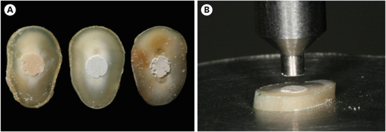
- Aline Borburema Neves, Laísa Inara Gracindo Lopes, Tamiris Gomes Bergstrom, Aline Saddock Sá da Silva, Ricardo Tadeu Lopes, Aline de Almeida Neves
- Restor Dent Endod 2021;46(4):e57. Published online October 29, 2021
- DOI: https://doi.org/10.5395/rde.2021.46.e57
-
Abstract
PDFPubReaderePub
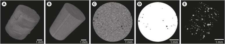
Objectives This study aimed to compare and evaluate the porosity and pore size distribution of high-viscosity glass ionomer cements (HVGICs) and conventional glass ionomer cements (GICs) using micro-computed tomography (micro-CT).
Materials and Methods Forty cylindrical specimens (
n = 10) were produced in standardized molds using HVGICs and conventional GICs (Ketac Molar Easymix, Vitro Molar, MaxxionR, and Riva Self-Cure). The specimens were prepared according to ISO 9917-1 standards, scanned in a high-energy micro-CT device, and reconstructed using specific parameters. After reconstruction, segmentation procedures, and image analysis, total porosity and pore size distribution were obtained for specimens in each group. After checking the normality of the data distribution, the Kruskal-Wallis test followed by the Student-Newman-Keuls test was used to detect differences in porosity among the experimental groups with a 5% significance level.Results Ketac Molar Easymix showed statistically significantly lower total porosity (0.15%) than MaxxionR (0.62%), Riva (0.42%), and Vitro Molar (0.57%). The pore size in all experimental cements was within the small-size range (< 0.01 mm3), but Vitro Molar showed statistically significantly more pores/defects with a larger size (> 0.01 mm3).
Conclusions Major differences in porosity and pore size were identified among the evaluated GICs. Among these, the Ketac Molar Easymix HVGIC showed the lowest porosity and void size.
-
Citations
Citations to this article as recorded by- The effect of contouring instruments on immediate quality and porosity of direct restorations
Carlos Soler-Tornero, Pekka Toivonen, Jaakko Suorsa, Sakari S. Karhula, Simo Saarakkala, Vuokko Anttonen, Jukka Leinonen
Clinical Oral Investigations.2025;[Epub] CrossRef - Impact of spacers and thermocycling on porosity and gaps in class II endodontic temporary restorations evaluated by microcomputed tomography
Fahda N. Algahtani, Manal Alkadi, Hiba R. Talic, Sarah S. AlShalawi, Lujain M. Alqarni, Reem M. Barakat, Rasha Haridy, Sara M. ElKhateeb, Rahaf A. Almohareb
Scientific Reports.2025;[Epub] CrossRef - Influence of Human Blood Contamination on Microhardness of Glass-Ionomer Cements and Glass-Hybrid Material
Katarina Franić, Ana Brundić, Jurica Matijević, Ana Ivanišević, Ivana Miletić, Anja Baraba
Materials.2025; 18(17): 4075. CrossRef - Effect of crown seating methods on the remnant cement in the subgingival region of a cement-retained implant crown
Fanghui Ji, Ji Suk Shim, Jeongyol Lee, Hwiseong Oh, Jae Jun Ryu
Scientific Reports.2024;[Epub] CrossRef - Enhancing Wear Resistance in Glass Ionomer Cement through Green-mediated Chitosan-, Titanium-, Zirconium-, and Hydroxyapatite-based Nanocomposites: An Analysis before and after Chewing Simulator Endurance
Srinavasa Surya Sitaram, Jessy Paulraj, Subhabrata Maiti, Rajeshkumar Shanmugam
International Journal of Clinical Pediatric Dentistry.2024; 17(11): 1229. CrossRef - The effect of mesoporous silica doped with silver nanoparticles on glass ionomer cements; physiochemical, mechanical and ion release analysis
Syed Saad Bin Qasim, Ali Bmuajdad
BMC Oral Health.2024;[Epub] CrossRef - Hyperbaric Pressure Effect on Dental Luting Cements
Secil OZKAN ATA, Nazım ATA, Rıfat UGURLUTAN
Journal of Basic and Clinical Health Sciences.2023; 7(1): 464. CrossRef - In Vitro Comparison of Differences in Setting Time of Premixed Calcium Silicate-Based Mineral Trioxide Aggregate According to Moisture Content of Gypsum
Hyun-Jin Kim, Jun-Seok Lee, Dong-Hoon Gwak, Yong-Seok Ko, Chun-Il Lim, Seung-Youl Lee
Materials.2023; 17(1): 35. CrossRef - Adhesion and Surface Roughness of Apatite-Containing Carbomer and Improved Ionically Bioactive Resin Compared to Glass Ionomers
Handan Yıldırım Işık, Aylin Çilingir
Journal of Functional Biomaterials.2023; 14(7): 367. CrossRef - An influence of finishing procedures and protective coating on the ultrastructure of conventional and hybrid glass ionomer cement restorations
Antonije Stankovic, Jelena Popovic, Marija Nikolic, Aleksandar Mitic, Nenad Stosic, Radomir Barac, Aleksandra Milovanovic
Stomatoloski glasnik Srbije.2023; 70(3): 138. CrossRef - Effect of aging on mechanical and antibacterial properties of fluorinated graphene reinforced glass ionomer: In vitro study
Suzan Khaled Arafa, Dalia Ibrahim Sherief, Mohamed Salah Nassif
Journal of the Mechanical Behavior of Biomedical Materials.2023; 142: 105803. CrossRef
- The effect of contouring instruments on immediate quality and porosity of direct restorations
- 3,493 View
- 22 Download
- 8 Web of Science
- 11 Crossref
- The effect of using nanoparticles in bioactive glass on its antimicrobial properties
- Maram Farouk Obeid, Kareim Moustafa El-Batouty, Mohammed Aslam
- Restor Dent Endod 2021;46(4):e58. Published online October 29, 2021
- DOI: https://doi.org/10.5395/rde.2021.46.e58
-
Abstract
PDFPubReaderePub
Objectives This study addresses the effect of using nanoparticles (np) on the antimicrobial properties of bioactive glass (BAG) when used in intracanal medicaments against
Enterococcus faecalis (E. faecalis ) biofilms.Materials and Methods E. faecalis biofilms, grown inside 90 root canals for 21 days, were randomly divided into 4 groups according to the antimicrobial regimen followed (n = 20; BAG-np, BAG, calcium hydroxide [CaOH], and saline). After 1 week, residual live bacteria were quantified in terms of colony-forming units (CFU), while dead bacteria were assessed with a confocal laser scanning microscope.Results Although there was a statistically significant decrease in the mean CFU value among all groups, the nano-group performed the best. The highest percentage of dead bacteria was detected in the BAG-np group, with a significant difference from the BAG group.
Conclusions The reduction of particle size and use of a nano-form of BAG improved the antimicrobial properties of the intracanal treatment of
E. faecalis biofilms-
Citations
Citations to this article as recorded by- Bioactive Glass in Dentistry: Mechanisms, Clinical Applications, and Future Perspectives
Madina A. Kurmanalina
West Kazakhstan Regenerative Medicine Journal.2026; 14(1): 28. CrossRef - Comparative assessment of the antimicrobial efficacy of bioactive glass with conventionally used intracanal medicaments: a systematic review
Ammar Haideri, Pradeep Solete, Gargi Gandhi, Vasaki Arunachalam, Surendar Ramamoorthi, Ahmed El-Kabbaney
Tanta Dental Journal.2026; 23(2): 191. CrossRef - Size matters: Radiation shielding superiority of borate glasses with nano vs. micro ZnO
Aljawhara H. Almuqrin, M.I. Sayyed, M. Elsafi
Nuclear Engineering and Technology.2025; 57(9): 103614. CrossRef - Effect of Chitosan and bioactive glass nanomaterials as intracanal medicaments on TGF-β1 release from intraradicular dentin
Sarah Salah Hashem, Mohammed M. Khalefa, Mahmoud Hassan Mohamed, Hemat M. ELSheikh, Fatma Abd El-Rahman Taher
BMC Oral Health.2025;[Epub] CrossRef - Effect of Er: YAG laser, phthalocyanine activated photodynamic therapy, and bioactive glass nanoparticles on smear layer removal and push out bond strength of quartz fiber posts to canal dentin: a SEM assessment
Okba Mahmoud, Erum Zain
Frontiers in Dental Medicine.2025;[Epub] CrossRef - Advancements in Root Canal Therapy: Translational Innovations and the Role of Nanoparticles in Endodontic Treatment
Noha M. Badawi, Mohamed M. Kataia, Hadeel A. Mousa, Mozhgan Afshari
Journal of Nanotechnology.2025;[Epub] CrossRef - Propolis in Endodontics—Unveiling Its Therapeutic Potential: A Narrative Review
Poorani Durai, Santha Devy A, Mithila Mohan, Harish Ramalingam, Shasidharan P, Rahul Chaurasia M
World Journal of Dentistry.2025; 16(10): 959. CrossRef - Application of Nanomaterials in Endodontics
Farzaneh Afkhami, Yuan Chen, Laurence J. Walsh, Ove A. Peters, Chun Xu
BME Frontiers.2024;[Epub] CrossRef - Antimicrobial efficacy of newly prepared nano-tricalcium silicate-58s bioactive glass-based endodontic sealer
Nawal Atiya Al-Sabawi, Sawsan Hameed Al-Jubori
Endodontology.2024; 36(2): 168. CrossRef - Antimicrobial Effects of Formulations of Various Nanoparticles and Calcium Hydroxide as Intra-canal Medications Against Enterococcus faecalis: A Systematic Review
Seema H Bukhari, Dax Abraham, Shakila Mahesh
Cureus.2024;[Epub] CrossRef - Effect of nanoparticles on antibacterial efficacy of intracanal medicament: A scoping review
Alpa Gupta, Arundeep Singh, Vivek Aggarwal
Endodontology.2023; 35(4): 283. CrossRef - Physical properties, marginal adaptation and bioactivity of an experimental mineral trioxide aggregate-like cement modified with bioactive materials
Abigailt Flores-Ledesma, Adriana Tejeda-Cruz, María A. Moyaho-Bernal, Ana Wintergerst, Yoshamin A. Moreno-Vargas, Jacqueline A. Rodríguez-Chávez, Carlos E. Cuevas-Suárez, Kenya Gutiérrez-Estrada, Jesús A. Arenas-Alatorre
Journal of Oral Science.2023; 65(2): 141. CrossRef - Nanopartículas antimicrobianas en endodoncia: Revisión narrativa
Gustavo Adolfo Tovar Rangel , Fanny Mildred González Sáenz , Ingrid Ximena Zamora Córdoba , Lina María García Zapata
Revista Estomatología.2023;[Epub] CrossRef
- Bioactive Glass in Dentistry: Mechanisms, Clinical Applications, and Future Perspectives
- 2,406 View
- 33 Download
- 6 Web of Science
- 13 Crossref
- Comparative analysis of bond strength to root dentin and compression of bioceramic cements used in regenerative endodontic procedures
- Maykely Naara Morais Rodrigues, Kely Firmino Bruno, Ana Helena Gonçalves de Alencar, Julyana Dumas Santos Silva, Patrícia Correia de Siqueira, Daniel de Almeida Decurcio, Carlos Estrela
- Restor Dent Endod 2021;46(4):e59. Published online November 9, 2021
- DOI: https://doi.org/10.5395/rde.2021.46.e59
-
Abstract
PDFPubReaderePub
Objectives This study compared the Biodentine, MTA Repair HP, and Bio-C Repair bioceramics in terms of bond strength to dentin, failure mode, and compression.
Materials and Methods Fifty-four slices obtained from the cervical third of 18 single-rooted human mandibular premolars were randomly distributed (
n = 18). After insertion of the bioceramic materials, the push-out test was performed. The failure mode was analyzed using stereomicroscopy. Another set of cylindrically-shaped bioceramic samples (n = 10) was prepared for compressive strength testing. The normality of data distribution was analyzed using the Shapiro-Wilk test. The Kruskal-Wallis and Friedman tests were used for the push-out test data, while compressive strength was analyzed with analysis of variance and the Tukey test, considering a significance level of 0.05.Results Biodentine presented a higher median bond strength value (14.79 MPa) than MTA Repair HP (8.84 MPa) and Bio-C Repair (3.48 MPa), with a significant difference only between Biodentine and Bio-C Repair. In the Biodentine group, the most frequent failure mode was mixed (61%), while in the MTA Repair HP and Bio-C Repair groups, it was adhesive (94% and 72%, respectively). Biodentine showed greater resistance to compression (29.59 ± 8.47 MPa) than MTA Repair HP (18.68 ± 7.40 MPa) and Bio-C Repair (19.96 ± 3.96 MPa) (
p < 0.05).Conclusions Biodentine showed greater compressive strength than MTA Repair HP and Bio-C Repair, and greater bond strength than Bio-C Repair. The most frequent failure mode of Biodentine was mixed, while that of MTA Repair HP and Bio-C Repair was adhesive.
-
Citations
Citations to this article as recorded by- Evaluation of marginal adaptation and bond strength of apical root canal plugs using different bioceramic cements
Michel Sena Fernandes Faria Lima, Alberto Nogueira da Gama Antunes, Kênia Maria Pereira Soares de Toubes, Fábio Fernandes Borém Bruzinga, Camila de Sousa Caneschi, Luís Fernando dos Santos Alves Morgan, Frank Ferreira Silveira
BMC Oral Health.2026;[Epub] CrossRef - Assessment of Release of Calcium Ions from Apical Plugs of Different Bioceramic Sealers in Regenerative Endodontics for Immature Permanent Teeth
Alok Dubey, Nischitha Naik, Bhavana Sujanamulk, Mushir Mulla, Munaz Mulla, Alkananda Sahoo, Mohamed T Salama, Murali Patla S Bhat
World Journal of Dentistry.2026; 17(1): 69. CrossRef - Obturation quality analysis of furcation perforations repaired with different magnifications and biomaterials
Mustafa Mert Tulgar, Yağmur Kılıç, Merve Işık Aydın, Ali Keleş
BMC Oral Health.2026;[Epub] CrossRef - Comparison of Sealing Ability of Biodentine, Biostructure Mineral Trioxide Aggregate, Theracal LC, and Bio-C Repair as Perforation Repair Material: An Inverted Laser Scanning Confocal Microscopic Study
Langpoklakpam Monica Devi, Gunjan Yadav, Amit Rai, Sonali Saha, Kavita Dhinsa, Anshul Sharma
International Journal of Clinical Pediatric Dentistry.2026; 19(7): 881. CrossRef - Comparación de la resistencia compresiva entre el Agregado Trióxido Mineral y BiodentineTM en perforaciones de furca de molares inferiores permanentes
Jheymy Gerardo Huatuco-Granda, John Paul Torres-Navarro, Rosa Josefina Roncal-Espinoza
Revista Facultad de Odontología.2024;[Epub] CrossRef - Effects of different calcium-silicate based materials on fracture resistance of immature permanent teeth with replacement root resorption and osteoclastogenesis
Gabriela Leite de Souza, Gabrielle Alves Nunes Freitas, Maria Tereza Hordones Ribeiro, Nelly Xiomara Alvarado Lemus, Carlos José Soares, Camilla Christian Gomes Moura
Restorative Dentistry & Endodontics.2023;[Epub] CrossRef - Evaluation the Marginal Adaptation for the Bio C Repair and Other Root end Filling Material by Using Scanning Electron Microscope (A Comparative In Vitro Study)
Fatimah HAMADHİ, Zainab M.
Cumhuriyet Dental Journal.2023; 26(3): 261. CrossRef - Dentin Bond Strength of Calcium Silicate-Based Materials: A Systematic Review of In Vitro Studies
Natalia Radulica, José Luis Sanz, Adrián Lozano
Applied Sciences.2023; 14(1): 104. CrossRef - Evaluation Of The Push-out Bond Strength Of The Bio-C Repair And Compare It With The Mineral Trioxide Aggregate And Amalgam When Used As Root-end Filling Material
Fatimah R. Hammadi, Zainab M Abdul-Ameer
Dental Hypotheses.2023; 14(2): 62. CrossRef - Effect of different root canal irrigants on push-out bond strength of two novel root-end filling materials
Nada Omar, Rasha M. Abdelraouf, Tamer M. Hamdy
BMC Oral Health.2023;[Epub] CrossRef - Effect of irrigation systems on the bond strength of calcium-silicate-based cement used as pulp barrier in regenerative endodontic treatment
Cihan Hascizmeci, Burak Buldur
Journal of Adhesion Science and Technology.2023; 37(23): 3393. CrossRef
- Evaluation of marginal adaptation and bond strength of apical root canal plugs using different bioceramic cements
- 4,207 View
- 87 Download
- 6 Web of Science
- 11 Crossref
- Spectrophotometric evaluation of restorative composite shades and their match with a classical shade guide
- Rafael Melara, Luciana Mendonça, Fábio Herrmann Coelho-de-Souza, Juliana Nunes Rolla, Luciano de Souza Gonçalves
- Restor Dent Endod 2021;46(4):e60. Published online November 12, 2021
- DOI: https://doi.org/10.5395/rde.2021.46.e60
-
Abstract
PDFPubReaderePub
Objectives The aim of this study was to verify the match between 5 shades of composites from different manufacturers with a shade guide and among the systems using a portable spectrophotometer.
Materials and Methods Shade measurements were performed on specimens of Z350 XT (3M ESPE), Charisma Diamond (Heraeus Kulzer GmbH), Esthet X-HD (Dentsply Caulk), and Empress Direct (Ivoclar-Vivadent) for shades A1, A2, A3, B1, and C3 using a Vita Easyshade spectrophotometer (Vita Zahnfabrik) against a white background. Corresponding shades of Vitapan Classical (Vita Zahnfabrik) guide were measured likewise and shade variation (ΔE) was calculated based on International Commission on Illumination L*a*b* parameters. The ΔE of the composites in each shade was compared by one-way analysis of variance and Tukey's
post hoc test (α = 0.05).Results All composites presented ΔE > 3.7 compared with the shade guide. Variation in shades A3, B1, and C3 was significantly different for all composites. ΔE of Z350 XT was significantly lower for A1 than for the other shades, whereas ΔE of Z350 XT and Charisma Diamond were significantly lower for A2 than for the other shades.
Conclusions No composite shade matched with the shade guide. Equivalent shades of the restorative composite from different manufacturers may show clinically noticeable ΔE.
-
Citations
Citations to this article as recorded by- Influence of different finishing and polishing protocols of composite CAD CAM blocks on surface roughness and biological response of gingival mesenchymal stem cells
Mohamed F. Haridy, Mohamed Shamel, Raghda A. Khalil, Ahmed Refaat Mohamed, Hoda Fouda, Hend S. Ahmed
Odontology.2026; 114(3): 1320. CrossRef - Análise espectrofotométrica da cor de resinas compostas e sua correspondência com a escala de referência VITA Classical
Gardenia Mascarenhas de Oliveira, Marta Conceição Oliveira Silva, Natan dos Anjos Nery de Oliveira, Maria Fernanda Moreira Carvalho Caxico, Lucas Gomes Pereira, Leandro do Rozário Teixeira, Marcus Vinícius Santos da Silva, Iuri Muniz Pepe
Cuadernos de Educación y Desarrollo.2026; 18(1): e10592. CrossRef - Baseline color-matching in anterior non-carious cervical lesions of patients of two single-shade resin composites: a randomized clinical trial
Ayşe Nur Doğan, Soley Arslan
Odontology.2026;[Epub] CrossRef - Evaluation of color blending effect of a single-shade resin composite with hybrid ceramics: an in vitro study
Jongchan Lee, Jinsoo Ahn, Sun-Young Kim
BMC Oral Health.2026;[Epub] CrossRef - A Multidimensional Analysis of Shade Selection Difficulty for Indirect Restorations Among Dental Students and Professionals
Roxana-Ionela Vasluianu, Andreas Katsonis, Monica Silvia Tatarciuc, Anca Mihaela Vitalariu, Adina Oana Armencia, Andrea-Simoni Katsoni, Panagiotis Perperidis, Catalina Cioloca Holban, Irina Gradinaru, Ovidiu Stamatin, Magda Ecaterina Antohe
Dentistry Journal.2026; 14(4): 234. CrossRef - Assessment of Color Accuracy in A2-Shade Composite Resins Using CIEDE2000: Conventional and 3D-Printed Materials
Gülçin Cagay Sevencan, Hilal Gülgezen Aydın
Journal of International Dental Sciences.2026; 12(1): 34. CrossRef - Shade Matching and Color Stability of Chameleon Effect Composites in Class V Restorations: An In-Vitro Colorimetric Study after Cola Immersion
Youssef Znait, Karim Corbani, Carlos Khairallah, Carina Mehanna, Louis Hardan, Fadi Hammoud, Rim Bourgi, Cynthia Kassis
Journal of Dental Health and Oral Research.2026; 7(2): 1. CrossRef - Instrumental and visual evaluation of the color adjustment potential of a recently introduced single‑shade composite resin versus multishade composite resins
Jiakang Zhu, Yue Xu, Mengxun Li, Cui Huang
The Journal of Prosthetic Dentistry.2025; 134(3): 832. CrossRef - Evaluation of the roughness, color match, and color stability of two monochromatic composite resins: a randomized controlled laboratory study
Iara Campos Santana, Sabrina Sobral de Oliveira, Karolina Pena Botelho, Renan Leonardi de Oliveira Rigotti, José Cristiano Ramos Glória, Adriana Maria Botelho, Dhelfeson Willya Douglas-de-Oliveira, Karine Taís Aguiar Tavano
BMC Oral Health.2025;[Epub] CrossRef - Instrumental and Visual Evaluation of the Chameleon Effect of Single-shaded Composite Resins
RM Adiguzel, LK Kose, N Arhun
Operative Dentistry.2024; 49(4): 432. CrossRef - Color Stability of Bioactive Restorative Material vs Nanohybrid Resin Composite: An In Vitro Study
Esraa H Saber, Mohsen H Abielhassan, Yasser A Abed, Shereen E Fahim
The Journal of Contemporary Dental Practice.2024; 25(3): 221. CrossRef - A system for reliable composite shade matching: Custom shade tabs and an intra‐oral mockup
Adamo Notarantonio, Amanda Seay
Journal of Esthetic and Restorative Dentistry.2023; 35(5): 787. CrossRef - Color Stability of Bioactive Restorative Materials After Immersion in Various Media
Shara I Sajini, Ali B Mushayt, Talal A Almutairi, Roaa Abuljadayel
Journal of International Society of Preventive and Community Dentistry.2022; 12(4): 418. CrossRef
- Influence of different finishing and polishing protocols of composite CAD CAM blocks on surface roughness and biological response of gingival mesenchymal stem cells
- 2,891 View
- 41 Download
- 11 Web of Science
- 13 Crossref
Case Report
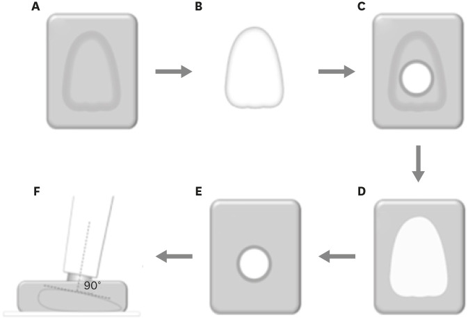
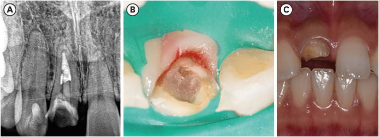
- Fiber-reinforced composite post removal using guided endodontics: a case report
- Changgi Cho, Hyo Jin Jo, Jung-Hong Ha
- Restor Dent Endod 2021;46(4):e50. Published online September 23, 2021
- DOI: https://doi.org/10.5395/rde.2021.46.e50
-
Abstract
PDFPubReaderePub
Although several techniques have been proposed to remove fiber-reinforced composite (FRC) post, no safe and efficient technique has been established. Recently, a guided endodontics technique has been introduced in cases of pulp canal obliteration. This study describes 2 cases of FRC post removal from maxillary anterior teeth using this guided endodontics technique with a dental operating microscope. Optically scanned data set from plaster cast model was superimposed with the data set of cone-beam computed tomography. By implant planning software, the path of a guide drill was selected. Based on them, a customized stent was fabricated and utilized to remove the FRC post. Employing guided endodontics, the FRC post was removed quickly and safely with minimizing the loss of the remaining tooth structure. The guided endodontics was a useful option for FRC post removal.
-
Citations
Citations to this article as recorded by- Comparing the Effectiveness of a Robotic and Dynamic Navigation System in Fiber Post removal: An In Vitro Study
Duo Zhou, Fulu Xu, Jiayun Dai, Xingyang Wang, Yifan Ping, Juan Wang
Journal of Endodontics.2026; 52(2): 261. CrossRef - Robot-assisted haptic guidance in endodontics: A pilot study evaluating efficiency and tooth structure preservation
Roshanak Momen, Joshua Dale, Mithun Suresh, Theodore D. Ravenel, Julie Marshall, Chin-Lo Hahn
Journal of Dentistry.2026; 173: 106854. CrossRef - Deviation analysis of guided fiber post removal using an assembled sleeveless guide system: A case series
Jingqi Zhu, Siyi Mo, Yuan Li, Yaojun Zhang, Xutong Song, Jingwen Liu, Ye Cao, Xiaoxiang Xu
The Journal of Prosthetic Dentistry.2026;[Epub] CrossRef - Static‐Guided Endodontics for Complex Intracanal Obstructions and Zirconia Post Removal: Report on Three Cases
Xinxuan Wang, Xuguang Li, Baicheng Yi
Clinical Case Reports.2026;[Epub] CrossRef - Application of 3D-printed resin guides for the removal of molar fiber posts
Yumin Wu, Lumei Huang, Bing Ge, Yuhang Zhang, Juan Zhang, Haifeng Xie, Ye Zhu, Chen Chen
Journal of Dentistry.2025; 153: 105462. CrossRef - Guided Removal of Long and Short Fiber Posts Using Endodontic Static Guides: A Case Report
Sahar Shafagh, Mamak Adel, Atiyeh Sabzpai
Clinical Case Reports.2025;[Epub] CrossRef - Guided versus non-guided fiber post removal: A systematic review and meta-analysis of the accuracy, efficiency, and dentin preservation of static navigation techniques in the removal of fiber posts
Mohamad Elabdalla, Farshad Khosraviani, Shahryar Irannejadrankouhi, Niloofar Ghadimi, Turgut Yağmur Yalçın, Shaheen Wathiq Tawfeeq Al Hajaj, Mahmood Dashti
The Journal of Prosthetic Dentistry.2025; 134(3): 630.e1. CrossRef - Top 100 Most-cited Scientific Articles in Guided Endodontic 2018–2024: A Bibliometric Analysis
Gustavo Adrián Morales Valladares, Raquel Esmeralda Guillén Guillén, Martha Elena Gallegos Intriago, Mary Yussely Burgos Barreiro, Claudia Jhelissa Campos Vélez, Andrés Alexander Castillo Chacón, Silvana Beatriz Terán Ayala
The Open Dentistry Journal.2025;[Epub] CrossRef - Nonsurgical Management of a Tooth With Intracanal Fiber Post and Periapical Lesion Using Guided Endodontic Technique
Mamak Adel, Zohreh Asgari
Clinical Case Reports.2025;[Epub] CrossRef - Impact of Guided Endodontics on the Success of Endodontic Treatment: An Umbrella Review of Systematic Reviews and Meta-Analyses
Aakansha Puri, Dax Abraham, Alpa Gupta
Cureus.2024;[Epub] CrossRef - Endodontia guiada por tomografia computadorizada de feixe cônico
Maysa Gaudereto Laurindo, Celso Neiva Campos, Anamaria Pessoa Pereira Leite, Paola Cantamissa Rodrigues Ferreira
Cadernos UniFOA.2024; 19(54): 1. CrossRef - Removal of fiber posts using conventional versus guided endodontics: a comparative study of dentin loss and complications
R. Krug, F. Schwarz, C. Dullin, W. Leontiev, T. Connert, G. Krastl, F. Haupt
Clinical Oral Investigations.2024;[Epub] CrossRef - Accuracy and Efficiency of the Surgical-Guide-Assisted Fiber Post Removal Technique for Anterior Teeth: An Ex Vivo Study
Ryota Ito, Satoshi Watanabe, Kazuhisa Satake, Ryuma Saito, Takashi Okiji
Dentistry Journal.2024; 12(10): 333. CrossRef - Endodontic management of severely calcified mandibular anterior teeth using guided endodontics: A report of a case and a review of the literature
Mina Davaji, Sahar Karimpour
Saudi Endodontic Journal.2024; 14(2): 245. CrossRef - A laboratory study comparing the static navigation technique using a bur with a conventional freehand technique using ultrasonic tips for the removal of fibre posts
Francesc Abella Sans, Zeena Tariq Alatiya, Gonzalo Gómez Val, Venkateshbabu Nagendrababu, Paul Michael Howell Dummer, Fernando Durán‐Sindreu Terol, Juan Gonzalo Olivieri
International Endodontic Journal.2024; 57(3): 355. CrossRef - A three‐dimensional printed assembled sleeveless guide system for fiber‐post removal
Yang Xue, Lei Zhang, Ye Cao, Yongsheng Zhou, Qiufei Xie, Xiaoxiang Xu
Journal of Prosthodontics.2023; 32(2): 178. CrossRef - Accuracy of a 3D printed sleeveless guide system used for fiber post removal: An in vitro study
Siyi Mo, Yongwei Xu, Lei Zhang, Ye Cao, Yongsheng Zhou, Xiaoxiang Xu
Journal of Dentistry.2023; 128: 104367. CrossRef - Expert consensus on digital guided therapy for endodontic diseases
Xi Wei, Yu Du, Xuedong Zhou, Lin Yue, Qing Yu, Benxiang Hou, Zhi Chen, Jingping Liang, Wenxia Chen, Lihong Qiu, Xiangya Huang, Liuyan Meng, Dingming Huang, Xiaoyan Wang, Yu Tian, Zisheng Tang, Qi Zhang, Leiying Miao, Jin Zhao, Deqin Yang, Jian Yang, Junqi
International Journal of Oral Science.2023;[Epub] CrossRef - Knowledge, attitude, practice and perception survey on post and core restorations
Aruna Kumari Veronica, Shamini Sai, Anand V Susila
Endodontology.2023; 35(3): 228. CrossRef
- Comparing the Effectiveness of a Robotic and Dynamic Navigation System in Fiber Post removal: An In Vitro Study
- 6,142 View
- 123 Download
- 14 Web of Science
- 19 Crossref
 First
First Prev
Prev

