
Previous issues
- Page Path
- HOME > Browse articles > Previous issues
- Volume 45 (4); November 2020
-


Research Articles
- Flow characteristics and alkalinity of novel bioceramic root canal sealers
- Anastasios Katakidis, Konstantinos Sidiropoulos, Elisabeth Koulaouzidou, Christos Gogos, Nikolaos Economides
- Restor Dent Endod 2020;45(4):e42. Published online August 18, 2020
- DOI: https://doi.org/10.5395/rde.2020.45.e42
-
 Abstract
 PDF
PDF PubReader
 ePub
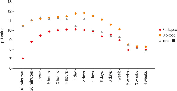
ePub Objective This study aimed to examine the physical properties (pH and flow) of 2 novel bioceramic sealers.
Materials and Methods The tested sealers were a calcium hydroxide sealer (Sealapex) and 2 bioceramic sealers (BioRoot RCS and TotalFill BC Sealer). Flow measurements were conducted according to ISO 6876/2012, with a press method of 0.05 mL of sealer. The pH of fresh samples was tested immediately after manipulation, while set samples were stored for 3 times the recommended setting time. The predetermined time intervals ranged from 3 minutes to 24 hours for fresh samples and from 10 minutes to 7 days and 4 weeks for the set samples. Analysis of variance was performed, with
p = 0.05 considered indicating significance.Results The mean flow values were 26.99 mm for BioRoot, 28.19 for Sealapex, and 30.8 mm for TotalFill BC Sealer, satisfying the ISO standard. In the set samples, BioRoot RCS had higher pH values at 24 hours to 1 week after immersion in distilled water. At 2 weeks, both bioceramic sealers had similar pH values, greater than that of Sealapex. In the fresh samples, the bioceramic sealers had significantly higher initial pH values than Sealapex (
p < 0.05). At 24 hours post-immersion, all sealers showed an alkaline pH, with the highest pH observed for TotalFill.Conclusions The TotalFill BC Sealer demonstrated the highest flow. The bioceramic sealers initially presented higher alkaline activity than the polymeric calcium hydroxide sealer. However, at 3 and 4 weeks post-immersion, all sealers had similar pH values.
-
Citations
Citations to this article as recorded by
- In vitro comparative evaluation of physicochemical and mechanical properties, cytocompatibility, and antimicrobial efficacy of various bioceramic root canal sealers
Fushi Wang, Jiaxing Li, Jingjing Wan, Siyuan Li, Shijia Tang, Li Wang, Liuyan Meng
Ceramics International.2026; 52(7): 9561. CrossRef - Comparative analysis between resin-based root canal sealer and recent bioceramic-based root canal sealers using MicroCT, film thickness, and solubility
Amira Galal Ismail, Manar M. Galal, Tamer M. Hamdy
Journal of Oral Biology and Craniofacial Research.2026; 16(2): 101400. CrossRef - Setting Characteristics, Solubility, Bioactivity and Interaction with Dentin of Four Calcium Silicate-Based Endodontic Sealers
Areti Dimitra Vrochari, Anastasia Agrafioti, Maria Dimitriadi, George Eliades
Journal of Functional Biomaterials.2026; 17(4): 192. CrossRef - Cryotherapy-Driven Modulation of Postoperative Pain in Single-Visit Endodontic Treatment Across Different Obturation Materials: A Retrospective Study
Kaan Ilıcalı, Ahter Şanal Çıkman, Özge Başar
Journal of Clinical Medicine.2026; 15(10): 3899. CrossRef - Functional and Bioactive Performance of Premixed Bioceramic Sealers with Warm Obturation: A Scoping Review
Patryk Wiśniewski, Stanisław Krokosz, Małgorzata Pietruska, Anna Zalewska
Gels.2025; 11(11): 932. CrossRef - Physicochemical properties of AH plus bioceramic sealer, Bio-C Sealer, and ADseal root canal sealer
Tamer M. Hamdy, Manar M. Galal, Amira Galal Ismail, Shehabeldin Saber
Head & Face Medicine.2024;[Epub] CrossRef - Characterization and Assessment of Physical Properties of 3 Single Syringe Hydraulic Cement–based Sealers
Veksina Raman, Josette Camilleri
Journal of Endodontics.2024; 50(3): 381. CrossRef - The Impact of Silver Nanoparticles on Dentinal Tubule Penetration of Endodontic Bioceramic Sealer
Sundus Bukhary, Sarah Alkahtany, Amal Almohaimede, Nourah Alkhayatt, Shahad Alsulaiman, Salma Alohali
Applied Sciences.2024; 14(24): 11639. CrossRef - Influence of root canal moisture on the penetration of TotalFill bioceramic sealer into the dentinal tubules: A confocal laser scanning microscopy study
Archika M Singh, Tarek M Elsewify, Walid S El-Sayed, Husam H Nuawafleh, Ranya F Elemam, Bassem M Eid
Saudi Endodontic Journal.2024; 14(2): 187. CrossRef - Unusual Canal Morphology in Mandibular Premolars With Two Distal and One Mesial Canal: A Case Series
Jinesh A, Sanjana Jayakumar Nair, Saurabh Gupta, Harsh Chansoria, Gaurav Rawat
Cureus.2024;[Epub] CrossRef - A scientometric, bibliometric, and thematic map analysis of hydraulic calcium silicate root canal sealers
Anastasios Katakidis, Konstantinos Kodonas, Anastasia Fardi, Christos Gogos
Restorative Dentistry & Endodontics.2023;[Epub] CrossRef - Thermal, chemical and physical analysis of VDW.1Seal, Fill Root ST, and ADseal root canal sealers
Shehabeldin Saber, Manar M. Galal, Amira Galal Ismail, Tamer M. Hamdy
Scientific Reports.2023;[Epub] CrossRef - α-tricalcium phosphate/fluorapatite-based cement - promising dental root canal filling material
Abdul Kazuz, Zeljko Radovanovic, Djordje Veljovic, Vesna Kojic, Dimitar Jakimov, Tamara Vlajic-Tovilovic, Vesna Miletic, Rada Petrovic, Djordje Janackovic
Processing and Application of Ceramics.2022; 16(1): 22. CrossRef
- In vitro comparative evaluation of physicochemical and mechanical properties, cytocompatibility, and antimicrobial efficacy of various bioceramic root canal sealers
- 3,720 View
- 44 Download
- 13 Crossref

- Effect of phytic acid as an endodontic chelator on resin adhesion to sodium hypochlorite-treated dentin
- Mohannad Nassar, Noriko Hiraishi, Md. Sofiqul Islam, Maria JRH. Romero, Masayuki Otsuki, Junji Tagami
- Restor Dent Endod 2020;45(4):e44. Published online August 24, 2020
- DOI: https://doi.org/10.5395/rde.2020.45.e44
-
Abstract
PDFPubReaderePub
Objectives Phytic acid (IP6), a naturally occurring agent, has been previously reported as a potential alternative to ethylenediaminetetraacetic acid (EDTA). However, its effect on adhesion to sodium hypochlorite (NaOCl)-treated dentin and its interactions with NaOCl have not been previously reported. Thus, in this study, the effects of IP6 on resin adhesion to NaOCl-treated dentin and the failure mode were investigated and the interactions between the used agents were analyzed.
Materials and Methods Micro-tensile bond strength (µTBS) testing was performed until failure on dentin treated with either distilled water (control), 5% NaOCl, or 5% NaOCl followed with chelators: 17% EDTA for 1 minute or 1% IP6 for 30 seconds or 1 minute. The failed specimens were assessed under a scanning electron microscope. The reaction of NaOCl with EDTA or IP6 was analyzed in terms of temperature, pH, effervescence, and chlorine odor, and the effects of the resulting mixtures on the color of a stained paper were recorded.
Results The µTBS values of the control and NaOCl with chelator groups were not significantly different, but were all significantly higher than that of the group treated with NaOCl only. In the failure analysis, a distinctive feature was the presence of resin tags in samples conditioned with IP6 after treatment with NaOCl. The reaction of 1% IP6 with 5% NaOCl was less aggressive than the reaction of the latter with 17% EDTA.
Conclusions IP6 reversed the adverse effects of NaOCl on resin-dentin adhesion without the chlorine-depleting effect of EDTA.
-
Citations
Citations to this article as recorded by- The Effect of Chemical Surface Modification on the Repair Bond Strength of Resin Composite: An In Vitro Study
Md Sofiqul Islam, Shadi El Bahra, Smriti Aryal A C, Vivek Padmanabhan, Abdulaziz Al Tawil, Ihab Saleh, Muhammed Mustahsen Rahman, Upoma Guha
Polymers.2025; 17(4): 513. CrossRef - Advancing Adhesive Strategies for Endodontically Treated Teeth—Part I: Impact of Endodontic Irrigation Protocols on the Chemical Composition and Structural Integrity of Coronal Dentin
Joana A. Marques, Rui I. Falacho, Sara Fateixa, Francisco Caramelo, João Miguel Santos, João Rocha, Markus B. Blatz, João Carlos Ramos, Paulo J. Palma
Journal of Esthetic and Restorative Dentistry.2025; 37(7): 1848. CrossRef - Effect of collagen crosslinkers on sodium hypochlorite treated dentin bond strength: a systematic review and meta-analysis
Weiqing Zhou, Shuting Feng, Xiaojun Chu, Shuaimei Xu, Xiongqun Zeng
Frontiers in Bioengineering and Biotechnology.2025;[Epub] CrossRef - Advancing Adhesive Strategies for Endodontically Treated Teeth—Part II: Dentin Sealing Before Irrigation Increases Long‐Term Microtensile Bond Strength to Coronal Dentin
Joana A. Marques, Rui I. Falacho, Gabriela Almeida, Francisco Caramelo, João Miguel Santos, João Rocha, Markus B. Blatz, João Carlos Ramos, Paulo J. Palma
Journal of Esthetic and Restorative Dentistry.2025; 37(7): 1865. CrossRef - Effects of phytic acid and etidronic acid using continuous and sequential chelation on the removal of smear layer, dentin microhardness, and push-out bond strength of calcium silicate-based cement
Ecehan Hazar, Ahmet Hazar
BMC Oral Health.2025;[Epub] CrossRef - Comparative evaluation of free available chlorine in sodium hypochlorite solutions admixed with novel chelating agents
Somya Tyagi, Sonali Taneja, Kandasamy Nagarajan, Divya Chowdhary
Endodontology.2025; 37(2): 188. CrossRef - Effect of different chelating agents, with and without activation, including XP-endo Finisher, on root dentin microhardness: An in vitro study
Mahmoud Mohamed A. Sherif, Mai Hamdy Ragab, Marwa ElSayed Sharaan
Saudi Endodontic Journal.2025; 15(3): 282. CrossRef - Oracle of phytic acid in dental panacea – Insight into properties, therapeutic effect, regeneration, materials interaction and oral physiology
Ummey Salma, C. Pushpalatha, SV. Sowmya, Dominic Augustine, Ahmed Alamoudi, Bassam Zidane, Nassreen Hassan Mohammad Albar, Shilpa Bhandi
The Saudi Dental Journal.2024; 36(8): 1093. CrossRef - In Vitro Bond Strength of Dentin Treated with Sodium Hypochlorite: Effects of Antioxidant Solutions
Guillermo Grazioli, Elisa de León Cáceres, Romina Tessore, Rafael Lund, Ana Monjarás-Ávila, Monika Lukomska-Szymanska, Louis Hardan, Rim Bourgi, Carlos Cuevas-Suárez
Antioxidants.2024; 13(9): 1116. CrossRef - Is a mix – A fix? “A microscopic analysis of depth of penetration of three combinations of irrigants”
Yantrapragada Lakshmi Sunanda, Krishna Prasad Parvathaneni, T. B. V. G. Raju, Abitha Seshadri, Nadimpalli Mahendra Varma, Gowtam Dev Dondapati
Journal of Conservative Dentistry and Endodontics.2024; 27(2): 186. CrossRef - Effect of phytic acid on dentinal collagen solubilization and its binding and debinding potentials to dentin
Diletta Forgione, Mohannad Nassar, Roda Seseogullari-Dirihan, Ahmed Jamleh, Arzu Tezvergil-Mutluay
Journal of Dentistry.2023; 128: 104361. CrossRef - Application of Inositol Hexaphosphate and Inositol in Dental Medicine: An Overview
Ana Druzijanic, Mare Kovic, Marija Roguljic, Livia Cigic, Martina Majstorovic, Ivana Vucenik
Biomolecules.2023; 13(6): 913. CrossRef - Ex-vivo study about antimicrobial effectiveness of phytic acid against Enterococcus faecalis into root canals
Giulia BOSCHI, Giorgio PICCINELLI, Carlo BONFANTI, Stefano A. SALGARELLO
Minerva Dental and Oral Science.2023;[Epub] CrossRef - Effect of phytic acid on bond strength and interfacial integrity of universal adhesive to deep dentin
Ahmed Mostafa Attia, Ahmed Fawzy Abo-Elezz, Rehab Khalil Safy
Brazilian Dental Journal.2022; 33(5): 116. CrossRef - Resin-Based Cement Applied to Enamel and Dentin Pre-Treated with Phytic Acid: An In Vitro Study
Mohannad Nassar, Md. Sofiqul Islam, Smriti Aryal A C, Hatem Mostafa El-Damanhoury, Salvatore Sauro, Noriko Hiraishi
Applied Sciences.2021; 11(24): 11976. CrossRef - Postspace pretreatment with 17% ethylenediamine tetraacetic acid, 7% maleic acid, and 1% phytic acid on bond strength of fiber posts luted with a self-adhesive resin cement
PriyaC Yadav, Ramya Raghu, Ashish Shetty, Subhashini Rajasekhara
Journal of Conservative Dentistry.2021; 24(6): 558. CrossRef - Phytic Acid: Properties and Potential Applications in Dentistry
Mohannad Nassar, Rania Nassar, Husain Maki, Abdullah Al-Yagoob, Mahmood Hachim, Abiola Senok, David Williams, Noriko Hiraishi
Frontiers in Materials.2021;[Epub] CrossRef
- The Effect of Chemical Surface Modification on the Repair Bond Strength of Resin Composite: An In Vitro Study
- 2,962 View
- 27 Download
- 17 Crossref
- A new phantom to evaluate the tissue dissolution ability of endodontic irrigants and activating devices
- Kimia Khoshroo, Brinda Shah, Alexander Johnson, John Baeten, Katherine Barry, Mohammadreza Tahriri, Mohamed S. Ibrahim, Lobat Tayebi
- Restor Dent Endod 2020;45(4):e45. Published online August 24, 2020
- DOI: https://doi.org/10.5395/rde.2020.45.e45
-
Abstract
PDFPubReaderePub
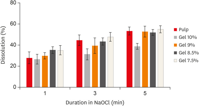
Objective The aim of this study was to introduce a gelatin/bovine serum albumin (BSA) tissue standard, which provides dissolution properties identical to those of biological tissues. Further, the study evaluated whether the utilization of endodontic activating devices led to enhanced phantom dissolution rates.
Materials and Methods Bovine pulp tissue was obtained to determine a benchmark of tissue dissolution. The surface area and mass of samples were held constant while the ratio of gelatin and BSA were varied, ranging from 7.5% to 10% gelatin and 5% BSA. Each sample was placed in an individual test tube that was filled with an appropriate sodium hypochlorite solution for 1, 3, and 5 minutes, and then removed from the solution, blotted dry, and weighed again. The remaining tissue was calculated as the percent of initial tissue to determine the tissue dissolution rate. A radiopaque agent (sodium diatrizoate) and a fluorescent dye (methylene blue) were added to the phantom to allow easy quantification of phantom dissolution in a canal block model when activated using ultrasonic (EndoUltra) or sonic (EndoActivator) energy.
Results The 9% gelatin + 5% BSA phantom showed statistically equivalent dissolution to bovine pulp tissue at all time intervals. Furthermore, the EndoUltra yielded significantly more phantom dissolution in the canal block than the EndoActivator or syringe irrigation.
Conclusions Our phantom is comparable to biological tissue in terms of tissue dissolution and could be utilized for
in vitro tests due to its injectability and detectability.-
Citations
Citations to this article as recorded by- Evaluation of pulp tissue dissolving efficiency of sodium and calcium hypochlorite solutions activated by ultrasonics and laser: an in vitro study
Oznur Ozturk, Ozgur Genc Sen
BMC Oral Health.2024;[Epub] CrossRef
- Evaluation of pulp tissue dissolving efficiency of sodium and calcium hypochlorite solutions activated by ultrasonics and laser: an in vitro study
- 2,370 View
- 12 Download
- 1 Crossref
- A cone-beam computed tomography study of the prevalence and location of the second mesiobuccal root canal in maxillary molars
- Seong-Ju Lee, Eun-Hye Lee, Se-Hee Park, Kyung-Mo Cho, Jin-Woo Kim
- Restor Dent Endod 2020;45(4):e46. Published online September 3, 2020
- DOI: https://doi.org/10.5395/rde.2020.45.e46
-
Abstract
PDFPubReaderePub
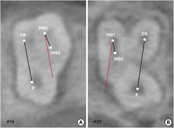
Objectives This study aimed to investigate the incidence and location of the second mesiobuccal root (MB2) canal in maxillary molars with the aid of various measuring points and lines using cone-beam computed tomography (CT).
Materials and Methods A total of 205 images of patients who underwent cone-beam CT examinations between 2011 and 2015 as part of their dental diagnosis and treatment were included. There were 76 images of the maxillary first molar and 135 images of the maxillary second molar. Canal orifices were detected at −1 mm from the top of the pulpal floor on cone-beam CT images. Image assessment was performed by 2 observers in reformatted image planes using software. Assessments included measurement of the distance between the MB1 and MB2 canals, and the angles between the lines connecting the MB1-MB2 and distobuccal (DB)-palatal (P) canals. The data were analyzed using the student's
t -test.Results The prevalence of the MB2 canal was 86.8% in the first molar and 28.9% in the second molar. The angle between the lines connecting the MB1-MB2 and DB-P canals was 2.3° ± 5.7° in the first molar and −3.95° ± 7.73° in the second molar. The distance between the MB1 and MB2 canals was 2.1 ± 0.44 mm in the first molar and 1.98 ± 0.42 mm in the second molar.
Conclusions The angles between the lines connecting the MB1-MB2 and DB-P canals was almost parallel. These findings may aid in the prediction of the location of the MB2 canal orifice.
-
Citations
Citations to this article as recorded by- Study on the Geometric Location Method of the Danger Zone in the Mesial Roots of Mandibular First Molars
Jinjie Yan, Yuanling Peng, Jing Yang, Jie Liu, Linxian Wang, Tingyuan Zhao, Jian Zhang, Kehua Que
Journal of Endodontics.2026; 52(3): 387. CrossRef - Comparative diagnostic accuracy of ChatGPT in second mesiobuccal canal detection
Mehmet Ali Altunkum, Sadullah Kaya
BMC Oral Health.2026;[Epub] CrossRef - Diagnostic Performance of Magnification and Ultrasonic Troughing in Detecting Second Mesiobuccal Canals: A Two-Factor Experimental Study
Mehmet Adiguzel, Furkan Ozeken
Cureus.2026;[Epub] CrossRef - Position of Second Mesiobuccal Canal Relative to Distobuccal and Palatal Canals of Maxillary Molars in an Iranian Population
Sina Mosadeghian, Azadeh Torkzadeh, Parisa Ranjbarian, Roya Asaadi
Journal of Research in Dental and Maxillofacial Sciences.2025; 10(1): 34. CrossRef - Machine Learning Models in the Detection of MB2 Canal Orifice in CBCT Images
Shishir Shetty, Meliz Yuvali, Ilker Ozsahin, Saad Al-Bayatti, Sangeetha Narasimhan, Mohammed Alsaegh, Hiba Al-Daghestani, Raghavendra Shetty, Renita Castelino, Leena R David, Dilber Uzun Ozsahin
International Dental Journal.2025; 75(3): 1640. CrossRef - EVALUATION OF THE PREVALENCE AND LOCATION OF SECOND MESIOBUCCAL CANALS IN 2100 UPPER FIRST AND SECOND MOLAR TEETH: A CONE BEAM COMPUTED TOMOGRAPHY STUDY
Bahar Kaplan, Özkan Adıgüzel, Ayşe Gül Öner Talmaç, Elif Meltem Aslan
İnönü Üniversitesi Sağlık Hizmetleri Meslek Yüksek Okulu Dergisi.2025; 13(3): 752. CrossRef - A novel method for the precise second mesiobuccal canal orifice location: A combined strategy for enhanced clinical practice
Yuhan Wang, Lingyun Li, Lu Zhang, Xiaoyan Wang
Journal of Dental Sciences.2025;[Epub] CrossRef - The Correlation between Intraorifice Distance and the Anatomical Characteristics of the Second Mesiobuccal Canal of Maxillary Molars: A CBCT Study
Isabella Perondi, Silvio Taschieri, Martino Baruffaldi, Roberto Fornara, Luca Francetti, Stefano Corbella, Deepa Gurunathan
International Journal of Dentistry.2024;[Epub] CrossRef - Endodontic management of type I maxillary first molar with two palatal roots using cone-beam computed tomography
Nuha Alghamdi
Dental Journal.2024; 57(1): 1. CrossRef - 3D geometric analysis of second mesiobuccal canal in permanent maxillary first molar tooth
Indrani Khadilkar, Divya Nangia, Amrita Chawla, Sidhartha Sharma, Vijay Kumar, Shalini Gupta, Ajay Logani
Australian Endodontic Journal.2023; 49(1): 140. CrossRef - Prevalence of mesiobuccal-2 canals in maxillary first and second molars among the Bruneian population—CBCT analysis
Hui Yi Onn, Malissa Siao Yun Abdullah Sikun, Hanif Abdul Rahman, Jagjit Singh Dhaliwal
BDJ Open.2022;[Epub] CrossRef - Location angle of second mesio-buccal canal in maxillary molars of an Indian population: an in vivo retrospective CBCT evaluation and proposal of a new classification
Kishor Vhorkate, Kulvinder Banga, Ajinkya M. Pawar, Shugufta Mir, Suraj Arora, Dian Agustin Wahjuningrum, Anuj Bhardwaj, Alexander Maniangat Luke
PeerJ.2022; 10: e14234. CrossRef - Maxillary molar root and canal morphology of Neolithic and modern Chinese
H.Y. Ren, K.Y. Kum, Y.S. Zhao, Y.J. Yoo, J.S. Jeong, Hiran Perinpanayagam, X.Y. Wang, G.J. Li, F. Wang, H. Fang, Y. Gu
Archives of Oral Biology.2021; 131: 105272. CrossRef
- Study on the Geometric Location Method of the Danger Zone in the Mesial Roots of Mandibular First Molars
- 4,917 View
- 64 Download
- 13 Crossref
- Physicochemical properties, cytotoxicity and penetration into dentinal tubules of sodium hypochlorite with and without surfactants
- Hernán Coaguila-Llerena, Isadora Barbieri, Mário Tanomaru-Filho, Renato de Toledo Leonardo, Ana Paula Ramos, Gisele Faria
- Restor Dent Endod 2020;45(4):e47. Published online September 10, 2020
- DOI: https://doi.org/10.5395/rde.2020.45.e47
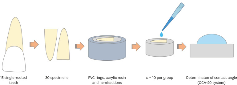
-
Abstract
PDFPubReaderePub
Objectives The aim of this study was to assess the physicochemical properties, cytotoxicity and penetration into dentinal tubules of ChlorCid™ Surf (3% sodium hypochlorite [NaOCl] with surfactant) in comparison to ChlorCid™ (3% NaOCl without surfactant).
Materials and Methods The physicochemical properties evaluated were pH, surface tension, free available chlorine (FAC) and contact angle. Cytotoxicity was evaluated in L929 fibroblasts exposed to the solutions by 3-(4,5-dimethylthiazol-2-yl)-2,5-diphenyl tetrazolium bromide and neutral red assays. Assessment of penetration into dentinal tubules was performed by staining single-rooted permanent human teeth with crystal violet (
n = 9), which were irrigated with the solutions and analyzed in cervical, middle and apical segments. Data were analyzed by one-way analysis of variance (ANOVA) and Tukey'spost -test, 2-way ANOVA and Bonferroni'spost -test ort -test (α = 0.05).Results ChlorCid™ Surf and ChlorCid™ FAC values were close to those indicated by the manufacturer. ChlorCid™ Surf showed lower surface tension and contact angle on dentin, and higher pH than ChlorCid™ (
p < 0.05). The penetration of ChlorCid™ Surf was higher in cervical and middle segments, compared with ChlorCid™ (p < 0.05). There was no difference in irrigant cytotoxicity (p > 0.05).Conclusions ChlorCid™ Surf showed lower surface tension, lower contact angle on root canal dentin, higher penetration into dentinal tubules and more alkaline pH, compared with ChlorCid™. However, both solutions showed similar cytotoxicity and FAC content.
-
Citations
Citations to this article as recorded by- Physicochemical and Biological Properties of the “All-In-One” Endodontic Irrigant Triton
Jesus Aranda, Elda Olivia Nobre de Souza, Arturo Javier Aranda Garcia, Renato de Toledo Leonardo, Ana Paula Ramos, Giampiero Rossi-Fedele, Gisele Faria
Journal of Endodontics.2026; 52(3): 421. CrossRef - Single‐ or two‐visits endodontic treatment: Impact of irrigants on surface roughness, microhardness, topography, and fracture resistance of root dentin
Yasmin Padoin, Sidnei Flores de Pellegrin, Duvan Cala Castillo, William Lemos Bevilaqua, Gabriel Kalil Rocha Pereira, Guilherme Pauletto
European Journal of Oral Sciences.2026;[Epub] CrossRef - Effects of penetration enhancers on the performance of irrigants for root canal disinfection
Yi Luo, Runze Liu, Pei Liu, Mengting Duan, Wei Fan, Bing Fan
Clinical Oral Investigations.2025;[Epub] CrossRef - Influence of post space disinfection protocols on the push-out bond strength of fiber posts luted with self-adhesive cement
Satheesh B. Haralur, Salem Ali Alqahtani, Khalid Salem Alqahtani, Mohammed A. Al-Qarni, Saeed M. AlQahtani
AIP Advances.2025;[Epub] CrossRef - Research methods assessing sodium hypochlorite cytotoxicity: A scoping review
Hernán Coaguila-Llerena, Luana Raphael da Silva, Gisele Faria
Heliyon.2024; 10(1): e23060. CrossRef - Amelioration in the sodium hypochlorite as root canal irrigant – A review
Preety Sehrawat
International Dental Journal of Student's Research.2024; 12(2): 65. CrossRef - Sonic-assisted antibacterial photodynamic therapy: a strategy for enhancing lateral canal disinfection
Yanhuang Wang, Lishan Lei, Jing Huang, Zhiyu Cai, Xiaojing Huang
BMC Oral Health.2024;[Epub] CrossRef - A Comparative Evaluation of Contact Angle and Depth of Penetration of Sodium Hypochlorite With Various Surfactants: An In Vitro Study
Shubhashini N, Krithika D, Akhilesh Gowda , Shruthi Nagaraja , Rhea S Mathew, Nivaskumar G A, Vinaychandra R
Cureus.2024;[Epub] CrossRef - Antibacterial efficacy of silver nanoparticles, sodium hypochlorite, chlorhexidine, and hypochlorous acid on dentinal surfaces infected with Enterococcus faecalis
Aysenur Oncu, Berkan Celikten, Betül Aydın, Gulin Amasya, Erkan Tuncay, Gamze Guney Eskiler, Leyla Açık, Fatma Semra Sevimay
Microscopy Research and Technique.2024; 87(9): 2094. CrossRef - Advances in the Role of Sodium Hypochlorite Irrigant in Chemical Preparation of Root Canal Treatment
Chen Cai, Xuan Chen, Yang Li, Qianzhou Jiang, Yeliz Guven
BioMed Research International.2023;[Epub] CrossRef - Effect of sodium hypochlorite-based formulations on the adhesion interface after fiber post cementation
Joatan Lucas de Sousa Gomes COSTA, Tatiane Miranda MANZOLI, João Felipe BESEGATO, Joissi Ferrari ZANIBONI, Eliane Cristina Gulin DE OLIVEIRA, Lucas David GALVANI, Andréa Abi Rached DANTAS, Luis Geraldo VAZ, Milton Carlos KUGA
Dental Materials Journal.2023; 42(6): 878. CrossRef - Physicochemical properties and penetration into dentinal tubules of calcium hypochlorite with surfactants
Hernán Coaguila-Llerena, Julia da Silva Toledo, Ana Paula Ramos, Gisele Faria
Brazilian Dental Journal.2022; 33(2): 1. CrossRef
- Physicochemical and Biological Properties of the “All-In-One” Endodontic Irrigant Triton
- 3,264 View
- 40 Download
- 12 Crossref
- Reference values for pulp oxygen saturation as a diagnostic tool in endodontics: a systematic review and meta-analysis
- Paula Lambert, Sergio Augusto Quevedo Miguens, Caroline Solda, Juliana Tomaz Sganzerla, Leandro Azambuja Reichert, Carlos Estrela, Fernando Branco Barletta
- Restor Dent Endod 2020;45(4):e48. Published online October 5, 2020
- DOI: https://doi.org/10.5395/rde.2020.45.e48
-
Abstract
PDFPubReaderePub
Objectives This systematic review aimed to identify mean oxygen saturation values (SpO2) using pulse oximetry in permanent maxillary anterior teeth.
Materials and Methods The MEDLINE, Scientific Electronic Library Online, Cochrane Central Register of Controlled Trials, EMBASE, and Literatura Latino Americana em Ciências da Saúde electronic databases were searched. Combinations and variations of “oximetry” AND “dental pulp test” were used as search terms. Studies reporting means and standard deviations of SpO2 values were included. Two reviewers independently extracted data following the Preferred Reporting Items for Systematic Reviews and Meta-Analyses checklist. Heterogeneity was assessed using the
I 2 statistic, and all analyses were performed using R software. Study quality was assessed using the Quality Assessment of Diagnostic Accuracy Studies-2 tool and the Newcastle-Ottawa scale.Results Of the 251 studies identified, 19 met the eligibility criteria and were included (total sample, 4,541 teeth). In the meta-analysis, the mean SpO2 values were 84.94% (95% confidence interval [CI], 84.85%–85.04%) for the central incisors, 89.29% (95% CI, 89.22%–89.35%) for the lateral incisors, and 89.20% (95% CI, 89.05%–89.34%) for the canines. The studies were predominantly low-quality due to the high risk of bias associated with the index test, unclear risk regarding patient selection, and concerns about outcome assessment.
Conclusions Although most studies were low-quality, the oxygen saturation levels in normal pulp could be established (minimum saturation, 77.52%). Despite the risk of bias of the included studies, the reference values reported herein are clinically relevant for assessments of changes in pulp status.
Trial Registration International Prospective Register of Systematic Reviews Identifier:
CRD42018085598 -
Citations
Citations to this article as recorded by- Lower dental pulp oxygen saturation in children with molar incisor hypomineralization: a cross-sectional study
Jade de Souza CAVALCANTE, Carlos ESTRELA, Fabrício Kitazono de CARVALHO, Francisco Wanderley Garcia de PAULA-SILVA, Manoel Damião de SOUSA-NETO, Kelly Fernanda MOLENA, Alexandra Mussolino de QUEIROZ
Brazilian Oral Research.2026;[Epub] CrossRef - Reference Values for Pulse Oximetry Testing in Permanent Teeth: A Systematic Review and Meta‐Analysis
Lilian Tietz, Theodoro Weissheimer, Cassiano Kuchenbecker Rösing, Marcus Vinicius Reis Só
International Endodontic Journal.2026;[Epub] CrossRef - Clinical Validation of Smartphone-Enabled Pulse Oximetry for Objective Pulp Vitality Assessment: A Diagnostic Accuracy Study
Celso Luiz Caldeira, Stephanie Isabel Diaz Zamalloa, Claudia Regina Guimaro Sakitani, Fernando Branco Barletta, Marinella Holzhausen
Journal of Endodontics.2025; 51(12): 1752. CrossRef - Laser Doppler Flowmetry and Continuous Tissue Oxygenation Monitoring: Best of Vitality Tests?
Herman J. J. Roeykens, Rani D’haese, Wolfgang Jacquet, Roeland J. G. De Moor, Stefan Vandeweghe
Oral.2025; 5(4): 83. CrossRef - Diagnostic accuracy of Transmitted-light plethysmography for the assessment of pulpal circulation in traumatized young permanent incisors
Satoko Kakino, Hiroaki Ohki, Kaori Kohi, Yuko Matsumura, Tsutomu Iwamoto
Scientific Reports.2025;[Epub] CrossRef - Future trends in endodontics
Foo Suanhow, Tawil Bill
Journal of Applied Biotechnology & Bioengineering.2024; 11(1): 1. CrossRef - Assessment of Pulpal Oxygen Saturation in Caries-free and Carious Maxillary Primary Central Incisors Using a Customized Dental Pulse Oximeter
Kranthi Reddy Kanumuru, Nancy Solomon, Hemalatha Ramkumar, Shankar Paulindraraj, Trophimus Gnanabagyan Jayakaran, Senthil Dakshinamoorthy
International Journal of Clinical Pediatric Dentistry.2023; 16(4): 560. CrossRef - Age-Related Variation of Pulpal Oxygen Saturation in Healthy Primary and Permanent Teeth in Children: A Clinical Study
Andreea Igna, Darian Rusu, Emilia Ogodescu, Ștefania Dinu, Marius Boariu, Adrian Voicu, Ștefan-Ioan Stratul
Journal of Clinical Medicine.2022; 12(1): 170. CrossRef - Pulp oxygen saturation measurement as a diagnostic tool for assessing pulp status in primary teeth: A systematic review and meta-analysis
Kanamarlapudi Venkata Saikiran, Deepa Gurunathan, Sainath Reddy Elicherla, Sreekanth Kumar Mallineni, Sivakumar Nuvvula
Journal of Indian Society of Pedodontics and Preventive Dentistry.2022; 40(4): 349. CrossRef - Diagnostic Value of Serum Chitinase‐3‐Like Protein 1 for Liver Fibrosis: A Meta‐analysis
Xiaoting Huang, Jialing Zhuang, Yongqiang Yang, Jiaxin Jian, Wen Ai, Chunyong Liu, Wenzhi Tang, Changyu Jiang, Yongshen He, Lesheng Huang, Se Peng, Jin Shui Pan
BioMed Research International.2022;[Epub] CrossRef - Assessment of Pulpal Status in Primary Teeth Following Direct Pulp Capping in an Experimental Canine Model
Andreea Igna, Cornel Igna, Mariana Ioana Miron, Larisa Schuszler, Roxana Dascălu, Mihaela Moldovan, Adrian Aristide Voicu, Carmen Darinca Todea, Marius Boariu, Maria-Alexandra Mârțu, Ștefan-Ioan Stratul
Diagnostics.2022; 12(8): 2022. CrossRef
- Lower dental pulp oxygen saturation in children with molar incisor hypomineralization: a cross-sectional study
- 3,227 View
- 43 Download
- 11 Crossref
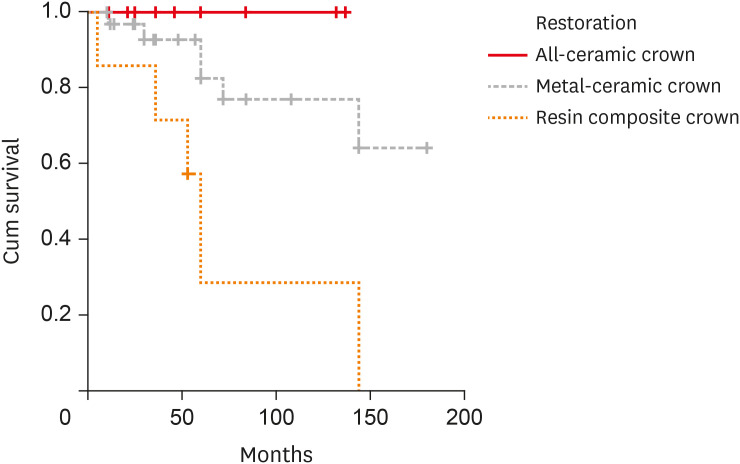
- Retrospective clinical and radiographic evaluation of restored endodontically treated teeth
- Paula Pontes Garcia, Aline Cappoani, Ricardo Susin Schelbauer, Gisele Maria Correr, Carla Castiglia Gonzaga
- Restor Dent Endod 2020;45(4):e49. Published online October 7, 2020
- DOI: https://doi.org/10.5395/rde.2020.45.e49
-
Abstract
PDFPubReaderePub
Objectives The aim of this study was to perform a clinical and radiographic analysis of endodontically treated teeth (ETT) restored with cast metal posts (CMPs) or prefabricated glass fiber posts (GFPs) and crowns.
Materials and Methods Fifty ETT were restored with 25 CMPs and 25 GFPs at a private dental clinic between 2001 and 2016. The restorations consisted of 12 all-ceramic crowns, 31 metal-ceramic crowns, and 7 composite resin crowns. Demographic data, type of teeth, type of post-and-core system, time of placement, crown restorations, the number of proximal contacts, the type of antagonist, and reports of any complications after post-and-core placement were recorded for each patient. Assessments were performed at baseline (radiographic) and follow-up (radiographic and clinical). Data were analyzed by the McNemar test, the Pearson χ2 test, and Kaplan-Meier survival curves (α = 0.05). The mean follow-up was 67.6 months.
Results No significant difference was observed for any of the radiographic parameters when the baseline and final radiographs were compared. In the clinical evaluation, anatomical form (
p = 0.009) and occlusion (p = 0.001) showed significant differences according to the type of crown restoration; specifically, metal-ceramic and all-ceramic crowns outperformed composite resin crowns.Conclusions CMPs and GFPs showed favorable results for restoring ETT after 6 years of follow-up. All-ceramic and metal-ceramic crowns showed higher survival rates and better clinical outcomes.
-
Citations
Citations to this article as recorded by- The Outcomes of Endodontically Treated Teeth Restored with Custom-Made Cast Post-and-Core Restorations: A Retrospective Cohort Study
Ahmed Ben Suleiman, Shivani Desai, Adam Tepperman, David Chvartszaid, Gevik Malkhassian, Effrat Habsha, Izchak Barzilay, Amir Azarpazhooh
Journal of Endodontics.2024; 50(3): 316. CrossRef - Effect of a circumferential ferrule on the survival and success of endodontically treated teeth restored with fiber posts: A systematic review and meta-analysis
Raghad A. Al-Dabbagh, Mohammed A. Sindi, Mohammed A. Sanari, Alaa I. Manna, Mona A. Al-Dabbagh
The Journal of Prosthetic Dentistry.2024; 132(6): 1251. CrossRef - Effect of Luting Cement Film Thickness on the Pull-Out Bond Strength of Endodontic Post Systems
Khalil Aleisa, Syed Rashid Habib, Abdul Sadekh Ansari, Ragad Altayyar, Shahad Alharbi, Sultan Ali S. Alanazi, Khalid Tawfik Alduaiji
Polymers.2021; 13(18): 3082. CrossRef
- The Outcomes of Endodontically Treated Teeth Restored with Custom-Made Cast Post-and-Core Restorations: A Retrospective Cohort Study
- 3,592 View
- 46 Download
- 3 Crossref
-
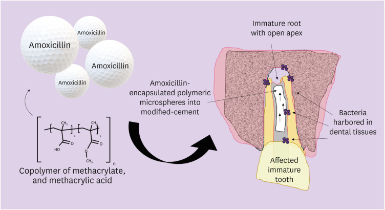
Incorporation of amoxicillin-loaded microspheres in mineral trioxide aggregate cement: an
in vitro study - Fábio Rocha Bohns, Vicente Castelo Branco Leitune, Isadora Martini Garcia, Bruna Genari, Nélio Bairros Dornelles, Silvia Stanisçuaski Guterres, Fabrício Aulo Ogliari, Mary Anne Sampaio de Melo, Fabrício Mezzomo Collares
- Restor Dent Endod 2020;45(4):e50. Published online October 7, 2020
- DOI: https://doi.org/10.5395/rde.2020.45.e50
-
Abstract
PDFPubReaderePub
Objectives In this study, we investigated the potential of amoxicillin-loaded polymeric microspheres to be delivered to tooth root infection sites via a bioactive reparative cement.
Materials and Methods Amoxicillin-loaded microspheres were synthesized by a spray-dray method and incorporated at 2.5% and 5% into a mineral trioxide aggregate cement clinically used to induce a mineralized barrier at the root tip of young permanent teeth with incomplete root development and necrotic pulp. The formulations were modified in liquid:powder ratios and in composition by the microspheres. The optimized formulations were evaluated
in vitro for physical and mechanical eligibility. The morphology of microspheres was observed under scanning electron microscopy.Results The optimized cement formulation containing microspheres at 5% exhibited a delayed-release response and maintained its fundamental functional properties. When mixed with amoxicillin-loaded microspheres, the setting times of both test materials significantly increased. The diametral tensile strength of cement containing microspheres at 5% was similar to control. However, phytic acid had no effect on this outcome (
p > 0.05). When mixed with modified liquid:powder ratio, the setting time was significantly longer than that original liquid:powder ratio (p < 0.05).Conclusions Lack of optimal concentrations of antibiotics at anatomical sites of the dental tissues is a hallmark of recurrent endodontic infections. Therefore, targeting the controlled release of broad-spectrum antibiotics may improve the therapeutic outcomes of current treatments. Overall, these results indicate that the carry of amoxicillin by microspheres could provide an alternative strategy for the local delivery of antibiotics for the management of tooth infections.
-
Citations
Citations to this article as recorded by- Functionalized calcium carbonate microparticles in ethyl cellulose films: A vehicle for sustained amoxicillin release for medical applications
Petru Niga, Simone Sala, Jenny Rissler, Lina Nyström, Anna Fureby, Ulla Elofsson, Joachim Schoelkopf, Roger Roth, Patrick Gane, Mehnath Sivaraj
PLOS One.2026; 21(4): e0320280. CrossRef - Local drug delivery for regeneration and disinfection in endodontics: A narrative review
Anu Elsa Swaroop, Sylvia Mathew, P. Harshini, Shruthi Nagaraja
Journal of Conservative Dentistry and Endodontics.2025; 28(2): 119. CrossRef - Modified Mineral Trioxide Aggregate—A Versatile Dental Material: An Insight on Applications and Newer Advancements
C. Pushpalatha, Vismaya Dhareshwar, S. V. Sowmya, Dominic Augustine, Thilla Sekar Vinothkumar, Apathsakayan Renugalakshmi, Amal Shaiban, Ateet Kakti, Shilpa H. Bhandi, Alok Dubey, Amulya V. Rai, Shankargouda Patil
Frontiers in Bioengineering and Biotechnology.2022;[Epub] CrossRef - Local Drug Delivery Systems for Vital Pulp Therapy: A New Hope
Ardavan Parhizkar, Saeed Asgary, Carlo Galli
International Journal of Biomaterials.2021; 2021: 1. CrossRef
- Functionalized calcium carbonate microparticles in ethyl cellulose films: A vehicle for sustained amoxicillin release for medical applications
- 2,465 View
- 16 Download
- 4 Crossref
- Comparison of vibration characteristics of file systems for root canal shaping according to file length
- Seong-Jun Park, Se-Hee Park, Kyung-Mo Cho, Hyo-Jin Ji, Eun-Hye Lee, Jin-Woo Kim
- Restor Dent Endod 2020;45(4):e51. Published online October 14, 2020
- DOI: https://doi.org/10.5395/rde.2020.45.e51
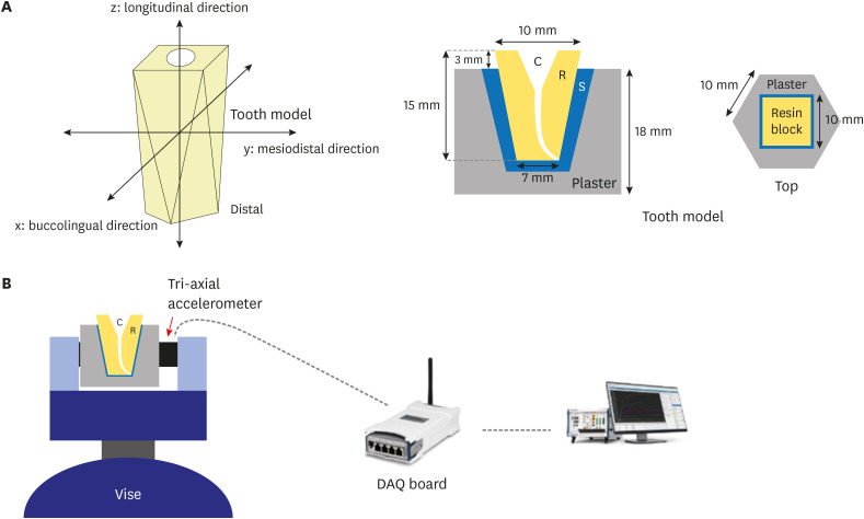
-
Abstract
PDFPubReaderePub
Objectives No studies have yet assessed vibration characteristics according to endodontic file length. Accordingly, the objective of the present study was to examine the vibration characteristics according to nickel-titanium file length and to compare these characteristics between different file systems.
Materials and Methods A total of 45 root canal models were divided into 3 experimental groups (
n = 15 each) based on the file system used (ProTaper Gold [PTG], ProTaper Next, or WaveOne Gold [WOG]). Each experimental group was further divided into 3 subgroups according to file length (21, 25, or 31 mm). An electric motor (X-SMART PLUS) was used in the experiment. For each file system, vibrations generated when using a size 25 file were measured and used to calculate the average vibration acceleration. The differences in vibrations were analyzed using 1-way analysis of variance and the Scheffépost hoc test with a confidence interval of 95%.Results In the PTG file system, significantly lower vibration acceleration was observed when using a 21-mm file than when using a 31-mm file. In the WOG file system, significantly stronger vibration acceleration was observed when using a 31-mm file than when using 21- or 25-mm files. Regardless of the file length, the WOG group exhibited significantly stronger vibration acceleration than the other 2 experimental groups.
Conclusions In clinical practice, choosing a file with the shortest length possible could help reduce vibrations. Additionally, consideration should be given to vibrations that could be generated when using WOG files with reciprocating motion.
-
Citations
Citations to this article as recorded by- Comparison vibration characteristics of several wireless endodontic handpieces
Bo-Kyung Lee, Yoon Lee, Se-Hee Park, Kyung-Mo Cho, Jin-Woo Kim
Journal of Dental Rehabilitation and Applied Science.2022; 38(2): 81. CrossRef
- Comparison vibration characteristics of several wireless endodontic handpieces
- 1,959 View
- 5 Download
- 1 Crossref
- Cytotoxicity and biocompatibility of high mol% yttria containing zirconia
- Gulsan Ara Sathi Kazi, Ryo Yamagiwa
- Restor Dent Endod 2020;45(4):e52. Published online October 14, 2020
- DOI: https://doi.org/10.5395/rde.2020.45.e52

-
Abstract
PDFPubReaderePub
Objectives Yttria-stabilized tetragonal phase zirconia has been used as a dental restorative material for over a decade. While it is still the strongest and toughest ceramic, its translucency remains as a significant drawback. To overcome this, stabilizing the translucency zirconia to a significant cubic crystalline phase by increasing the yttria content to more than 8 mol% (8YTZP). However, the biocompatibility of a high amount of yttria is still an important topic that needs to be investigated.
Materials and Methods Commercially available 8YTZP plates were used. To enhance cell adhesion, proliferation, and differentiation, the surface of the 8YTZP is sequentially polished with a SiC-coated abrasive paper and surface coating with type I collagen. Fibroblast-like cells L929 used for cell adherence and cell proliferation analysis, and mouse bone marrow-derived mesenchymal stem cells (BMSC) used for cell differentiation analysis.
Results The results revealed that all samples, regardless of the surface treatment, are hydrophilic and showed a strong affinity for water. Even the cell culture results indicate that simple surface polishing and coating can affect cellular behavior by enhancing cell adhesion and proliferation. Both L929 cells and BMSC were nicely adhered to and proliferated in all conditions.
Conclusions The results demonstrate the biocompatibility of the cubic phase zirconia with 8 mol% yttria and suggest that yttria with a higher zirconia content are not toxic to the cells, support a strong adhesion of cells on their surfaces, and promote cell proliferation and differentiation. All these confirm its potential use in tissue engineering.
-
Citations
Citations to this article as recorded by- Comparative Evaluation of the Effects of Yttrium Stabilized Zirconia Nanoparticles (YSZrO2- NP) and Porcelain Particles on Property Enhancement of Polymethylmethacrylate (PMMA) Denture Composites
Ihuoma V. Diwe, Henry E. Mgbemere, Olurotimi A. Adeleye, Bolanle Akinboboye
Journal of Biomimetics, Biomaterials and Biomedical Engineering.2026; 70: 21. CrossRef - DLP 3D printing of Y2O3-Doped alumina bioceramics: An integrated study on synthesis, phase evolution, mechanical properties, and biocompatibility
Abuzer Acikgoz, Bulent Aktas, Gokhan Demircan, Ruken Das, Zeynep Celik, Busra Ergin, Hatice Gumushan Aktas, Kemal Dogan
Materials Today Chemistry.2026; 53: 103503. CrossRef - Phase-separated Zr70-xAl12.5Fe17.5Yx (x = 0–25 at.%) metallic glasses with suitable mechanical properties for possible implant applications
Devinder Singh, Parthiban Ramasamy, Anna Sophie Jelinek, Verena Maier-Kiener, Rahul Bhattacharya, Zhuo Chen, Elham Sharifikolouei, Alessandro Calogero Scalia, Ziba Najmi, Andrea Cochis, Simon Fellner, Eray Yüce, Christoph Gammer, Zaoli Zhang, Jürgen Ecker
Journal of Materials Research and Technology.2025; 35: 6468. CrossRef - Rapid and straightforward preparation of sturdy ZrO2-Y2O3@N-GDs inorganic-organic nanohybrid for boosted biomedical applications
S. Kumaraguru, L. Ragunath, J. Suresh, K. Gopinath
Inorganic Chemistry Communications.2025; 182: 115433. CrossRef - Thermal barrier coatings of YSZ developed by plasma sprayed technique and its effective use in orthopedic and dental application
Aishwariya Rajendiran, Vijayalakshmi Uthirapathy
Journal of Materials Science: Materials in Medicine.2025;[Epub] CrossRef - Extreme temperature gradient promoting oxygen diffusion in yttria‐stabilized zirconia: A molecular dynamics study
Jian Guo, Yan Yin, Min Yi
Journal of the American Ceramic Society.2024; 107(10): 6783. CrossRef - Bioceramics: a review on design concepts toward tailor-made (multi)-functional materials for tissue engineering applications
Ritesh Kumar, Ipsita Pattanayak, Pragyan Aparajita Dash, Smita Mohanty
Journal of Materials Science.2023; 58(8): 3460. CrossRef - In Vitro Degradation of Mg-Doped ZrO2 Bioceramics at the Interface with Xerostom® Saliva Substitute Gel
Liliana Bizo, Marieta Mureşan-Pop, Réka Barabás, Lucian Barbu-Tudoran, Antonela Berar
Materials.2023; 16(7): 2680. CrossRef - Processing of gelatine coated composite scaffolds based on magnesium and strontium doped hydroxyapatite and yttria-stabilized zirconium oxide
Aleksa Galic, Tamara Matic, Natasa Obradovic, Zvezdana Bascarevic, Djordje Veljovic
Science of Sintering.2023; 55(4): 469. CrossRef - Biocompatibility of ZrO2 vs. Y-TZP Alloys: Influence of Their Composition and Surface Topography
Alex Tchinda, Laëtitia Chézeau, Gaël Pierson, Richard Kouitat-Njiwa, B H Rihn, Pierre Bravetti
Materials.2022; 15(13): 4655. CrossRef - Influence of oxygen vacancy compensation on the structure, electronic and mechanical properties of yttrium stabilized tetragonal zirconia
Zhou Fan, Yang Wang, Yidong Zhang, Jianyi Liu
Materials Science in Semiconductor Processing.2021; 135: 106082. CrossRef
- Comparative Evaluation of the Effects of Yttrium Stabilized Zirconia Nanoparticles (YSZrO2- NP) and Porcelain Particles on Property Enhancement of Polymethylmethacrylate (PMMA) Denture Composites
- 2,711 View
- 25 Download
- 11 Crossref
- Effect of post space preparation drills on the incidence of root dentin defects
- Thaíse Ayres Bezerra Zuli, Orlando Aguirre Guedes, Gislaine Figueiredo Zarza Arguello Gonçalves, Aurélio Rosa da Silva Júnior, Álvaro Henrique Borges, Andreza Maria Fábio Aranha
- Restor Dent Endod 2020;45(4):e53. Published online October 16, 2020
- DOI: https://doi.org/10.5395/rde.2020.45.e53
-
Abstract
PDFPubReaderePub
Objectives This study investigated the incidence of root dentin defects after the use of different post space preparation (PSP) drills.
Materials and Methods Seventy-two bovine incisors were selected and obtained 14-mm-long root sections. Twelve roots served as controls with no intervention (G1). The 60 root canals remaining were instrumented using the crown-down technique with the ProTaper Next system and obturated using the lateral condensation technique. Specimens were randomly distributed into 5 groups (
n = 12) according to the operative steps performed: G2, root canal instrumentation and filling (I+F); G3, I+F and PSP with Gates-Glidden drills; G4, I+F and PSP with Largo-Peeso reamers; G5, I+F and PSP with Exacto drill; and G6, I+F and PSP with WhitePost drill. Roots were sectioned at 3, 6, 9, and 12 mm from the apex, and digital images were captured. The presence of root dentin defects was recorded. Data were analyzed by the χ2 test, withp < 0.05 considered to indicate statistical significance.Results Root dentin defects were observed in 39.6% of the root sections. No defects were observed in G1. G5 had significantly more cracks and craze lines than G1, G2, and G3 (
p < 0.05), and more fractures than G1, G2, G3, and G4 (p < 0.05). When all root sections were analyzed together, significantly more defects were observed at the 12-mm level than at the 3-mm level (p < 0.05).Conclusions PSP drills caused defects in the root dentin. Gates-Glidden drills caused fewer root defects than Largo-Peeso reamers and Exacto drills.
-
Citations
Citations to this article as recorded by- Fracture Strength of CAD/CAM Endocrown and Post-Core Restorations with Fiber Strip Reinforcement in Mandibular Premolars
Kerem Yılmaz, Hakan Aydın, Zeynep Soylu, Özge Çiloğlu, Esma Fatıma Delican, Mehmet Mustafa Özarslan, Fehmi Gönüldaş
Journal of Functional Biomaterials.2026; 17(5): 248. CrossRef - Evaluation of dentinal crack formation during post space preparation using different fiber post systems with micro-computed tomography
Ayşe Nur Kuşuçar, Damla Kırıcı
BMC Oral Health.2025;[Epub] CrossRef - Fracture and Crack Behavior of Weakened Incisors Restored With Fiber Posts, Polyethylene Reinforcement, or 3D-Printed Endocrowns
Diana Codas-Duarte, Laís L Pelozo, Jardel F Mazzi-Chaves, Fabiane C Lopes-Olhê, Manoel D Sousa-Neto, Aline E Souza-Gabriel
Cureus.2025;[Epub] CrossRef - Selecting drill size for post space preparation based on final endodontic radiographs: An in vitro study
Farzaneh Farid, Julfikar Haider, Marjan Sadeghpour Shahab, Nika Rezaeikalantari
Technology and Health Care.2024; 32(4): 2575. CrossRef - Cone Beam Computed Tomography Analysis of Post Space in Bifurcated Premolars Using ParaPost and Peeso Reamer Drills
Abdulaziz Saleh Alqahtani, Omar Nasser Almonabhi, Abdulmajeed Moh. Almutairi, Reem R. Alnatsha
The Open Dentistry Journal.2024;[Epub] CrossRef - A Comparative Evaluation of Real-Time Guided Dynamic Navigation and Conventional Techniques for Post Space Preparation During Post Endodontic Management: An In Vitro Study
Sherifa Shervani, Sihivahanan Dhanasekaran, Vijay Venkatesh
Cureus.2024;[Epub] CrossRef - The effect of ultrasonic vibration protocols for cast post removal on the incidence of root dentin defects
Giulliano C. Serpa, Orlando A. Guedes, Neurinelma S. S. Freitas, Julio A. Silva, Carlos Estrela, Daniel A. Decurcio
Journal of Oral Science.2023; 65(3): 190. CrossRef
- Fracture Strength of CAD/CAM Endocrown and Post-Core Restorations with Fiber Strip Reinforcement in Mandibular Premolars
- 3,479 View
- 39 Download
- 7 Crossref
- Effects of zinc oxide and calcium–doped zinc oxide nanocrystals on cytotoxicity and reactive oxygen species production in different cell culture models
- Gabriela Leite de Souza, Camilla Christian Gomes Moura, Anielle Christine Almeida Silva, Juliane Zacour Marinho, Thaynara Rodrigues Silva, Noelio Oliveira Dantas, Jéssica Fernanda Sena Bonvicini, Ana Paula Turrioni
- Restor Dent Endod 2020;45(4):e54. Published online October 19, 2020
- DOI: https://doi.org/10.5395/rde.2020.45.e54
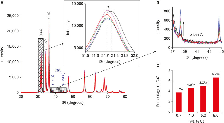
-
Abstract
PDFPubReaderePub
Objectives This study aimed to synthesize nanocrystals (NCs) of zinc oxide (ZnO) and calcium ion (Ca2+)-doped ZnO with different percentages of calcium oxide (CaO), to evaluate cytotoxicity and to assess the effects of the most promising NCs on cytotoxicity depending on lipopolysaccharide (LPS) stimulation.
Materials and Methods Nanomaterials were synthesized (ZnO and ZnO:xCa, x = 0.7; 1.0; 5.0; 9.0) and characterized using X-ray diffractometry, scanning electron microscopy, and methylene blue degradation. SAOS-2 and RAW 264.7 were treated with NCs, and evaluated for viability using the MTT assay. NCs with lower cytotoxicity were maintained in contact with LPS-stimulated (+LPS) and nonstimulated (−LPS) human dental pulp cells (hDPCs). Cell viability, nitric oxide (NO), and reactive oxygen species (ROS) production were evaluated. Cells kept in culture medium or LPS served as negative and positive controls, respectively. One-way analysis of variance and the Dunnett test (α = 0.05) were used for statistical testing.
Results ZnO:0.7Ca and ZnO:1.0Ca at 10 µg/mL were not cytotoxic to SAOS-2 and RAW 264.7. +LPS and −LPS hDPCs treated with ZnO, ZnO:0.7Ca, and ZnO:1.0Ca presented similar NO production to negative control (
p > 0.05) and lower production compared to positive control (p < 0.05). All NCs showed reduced ROS production compared with the positive control group both in +LPS and −LPS cells (p < 0.05).Conclusions NCs were successfully synthesized. ZnO, ZnO:0.7Ca and ZnO:1.0Ca presented the highest percentages of cell viability, decreased ROS and NO production in +LPS cells, and maintenance of NO production at basal levels.
-
Citations
Citations to this article as recorded by- Waste-derived Ca and Zn-based bimetallic (Ca/Zn) nanorods encapsulated chitosan-based haemostatic dressing bandage: A step towards waste to bandages
Pooja Thakur, Rishabh Anand Omar, Neetu Talreja, Divya Chauhan, Mohammad Ashfaq
Journal of Industrial and Engineering Chemistry.2025; 143: 327. CrossRef - Europium and calcium-co-doped TiO2 nanocrystals: tuning the biocompatibility and luminescence traceability of Drosophila melanogaster
Jerusa Maria de Oliveira, Larissa Iolanda M. de Almeida, Francisco Rubens Alves dos Santos, João Paulo S. de Carvalho, Amanda I. dos S. Barbosa, Marcus Andrei R. F. da Costa, Vanessa Tomaz Maciel, Gabriela L. de Souza, Alysson N. Magalhães, Marcos V. Verm
Environmental Science: Nano.2025; 12(1): 835. CrossRef - Development and evaluation of capsules loaded with red propolis extract and metallic nanoparticles using the ionic gelation method
Ilza Fernanda Barboza Duarte Rodrigues, Jéssica Maria Pereira, Lívia Maria Santos de Lima, Kathleen Gomes Lins Silva, Melissa Rosa Silva, Valdemir da Costa Silva, Salvana Priscylla Manso Costa, Ticiano Gomes do Nascimento, Adeildo Junior de Oliveira, John
Journal of Apicultural Research.2025; 64(4): 1151. CrossRef - Structural, optical, and magnetic behavior and the nucleation of a Griffiths-like phase in (Ca,V)-doped ZnO nanoparticles
S. Mrabet, N. Ihzaz, M. N. Bessadok, C. Vázquez-Vázquez, M. Alshammari, O. M. Lemine, D. Ananias, L. El Mir
Dalton Transactions.2025; 54(18): 7400. CrossRef - The effect of iron oxide synergism on the structural and magnetic properties of iron-doped ZnO
Adenilson F. dos Santos, Angela Marta da Silva, Thaís Karine de Lima, Noelio O. Dantas, Marcio A. Correa, Anielle Christine A. Silva
Next Materials.2025; 9: 101047. CrossRef - IN VITRO EVALUATION OF THE ANTI-LEISHMANIAL ROLE OF MILTEFOSINE-LOADED MESOPORUSZNO NANOPARTICLES IN RAW 264.7 MACROPHAGES
PARAG GHOSH, DILEEP KUMAR BHARATI, DIBYA DAS, SUBAS CHANDRA DINDA, ANIRBANDEEP BOSE
Asian Journal of Pharmaceutical and Clinical Research.2025; : 231. CrossRef - Development of antibacterial dual-cure dental resin composites via tetrapod-shaped zinc oxide incorporation
Hwalim Lee, Yu-Jin Kim, Ye-Jin Yang, Jung-Hwan Lee, Hae-Hyoung Lee
Dental Materials.2024; 40(11): 1762. CrossRef - Investigation on the non-linear behaviour of silicon nanowires and assessment of the biosensing potential
M M A Hakim
Engineering Research Express.2023; 5(2): 025017. CrossRef - Evaluation of Cytotoxicity, Cell Attachment, and Elemental Characterization of Three Calcium Silicate-Based Sealers
Anahi de Paula Melo, Camila Maria Peres de Rosatto, Danilo Cassiano Ferraz, Gabriela Leite de Souza, Camilla Christian Gomes Moura
Materials.2023; 16(20): 6705. CrossRef - Metallic Nanoparticles: A New Frontier in the Fight Against Leishmaniasis
Rhanoica Oliveira Guerra, José Rodrigues do Carmo Neto, Tarcísio de Albuquerque Martins, Thaís Soares Farnesi de-Assunção, Virmondes Rodrigues Junior, Carlo José Freire de Oliveira, Anielle Christine Almeida Silva, Marcos Vinicius da Silva
Current Medicinal Chemistry.2022; 29(26): 4547. CrossRef - In situ synthesis of zinc oxide/selenium composite for UV blocker application
Chaoqun Xia, Shi Liu, Baining Cui, Mingjun Li, Hongshui Wang, Chunyong Liang, Phong A. Tran, Yan Wang, Huan Zhou, Lei Yang
International Journal of Applied Ceramic Technology.2022;[Epub] CrossRef - Biocompatibility and Connectivity of Semiconductor Nanostructures for Cardiac Tissue Engineering Applications
Roberto Gaetani, Yuriy Derevyanchuk, Andrea Notargiacomo, Marialilia Pea, Massimiliano Renzi, Elisa Messina, Fabrizio Palma
Bioengineering.2022; 9(11): 621. CrossRef - Calcium-doped zinc oxide nanocrystals as an innovative intracanal medicament: a pilot study
Gabriela Leite de Souza, Thamara Eduarda Alves Magalhães, Gabrielle Alves Nunes Freitas, Nelly Xiomara Alvarado Lemus, Gabriella Lopes de Rezende Barbosa, Anielle Christine Almeida Silva, Camilla Christian Gomes Moura
Restorative Dentistry & Endodontics.2022;[Epub] CrossRef
- Waste-derived Ca and Zn-based bimetallic (Ca/Zn) nanorods encapsulated chitosan-based haemostatic dressing bandage: A step towards waste to bandages
- 2,676 View
- 14 Download
- 13 Crossref
- Assessment of the radiant emittance of damaged/contaminated dental light-curing tips by spectrophotometric methods
- Abdulrahman A. Balhaddad, Isadora Garcia, Fabrício Collares, Cristopher M. Felix, Nisha Ganesh, Qoot Alkabashi, Ward Massei, Howard Strassler, Mary Anne Melo
- Restor Dent Endod 2020;45(4):e55. Published online November 3, 2020
- DOI: https://doi.org/10.5395/rde.2020.45.e55
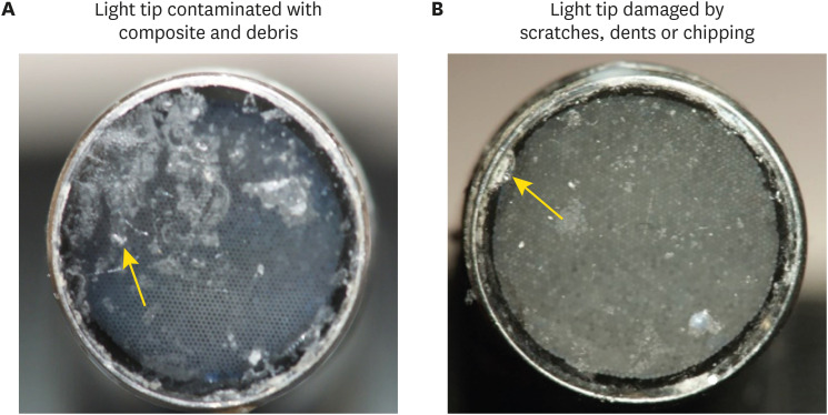
-
Abstract
PDFPubReaderePub
Objectives This study investigated the effects of physically damaged and resin-contaminated tips on radiant emittance, comparing them with new undamaged, non-contaminated tips using 3 pieces of spectrophotometric laboratory equipment.
Materials and Methods Nine tips with damage and/or resin contaminants from actual clinical situations were compared with a new tip without damage or contamination (control group). The radiant emittance was recorded using 3 spectrophotometric methods: a laboratory-grade thermopile, a laboratory-grade integrating sphere, and a portable light collector (checkMARC).
Results A significant difference between the laboratory-grade thermopile and the laboratory-grade integrating sphere was found when the radiant emittance values of the control or damaged/contaminated tips were investigated (
p < 0.05), but both methods were comparable to checkMARC (p > 0.05). Regardless of the method used to quantify the light output, the mean radiant emittance values of the damaged/contaminated tips were significantly lower than those of the control (p < 0.05). The beam profile of the damaged/contaminated tips was less homogeneous than that of the control.Conclusions Damaged/contaminated tips can reduce the radiant emittance output and the homogeneity of the beam, which may affect the energy delivered to composite restorations. The checkMARC spectrophotometer device can be used in dental offices, as it provided values close to those produced by a laboratory-grade integrated sphere spectrophotometer. Dentists should assess the radiant emittance of their light-curing units to ensure optimal curing in photoactivated, resin-based materials.
-
Citations
Citations to this article as recorded by- Effect of damage or contamination to the tips of 200 light-curing units
Abdulrahman A. Balhaddad, Afnan O. Al-Zain, Hassan A. Alyami, Husain A. Almakrami, Osama A. Alsulaiman, Eman H. Ismail, Richard B. Price, Ahmed A. Alsulaiman
BMC Oral Health.2025;[Epub] CrossRef - The Performance of Light-curing Units Used in Different Clinics at Aseer Region, Saudi Arabia: A Cross-sectional Study
Mohammed M Al Moaleem, Ghadeer S Alwadai, Nada A Alamoudi, Naif N Abogazalah, Saleh A Alqahtani, Faisal H Alshehri, Wafa H Alaajam, Mohammad A Alamri, Amjad Y Alhaydan
The Journal of Contemporary Dental Practice.2025; 26(8): 784. CrossRef - Evaluation of Radiant Power of the Light Curing Units Used in Clinics at Governmental and Privates Dental Faculties
Sami Ali Hasan, Ibrahim Al-Shami, Mohsen Al-Hamzi, Ghadeer Alwadai, Nada Alamoudi, Saleh Alqahtani, Arwa Daghrery, Wafa Alaajam, Mansoor Shariff, Hussain Kinani, Mohammed Al Moaleem
Medical Devices: Evidence and Research.2024; Volume 17: 301. CrossRef - Evaluation of the information provided in the instruction manuals of dental light‐curing units
Afnan O. Al‐Zain, Eman H. Ismail, Abdulrahman A. Balhaddad, Osamah Toras, Yousif Alharthy, Rafa Alsultan, Abeer Alrossais, Richard B. Price
Journal of Esthetic and Restorative Dentistry.2024; 36(10): 1466. CrossRef - Utilizing Light Cure Units: A Concise Narrative Review
Fatin A. Hasanain, Hani M. Nassar
Polymers.2021; 13(10): 1596. CrossRef - Improper Light Curing of Bulkfill Composite Drives Surface Changes and Increases S. mutans Biofilm Growth as a Pathway for Higher Risk of Recurrent Caries around Restorations
Haifa Maktabi, Maria Salem Ibrahim, Abdulrahman A. Balhaddad, Qoot Alkhubaizi, Isadora Martini Garcia, Fabrício Mezzomo Collares, Howard Strassler, Ana Paula P. Fugolin, Carmem S. Pfeifer, Mary Anne S. Melo
Dentistry Journal.2021; 9(8): 83. CrossRef
- Effect of damage or contamination to the tips of 200 light-curing units
- 2,321 View
- 8 Download
- 6 Crossref
Case Report
- Bioblock technique to treat severe internal resorption with subsequent periapical pathology: a case report
- Márk Fráter, Tekla Sáry, Sufyan Garoushi
- Restor Dent Endod 2020;45(4):e43. Published online August 18, 2020
- DOI: https://doi.org/10.5395/rde.2020.45.e43
-
Abstract
PDFPubReaderePub
A variety of therapeutic modalities can be used for the endodontic treatment of a traumatized tooth with internal root resorption (IRR). The authors present a case report of the successful restoration of a traumatized upper central incisor that was weakened due to severe IRR and subsequent periapical lesion formation. A 20-year-old female patient was referred to our clinic with severe internal resorption and subsequent periapical pathosis destroying the buccal bone wall. Root canal treatment had been initiated previously at another dental practice, but at that time, the patient's condition could not be managed even with several treatments. After cone-beam computed tomography imaging and proper chemomechanical cleaning, the tooth was managed with a mineral trioxide aggregate plug followed by root canal filling using short fiber-reinforced composite, known as the Bioblock technique. This report is the first documentation of the use of the Bioblock technique in the restoration of a traumatized tooth. The Bioblock technique appears to be ideal for restoring wide irregular root canals, as in cases of severe internal resorption, because it can uniquely fill out the hollow irregularities of the canal. However, further long-term clinical investigations are required to provide additional information about this new technique.
-
Citations
Citations to this article as recorded by- Üvegszálas fogászati kompozit tömőanyag keménysége a gyökércsatornában: nanoindentációs vizsgálat
András Jakab, Kata Lilla Vánkay, Tamás Tarjányi, Gábor Gulyás, Krisztián Bali, Pál Patrik Dézsi, Márton Sámi, Márk Fráter
Fogorvosi Szemle.2024; 117(2): 47. CrossRef - Evaluation of microhardness of short fiber-reinforced composites inside the root canal after different light curing methods – An in vitro study
Márk Fráter, János Grosz, András Jakab, Gábor Braunitzer, Tamás Tarjányi, Gábor Gulyás, Krisztián Bali, Paula Andrea Villa-Machado, Sufyan Garoushi, András Forster
Journal of the Mechanical Behavior of Biomedical Materials.2024; 150: 106324. CrossRef - Imaging techniques and various treatment modalities used in the management of internal root resorption: A systematic review
R. S Digholkar, S D Aggarwal, P S Kurtarkar, P. B Dhatavkar, V L Neil, D N Agarwal
Endodontology.2023; 35(2): 85. CrossRef - The Impact of the Preferred Reporting Items for Case Reports in Endodontics (PRICE) 2020 Guidelines on the Reporting of Endodontic Case Reports
Sofian Youssef, Phillip Tomson, Amir Reza Akbari, Natalie Archer, Fayjel Shah, Jasmeet Heran, Sunmeet Kandhari, Sandeep Pai, Shivakar Mehrotra, Joanna M Batt
Cureus.2023;[Epub] CrossRef - Fatigue performance of endodontically treated premolars restored with direct and indirect cuspal coverage restorations utilizing fiber-reinforced cores
Márk Fráter, Tekla Sáry, Janka Molnár, Gábor Braunitzer, Lippo Lassila, Pekka K. Vallittu, Sufyan Garoushi
Clinical Oral Investigations.2022; 26(4): 3501. CrossRef
- Üvegszálas fogászati kompozit tömőanyag keménysége a gyökércsatornában: nanoindentációs vizsgálat
- 4,655 View
- 122 Download
- 5 Crossref
 First
First Prev
Prev

