Warning: mkdir(): Permission denied in /home/virtual/lib/view_data.php on line 81

Warning: fopen(upload/ip_log/ip_log_2024-12.txt): failed to open stream: No such file or directory in /home/virtual/lib/view_data.php on line 83

Warning: fwrite() expects parameter 1 to be resource, boolean given in /home/virtual/lib/view_data.php on line 84
Incorporation of amoxicillin-loaded microspheres in mineral trioxide aggregate cement: an in vitro study
Skip Navigation
Skip to contents

Restor Dent Endod : Restorative Dentistry & Endodontics

OPEN ACCESS

Articles

Page Path
HOME > Restor Dent Endod > Volume 45(4); 2020 > Article
Research Article Incorporation of amoxicillin-loaded microspheres in mineral trioxide aggregate cement: an in vitro study
Fábio Rocha Bohns1orcid, Vicente Castelo Branco Leitune1orcid, Isadora Martini Garcia1orcid, Bruna Genari1,2orcid, Nélio Bairros Dornelles1orcid, Silvia Stanisçuaski Guterres3orcid, Fabrício Aulo Ogliari4orcid, Mary Anne Sampaio de Melo5,6orcid, Fabrício Mezzomo Collares1orcid

DOI: https://doi.org/10.5395/rde.2020.45.e50
Published online: October 7, 2020

1Department of Dental Materials, School of Dentistry, Federal University of Rio Grande do Sul, Porto Alegre, RS, Brazil.

2Department of Orthodontics and Biomaterials, Centro Universitário UDF, Brasília, DF, Brazil.

3Cosmetology Laboratory, School of Pharmaceutical Sciences, Federal University of Rio Grande do Sul, Porto Alegre, RS, Brazil.

4Yller Biomaterials, Pelotas, RS, Brazil.

5Program in Biomedical Sciences, University of Maryland School of Dentistry, Baltimore, MD, USA.

6Division of Operative Dentistry, Department of General Dentistry, University of Maryland School of Dentistry, Baltimore, MD, USA.

Correspondence to Mary Anne Sampaio de Melo, DDS, MSc, PhD. Associate Professor, Division of Operative Dentistry, Department of General Dentistry, University of Maryland School of Dentistry, 650 W Baltimore St, Baltimore, MD 21201, USA. mmelo@umaryland.edu
Correspondence to Fabrício Mezzomo Collares, DDS, MSc, PhD. Associate Professor, Department of Dental Materials, School of Dentistry, Federal University of Rio Grande do Sul, Rua Ramiro Barcelos, 2492 Rio Branco, Porto Alegre, RS 90035-003, Brazil. fabricio.collares@ufrgs.br
• Received: March 8, 2020   • Revised: May 11, 2020   • Accepted: May 26, 2020

Copyright © 2020. The Korean Academy of Conservative Dentistry

This is an Open Access article distributed under the terms of the Creative Commons Attribution Non-Commercial License (https://creativecommons.org/licenses/by-nc/4.0/) which permits unrestricted non-commercial use, distribution, and reproduction in any medium, provided the original work is properly cited.

  • 25 Views
  • 0 Download
prev next
  • Objectives
    In this study, we investigated the potential of amoxicillin-loaded polymeric microspheres to be delivered to tooth root infection sites via a bioactive reparative cement.
  • Materials and Methods
    Amoxicillin-loaded microspheres were synthesized by a spray-dray method and incorporated at 2.5% and 5% into a mineral trioxide aggregate cement clinically used to induce a mineralized barrier at the root tip of young permanent teeth with incomplete root development and necrotic pulp. The formulations were modified in liquid:powder ratios and in composition by the microspheres. The optimized formulations were evaluated in vitro for physical and mechanical eligibility. The morphology of microspheres was observed under scanning electron microscopy.
  • Results
    The optimized cement formulation containing microspheres at 5% exhibited a delayed-release response and maintained its fundamental functional properties. When mixed with amoxicillin-loaded microspheres, the setting times of both test materials significantly increased. The diametral tensile strength of cement containing microspheres at 5% was similar to control. However, phytic acid had no effect on this outcome (p > 0.05). When mixed with modified liquid:powder ratio, the setting time was significantly longer than that original liquid:powder ratio (p < 0.05).
  • Conclusions
    Lack of optimal concentrations of antibiotics at anatomical sites of the dental tissues is a hallmark of recurrent endodontic infections. Therefore, targeting the controlled release of broad-spectrum antibiotics may improve the therapeutic outcomes of current treatments. Overall, these results indicate that the carry of amoxicillin by microspheres could provide an alternative strategy for the local delivery of antibiotics for the management of tooth infections.
Bacterial pathogens can reach the dental pulp, compromising the inner section of a tooth that contains the nerve tissue and blood vessels [1]. As a result of carious lesions or tooth fractures, bacteria gain entry into the pulp of the tooth and can be sheltered into dentinal tubules and root canal [2]. These microorganisms and their byproducts are responsible for initiating/maintaining a periapical inflammatory process of the pulp tissue that often become necrotic [3]. As the infection progresses, bacteria spread down the root, and through the apical foramen, reach the apex of the tooth root [4]. In cases of tooth infections in young patients, the affected tooth can be immature with incompletely formed roots. The treatment that met the standard of care for infected immature teeth relies on the use of a specific reparative material to neutralize the bacteria and to stimulate the root end closure process by forming a mineralized apical barrier [5,6].
The failure to induce root-end closure (apexification) is often caused by the persistence of microorganisms in the root canal system [7]. Mineral trioxide aggregate (MTA)-cements are Portland-like cement compounded by tricalcium silicate, tricalcium aluminate, tricalcium oxide, silicate oxide, and bismuth oxide [8]. This class of material has outstanding bioactivity with substantial calcium ions release and satisfactory performance as sealing material for root canal treatment [9]. Regrettably, most sealing materials, including MTA-based root canal sealers, lack of substantial antibacterial properties [10]. Some of them that were found to have bacteriostatic behavior, only retain this property for few days or lose this quality after the setting of the material [11,12]. As these materials have intimate contact with the root canal structures and tissues, it would be beneficial if sealing materials retained their antibacterial properties.
Polymer-based drug delivery systems provide a robust technology platform to enable sustained, spatiotemporally controlled drug release [13]. Microspheres have long been used in drug delivery applications because of their managed release capabilities [14]. Microsphere-based strategies have attracted attention in Dentistry because these materials may provide controlled release of bioactive molecules to promote disinfection and consequent healing of surrounds tissues [15]. Microspheres are organic or inorganic, spherical, free-flowing particles ranging from 1 to 1,000 μm in diameter that may carry drugs or bioactive molecules [16]. In medicine, they have been extensively used in drug delivery/targeting applications mainly because of their ability to enhance the efficacy of the associated drug by providing both a large surface area–to–volume ratio for drug release and spatial and temporal control over release [17].
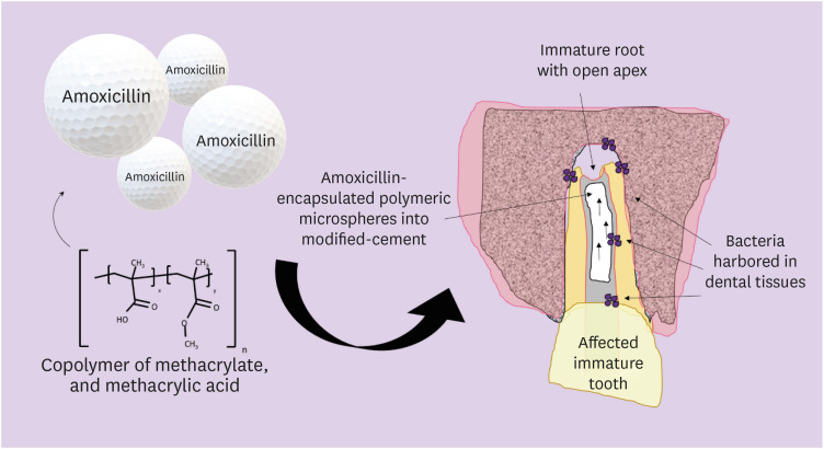
Poly-methacrylic acid-co-methyl methacrylates are copolymers of methacrylate and methacrylic acid, and its use to delayed drug delivery are well reported [18]. This class of synthetic polymers, as known as Eudragits, presents variations in their proportion of neutral, alkaline, or acid groups, resulting in different physicochemical properties, including controlled release [19]. Overall, β-lactam antibiotics are used as the first option for the treatment of endodontic infections [20]. Amoxicillin is a broad-spectrum antibiotic with lower rates of bacterial resistance and the risk of side effects in endodontics [21]. The addition of the antibiotics system with a delayed-release profile to MTA could be a promising approach to improve the treatment outcomes (Figure 1).
Figure 1

Illustrative scheme of a possible approach for the proposed bioactive dental cement with amoxicillin-loaded polymeric microspheres.
rde-45-e50-g001.jpg
Based on it, the present study investigated a simple and effective approach for the incorporation of amoxicillin-loaded polymeric microspheres into a modified formulation of bioactive cement and evaluated the resulting drug release profile and the effect on clinically relevant physical and mechanical properties.
Amoxicillin microspheres preparation
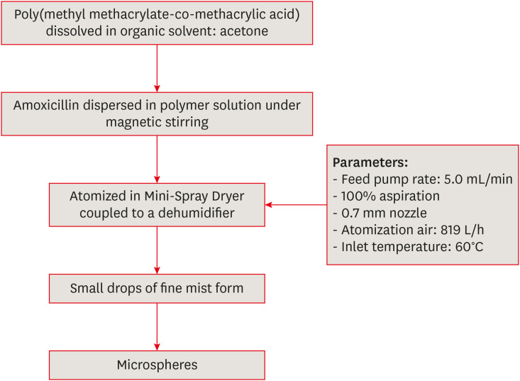
Microspheres loaded with amoxicillin (MSAMOX) were prepared with poly(methacrylic acid-co-methyl methacrylate [Eudragit S100]), and trihydrate amoxicillin following a previous study [22]. Briefly, the polymer was first dissolved in acetone (200 mL), and subsequently, the amoxicillin was added under magnetic stirring at 25°C. The resultant solution was spray-dried using a Mini-Spray Dryer B-290 (Buchi, Flawi, Switzerland) coupled to a dehumidifier (B-292, Buchi) using the following parameters: feed pump rate of 5.0 mL min−1, 100% aspiration, 0.7 mm nozzle, atomization air at 819 Lh−1, and inlet temperature of 60°C with a resulting outlet temperature of approximately 40°C. Figure 2 summarizes the synthesis route for the microspheres. Morphological characteristics of microspheres of amoxicillin were evaluated using scanning electron microscopy (SEM; JSM 6060, Jeol, Tokyo, Japan) with a voltage of 15 kW.
Figure 2

Schematic demonstration of the synthesis route for microsphere preparation by spray-dry method.
rde-45-e50-g002.jpg
Morphological characterization
SEM was used to investigate the morphology and shell thickness of the prepared microspheres. After drying for 48 hours in a desiccator, the samples were then vacuum-coated with a thin layer of gold for 90 seconds to obtain scanning electron microscope images. These particles were examined using SEM at 5 kV.
Composition of mineral trioxide aggregate formulations
Synthetized MSAMOX presented a mean size of 2.664 μm, d10 of 1.369 μm, d50 of 2.430 nm, and d90 of 4.315 μm according to a previous study [22]. To prepare the groups of MTA, MSAMOX was mixed with the MTA at the weight percentages of 0%, 2.5%, and 5%. They were selected following favorable results of pilot tests. The MTA (WMTA Angelus, Angelus, Londrina, PR, Brazil) was used as parent material for the MSAMOX's incorporation. Preliminary studies were performed to tune the optimal liquid/powder ratio able to reach an acceptable homogenization of 5.0 wt% of MSAMOX. The resulting homogeneity was achieved at a liquid/powder ratio of 1.16 mL/g. Based on these screening results, 2 formulations were used as a control: 1) cement mixed with deionized water using the manufacturer's instruction at a liquid/powder ratio of 0.30 mL/g, (Controlratio 0.3) and 2) cement mixed with deionized water at a liquid/powder ratio of 1.16 mL/g (Controlratio 1.16). The experimental cements were prepared by mixing the tested MSAMOX concentrations with deionized water at a liquid/powder ratio of 1.16. Table 1 describes the chemical compositions of formulations assessed in this study.
Table 1

Description of the composition from each group formulated in this study

Group Description Liquid-to-powder ratio
Controlratio 0.3 Tricalcium silicate, dicalcium silicate, tricalcium aluminate, calcium oxide, bismuth oxide 0.30 mL/g
Controlratio 1.16 Cement at the liquid-to-powder ratio of 1.16 1.16 mL/g
MSAMOX at 2.5% Cement at the liquid-to-powder ratio of 1.16 + 2.5% microspheres loaded with amoxicillin 1.16 mL/g
MSAMOX at 5% Cement at the liquid-to-powder ratio of 1.16 + 5% microspheres loaded with amoxicillin 1.16 mL/g
Amoxicillin release profile
The group with the highest concentration of amoxicillin (MSAMOX at 5%) was used to evaluate the drug release profile. To prepare the samples (n = 4), the cement was mixed and poured into cylindrical silicone molds measuring 5.5 mm of diameter and 2.5 mm of height. A polyester strip was placed on the top of each mold, and a load of 11 N was applied during 60 seconds to obtain a flat surface on each sample. All samples were stored in an incubator at 37°C for 7 days. The samples were put separately inside acetate membranes from Millipore (Millipore Corp., Darmstadt, Germany) with a pore size of 0.46 µm previously conditioned with deionized water for 30 minutes. The samples were immersed in 80 mL of simulated body fluid (SBF) prepared according to a previous protocol, followed by bath stirring (Quimiserv, EH4basic, Kika WERKE) [23]. After 2-, 6-, 12-, 24-, 48-, 72-, and 96 hours, 1 mL of SBF was collected and stored in Eppendorf tubes appropriately numbered; the same volume of SBF was replaced into the solution to avoid changes on fluid's concentration.
Setting time
The setting time of the test materials was determined by using the method recommended by ASTM C 266, also known as the Gillmore needle test with slight modifications [24]. To prepare the samples (n = 3) for setting time analysis, cylindrical silicone molds measuring 9 mm in diameter and 1.5 mm in height were used. The particles were incorporated into the mixture, and each cement was inserted into the silicone molds until it was filled. The excesses of the materials were removed, and a polyester strip was placed over each sample to obtain a flat surface. The samples were put in an incubator at 37°C and, after 120 seconds of the onset of the mixing, the initial mark was made using a Gillmore needle weighing 100 g and with a tip measuring 2 mm in diameter; this procedure was repeated at 60 seconds intervals and the values of initial setting time were recorded at the moment when the needle could not mark the sample surface.
Diametral tensile strength
The samples (n = 3) for diametral tensile strength (DTS) tests were prepared with cylindrical silicone molds measuring 9 mm in diameter and 1.5 mm in height. Each group was mixed and poured into the molds; the excess of the material was removed, and a polyester strip was put over the samples to secure a flat surface. All samples were allowed to set in an incubator at 37°C for 7 days. DTS of the samples was conducted on a universal testing machine Shimadzu (EZ-SX, Shimadzu, Japan) at a loading rate of 1 mm/min. DTS of the specimens were calculated using the relationship:
DTS=2Pπbw
Where: P is the maximum load (N), b is the diameter (mm), and w is the thickness (mm) of the measured specimen.
Statistical analysis
The software SigmaPlot, version 12.0 (Systat Software, Inc., San Jose, CA, USA) was used to data analysis. One way analysis of variance (ANOVA) and Tukey tests were used to evaluate the statistical significance data of setting time and DTS. Prematurely failure of specimens was included in the mean with the value of 0.18 MPa (mean value between zero and the minimum value observed in the study). A significant statistical difference was considered at p < 0.05.
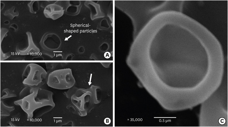
Figure 3 shows SEM images of the microspheres fabricated by the spray-dry method. Figure 3A shows Spherical-shaped particles with a diameter ranging from 2 µm to 5 µm. The microspheres with its mashed shell indicate that the microspheres were not solid capsules but consist of a single internal cavity, which was occupied by amoxicillin. The non-spherical appearance of some of the particles can be due to the applied cutting force. The microspheres present irregular morphology and various size distribution. Some of these microspheres were also seen to be interconnected (arrow; Figure 3B). This is thought to have occurred as a result of liquid residue remaining between the microspheres and polymeric interconnection between the particles. The acetone subsequently acted as a solvent on minimal quantities of the shell surface, which is clearly indicated in Figure 3C.
Figure 3

Scanning electron microscopy images of amoxicillin-loaded microspheres.

rde-45-e50-g003.jpg
The amoxicillin release profile of MSAMOX mixed with the reparative cement at the weight percentages of 5% is shown in Table 2. The release profile demonstrated that the MSAMOX effectively retarded the drug release after 7 days of storage and showed an approximately 17% of drug release after 96 hours of evaluation.
Table 2

Amoxicillin release profile of 5% microspheres loaded with amoxicillin

Collection periods (hr) Amoxicillin release (%)
2 0.00
6 0.00
12 0.00
24 0.00
48 0.00
72 0.00
96 16.68
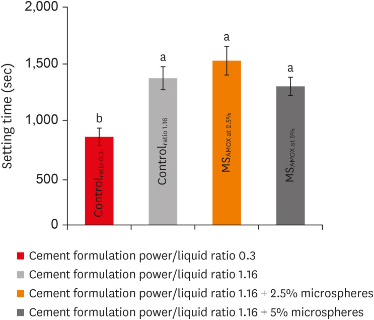
Figure 4 displays the initial setting time of the reparative cement with and without MSAMOX. One way ANOVA and Tukey tests showed that the modification of power: liquid ratio of MSAMOX significantly prolonged the initial setting time from 805 seconds to 1,433 seconds. When adding MSAMOX at 2.5%, only slightly prolonged the initial setting time of 83 seconds was noted concerning Controlratio 1.16. Increasing the concentration of MSAMOX revealed an initial setting time of 1,417 seconds, similar to the Controlratio 1.16. All the experimental formulations were statistically different from the Controlratio 0.3 (p < 0.05).
Figure 4

Setting times of tested cement formulations as the microsphere concentration increases. Values followed by the same letters are not significantly different (p > 0.05).
rde-45-e50-g004.jpg
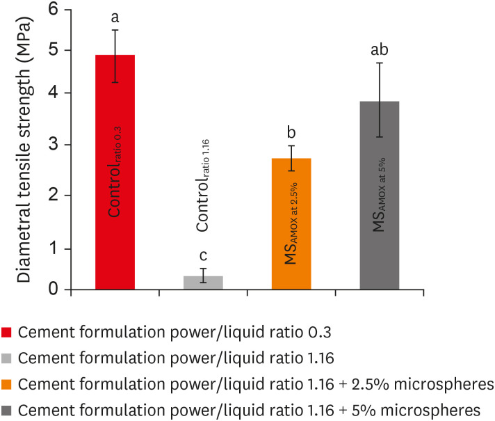
The means and standard deviations of the compressive strength of the groups are shown in Figure 5. One way ANOVA and Tukey tests indicated statistically significant different values among groups. The compressive strength values of the Controlratio 1.16 were significantly lower than the Controlratio 0.3 (p < 0.05). Overall, the incorporation of MSAMOX has improved the compressive strength results. MSAMOX at 2.5% and MSAMOX at 5% has shown an increase of 90.8% and 92.8%, respectively, in relation to Controlratio 1.16. Comparing to the Controlratio 0.3, the MSAMOX at 5% has statistically similar.
Figure 5

Diametral tensile strength (DTS) of the tested cement formulations according to microsphere concentration. Values followed by the same letters are not significantly different (p > 0.05).
rde-45-e50-g005.jpg
In the present study, MSAMOX were successfully synthesized and incorporated in a tuned formulation to grant delays in the amoxicillin release profile for the first time. With this approach, the prolonged antimicrobial effect during extended periods could increase the killing of remaining bacteria, which improve the healing tissue integrity and cellular function. Therefore, recent interests and significant efforts in preventing and actively treating tooth infections by directly targeting infection causative agents through direct application of antimicrobial agents either alone or loaded into dental materials. These have the advantage of overcoming challenges such as poor, locally delivered antibiotics [25]. Single-visit apexification using calcium silicate cements has been successfully used for the endodontic treatment of teeth with necrotic pulps and open apices [26].
Here, MSAMOX effectively delayed the drug release when incorporated in the regenerative cement. At the measuring time of 96 hours, the amoxicillin release profile showed 16.68% of drug release but showed no release at previous measurements (Table 2). Similar results have been obtained by other researchers when using of acrylate-based polymers as a delayed carrier [26]. The microspheres obtained are monolithic particles retained in a porous or solid polymer matrix, differently from microcapsules that consist of a solid or liquid drug container, surrounded by a polymeric membrane [27,28]. The primary process of drug release from microspheres relays on diffusion, biodegradation, desorption of the adsorbed drug from the surface [14].
MSAMOX can be prepared by solid-state reaction, co-precipitation, sol-gel method, and spray-dry routes [16]. In the current study, a facile method obtained the loaded microspheres using a Eudragit S100. The used copolymer solubilizes at pH 7. However, the alkaline medium induced by Ca2+ released from MTA-cements may delay its degradation, retarding the release of the drug [29,30]. The MTA's ability to increase the local pH makes this material very favorable as a vehicle for polymeric microspheres.
The setting behavior of MTA is driven by many variables such as the fineness of the particles, the water/powder ratio, temperature, humidity of application site, and the addition of substances [31]. As a Portland cement, MTA absorbs water. The water trapped in the cement mix and saturated by calcium hydroxide is then released to the environment [32]. High water content and the presence of amoxicillin microspheres may have affected the hydration mechanism of the used MTA-base cement, increasing the setting times of experimental groups. Fridland and Rosado [32] found that the physical properties of MTA changed when mixed with different water-to-powder proportions [33]. However, the amount of water incorporated in the mix is limited by its loss of consistency in the presence of excessive liquid. The modified MTA-base cement had a higher liquid-to-powder ratio in comparison with MTA, which needs more mixing liquid to achieve desired consistency to work. The targeted formulation of MSAMOX at 5% presented suitable handing properties at the water-to-powder ratio of 1.16.
The liquid-to-powder ratio can also determine the mechanical properties of MTA-base cement. A higher liquid-to-powder ratio gives rise to an increasing degree of water pore volume, porosity, and solubility, which consequently can influence the mechanical characteristics of the cement. In our study, the incorporation of MSAMOX has improved the compressive strength results. As amoxicillin has hydrophilic characteristics, its addition to the mixture may have contributed to absorbing water, remaining less unreacted water in the cement bulk, significantly increasing the strength of the targeted formulation of MSAMOX at 5% [34]. Also, the smooth and round shape of microspheres works as a distributor of stress, which may contribute to the significant increase in mechanical property. The MSAMOX at 5% presenting adequate strength can get the benefit of the humid environment because of the inherent hydrophilic nature. Further studies to investigate the in vivo behavior of MTA-base cement with synthesized MSAMOX at 5% seem relevant.
In this work, we have tuned the formulation of a bioactive cement with polymeric microspheres to impart delayed release of antibiotics as a potential drug delivery system to treat local tooth infections. The resulting synthesis is facile, reproducible, and highly reliable. The incorporation of polymeric microspheres at the 5 wt% conveys delayed-release without significant detrimental effects of the physical and mechanical behavior of the material. The possibility of slowing the release of the antibiotics delivered at local sites of infection could find potential applications within dental materials that are in close contact with dental tissues.
The authors gratefully acknowledge Microscopy and Microanalysis Center (Federal University of Rio Grande do Sul) for the microscopy analyses and Angelus for providing the MTA used in this research. This study was financed in part by the Coordenação de Aperfeiçoamento de Pessoal de Nível Superior-Brasil (CAPES)—Finance Code 001—scholarship.

Conflict of Interest: No potential conflict of interest relevant to this article was reported.

Author Contributions:

  • Conceptualization: Leitune VCB, Ogliari FA, Collares FM.

  • Formal analysis: Bohns FR, Genari B, Dornelles NB Júnior, Collares FM.

  • Investigation: Bohns FR, Leitune VCB, Genari B, Dornelles NB Júnior, Guterres SS, Ogliari FA, Collares FM.

  • Project administration: Ogliari FA, Collares FM.

  • Resources: Ogliari FA, Collares FM.

  • Supervision: Leitune VCB, Ogliari FA, Collares FM.

  • Visualization: Bohns FR, Garcia IM, Melo MAS, Collares FM.

  • Writing - original draft: Bohns FR, Garcia IM, Collares FM.

  • Writing - review & editing: Melo MAS, Collares FM.

  • 1. Ricucci D, Loghin S, Niu LN, Tay FR. Changes in the radicular pulp-dentine complex in healthy intact teeth and in response to deep caries or restorations: a histological and histobacteriological study. J Dent 2018;73:76-90. ArticlePubMed
  • 2. Ricucci D, Siqueira JF Jr, Loghin S, Lin LM. Pulp and apical tissue response to deep caries in immature teeth: a histologic and histobacteriologic study. J Dent 2017;56:19-32. ArticlePubMed
  • 3. Farges JC, Alliot-Licht B, Renard E, Ducret M, Gaudin A, Smith AJ, Cooper PR. Dental pulp defence and repair mechanisms in dental caries. Mediators Inflamm 2015;2015:230251. ArticlePubMedPMCPDF
  • 4. Martin FE. Carious pulpitis: microbiological and histopathological considerations. Aust Endod J 2003;29:134-137. PubMed
  • 5. Guerrero F, Mendoza A, Ribas D, Aspiazu K. Apexification: a systematic review. J Conserv Dent 2018;21:462-465. ArticlePubMedPMC
  • 6. Flanagan TA. What can cause the pulps of immature, permanent teeth with open apices to become necrotic and what treatment options are available for these teeth. Aust Endod J 2014;40:95-100. ArticlePubMed
  • 7. Conde MCM, Chisini LA, Sarkis-Onofre R, Schuch HS, Nör JE, Demarco FF. A scoping review of root canal revascularization: relevant aspects for clinical success and tissue formation. Int Endod J 2017;50:860-874. ArticlePubMedPDF
  • 8. Felippe WT, Felippe MCS, Rocha MJC. The effect of mineral trioxide aggregate on the apexification and periapical healing of teeth with incomplete root formation. Int Endod J 2006;39:2-9. PubMed
  • 9. Pace R, Giuliani V, Nieri M, Di Nasso L, Pagavino G. Mineral trioxide aggregate as apical plug in teeth with necrotic pulp and immature apices: a 10-year case series. J Endod 2014;40:1250-1254. ArticlePubMed
  • 10. Torabinejad M, Nosrat A, Verma P, Udochukwu O. Regenerative endodontic treatment or mineral trioxide aggregate apical plug in teeth with necrotic pulps and open apices: a systematic review and meta-analysis. J Endod 2017;43:1806-1820. ArticlePubMed
  • 11. Mohammadi Z, Shalavi S. Effect of hydroxyapatite and bovine serum albumin on the antibacterial activity of MTA. Iran Endod J 2011;6:136-139. PubMedPMC
  • 12. Kim RJY, Kim MO, Lee KS, Lee DY, Shin JH. An in vitro evaluation of the antibacterial properties of three mineral trioxide aggregate (MTA) against five oral bacteria. Arch Oral Biol 2015;60:1497-1502. PubMed
  • 13. Piluso S, Soultan AH, Patterson J. Molecularly engineered polymer-based systems in drug delivery and regenerative medicine. Curr Pharm Des 2017;23:281-294. ArticlePubMed
  • 14. Merkle HP. Drug delivery's quest for polymers: where are the frontiers? Eur J Pharm Biopharm 2015;97:293-303. ArticlePubMed
  • 15. Cuppini M, Zatta KC, Mestieri LB, Grecca FS, Leitune VCB, Guterres SS, Collares FM. Antimicrobial and anti-inflammatory drug-delivery systems at endodontic reparative material: synthesis and characterization. Dent Mater 2019;35:457-467. ArticlePubMed
  • 16. Ghosh Dastidar D, Saha S, Chowdhury M. Porous microspheres: synthesis, characterisation and applications in pharmaceutical & medical fields. Int J Pharm 2018;548:34-48. ArticlePubMed
  • 17. Gupta V, Khan Y, Berkland CJ, Laurencin CT, Detamore MS. Microsphere-based scaffolds in regenerative engineering. Annu Rev Biomed Eng 2017;19:135-161. ArticlePubMedPMC
  • 18. Chawla A, Sharma P, Pawar P. Eudragit S-100 coated sodium alginate microspheres of naproxen sodium: formulation, optimization and in vitro evaluation. Acta Pharm 2012;62:529-545. PubMed
  • 19. Kaur G, Grewal J, Jyoti K, Jain UK, Chandra R, Madan J. Oral controlled and sustained drug delivery systems: concepts, advances, preclinical, and clinical status. In: Grumezescu AM, editor. Drug targeting and stimuli sensitive drug delivery systems. Norwich, NY: William Andrew Publishing; 2018. p. 567-626.
  • 20. Bolfoni MR, Pappen FG, Pereira-Cenci T, Jacinto RC. Antibiotic prescription for endodontic infections: a survey of Brazilian endodontists. Int Endod J 2018;51:148-156. ArticlePubMedPDF
  • 21. Segura-Egea JJ, Gould K, Şen BH, Jonasson P, Cotti E, Mazzoni A, Sunay H, Tjäderhane L, Dummer PM. Antibiotics in endodontics: a review. Int Endod J 2017;50:1169-1184. PubMed
  • 22. Dornelles NB, Collares FM, Genari B, de Souza Balbinot G, Samuel SMW, Arthur RA, Visioli F, Guterres SS, Leitune VCB. Influence of the addition of microsphere load amoxicillin in the physical, chemical and biological properties of an experimental endodontic sealer. J Dent 2018;68:28-33. ArticlePubMed
  • 23. Kokubo T, Takadama H. How useful is SBF in predicting in vivo bone bioactivity? Biomaterials 2006;27:2907-2915. ArticlePubMed
  • 24. Ha WN, Nicholson T, Kahler B, Walsh LJ. Methodologies for measuring the setting times of mineral trioxide aggregate and Portland cement products used in dentistry. Acta Biomater Odontol Scand 2016;2:25-30. ArticlePubMedPMCPDF
  • 25. Parhizkar A, Nojehdehian H, Asgary S. Triple antibiotic paste: momentous roles and applications in endodontics: a review. Restor Dent Endod 2018;43:e28. ArticlePubMedPMCPDF
  • 26. Lee G, Chung C, Kim S, Shin SJ. Observation of an extracted premolar 2.5 years after mineral trioxide aggregate apexification using micro-computed tomography. Restor Dent Endod 2019;45:e4. ArticlePubMedPMCPDF
  • 27. Cartagena AF, Esmerino LA, Polak-Junior R, Olivieri Parreiras S, Domingos Michél M, Farago PV, Campanha NH. New denture adhesive containing miconazole nitrate polymeric microparticles: antifungal, adhesive force and toxicity properties. Dent Mater 2017;33:e53-e61. ArticlePubMed
  • 28. Gavini E, Bonferoni MC, Rassu G, Obinu A, Ferrari F, Giunchedi P. Biodegradable microspheres as intravitreal delivery systems for prolonged drug release. what is their eminence in the nanoparticle era? Curr Drug Deliv 2018;15:930-940. ArticlePubMed
  • 29. Duarte MA, Demarchi AC, Yamashita JC, Kuga MC, Fraga SC. pH and calcium ion release of 2 root-end filling materials. Oral Surg Oral Med Oral Pathol Oral Radiol Endod 2003;95:345-347. PubMed
  • 30. Javid B, Panahandeh N, Torabzadeh H, Nazarian H, Parhizkar A, Asgary S. Bioactivity of endodontic biomaterials on dental pulp stem cells through dentin. Restor Dent Endod 2019;45:e3. ArticlePubMedPMCPDF
  • 31. Mestieri LB, Zaccara IM, Pinheiro LS, Barletta FB, Kopper PMP, Grecca FS. Cytocompatibility and cell proliferation evaluation of calcium phosphate-based root canal sealers. Restor Dent Endod 2019;45:e2. ArticlePubMedPMCPDF
  • 32. Fridland M, Rosado R. Mineral trioxide aggregate (MTA) solubility and porosity with different water-to-powder ratios. J Endod 2003;29:814-817. ArticlePubMed
  • 33. Basturk FB, Nekoofar MH, Günday M, Dummer PM. The effect of various mixing and placement techniques on the compressive strength of mineral trioxide aggregate. J Endod 2013;39:111-114. ArticlePubMed
  • 34. Bouyarmane H, El Hanbali I, El Karbane M, Rami A, Saoiabi A, Saoiabi S, Masse S, Coradin T, Laghzizil A. Parameters influencing ciprofloxacin, ofloxacin, amoxicillin and sulfamethoxazole retention by natural and converted calcium phosphates. J Hazard Mater 2015;291:38-44. ArticlePubMed

Tables & Figures

REFERENCES

    Citations

    Citations to this article as recorded by  

      • ePub LinkePub Link
      • Cite
        CITE
        export Copy Download
        Close
        Download Citation
        Download a citation file in RIS format that can be imported by all major citation management software, including EndNote, ProCite, RefWorks, and Reference Manager.

        Format:
        • RIS — For EndNote, ProCite, RefWorks, and most other reference management software
        • BibTeX — For JabRef, BibDesk, and other BibTeX-specific software
        Include:
        • Citation for the content below
        Incorporation of amoxicillin-loaded microspheres in mineral trioxide aggregate cement: an in vitro study
        Restor Dent Endod. 2020;45(4):e50  Published online October 7, 2020
        Close
      • XML DownloadXML Download
      Figure
      • 0
      • 1
      • 2
      • 3
      • 4
      Incorporation of amoxicillin-loaded microspheres in mineral trioxide aggregate cement: an in vitro study
      Image Image Image Image Image
      Figure 1 Illustrative scheme of a possible approach for the proposed bioactive dental cement with amoxicillin-loaded polymeric microspheres.
      Figure 2 Schematic demonstration of the synthesis route for microsphere preparation by spray-dry method.
      Figure 3 Scanning electron microscopy images of amoxicillin-loaded microspheres.
      Figure 4 Setting times of tested cement formulations as the microsphere concentration increases. Values followed by the same letters are not significantly different (p > 0.05).
      Figure 5 Diametral tensile strength (DTS) of the tested cement formulations according to microsphere concentration. Values followed by the same letters are not significantly different (p > 0.05).
      Incorporation of amoxicillin-loaded microspheres in mineral trioxide aggregate cement: an in vitro study

      Description of the composition from each group formulated in this study

      GroupDescriptionLiquid-to-powder ratio
      Controlratio 0.3Tricalcium silicate, dicalcium silicate, tricalcium aluminate, calcium oxide, bismuth oxide0.30 mL/g
      Controlratio 1.16Cement at the liquid-to-powder ratio of 1.161.16 mL/g
      MSAMOX at 2.5%Cement at the liquid-to-powder ratio of 1.16 + 2.5% microspheres loaded with amoxicillin1.16 mL/g
      MSAMOX at 5%Cement at the liquid-to-powder ratio of 1.16 + 5% microspheres loaded with amoxicillin1.16 mL/g

      Amoxicillin release profile of 5% microspheres loaded with amoxicillin

      Collection periods (hr)Amoxicillin release (%)
      20.00
      60.00
      120.00
      240.00
      480.00
      720.00
      9616.68
      Table 1 Description of the composition from each group formulated in this study

      Table 2 Amoxicillin release profile of 5% microspheres loaded with amoxicillin


      Restor Dent Endod : Restorative Dentistry & Endodontics
      Close layer
      TOP