
Previous issues
- Page Path
- HOME > Browse articles > Previous issues
- Volume 47 (1); February 2022
-


Editorial
- Appreciation to reviewers and announcement of a new collaboration
- Kyung-San Min
- Restor Dent Endod 2022;47(1):e14. Published online February 18, 2022
- DOI: https://doi.org/10.5395/rde.2022.47.e14
- 1,222 View
- 28 Download

Review Article
- Outcomes of the GentleWave system on root canal treatment: a narrative review
- Hernán Coaguila-Llerena, Eduarda Gaeta, Gisele Faria
- Restor Dent Endod 2022;47(1):e11. Published online February 14, 2022
- DOI: https://doi.org/10.5395/rde.2022.47.e11
-
 Abstract
 PDF
PDF PubReader
 ePub
ePub This study aimed to describe the outcomes of the GentleWave system (GW) (Sonendo) on root canal treatment. Published articles were collected from scientific databases (MEDLINE/PubMed platform, Web of Science, Scopus, Science Direct and Embase). A total of 24 studies were collected from August/2014 to July/2021, 20
in vitro and 4 clinical. GW System was not associated with extrusion of the irrigant, promoted faster organic dissolution than conventional syringe irrigation (CSI), passive ultrasonic irrigation (PUI) continuous ultrasonic irrigation (CUI) and EndoVac, reduced more bacterial DNA and biofilm than PUI and CUI, promoted higher penetration of sodium hypochlorite into dentinal tubules than PUI and CUIin vitro , and removed more intracanal medication than CSI and PUI. GW was able to remove pulp tissue and calcifications. Moreover, its ability to remove hard-tissue debris and smear layer was better than that of CSI, and its ability to remove root canal obturation residues was lower or similar to that of PUI, and similar to that of CSI and EndoVac. Regarding root canal obturation of minimally instrumented molar canals, GW was associated with high-quality obturation. Clinically, the success rate of endodontic treatment using GW was 97.3%, and the short-term postoperative pain in the GW group was not different from CSI. Further research, mainly clinical, is needed to establish whether GW has any advantages over other available irrigation methods.-
Citations
Citations to this article as recorded by
- Clinical and Laboratory Insights Into the GentleWave System: A Scoping Review
Lucas Peixoto de Araújo, Bruna Cavalcante Chaves de Araújo, Felipe Immich, Bruno das Neves Cavalcanti, Neville J. McDonald
Journal of Endodontics.2026; 52(2): 212. CrossRef - Efficacy of Supplementary Irrigation Methods Against Bacterial Biofilm‐Infected Root Canals Prepared With Minimally Invasive and Conventional Techniques
Giuliana Soimu, Abhishek Parolia, Anelise V. Masiero, Fang Qian, Thomas Moninger, Jeffrey A. Banas, Fabricio B. Teixeira
Australian Endodontic Journal.2026; 52(1): 46. CrossRef - Effectiveness of the GentleWave system in root canal disinfection: a systematic review
Sıla Nur Usta, Eda Doğuş, Mustafa Gündoğar
BMC Oral Health.2026;[Epub] CrossRef - GentleWave versus Established Irrigation Techniques: Current Evidence from a Scoping Review
Mohmed Isaqali Karobari, Abdul Habeeb Adil, Niher Tabassum Snigdha, Emmanuel João Nogueira Leal da Silva
Journal of Endodontics.2026; 52(5): 713. CrossRef - Endodontic bioceramics: current and futurity aspects
Roma M, Karthik Shetty, Laxmish Mallya, Krishna Prasad Shetty
Frontiers in Oral Health.2026;[Epub] CrossRef - Modern Endodontic Irrigation and Activation: From Disinfection to Dentin Substrate Conditioning—A Narrative Review
Angelo Aliberti, Mirko Piscopo, Roberta Gasparro, Gilberto Sammartino, Oreste Trosino, Francesco Riccitiello, Pietro Ausiello
Applied Sciences.2026; 16(10): 4593. CrossRef - Comparative Evaluation of Herbal Irrigants on Fracture Resistance of Endodontically Treated Teeth: An In Vitro Study
Sandhya K Punia, Shanya Goyal, Yogender Kumar, Deepak Sharma, Azhar Zeya, Vikas Pandey
Dental Journal of Advance Studies.2026; 14(1): 49. CrossRef - A retrospective pragmatic clinical study of healing and predictors of success following gentlewave and conventional endodontic irrigation
Ather Amber, Dybdal-Hargreaves Nicholas, Thakkar Shilpa, Slykhous Rene, Chrepa Vanessa, Carrico Caroline, Nikita B. Ruparel
Clinical Oral Investigations.2026;[Epub] CrossRef - Use of the gentlewave system in endodonticsUse of the gentlewave system in endodontics
Daiana Jacobi Lazzarotto, Mayara Colpo Prado, Lara Dotto, Rafael Sarkis-Onofre
Brazilian Journal of Oral Sciences.2025; 24: e254250. CrossRef - A Comparison Between Multisonic and Ultrasonic Irrigant Activation Techniques for Multispecies Biofilm Removal During Root Canal Disinfection: A Systematic Review
Preethi Varadan, Sangavi Ra, Mathan R Rajendran
Cureus.2025;[Epub] CrossRef - Improving fluid dynamics during root canal irrigation
Geeta Asthana, Sadhna Manglani, Rajashree Tamuli
Journal of Conservative Dentistry and Endodontics.2025; 28(6): 595. CrossRef - Bibliometric analysis of the GentleWave system: trends, collaborations, and research gaps
Raimundo Sales de Oliveira Neto, Thais de Moraes Souza, João Vitor Oliveira de Amorim, Thaine Oliveira Lima, Guilherme Ferreira da Silva, Rodrigo Ricci Vivan, Murilo Priori Alcalde, Marco Antonio Hungaro Duarte
Restorative Dentistry & Endodontics.2025; 50(2): e17. CrossRef - Effectiveness of the iVac System Compared to Conventional Irrigation and Ultrasonic Activation in Reducing Microbial Biofilm, Lipopolysaccharides and Apical Extrusion
Brenda P. F. A. Gomes, Ana B. S. Lopes, Emelly Aveiro, Lidiane M. Louzada, Ederaldo P. Godoi‐Junior, Pedro I. G. Fagundes, Esdras G. Alves‐Silva, Antônio A. L. Moura‐Filho, Rodrigo Arruda‐Vasconcelos, Juliana D. Bronzato
Australian Endodontic Journal.2025; 51(3): 598. CrossRef - Influence of Ultrasonic Activation of Endodontic Irrigants on Microbial Reduction and Postoperative Pain: A Scoping Review of In Vivo Studies
Jacob Marx, Corban Ward, Bayler Gunnell, Zachary Marx, Alicia Parry, Samuel Dyal, Amir Mohajeri, Man Hung
Dentistry Journal.2025; 13(10): 459. CrossRef - The effect of ultrasonic and multisonic irrigation on root canal microbial communities: An ex vivo study
Ki Hong Park, Ronald Ordinola‐Zapata, W. Craig Noblett, Bruno P. Lima, Christopher Staley
International Endodontic Journal.2024; 57(7): 895. CrossRef - An Experimental Anatomic CBCT Study on the Correlations Between MB1 and MB2 of the Mesio-Vestibular Root of the Upper First Molars
Luca Fiorillo, Cesare D’Amico, Giusy Rita Maria La Rosa, Francesco Calanna, Alfio Pappalardo, Eugenio Pedullà
Journal of Craniofacial Surgery.2024; 35(2): 672. CrossRef - Bioceramics in Endodontics: Updates and Future Perspectives
Xu Dong, Xin Xu
Bioengineering.2023; 10(3): 354. CrossRef - Comparative analysis of the effectiveness of modern irrigants activation techniques in the process of mechanical root canal system treatment (Literature review)
Anatoliy Potapchuk, Vasyl Almashi, Arsenii Horzov, Victor Buleza
InterConf.2023; (34(159)): 200. CrossRef - Evaluation of machine-assisted irrigation on removal of intracanal biofilm and extrusion of sodium hypochlorite using a three-dimensionally printed root canal model
Ji-Yoon Shin, Mi-Ah Kim, Hee-Jin Kim, Prasanna Neelakantan, Mi-Kyung Yu, Kyung-San Min
Journal of Oral Science.2023; 65(3): 158. CrossRef - Analysis of the efficiency of sound impact on the system of canals of the tooth root: A laboratory study
Anatolii A. Adamchik, Valerii V. Tairov, Irina O. Kamyshnikova, Ekaterina S. Zaporozhskaya-Abramova, Zhanna V. Solovyeva, Viktoria A. Ivashchenko, Natalia V. Lapina, Armenak V. Arutyunov, Olga N. Risovannaya, Ksenia D. Kirsch, Valeria D. Golubina
Russian Journal of Dentistry.2023; 27(4): 261. CrossRef - Comparative analysis of the effectiveness of modern irrigants activation techniques in the protocol of chemomechanical root canal system treatment (literature review)
A. Potapchuk, V. Almashi, Y. Rak, Y. Melnyk, V. Buleza, A. Horzov
SUCHASNA STOMATOLOHIYA.2023; 114(3): 4. CrossRef - Multispecies biofilm removal by a multisonic irrigation system in mandibular molars
Hernán Coaguila‐Llerena, Ronald Ordinola‐Zapata, Christopher Staley, Matthew Dietz, Ruoqiong Chen, Gisele Faria
International Endodontic Journal.2022; 55(11): 1252. CrossRef
- Clinical and Laboratory Insights Into the GentleWave System: A Scoping Review
- 5,756 View
- 99 Download
- 15 Web of Science
- 22 Crossref
Research Articles
- Shape and anatomical relationship of the mental foramen to the mandibular premolars in an Indian sub-population: a retrospective CBCT analysis
- Komal Sheth, Kulvinder Singh Banga, Ajinkya M. Pawar, James L. Gutmann, Hyeon-Cheol Kim
- Restor Dent Endod 2022;47(1):e1. Published online December 13, 2021
- DOI: https://doi.org/10.5395/rde.2022.47.e1
-
Abstract
PDFPubReaderePub
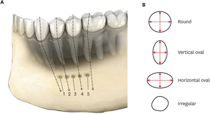
Objectives This study assessed the shape and anatomical relationship of the mental foramen (MF) to mandibular posterior teeth in an Indian sub-population.
Materials and Methods In total, 475 existing cone-beam computed tomography records exhibiting 950 MFs and including the bilateral presence of mandibular premolars and first molars were assessed. Images were evaluated 3-dimensionally to ascertain the position, shape, and anatomical proximity of MFs to mandibular teeth. The position and shape of MFs were measured and calculated. The Pythagorean theorem was used to calculate the distance between the root apex of the mandibular teeth and the MF.
Results MFs exhibited a predominantly round shape (left: 67% and right: 65%) followed by oval (left: 30% and right: 31%) in both males and females and in different age groups. The root apices of mandibular second premolars (left: 71% and right: 62%) were closest to the MF, followed by distal to the first premolars and mesial to the second premolars. The mean vertical distance between the MF and the nearest tooth apex calculated on sagittal sections was 2.20 mm on the right side and 2.32 mm on the left side; no significant difference was found according to sex or age. The distance between the apices of the teeth and the MF was ≥ 4 mm (left; 4.09 ± 1.27 mm and right; 4.01 ± 1.15 mm).
Conclusions These findings highlight the need for clinicians to be aware of the location of the MF in treatment planning and while performing non-surgical and surgical endodontic procedures.
-
Citations
Citations to this article as recorded by- Morphometric analysis of mental foramen in retained cadaveric specimens of mandibles of Sri Lankan population
Dadallage Tharanga De Silva, Usliyanage Clifford Priyantha Perera
Anatomical Science International.2026; 101(3): 340. CrossRef - Anatomical and radiographic assessment of variations of the mental foramen and their impact on success of local anaesthesia administration
Isratul Jannat, M. Ummay Salma, Nipu Rani Chowdhury, Kulsum Nahar, Dilruba Binte Mostafa, Khandokar Emanuzzaman Emon, Shahela Sarmin
International Journal of Research in Medical Sciences.2026; 14(3): 823. CrossRef - Clinical Implications of the Localization and Morphological Variability of the Mental Foramen—A Systematic Review
Mariola Krzykawska-Krupska, Janusz Pach, Piotr Regulski, Jacek Tomczyk, Izabela Strużycka, Kazimierz Szopiński, Katarzyna Osipowicz, Anna Pogorzelska
Diagnostics.2026; 16(5): 779. CrossRef - Optimising Treatment Strategies: Labial versus Labio-inferior Plating Using Three-dimensional Miniplates for Mandibular Symphysis and Parasymphysis Fractures
Akash P Muralidharan, Kalyani Bhate, K Mithun Nilgiri, Sumithra S Nair, Lakshmi Shetty, Rose Johnson
Advances in Human Biology.2025; 15(2): 242. CrossRef - A Cross-Sectional CBCT Study of Anterior Loop, Accessory Mental Foramen, and Lingual Foramina in Patients’ Mandibles: Implications for Safer Implant Planning
Abbas Shokri, Mohammad Mahdi Maleki, Leili Tapak
Journal of Maxillofacial and Oral Surgery.2025;[Epub] CrossRef - Radiographic Recognition of Mental Nerve for Secured Dental Implant Placement by Cone-Beam Computed Tomography in Mosul City Population
Asmaa B. Al-Saffar, Mekdad H. Alrigbo, Rawaa Y. Al-Rawee
Journal of Craniofacial Surgery.2024; 35(7): 2049. CrossRef - Accuracy of Implant Size Prediction Based on Edentulous Ridge Dimension on Cone-beam Computed Tomography - A Retrospective Study
Hunter R. Jolicoeur, Gerard A. Camargo, Tamara G. Stephenson, Wenjian Zhang
Annals of Maxillofacial Surgery.2024; 14(2): 187. CrossRef - Mental Foramenin Panoramik Radyografi ve Konik Işınlı Bilgisayarlı Tomografi Görüntüleri Üzerinde Morfolojik Analizi
Ezgi UZUN, Burak Kerem APAYDIN, Ayşen TİL
Selcuk Dental Journal.2023; 10(3): 540. CrossRef - Evaluation of the Possible Relationship between the Curvature and
Horizontal Course of the Inferior Alveolar Canal
Cansu G. Koca, M. Fatih Çiçek, Sanaz Sadry, Ozan Yenidünya, Fatma Akkoca Kaplan, Aras Erdil
Current Medical Imaging Formerly Current Medical Imaging Reviews.2023;[Epub] CrossRef
- Morphometric analysis of mental foramen in retained cadaveric specimens of mandibles of Sri Lankan population
- 3,979 View
- 70 Download
- 10 Web of Science
- 9 Crossref
- How do imaging protocols affect the assessment of root-end fillings?
- Fernanda Ferrari Esteves Torres, Reinhilde Jacobs, Mostafa EzEldeen, Karla de Faria-Vasconcelos, Juliane Maria Guerreiro-Tanomaru, Bernardo Camargo dos Santos, Mário Tanomaru-Filho
- Restor Dent Endod 2022;47(1):e2. Published online December 15, 2021
- DOI: https://doi.org/10.5395/rde.2022.47.e2
-
Abstract
PDFPubReaderePub
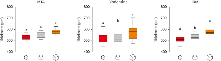
Objectives This study investigated the impact of micro-computed tomography (micro-CT)-based voxel size on the analysis of material/dentin interface voids and thickness of different endodontic cements.
Materials and Methods Following root-end resection and apical preparation, maxillary premolars were filled with mineral trioxide aggregate (MTA), Biodentine, and intermediate restorative material (IRM) (
n = 24). The samples were scanned using micro-CT (SkyScan 1272; Bruker) and the cement/dentin interface and thickness of materials were evaluated at voxel sizes of 5, 10, and 20 µm. Analysis of variance and the Tukey test were conducted, and the degree of agreement between different voxel sizes was evaluated using the Bland and Altman method (p < 0.05).Results All materials showed an increase in thickness from 5 to 10 and 20 µm (
p < 0.05). When evaluating the interface voids, materials were similar at 5 µm (p > 0.05), while at 10 and 20 µm Biodentine showed the lowest percentage of voids (p < 0.05). A decrease in the interface voids was observed for MTA and IRM at 20 µm, while Biodentine showed differences among all voxel sizes (p < 0.05). The Bland-Altman plots for comparisons among voxel sizes showed the largest deviations when comparing images between 5 and 20 µm.Conclusions Voxel size had an impact on the micro-CT evaluation of thickness and interface voids of endodontic materials. All cements exhibited an increase in thickness and a decrease in the void percentage as the voxel size increased, especially when evaluating images at 20 µm.
-
Citations
Citations to this article as recorded by- Effect of ultrasonic activation of endodontic sealers on root canal filling quality during the single-cone obturation procedure: a systematic review and meta-analysis of laboratory-based studies
Shuting Feng, Weiqing Zhou, Xiaojun Chu, Shuaimei Xu, Xiongqun Zeng
Odontology.2025; 113(4): 1380. CrossRef - Marginal Adaptation and Porosity of a Novel MTA Brand Applied as Root-End Filling Material: A Micro-CT Study
Yaneta Kouzmanova, Ivanka Dimitrova
Applied Sciences.2024; 14(7): 2758. CrossRef - Supplementary methods for filling material removal: A systematic review and meta-analysis of micro-CT imaging studies
Bruna Venzke Fischer, Taynara Santos Goulart, Filipe Colombo Vitali, Diego Leonardo de Souza, Cleonice da Silveira Teixeira, Lucas da Fonseca Roberti Garcia
Journal of Dentistry.2024; 151: 105445. CrossRef
- Effect of ultrasonic activation of endodontic sealers on root canal filling quality during the single-cone obturation procedure: a systematic review and meta-analysis of laboratory-based studies
- 3,047 View
- 30 Download
- 3 Web of Science
- 3 Crossref
- Comparison of shaping ability of the Reciproc Blue and One Curve with or without glide path in simulated S-shaped root canals
- Vincenzo Biasillo, Raffaella Castagnola, Mauro Colangeli, Claudia Panzetta, Irene Minciacchi, Gianluca Plotino, Simone Staffoli, Luca Marigo, Nicola Maria Grande
- Restor Dent Endod 2022;47(1):e3. Published online December 28, 2022
- DOI: https://doi.org/10.5395/rde.2022.47.e3
-
Abstract
PDFPubReaderePub
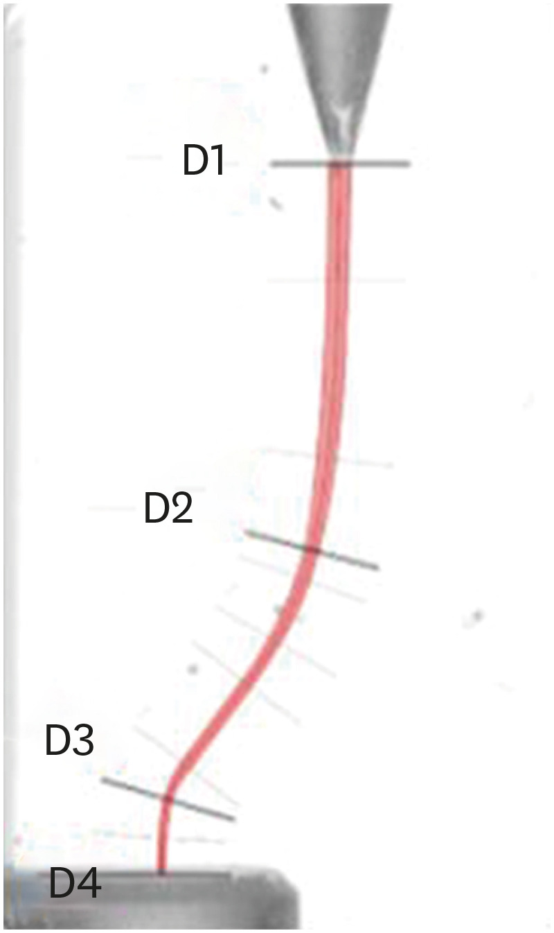
Objectives This study aimed to assess the impact of a glide-path on the shaping ability of 2 single-file instruments and to compare the centering ability, maintenance of original canal curvatures and area of instrumentation in simulated S-shaped root canals.
Materials and Methods Forty simulated S-shaped root canals were used and were prepared with One Curve (group OC), One G and OC (group GOC), Reciproc Blue (group RB) and R-Pilot and RB (group PRB) and scanned before and after instrumentation. The images were analyzed using AutoCAD. After superimposing the samples, 4 levels (D1, D2, D3, and D4) and 2 angles (Δ1 and Δ2) were established to evaluate the centering ability and modification of the canal curvatures. Then, the area of instrumentation (ΔA) was measured. The data were analyzed using 2-way analysis of variance and Tukey's test for multiple comparisons (
p < 0.05).Results Regarding the centering ability in the apical part (D3, D4), the use of the glide-path yielded better results than the single-file groups. Among the groups at D4, OC showed the worst results (
p < 0.05). The OC system removed less material (ΔA) than the RB system, and for Δ1, OC yielded a worse result than RB (p < 0.05).Conclusions The glide-path improved the centering ability in the apical part of the simulated S-shaped canals. The RB system showed a better centering ability in the apical part and major respect of the canal curvatures compared with OC system.
-
Citations
Citations to this article as recorded by- Evaluation of Apical Debris Extrusion and the Remaining Canal Material during Retreatment of a Bioceramic Sealer by the XP-endo Finisher File System, Followed by Various Supplementary Methods: An in Vitro Study
Paras Mull Gehlot, Parvathi Sudeep, Annapoorna B Mariswamy
World Journal of Dentistry.2025; 15(10): 837. CrossRef - Shaping Ability of Rotary NiTi Systems in S‐Shaped Root Canals of Mandibular Molars
Renata M. S. Leal, Emmanuel J. N. L. Silva, Maria C. B. P. Campos, Clarissa T. Rodrigues, Marco A. H. Duarte, Bruno C. Cavenago
Australian Endodontic Journal.2025; 51(1): 133. CrossRef - Comparison of Debris Extrusion and Preparation Time by Traverse, R‐Motion Glider C, and Other Glide Path Systems in Severely Curved Canals
Taher Al Omari, Layla Hassouneh, Khawlah Albashaireh, Alaa Dkmak, Rami Albanna, Ali Al-Mohammed, Ahmed Jamleh, Lucas da Fonseca Roberti Garcia
International Journal of Dentistry.2025;[Epub] CrossRef - Glide Path – An Ineluctable Route for Successful Endodontic Mechanics: A Literature Review
Mahima Bharat Mehta, Anupam Sharma, Aniket Jadhav, Aishwarya Handa, Abhijit Bajirao Jadhav, Ashwini A. Narayanan
Journal of the International Clinical Dental Research Organization.2024; 16(2): 101. CrossRef - Screw-in force, torque generation, and performance of glide-path files with three rotation kinetics
Jee-Yeon Woo, Ji-Hyun Jang, Seok Woo Chang, Soram Oh
Odontology.2024; 112(3): 761. CrossRef - Glide Path in Endodontics: A Literature Review of Current Knowledge
Vlad Mircea Lup, Giulia Malvicini, Carlo Gaeta, Simone Grandini, Gabriela Ciavoi
Dentistry Journal.2024; 12(8): 257. CrossRef - In Vitro Research Methods Used to Evaluate Shaping Ability of Rotary Endodontic Files—A Literature Review
Ranya F. Elemam, Ana Mano Azul, João Dias, Khaled El Sahli, Renato de Toledo Leonardo
Dentistry Journal.2024; 12(10): 334. CrossRef - Endodontic glide path - importance and performance techniques
Milica Jovanovic-Medojevic, Мiljan Stosic, Vanja Opacic-Galic, Violeta Petrovic
Srpski arhiv za celokupno lekarstvo.2023; 151(5-6): 380. CrossRef
- Evaluation of Apical Debris Extrusion and the Remaining Canal Material during Retreatment of a Bioceramic Sealer by the XP-endo Finisher File System, Followed by Various Supplementary Methods: An in Vitro Study
- 4,208 View
- 57 Download
- 8 Web of Science
- 8 Crossref
- Effects of dentin surface preparations on bonding of self-etching adhesives under simulated pulpal pressure
- Chantima Siriporananon, Pisol Senawongse, Vanthana Sattabanasuk, Natchalee Srimaneekarn, Hidehiko Sano, Pipop Saikaew
- Restor Dent Endod 2022;47(1):e4. Published online December 28, 2021
- DOI: https://doi.org/10.5395/rde.2022.47.e4
-
Abstract
PDFPubReaderePub
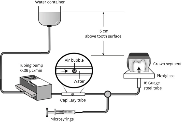
Objectives This study evaluated the effects of different smear layer preparations on the dentin permeability and microtensile bond strength (µTBS) of 2 self-etching adhesives (Clearfil SE Bond [CSE] and Clearfil Tri-S Bond Universal [CTS]) under dynamic pulpal pressure.
Materials and Methods Human third molars were cut into crown segments. The dentin surfaces were prepared using 4 armamentaria: 600-grit SiC paper, coarse diamond burs, superfine diamond burs, and carbide burs. The pulp chamber of each crown segment was connected to a dynamic intra-pulpal pressure simulation apparatus, and the permeability test was done under a pressure of 15 cmH2O. The relative permeability (%P) was evaluated on the smear layer-covered and bonded dentin surfaces. The teeth were bonded to either of the adhesives under pulpal pressure simulation, and cut into sticks after 24 hours water storage for the µTBS test. The resin-dentin interface and nanoleakage observations were performed using a scanning electron microscope. Statistical comparisons were done using analysis of variance and
post hoc tests.Results Only the method of surface preparation had a significant effect on permeability (
p < 0.05). The smear layers created by the carbide and superfine diamond burs yielded the lowest permeability. CSE demonstrated a higher µTBS, with these values in the superfine diamond and carbide bur groups being the highest. Microscopic evaluation of the resin-dentin interface revealed nanoleakage in the coarse diamond bur and SiC paper groups for both adhesives.Conclusions Superfine diamond and carbide burs can be recommended for dentin preparation with the use of 2-step CSE.
-
Citations
Citations to this article as recorded by- Effect of smear layer pretreatment with EDTA and sodium hypochlorite on the dentin bond durability of universal adhesives
Thanawat Ruaydee, Chantida Pawaputanon Na Mahasarakham, Vanthana Sattabanasuk, Pipop Saikaew
Frontiers in Dental Medicine.2026;[Epub] CrossRef - Determination of marginal permeability of restorations in the cervical region using a universal adhesive system: a randomized controlled open-label laboratory study
Svetlana N. Razumova, Anzhela S. Brago, Oxana R. Ruda, Artur G. Talandis, Lamara M. Khaskhanova, Ruzanna M. Bragunova, Bohdan O. Pecherskyi
Russian Journal of Dentistry.2026; 30(2): 113. CrossRef - Catechol–Phosphonate–Augmented Universal Adhesive for Hydrolysis-Resistant Dentin Bonds: A µTBS and Spectroscopic Study
Rabeia J. Khalil, Suha K. Ibrahim, Athraa H. Madhat, Ali H. Tawfieq
European Journal of Dentistry.2026;[Epub] CrossRef - The effect of different adhesive strategies and diamond burs on dentin bond strength of universal resin cements
Chavakorn Atsavathavornset, Pipop Saikaew, Choltacha Harnirattisai, Hidehiko Sano
Clinical Oral Investigations.2025;[Epub] CrossRef - Universal adhesive systems in dentistry: A narrative review
Svetlana N. Razumova, Anzhela S. Brago, Oxana R. Ruda, Zoya A. Guryeva, Elvira V. Adzhieva
Russian Journal of Dentistry.2024; 28(5): 512. CrossRef - Delayed light activation of resin composite affects the bond strength of adhesives under dynamic simulated pulpal pressure
Nattaporn Sukprasert, Choltacha Harnirattisai, Pisol Senawongse, Hidehiko Sano, Pipop Saikaew
Clinical Oral Investigations.2022; 26(11): 6743. CrossRef
- Effect of smear layer pretreatment with EDTA and sodium hypochlorite on the dentin bond durability of universal adhesives
- 4,351 View
- 65 Download
- 4 Web of Science
- 6 Crossref
- A 3-year retrospective study of clinical durability of bulk-filled resin composite restorations
- Muhittin Ugurlu, Fatmanur Sari
- Restor Dent Endod 2022;47(1):e5. Published online December 30, 2021
- DOI: https://doi.org/10.5395/rde.2022.47.e5
-
Abstract
PDFPubReaderePub
Objectives This study aimed to assess the clinical longevity of a bulk-fill resin composite in Class II restorations for 3-year.
Materials and Methods Patient record files acquired from the 40 patients who were treated due to needed 2 similar sizes Class II composite restorations were used for this retrospective study. In the experimental cavity, the flowable resin composite SDR was inserted in the dentinal part as a 4 mm intermediate layer. A 2 mm coverage layer with a nano-hybrid resin composite (CeramX) was placed on SDR. The control restoration was performed by an incremental technique of 2 mm using the nano-hybrid resin composite. The restorations were blindly assessed by 2 calibrated examiners using modified United States Public Health Service criteria at baseline and 1, 2, and 3 years. The data were analyzed using non-parametric tests (
p = 0.05).Results Eighty Class II restorations were evaluated. After 3-years, 4 restorations (5%) failed, 1 SDR + CeramX, and 3 CeramX restorations. The annual failure rate (AFR) of the restorations was 1.7%. The SDR + CeramX group revealed an AFR of 0.8%, and the CeramX group an AFR of 2.5% (
p > 0.05). Regarding anatomical form and marginal adaptation, significant alterations were observed in the CeramX group after 3-years (p < 0.05). The changes in the color match were observed in each group over time (p < 0.05).Conclusions The use of SDR demonstrated good clinical durability in deep Class II resin composite restorations.
-
Citations
Citations to this article as recorded by- Bond Strength of Bulk-Fill Resin Repairs: Impact of Surface and Adhesive Protocols
Samuel Eleutério Paiva Sousa, Fiorella Elizabeth Arévalo Tarrillo, Maria Paula Novaes Camargo Manna, Sandra Ribeiro de Barros da Cunha, Maria Ângela Pita Sobral
Odovtos - International Journal of Dental Sciences.2026; 28(2): 204. CrossRef - Evaluation of Surface Roughness and Microhardness of New Generation Bulk-Fill Composites
Zehra SÜSGÜN YILDIRIM, Ezgi SONKAYA, Zeliha Gonca BEK KÜRKLÜ
Cumhuriyet Dental Journal.2023; 26(2): 180. CrossRef - Damping Behaviour and Mechanical Properties of Restorative Materials for Primary Teeth
Thomas Niem, Roland Frankenberger, Stefanie Amend, Bernd Wöstmann, Norbert Krämer
Materials.2022; 15(21): 7698. CrossRef
- Bond Strength of Bulk-Fill Resin Repairs: Impact of Surface and Adhesive Protocols
- 5,067 View
- 45 Download
- 2 Web of Science
- 3 Crossref
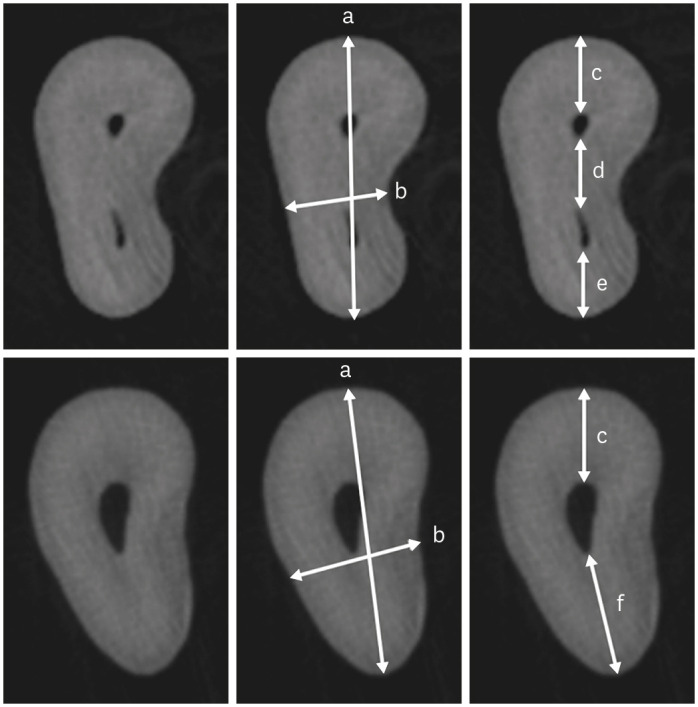
- Morphological characteristics of the mesiobuccal root in the presence of a second mesiobuccal canal: a micro-CT study
- Lucas P. Lopes Rosado, Matheus Lima Oliveira, Karla Rovaris, Deborah Queiroz Freitas, Frederico Sampaio Neves
- Restor Dent Endod 2022;47(1):e6. Published online January 18, 2022
- DOI: https://doi.org/10.5395/rde.2022.47.e6
-
Abstract
PDFPubReaderePub
Objectives This study investigated the internal morphology of mesiobuccal (MB) roots of maxillary molars with a second mesiobuccal (MB2) canal.
Materials and Methods Forty-seven maxillary first or second molars from Brazilians were scanned using micro-computed tomography. The following measurements were obtained from the MB roots: root thickness, root width, and dentin thickness of the buccal aspect of the first mesiobuccal (MB1) canal, between the MB1 and MB2 canals, and the palatal aspect of the MB2 and MB1 canals at 3 mm from the root apex and in the furcation region. For statistical analysis, the Student’s
t -test and analysis of variance with thepost-hoc Tukey test were used (α = 0.05).Results In maxillary molars with an MB2 canal, MB roots were significantly thicker (
p = 0.0014) and narrower (p = 0.0016) than in maxillary molars without an MB2 canal. The dentin thickness of the palatal aspect of the MB1 canal was also significantly greater than that of MB roots without an MB2 canal at 3 mm from the root apex (p = 0.0007) and in the furcation region (p < 0.0001). In the furcation region of maxillary molars with an MB2 canal, the dentin thickness between the MB1 and MB2 canals was significantly smaller than that in the buccal and palatal aspects (p < 0.0001).Conclusions The internal morphology of MB roots of maxillary molars with an MB2 canal revealed differences in dentin thickness, root diameter, and distance between the canals when compared with maxillary molars without an MB2 canal.
-
Citations
Citations to this article as recorded by- Association between lingual canal detection and buccolingual root width in mandibular anterior teeth: a retrospective CBCT Study
Önder Çam, Melis Oya Ateş, Ali Keleş
BMC Oral Health.2026;[Epub] CrossRef - Effectiveness and safety of three NiTi systems in endodontic retreatment of MB1 and MB2 root canals: a micro-CT and CBCT combined analysis
Airton Oliveira Santos-Junior, Rocharles Cavalcante Fontenele, Karina Ines Medina Carita Tavares, Fernanda Ferrari Esteves Torres, Jáder Camilo Pinto, Pedro Luis Busto Rosim, Andréa Gonçalves, Marco Antonio Hungaro Duarte, Juliane Maria Guerreiro-Tanomaru
Clinical Oral Investigations.2025;[Epub] CrossRef - Cone-beam computed tomography evaluation of root and canal morphology of maxillary molars in a Chinese kazakh population
Shuchun Yang, Chenye Li, Hui Shi, Ming Liu, Xu Wang
BMC Oral Health.2025;[Epub] CrossRef - Can maxillary molar dimensions predict the presence of the second mesiobuccal canal?
Lucas P. Lopes Rosado, Deborah Queiroz Freitas, Karla Rovaris, Matheus L. Oliveira, Frederico Sampaio Neves
Oral Radiology.2023; 39(3): 482. CrossRef - Can the detection of second mesiobuccal canals be enhanced based on the volume of adjacent canals?
Lucas P. Lopes Rosado, Deborah Q. Freitas, Karla Rovaris, Matheus L. Oliveira, Frederico S. Neves
Archives of Oral Biology.2023; 146: 105604. CrossRef - Assessment of the coronal root canal morphology of permanent maxillary first molars using digital 3D-reconstruction technology based on micro-computed tomography data
Mudan Wang, Yuxuan Gao, Qi Deng, Yuan Gao, Dongzhe Song, Dingming Huang
Journal of Dental Sciences.2023; 18(2): 586. CrossRef
- Association between lingual canal detection and buccolingual root width in mandibular anterior teeth: a retrospective CBCT Study
- 2,627 View
- 48 Download
- 7 Web of Science
- 6 Crossref
- In-office dental bleaching with violet light emitting diode: bleaching efficacy and pulpal temperature rise
- Brunna Katyuscia de Almeida Guanaes, Talyta Neves Duarte, Gisele Maria Correr, Marina da Rosa Kaizer, Carla Castiglia Gonzaga
- Restor Dent Endod 2022;47(1):e7. Published online February 3, 2022
- DOI: https://doi.org/10.5395/rde.2022.47.e7

-
Abstract
PDFPubReaderePub
Objectives This study evaluated the bleaching efficacy of different in-office protocols associated with violet light emitting diode (V-LED), and measured the pulpal temperature rise caused by V-LED with or without gel application.
Materials and Methods Bovine incisors were distributed in 4 groups (
n = 10): VL – V-LED; HP – 35% hydrogen peroxide (control); HYB – hybrid protocol, V-LED applied without gel for 10 irradiation cycles followed by V-LED applied with gel for another 10 irradiation cycles; and HPVL – gel and V-LED applied for 20 irradiation cycles. Three bleaching sessions were performed with 7-day intervals. Bleaching efficacy was evaluated withE 00 and ΔWID . Data were recorded at baseline, 7, 14, 21 and 70 days. For pulpal temperature rise, thermocouples were placed inside the pulp chamber of human incisors. To determine intrapulpal temperature, the teeth were irradiated with V-LED with or without application of bleaching gel. Color difference data were analyzed by 2-way repeated measures ANOVA and Tukey’s test. Pulpal temperature was analyzed byt -test (α = 5%).Results VL exhibited lower color (
E 00) and whiteness changes (ΔWID ) than the other groups. HPVL presented higher color change values than HYB. HYB and HPVL showed not different ΔWID values; and HP showed the highest whiteness changes at all times. There were significant differences comparing ΔT with gel (8.9°C) and without gel application (7.2°C).Conclusions HPLV was more efficient than HYB. The 2 protocols with VL showed similar results to control. Gel application combined with VL promoted higher pulpal temperature than to the no gel group.
-
Citations
Citations to this article as recorded by- Inverse Heat Conduction Estimation of Heat Flux in Human Dentin from Dental Curing Lights Using the Conjugate Gradient Method
Ahmad Soori, Farshad Kowsary, Shadab Safarzadeh Khosroshahi, Mohammad Vahedi
International Journal of Thermophysics.2026;[Epub] CrossRef - Illuminating the evidence: A comprehensive review of light-assisted in-office tooth bleaching
Márcia V.G.B. Queiroz, Rafael Dascanio, Vinicius H. Hutemma, Diogo A. Chiovetto, Adriano F. Lima, Jorge R. Soto-Montero, Matheus Kury
Journal of Dentistry.2026; 171: 106707. CrossRef - Spectrophotometric Evaluation of Laser-Assisted Dental Bleaching Using Erbium-Doped Yttrium Aluminum Garnet (Er:YAG) and Diode Lasers at Different Wavelengths: An In Vitro Study
Esraa Ihssan Alshibli, Omar H. Hamadah, Mohammad Y. Hajeer
Cureus.2026;[Epub] CrossRef - Effect of antioxidant on tooth sensitivity after bleaching
Mohamed Nabil, Mostafa Mohamed Hasan, Eman Abd Elghany Shebl
Journal of Esthetic and Restorative Dentistry.2024; 36(3): 429. CrossRef - In-office Bleaching Activated With Violet LED: Effect on Pulpal and Tooth Temperature and Pulp Viability
NR Carlos, RT Basting, KR Kantovitz, ES Bronze-Uhle, PN Lisboa Filho, V Cavalli, RT Basting
Operative Dentistry.2024; 49(3): 262. CrossRef - Low and high hydrogen peroxide concentrations of in-office dental bleaching associated with violet light: an in vitro study
Isabela Souza Vardasca, Michael Willian Favoreto, Mylena de Araujo Regis, Taynara de Souza Carneiro, Emanuel Adriano Hul, Christiane Philippini Ferreira Borges, Alessandra Reis, Alessandro D. Loguercio, Carlos Francci
Clinical Oral Investigations.2024;[Epub] CrossRef - Bleaching efficacy of in-office bleaching with violet light using low-concentration hydrogen peroxide nanoparticulate photocatalyst gel: A randomized controlled trial
Gustavo Garcia Castro, Palena Araújo Pinto, Michael Willian Favoreto, Alessandra Reis, Maria Viviana-Mora, Rita de Cássia Mendonça de Miranda, Andres Felipe Milan Cardenas, Alessandro D. Loguercio, Rudys Rodolfo de Jesus Tavarez
Photodiagnosis and Photodynamic Therapy.2024; 50: 104410. CrossRef - Influence of Different Light-Activated Bleaching Gels on Pulp Chamber Temperature: An In Vitro Study
Mandana Karimi, Elmira Ataee, Ladan Ranjbar Omrani, Mahdi Abbasi, Elham Ahmadi
Avicenna Journal of Dental Research.2024; 16(4): 225. CrossRef - Continuous vs fractionated violet LED light protocols for dental bleaching: Evaluations of color change and temperature of the dental pulp and buccal surface
Mayanna Pacheco Trindade Najar, Luciana Hilel Rangel Barbosa, Natália Russo Carlos, Fabiana Mantovani Gomes França, Cecilia Pedroso Turssi, Waldemir Francisco Vieira-Junior, Roberta Tarkany Basting
Photodiagnosis and Photodynamic Therapy.2023; 42: 103631. CrossRef - Improved esthetic efficacy and reduced cytotoxicity are achieved with a violet LED irradiation of manganese oxide-enriched bleaching gels
Marlon Ferreira Dias, Beatriz Voss Martins, Rafael Antonio de Oliveira Ribeiro, Josimeri Hebling, Carlos Alberto de Souza Costa
Lasers in Medical Science.2022;[Epub] CrossRef
- Inverse Heat Conduction Estimation of Heat Flux in Human Dentin from Dental Curing Lights Using the Conjugate Gradient Method
- 4,483 View
- 46 Download
- 10 Web of Science
- 10 Crossref
- Comparison of instrumental methods for color change assessment of Giomer resins
- Luiza de Almeida Queiroz Ferreira, Rogéli Tibúrcio Ribeiro da Cunha Peixoto, Cláudia Silami de Magalhães, Tassiana Melo Sá, Monica Yamauti, Francisca Daniele Moreira Jardilino
- Restor Dent Endod 2022;47(1):e8. Published online February 3, 2022
- DOI: https://doi.org/10.5395/rde.2022.47.e8
-
Abstract
PDFPubReaderePub
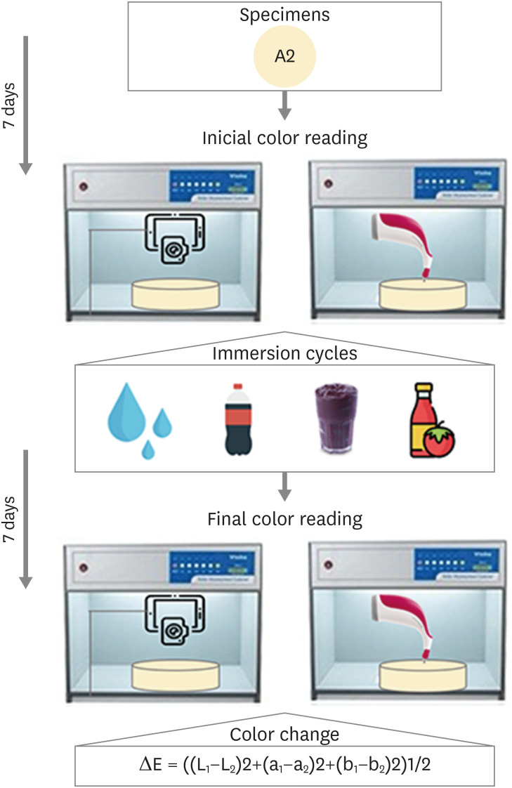
Objectives The aim of this study was to compare the color change of the Giomer resin composite (Beautifil-Bulk) by using photographs obtained with a smartphone (iPhone 6S) associated with Adobe Photoshop software (digital method), with the spectrophotometric method (Vita Easyshade) after immersion in different pigment solutions.
Materials and Methods Twenty resin composite samples with a diameter of 15.0 mm and thickness of 1.0 mm were confectioned in A2 color (
n = 5). Photographs and initial color readings were performed with a smartphone and spectrophotometer, respectively. Then, samples were randomly divided and subjected to cycles of immersion in distilled water (control), açai, Coke, and tomato sauce, 3 times a day, 20 minutes for 7 days. Later, new photographs and color readings were taken.Results The analysis (2-way analysis of variance, Holm-Sidak,
p < 0.05) demonstrated no statistical difference (p < 0.005) between the methods in all groups. Similar color changes were observed for all pigment solutions when using the spectrophotometric method. For the digital method, all color changes were clinically unacceptable, with distilled water and tomato sauce similar to each other and with statistical differences (p < 0.005) for Coke and açai.Conclusions Only the tomato sauce produced a color change above the acceptability threshold using both methods of color assessment. The spectrophotometric and digital methods produce different patterns of color change. According to our results, the spectrophotometric method is more recommended in color change assessment.
-
Citations
Citations to this article as recorded by- Are Sculptable Bulk‐Fill Composites Susceptible to Color Change: A Systematic Review
Jamieson Wong, Constance Yeo, Michelle The, Filip Taneski, Uros Josic, Lorenzo Breschi, Vesna Miletic
Journal of Esthetic and Restorative Dentistry.2026; 38(1): 70. CrossRef - The effects of mechanical and chemical degradation on the surface roughness, gloss, and color stability of bulk-fill resin composites
Merve Nezir, Hanife Altınışık, Esra Özyurt, Naz Bayar, Mediha Büyükgöze Dindar
BMC Oral Health.2025;[Epub] CrossRef - Color Image Expression through CIE L*a*b* System in Foods
Hyun-Woong Choi, Seong-Eun Park, Hong-Seok Son
Journal of the Korean Society of Food Science and Nutrition.2023; 52(2): 223. CrossRef
- Are Sculptable Bulk‐Fill Composites Susceptible to Color Change: A Systematic Review
- 3,768 View
- 49 Download
- 3 Web of Science
- 3 Crossref
- Effects of 3 different light-curing units on the physico-mechanical properties of bleach-shade resin composites
- Azin Farzad, Shahin Kasraei, Sahebeh Haghi, Mahboubeh Masoumbeigi, Hassan Torabzadeh, Narges Panahandeh
- Restor Dent Endod 2022;47(1):e9. Published online February 7, 2022
- DOI: https://doi.org/10.5395/rde.2022.47.e9
-
Abstract
PDFPubReaderePub
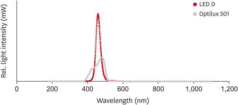
Objectives This study investigated the microhardness, flexural strength, and color stability of bleach-shade resin composites cured with 3 different light-curing units.
Materials and Methods In this
in vitro experimental study, 270 samples were fabricated of bleach and A2 shades of 3 commercial resin composites (Point 4, G-aenial Anterior, and Estelite Sigma Quick). Samples (n = 5 for each trial) were cured with Bluephase N, Woodpecker LED.D, and Optilux 501 units and underwent Vickers microhardness and flexural strength tests. The samples were tested after 24 hours of storage in distilled water. Color was assessed using a spectrophotometer immediately after preparation and 24 hours after curing. Data were analyzed using 3-way analysis of variance and the Tukey test (p ≤ 0.001).Results Samples cured with Optilux exhibited the highest and those cured with LED.D exhibited the lowest microhardness (
p = 0.023). The bleach shade of Point 4 composite cured with Optilux displayed the highest flexural strength, while the same composite and shade cured with Sigma Quick exhibited the lowest (p ≤ 0.001). The color change after 24 hours was greatest for the bleach shade of G-aenial cured with Bluephase N and least for the A2 shade of Sigma Quick cured with Optilux (p ≤ 0.001).Conclusions Light curing with polywave light-emitting diode (LED) yielded results between or statistically similar to those of quartz-tungsten-halogen and monowave LED in the microhardness and flexural strength of both A2 and bleach shades of resin composites. However, the brands of light-curing devices showed significant differences in color stability.
-
Citations
Citations to this article as recorded by- Mechanical Behaviour of Novel Nanohybrid Resin Composite Using Two Light Cure Systems
Ghada H. Naguib, Jumana Mazhar, Abeer Alnowaiser, Abdulghani Mira, Hisham Mously, Rabab Aljawi, Samar H. Abuzinadah, Mohamed T. Hamed
International Dental Journal.2025; 75(2): 1136. CrossRef - Repair Bond Strength of Aged Composite: Effect of Thermocycling and Surface Treatment
Sina Yarmoradian, Ladan Ranjbar Omrani, Elham Ahmadi, Niyousha Rafeie, Mahdi Abbasi, Nastaran Dabiri Shahabi
Journal of Research in Dental and Maxillofacial Sciences.2025; 10(3): 228. CrossRef - Evaluation of the Depth of Cure by Microhardness of Bulk-Fill Composites with Monowave and Polywave LED Light-Curing Units
Socratis Thomaidis, Dimitris Kampouropoulos, Maria Antoniadou, Afrodite Kakaboura
Applied Sciences.2024; 14(24): 11532. CrossRef - Effect of hard segment chemistry and structure on the self‐healing properties of UV‐curable coatings based on the urethane acrylates with built‐in Diels–Alder adduct
Paulina Bednarczyk, Karolina Mozelewska, Małgorzata Nowak, Joanna Klebeko, Joanna Rokicka, Paula Ossowicz‐Rupniewska
Journal of Applied Polymer Science.2023;[Epub] CrossRef - Effects of Dental Bleaching Agents on the Surface Roughness of Dental Restoration Materials
Alexandru Dan Popescu, Mihaela Jana Tuculina, Oana Andreea Diaconu, Lelia Mihaela Gheorghiță, Claudiu Nicolicescu, Cristian Niky Cumpătă, Cristiana Petcu, Jaqueline Abdul-Razzak, Ana Maria Rîcă, Ruxandra Voinea-Georgescu
Medicina.2023; 59(6): 1067. CrossRef - Effect of Polywave and Monowave Light Curing Units on Color Change of Composites Containing Trime-thylbenzoyl-Diphenyl-Phosphine Before and After Aging
Negar Madihi, Maryam Hoorizad ganjkar, Negin Nasoohi, Ali Kaboudanian Ardestani
Journal of Research in Dental and Maxillofacial Sciences.2023; 8(4): 249. CrossRef
- Mechanical Behaviour of Novel Nanohybrid Resin Composite Using Two Light Cure Systems
- 3,040 View
- 47 Download
- 4 Web of Science
- 6 Crossref
- Effect of irrigants on the color stability, solubility, and surface characteristics of calcium-silicate based cements
- Selen Küçükkaya Eren, Sevinc Askerbeyli Örs, Hacer Aksel, Şenay Canay, Duygu Karasan
- Restor Dent Endod 2022;47(1):e10. Published online February 10, 2022
- DOI: https://doi.org/10.5395/rde.2022.47.e10

-
Abstract
PDFPubReaderePub
Objectives This study aimed to investigate the color stability, solubility, and surface characteristics of 3 calcium silicate-based cements (CSCs) after immersion in different solutions.
Materials and Methods ProRoot white mineral trioxide aggregate (MTA), Biodentine, and Endosequence Root Repair Material (ERRM) were placed in cylindrical molds and stored at 37°C for 24 hours. Each specimen was immersed in distilled water, 5% sodium hypochlorite (NaOCl), 2% chlorhexidine, or 0.1% octenidine hydrochloride (OCT) for 24 hours. Color changes were measured with a spectrophotometer. Solubility was determined using an analytical balance with 10−5 g accuracy. The surface characteristics were analyzed using scanning electron microscopy and energy-dispersive spectroscopy. Data were analyzed using 2-way analysis of variance, the Tukey test, and the paired
t -test.Results MTA exhibited significant discoloration in contact with NaOCl (
p < 0.05). White precipitation occurred on the surfaces of Biodentine and ERRM after contact with the solutions, and none of the materials presented dark brown discoloration. All materials showed significant solubility after immersion in the solutions (p < 0.05), irrespective of the solution type (p > 0.05). The surface topography and elemental composition of the samples showed different patterns of crystal formation and precipitation depending on the solution type.Conclusions All materials presented some amount of solubility and showed crystal precipitation after contact with the solutions. Biodentine and ERRM are suitable alternatives to ProRoot MTA as they do not exhibit discoloration. The use of OCT can be considered safe for CSCs.
-
Citations
Citations to this article as recorded by- Time-dependent Tooth Color Changes Following Conventional, Silver-based, and Photodynamic Root Canal Irrigants: An In Vitro Study
Laila Mohamed Mohamed Kenawi, Mohamed Fattouh, Khaled Abid Althaqafi, Abla Arafa
The Open Dentistry Journal.2026;[Epub] CrossRef - Effects of interactions between sealers and irrigants on the physicochemical and surface characteristics of endodontic sealers
Hye-In Kim, Yeon-Ju You, Yemi Kim, Jin-Woo Kim, Young-Eun Jang
Clinical Oral Investigations.2026;[Epub] CrossRef - Effect of Addition of Zinc Oxide on Color Stability of a Calcium Silicate–Based Cement Containing Bismuth Oxide in the Presence of Blood and Sodium Hypochlorite
Mehrfam Khoshkhounejad, Noushin Shokouhinejad, Pegah Sarraf, MohammadHossein Mirisiahi, Hannah Wesley
International Journal of Dentistry.2026;[Epub] CrossRef - Effects of sodium hypochlorite and chlorhexidine on microhardness and solubility of mineral trioxide aggregate and bioceramic putty
Aryadi Aryadi, Celine Agatha Hadi, Wiena Widyastuti, Eko Fibryanto, Elline Elline, Eddy Eddy
Journal of Conservative Dentistry and Endodontics.2026; 29(5): 503. CrossRef - Influence of endodontic irrigants on hydraulic cements: solubility, color alteration and surface changes
Sıla Usta, Cangül Keskin, Ayşe Oktay, Emmanuel João Nogueira Leal Silva
European Oral Research.2026; 60(1): 230. CrossRef - Probiotics As An Adjunctive Therapy In Periodontal Diseases: A Systematic Review.
Anca Ioana Neacșa, Alexandru Vlasa
Acta Stomatologica Marisiensis Journal.2026; 9(1): 5. CrossRef - Chemical and in vivo analyses of calcium silicate‐based materials in bone and connective tissues
Ana Cristina Padilha Janini, Lauter Eston Pelepenko, Brenda Fornazaro Moraes, Victor Augusto Benedicto dos Santos, Matheus Barros‐Costa, Isabela Alvarenga Maciel dos Santos, Fábio Roberto de Souza Batista, Juliana de Aguiar Silveira Meira, Mariza Akemi Ma
International Endodontic Journal.2025; 58(3): 484. CrossRef - Topic: Perspectives on Success and Failure of Endodontic Treatments
Ilma Robo, Manola Kelmendi, Eva Habazaj, Kleves Elezi, Rialda Xhizdari, Nevila Alliu
SN Comprehensive Clinical Medicine.2025;[Epub] CrossRef - Bismuth release from endodontic materials: Proposed mechanisms for systemic circulation and organ accumulation
Lauter Eston Pelepenko, Benjamin Hewitt, Rodrigo Bueno de Oliveira, Brenda Fornazaro Moraes, Débora C. Coraça-Huber, Ana Cristina Padilha Janini, Marina Angélica Marciano
Journal of Hazardous Materials.2025; 494: 138580. CrossRef - Effect of Vital Pulp Therapy Biomaterials on Tooth Discolouration: A Review of the Literature
Maedeh Gilvari Sarshari, Kiana Shakeri, Ardavan Parhizkar, Naresh Kasoju
International Journal of Biomaterials.2025;[Epub] CrossRef - Data about application of chlorhexidine as a periodontal irrigant –
Systematic Review.
Ilma Robo, Manola Kelmendi , Eva Habazaj , Kristi Sulanjaku , Nevila Alliu
Acta Stomatologica Marisiensis Journal.2025; 8(1): 6. CrossRef - Biocompatibility of Hydraulic Calcium Silicate-Based Cement MTA FlowTM on Human Dental Pulp Stem Cells In Vitro
Paulius Tušas, Josette Camilleri, Milda Alksnė, Egidijus Šimoliūnas, Saulius Drukteinis, Eglė Marija Urbonė, Virginija Bukelskienė, Vygandas Rutkūnas, Vytautė Pečiulienė
Journal of Functional Biomaterials.2025; 16(7): 252. CrossRef - Evaluation of the Effects of Different Irrigation Solutions on MTA and Dentin Microhardness
Gokay Buyukcolpan, İdil Özden, Hesna Sazak Öveçoğlu
Clinical and Experimental Health Sciences.2025; 15(3): 524. CrossRef - Comparative Evaluation of the Intratubular Penetration Ability of Two Retrograde Obturation Techniques in Micro-Endodontic Surgical Procedure: An In Vitro Study with Confocal Laser Scanning Microscopy
Alberto Casino Alegre, Michell Ramírez López, Manuel Monterde Hernández, Susana Aranda Verdú, Jorge Rubio Climent, Antonio Pallarés Sabater
Dentistry Journal.2025; 13(11): 509. CrossRef - The outcome of combined use of iRoot BP Plus and iRoot SP for root-end filling in endodontic microsurgery: a randomized controlled trial
Xu Dong, Qin Su, Wen Li, Jinbo Yang, Dongzhe Song, Jing Yang, Xin Xu
Clinical Oral Investigations.2024;[Epub] CrossRef - How Does Ethylenediaminetetraacetic Acid Irrigation Affect Biodentine? A Multimethod Ex Vivo Study
Katarzyna Dąbrowska, Aleksandra Palatyńska-Ulatowska, Leszek Klimek
Materials.2024; 17(6): 1230. CrossRef - Color stability and solubility of Biodentine and NeoPutty in contact with different irrigation solutions
Sıla Nur Usta, Cangül Keskin
Restorative Dentistry & Endodontics.2024;[Epub] CrossRef - The impact of various irrigation solutions on the color stabilities of five calcium silicate cement: an in-vitro study
Aslı Soğukpınar Onsuren, Onur Kesici, Elif Uğurbekler Hündü
Selcuk Dental Journal.2024; 11(3): 313. CrossRef - Bioceramics in Endodontics: Updates and Future Perspectives
Xu Dong, Xin Xu
Bioengineering.2023; 10(3): 354. CrossRef - Effect of calcium silicate-based endodontic sealers on tooth color: A 3-year in vitro experimental study
Carmen Llena, Ana Herrero, Sandra Lloret, Martha Barraza, Jose Luis Sanz
Heliyon.2023; 9(2): e13237. CrossRef - Evaluation of the Shear Bond Strength of Four Bioceramic Materials with Different Restorative Materials and Timings
Abeer S. Alqahtani, Ayman M. Sulimany, Abdullah S. Alayad, Abdulaziz S. Alqahtani, Omar A. Bawazir
Materials.2022; 15(13): 4668. CrossRef
- Time-dependent Tooth Color Changes Following Conventional, Silver-based, and Photodynamic Root Canal Irrigants: An In Vitro Study
- 3,023 View
- 43 Download
- 16 Web of Science
- 21 Crossref
- Interplay of collagen and mast cells in periapical granulomas and periapical cysts: a comparative polarizing microscopic and immunohistochemical study
- Deepty Bansal, Mala Kamboj, Anjali Narwal, Anju Devi, Nisha Marwah
- Restor Dent Endod 2022;47(1):e12. Published online February 14, 2022
- DOI: https://doi.org/10.5395/rde.2022.47.e12
-
Abstract
PDFPubReaderePub
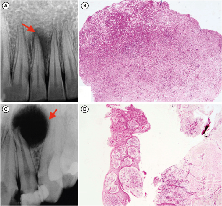
Objectives This pilot study aimed to establish the interrelationship between collagen and mast cells in periapical granulomas and periapical cysts.
Materials and Methods An observational cross-sectional study was conducted on the paraffin-embedded tissue sections of 68 specimens (34 periapical granulomas and 34 periapical cysts). The specimens were stained with picrosirius to observe collagen fiber birefringence and anti-tryptase antibody to evaluate the mast cell count immunohistochemically. The mean number and birefringence of collagen fibers, as well as the mean number of mast cells (total, granulated, and degranulated), and the mean inflammatory cell density were calculated. The data obtained were analyzed using the Kruskal Wallis test, Mann Whitney
U test, and Spearman correlation test (p < 0.05).Results The mean number of thick collagen fibers was higher in periapical cysts, while that of thin fibers was higher in granulomas (
p = 0.00). Cysts emitted orange-yellow to red birefringence, whereas periapical granulomas had predominantly green fibers (p = 0.00). The mean inflammatory cell density was comparable in all groups (p = 0.129). The number of total, degranulated, and granulated mast cells exhibited significant results (p = 0.00) in both groups. Thick cyst fibers showed significant inverse correlations with inflammation and degranulated mast cells (p = 0.041, 0.04 respectively).Conclusions Mast cells and inflammatory cells influenced the nature of collagen fiber formation and its birefringence. This finding may assist in the prediction of the nature, pathogenesis, and biological behavior of periapical lesions.
-
Citations
Citations to this article as recorded by- Chronic periapical lesions: clinical and morphological characterization of 508 cases
Talytha Barbosa da Rocha, Raelly Katharinne Lima de Meneses, Nathália Yvia Assis Henriques, Jonh Lennon Silva Cunha, Manuel Antonio Gordón-Núñez, Cassiano Francisco Weege Nonaka, Pollianna Muniz Alves
JORDI - Journal of Oral Diagnosis.2026;[Epub] CrossRef - The Mystifying Role of Mast Cells in the Pathogenesis of Periapical Pathologies - A Systematic Review
Mala Kamboj, Shashibala Malik, R. Keerthika, Anjali Narwal, Anju Devi, Gopikrishnan Vijayakumar, Adarsh Kumar
Journal of Endodontics.2025; 51(7): 845. CrossRef - Immunohistochemical Analysis of CD117 in the Mast Cells of Odontogenic Keratocysts
Sujatha Varma, Shameena PM, Plakkil Viswanathan Deepthi, Indu G
Cureus.2024;[Epub] CrossRef - Immunohistochemical evaluation of cyclooxygenase‐2 and mast cell density in periapical lesions
Shashibala Malik, Mala Kamboj, Anjali Narwal, Anju Devi
International Endodontic Journal.2023; 56(8): 980. CrossRef
- Chronic periapical lesions: clinical and morphological characterization of 508 cases
- 3,285 View
- 31 Download
- 2 Web of Science
- 4 Crossref
- Efficacy of reciprocating instruments and final irrigant activation protocols on retreatment of mesiobuccal roots of maxillary molars: a micro-CT analysis
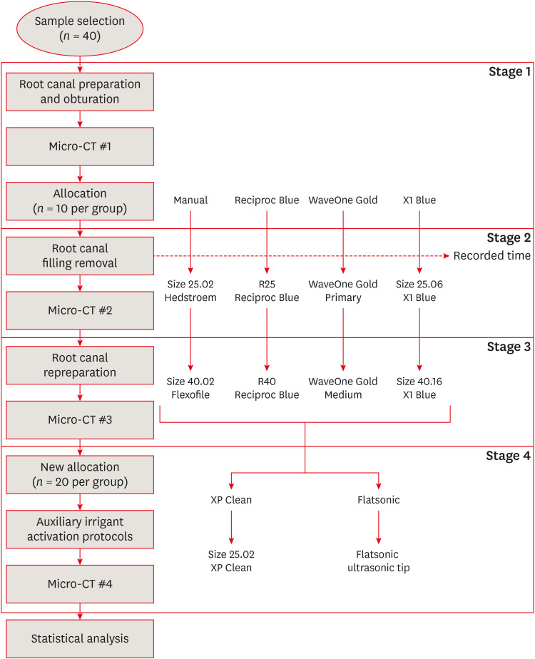
- Lilian Tietz, Renan Diego Furlan, Ricardo Abreu da Rosa, Marco Antonio Hungaro Duarte, Murilo Priori Alcalde, Rodrigo Ricci Vivan, Theodoro Weissheimer, Marcus Vinicius Reis Só
- Restor Dent Endod 2022;47(1):e13. Published online February 15, 2022
- DOI: https://doi.org/10.5395/rde.2022.47.e13
-
Abstract
PDFPubReaderePub
Objectives This study evaluated the efficacy of 3 reciprocating systems and the effects of 2 instruments for irrigant activation on filling material removal.
Materials and Methods Forty mesiobuccal roots of maxillary molars were prepared up to size 25.06 and obturated. Micro-computed tomography (micro-CT) examination #1 was performed. Teeth were then divided into 4 groups (
n = 10), according to the retreatment protocol: (1) manual, (2) Reciproc Blue, (3) WaveOne Gold, and (4) X1 Blue. Micro-CT examinations #2 and #3 were performed after filling removal and repreparation, respectively. Next, all teeth were divided into 2 new groups (n = 20) according to the irrigant activation protocol: XP Clean (XP Clean size 25.02) and Flatsonic (Flatsonic ultrasonic tip). Micro-CT examination #4 was performed after irrigant activation. Statistical analysis was performed with a significance level set at 5%.Results WaveOne Gold removed a significantly greater amount of filling material than the manual group (
p < 0.05). The time to reach the WL was similar for all reciprocating systems (p > 0.05). X1 Blue was faster than the manual group (p < 0.05). Only manual group improved the filling material removal after the repreparation stage (p < 0.05). Both activation protocols significantly improved the filling material removal (p < 0.05), without differences between them (p > 0.05).Conclusions None of the tested instruments completely removed the filling material. X1 Blue size 25.06 reached the working length in the shortest time. XP Clean and Flatsonic improved the filling material removal.
-
Citations
Citations to this article as recorded by- Impact of vehicle and insertion method on intratubular penetration of calcium hydroxide pastes: an in vitro confocal laser microscopy analysis
Mirela Barros, Daiara Da Silva Franco, Lidiane De Castro Pinto, Flaviana Bombarda De Andrade, Luciana Betti
European Oral Research.2026; 60(1): 202. CrossRef - Removal of Obturation Material from Mesial Roots and Isthmus of Mandibular Molars by Activation of Irrigating Solution During Endodontic Retreatment: Micro-CT Analysis
Eulália Mendes de Oliveira, Nathalie Murielly Rolim de Abreu, Alinne Patiery Pacífico Oliveira Feitosa, Ana Grasiela Limoeiro, Rodrigo Ricci Vivan, Frederico Barbosa de Sousa, Marco Antônio Húngaro Duarte, Bruno Carvalho de Vasconcelos
Pesquisa Brasileira em Odontopediatria e Clínica Integrada.2026;[Epub] CrossRef - Supplementary instrumentation did not enhance the removal of residual gutta-percha: a micro-computed tomography study
Selin Nur Ayaz, Meltem Kucuk, Deniz Yanık Nalbantoğlu, Ali Keles, Amine Yigit, Fugen Dagli Comert Tasman, Bekir Karabucak
Odontology.2025;[Epub] CrossRef - Supplementary methods for filling material removal: A systematic review and meta-analysis of micro-CT imaging studies
Bruna Venzke Fischer, Taynara Santos Goulart, Filipe Colombo Vitali, Diego Leonardo de Souza, Cleonice da Silveira Teixeira, Lucas da Fonseca Roberti Garcia
Journal of Dentistry.2024; 151: 105445. CrossRef - Evaluation of the Ability of 3 Reciprocating Instruments to Remove Obturation Material: A Micro–Computed Tomography Study
Fábio Luiz Cecagno, Alexandre Sigrist De Martin, Carlos Eduardo Fontana, Bruno Cavalini Cavenago, Wayne Martins Nascimento, Ana Grasiela da Silva Limoeiro, Carlos Eduardo da Silveira Bueno
Journal of Endodontics.2024; 50(3): 376. CrossRef - Comparative evaluation of cleaning efficiency of single file NiTi rotary system during root canal treatment procedure - A scanning electron microscope study
Ruchi Vashisht, Umesh Kumar, Swaty Jhamb, Ruchi Singla
Journal of Conservative Dentistry.2023; 26(3): 316. CrossRef - Influence of rotary and reciprocating kinematics on the accuracy of an integrated apex locator
Verônica de Almeida Gardelin, Júlia Itzel Acosta Moreno Vinholes, Renata Grazziotin‐Soares, Fernanda Geraldo Pappen, Fernando Branco Barletta
Australian Endodontic Journal.2023; 49(S1): 202. CrossRef
- Impact of vehicle and insertion method on intratubular penetration of calcium hydroxide pastes: an in vitro confocal laser microscopy analysis
- 3,671 View
- 51 Download
- 6 Web of Science
- 7 Crossref
 First
First Prev
Prev

