Articles
- Page Path
- HOME > Restor Dent Endod > Volume 47(1); 2022 > Article
- Research Article Comparison of shaping ability of the Reciproc Blue and One Curve with or without glide path in simulated S-shaped root canals
-
Vincenzo Biasillo1
 , Raffaella Castagnola1
 , Mauro Colangeli1
 , Claudia Panzetta2
 , Irene Minciacchi1
 , Gianluca Plotino3
 , Simone Staffoli4
 , Luca Marigo1
 , Nicola Maria Grande1
-
Restor Dent Endod 2022;47(1):e3.
DOI: https://doi.org/10.5395/rde.2022.47.e3
Published online: December 28, 2022
1Department of Operative Dentistry and Endodontics, Fondazione Policlinico Universitario A. Gemelli – IRCCS, School of Dentistry, Università Cattolica del Sacro Cuore, Rome, Italy.
2Physics Department, Università Cattolica del Sacro Cuore, Rome, Italy.
3Private practice, Rome, Italy.
4Department of Oral and Maxillo-Facial Science, Sapienza University of Rome, Rome, Italy.
- Correspondence to Raffaella Castagnola, DDS, PhD. Adjunt Professor, Department of Operative Dentistry and Endodontics, Fondazione Policlinico Universitario A. Gemelli – IRCCS, School of Dentistry, Università Cattolica del Sacro Cuore, Largo Agostino Gemelli 8, Rome 00168, Italy. raffaellacastagnola@inwind.it
Copyright © 2022. The Korean Academy of Conservative Dentistry
This is an Open Access article distributed under the terms of the Creative Commons Attribution Non-Commercial License (https://creativecommons.org/licenses/by-nc/4.0/) which permits unrestricted non-commercial use, distribution, and reproduction in any medium, provided the original work is properly cited.
Abstract
-
Objectives This study aimed to assess the impact of a glide-path on the shaping ability of 2 single-file instruments and to compare the centering ability, maintenance of original canal curvatures and area of instrumentation in simulated S-shaped root canals.
-
Materials and Methods Forty simulated S-shaped root canals were used and were prepared with One Curve (group OC), One G and OC (group GOC), Reciproc Blue (group RB) and R-Pilot and RB (group PRB) and scanned before and after instrumentation. The images were analyzed using AutoCAD. After superimposing the samples, 4 levels (D1, D2, D3, and D4) and 2 angles (Δ1 and Δ2) were established to evaluate the centering ability and modification of the canal curvatures. Then, the area of instrumentation (ΔA) was measured. The data were analyzed using 2-way analysis of variance and Tukey's test for multiple comparisons (p < 0.05).
-
Results Regarding the centering ability in the apical part (D3, D4), the use of the glide-path yielded better results than the single-file groups. Among the groups at D4, OC showed the worst results (p < 0.05). The OC system removed less material (ΔA) than the RB system, and for Δ1, OC yielded a worse result than RB (p < 0.05).
-
Conclusions The glide-path improved the centering ability in the apical part of the simulated S-shaped canals. The RB system showed a better centering ability in the apical part and major respect of the canal curvatures compared with OC system.
INTRODUCTION
MATERIALS AND METHODS
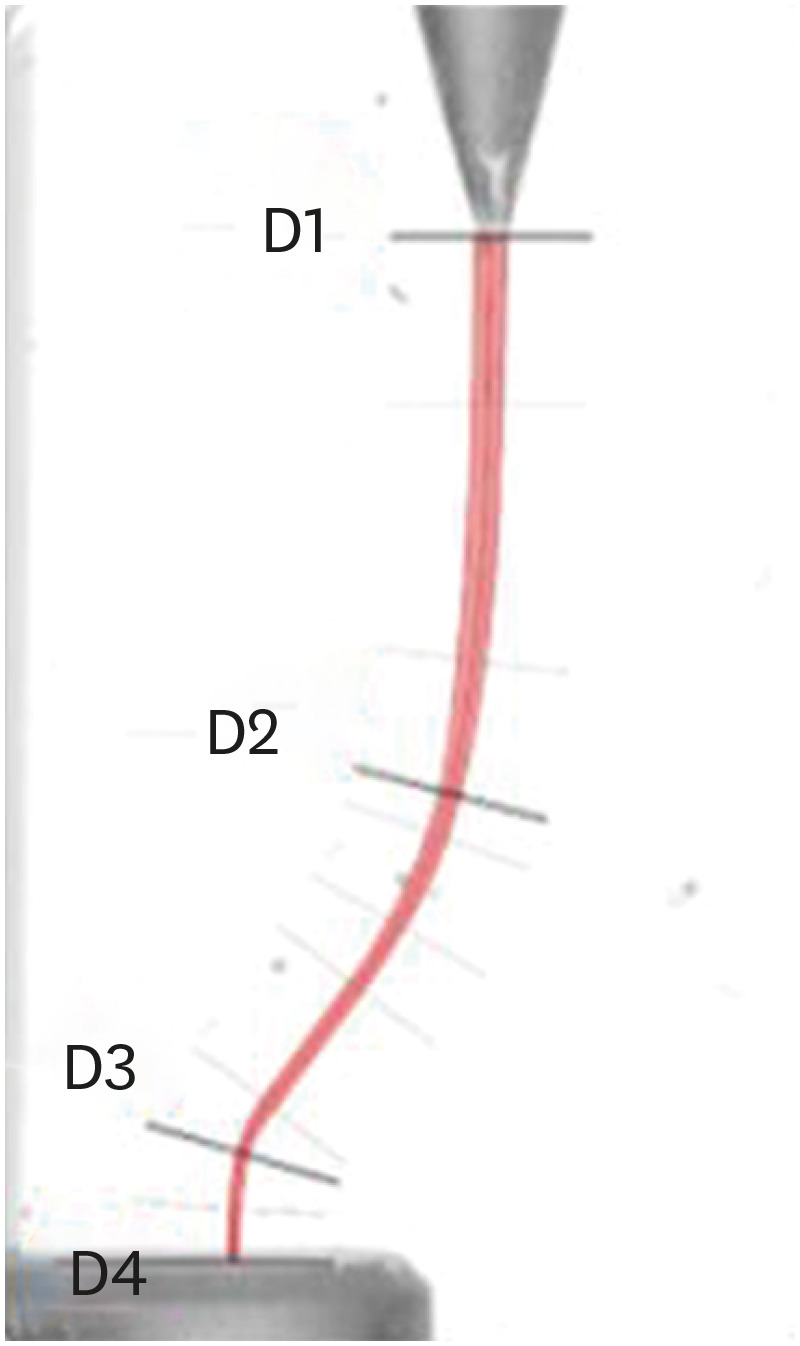
The 4 levels defined in the artificial canals: The beginning of artificial canal (D1); the center of the first curvature (D2); the center of the second curvature (D3); apex level (D4).

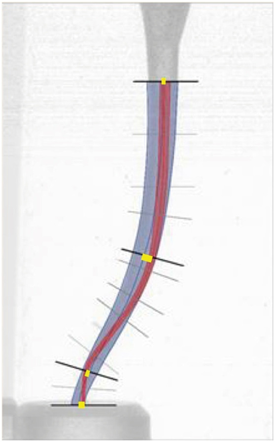
Superimposition of the artificial canal before (red) and after shaping (blue) and the measurement of centering ability (yellow line).

RESULTS
Centering ability
Deviation of original canal curvatures (°) and area of instrumentation (mm2)
DISCUSSION
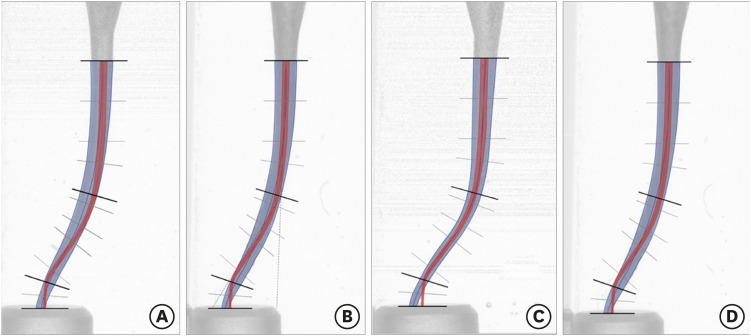
The 4 simulated resin blocks groups after superimposition, R-Pilot and Reciproc Blue (RPB) (A), Reciproc Blue (RB) (B), One Curve (OC) (C), One G and One Curve (GOC) (D).

CONCLUSIONS
-
Conflict of Interest: No potential conflict of interest relevant to this article was reported.
-
Author Contributions:
Conceptualization: Marigo L, Grande NM, Biasillo V, Castagnola R.
Data curation: Biasillo V, Castagnola R.
Formal analysis: Colangeli M, Panzetta C.
Investigation: Biasillo V, Grande NM.
Methodology: Grande NM, Marigo L, Biasillo V.
Resources: Staffoli S, Grande NM.
Supervision: Marigo L, Grande NM.
Validation: Staffoli S, Minciacchi I.
Writing - original draft: Biasillo V, Castagnola R.
Writing - review & editing: Plotino G.
- 1. Walia HM, Brantley WA, Gerstein H. An initial investigation of the bending and torsional properties of Nitinol root canal files. J Endod 1988;14:346-351.ArticlePubMed
- 2. Bürklein S, Hinschitza K, Dammaschke T, Schäfer E. Shaping ability and cleaning effectiveness of two single-file systems in severely curved root canals of extracted teeth: Reciproc and WaveOne versus Mtwo and ProTaper. Int Endod J 2012;45:449-461.ArticlePubMed
- 3. Plotino G, Grande NM, Testarelli L, Gambarini G. Cyclic fatigue of Reciproc and WaveOne reciprocating instruments. Int Endod J 2012;45:614-618.ArticlePubMed
- 4. De-Deus G, Arruda TEP, Souza EM, Neves A, Magalhães K, Thuanne E, Fidel RAS. The ability of the Reciproc R25 instrument to reach the full root canal working length without a glide path. Int Endod J 2013;46:993-998.ArticlePubMed
- 5. Zuolo ML, Carvalho MC, De-Deus G. Negotiability of second mesiobuccal canals in maxillary molars using a reciprocating system. J Endod 2015;41:1913-1917.ArticlePubMed
- 6. Yun HH, Kim SK. A comparison of the shaping abilities of 4 nickel-titanium rotary instruments in simulated root canals. Oral Surg Oral Med Oral Pathol Oral Radiol Endod 2003;95:228-233.ArticlePubMed
- 7. Schäfer E, Florek H. Efficiency of rotary nickel-titanium K3 instruments compared with stainless steel hand K-Flexofile. Part 1. Shaping ability in simulated curved canals. Int Endod J 2003;36:199-207.ArticlePubMedPDF
- 8. Peters OA. Current challenges and concepts in the preparation of root canal systems: a review. J Endod 2004;30:559-567.ArticlePubMed
- 9. Greene KJ, Krell KV. Clinical factors associated with ledged canals in maxillary and mandibular molars. Oral Surg Oral Med Oral Pathol 1990;70:490-497.ArticlePubMed
- 10. Schäfer E, Vlassis M. Comparative investigation of two rotary nickel-titanium instruments: ProTaper versus RaCe. Part 1. Shaping ability in simulated curved canals. Int Endod J 2004;37:229-238.ArticlePubMed
- 11. Plotino G, Nagendrababu V, Bukiet F, Grande NM, Veettil SK, De-Deus G, Aly Ahmed HM. Influence of negotiation, glide path, and preflaring procedures on root canal shaping-terminology, basic concepts, and a systematic review. J Endod 2020;46:707-729.ArticlePubMed
- 12. Ruddle CJ, Machtou P, West JD. Endodontic canal preparation: innovations in glide path management and shaping canals. Dent Today 2014;33:118-123.
- 13. Berutti E, Cantatore G, Castellucci A, Chiandussi G, Pera F, Migliaretti G, Pasqualini D. Use of nickel-titanium rotary PathFile to create the glide path: comparison with manual preflaring in simulated root canals. J Endod 2009;35:408-412.ArticlePubMed
- 14. Pasqualini D, Bianchi CC, Paolino DS, Mancini L, Cemenasco A, Cantatore G, Castellucci A, Berutti E. Computed micro-tomographic evaluation of glide path with nickel-titanium rotary PathFile in maxillary first molars curved canals. J Endod 2012;38:389-393.ArticlePubMed
- 15. Plotino G, Grande NM, Testarelli L, Gambarini G, Castagnola R, Rossetti A, Özyürek T, Cordaro M, Fortunato L. Cyclic fatigue of reciproc and reciproc blue nickel-titanium reciprocating files at different environmental temperatures. J Endod 2018;44:1549-1552.ArticlePubMed
- 16. De-Deus G, Silva EJNL, Vieira VTL, Belladonna FG, Elias CN, Plotino G, Grande NM. Blue thermomechanical treatment optimizes fatigue resistance and flexibility of the reciproc files. J Endod 2017;43:462-466.ArticlePubMed
- 17. Özyürek T, Uslu G, Gündoğar M, Yılmaz K, Grande NM, Plotino G. Comparison of cyclic fatigue resistance and bending properties of two reciprocating nickel-titanium glide path files. Int Endod J 2018;51:1047-1052.ArticlePubMedPDF
- 18. Staffoli S, Grande NM, Plotino G, Özyürek T, Gündoğar M, Fortunato L, Polimeni A. Influence of environmental temperature, heat-treatment and design on the cyclic fatigue resistance of three generations of a single-file nickel-titanium rotary instrument. Odontology 2019;107:301-307.ArticlePubMedPDF
- 19. Yılmaz K, Uslu G, Gündoğar M, Özyürek T, Grande NM, Plotino G. Cyclic fatigue resistances of several nickel-titanium glide path rotary and reciprocating instruments at body temperature. Int Endod J 2018;51:924-930.ArticlePubMedPDF
- 20. Cunningham CJ, Senia ES. A three-dimensional study of canal curvatures in the mesial roots of mandibular molars. J Endod 1992;18:294-300.ArticlePubMed
- 21. Berutti E, Negro AR, Lendini M, Pasqualini D. Influence of manual preflaring and torque on the failure rate of ProTaper rotary instruments. J Endod 2004;30:228-230.ArticlePubMed
- 22. Patiño PV, Biedma BM, Liébana CR, Cantatore G, Bahillo JG. The influence of a manual glide path on the separation rate of NiTi rotary instruments. J Endod 2005;31:114-116.ArticlePubMed
- 23. D'Amario M, Baldi M, Petricca R, De Angelis F, El Abed R, D'Arcangelo C. Evaluation of a new nickel-titanium system to create the glide path in root canal preparation of curved canals. J Endod 2013;39:1581-1584.ArticlePubMed
- 24. Roland DD, Andelin WE, Browning DF, Hsu GHR, Torabinejad M. The effect of preflaring on the rates of separation for 0.04 taper nickel titanium rotary instruments. J Endod 2002;28:543-545.ArticlePubMed
- 25. Peters OA, Peters CI, Schönenberger K, Barbakow F. ProTaper rotary root canal preparation: effects of canal anatomy on final shape analysed by micro CT. Int Endod J 2003;36:86-92.ArticlePubMedPDF
- 26. Abu Haimed A, Abuhaimed T, Dummer P, Bryant S. The root canal shaping ability of WaveOne and Reciproc versus ProTaper Universal and Mtwo rotary NiTi systems. Saudi Endod J 2017;7:8-15.Article
- 27. Yared G. Canal preparation with only one reciprocating instrument without prior hand filing: a new concept. Peterborough: Quality Endodontic Distributors Ltd; 2010. p. 1-8.
- 28. Uslu G, Özyürek T, Yılmaz K, Gündoğar M. Cyclic fatigue resistance of R-Pilot, HyFlex EDM and PathFile nickel-titanium glide path files in artificial canals with double (S-shaped) curvature. Int Endod J 2018;51:584-589.PubMed
- 29. Topçuoğlu HS, Düzgün S, Aktı A, Topçuoğlu G. Laboratory comparison of cyclic fatigue resistance of WaveOne Gold, Reciproc and WaveOne files in canals with a double curvature. Int Endod J 2017;50:713-717.ArticlePubMedPDF
- 30. Hulsmann M, Peters OA, Dummer PMH. Mechanical preparation of root canals: shaping goals, techniques and means. Endod Topics 2005;10:30-76.Article
- 31. Zhang L, Luo HX, Zhou XD, Tan H, Huang DM. The shaping effect of the combination of two rotary nickel-titanium instruments in simulated S-shaped canals. J Endod 2008;34:456-458.ArticlePubMed
- 32. Keskin C, Sarıyılmaz E, Demiral M. Shaping ability of Reciproc Blue reciprocating instruments with or without glide path in simulated S-shaped root canals. J Dent Res Dent Clin Dent Prospect 2018;12:63-67.ArticlePubMedPMCPDF
- 33. Bürklein S, Poschmann T, Schäfer E. Shaping ability of different nickel-titanium systems in simulated S-shaped canals with and without glide path. J Endod 2014;40:1231-1234.ArticlePubMed
- 34. Navós BV, Hoppe CB, Mestieri LB, Böttcher DE, Só MVR, Grecca FS. Centering and transportation: in vitro evaluation of continuous and reciprocating systems in curved root canals. J Conserv Dent 2016;19:478-481.ArticlePubMedPMC
- 35. Jain A, Asrani H, Singhal AC, Bhatia TK, Sharma V, Jaiswal P. Comparative evaluation of canal transportation, centering ability, and remaining dentin thickness between WaveOne and ProTaper rotary by using cone beam computed tomography: an in vitro study. J Conserv Dent 2016;19:440-444.ArticlePubMedPMC
- 36. Yao J, Sun Y, Yang M, Duan Y. Chemistry, physics and biology of graphene-based nanomaterials: New horizons for sensing, imaging and medicine. J Mater Chem 2012;22:14313-14329.Article
- 37. Elashiry MM, Saber SE, Elashry SH. Comparison of shaping ability of different single-file systems using microcomputed tomography. Eur J Dent 2020;14:70-76.ArticlePubMedPMC
- 38. Özyürek T, Yılmaz K, Uslu G. Shaping ability of reciproc, waveone gold, and hyflex edm single-file systems in simulated s-shaped canals. J Endod 2017;43:805-809.ArticlePubMed
- 39. Dhingra A, Kochar R, Banerjee S, Srivastava P. Comparative evaluation of the canal curvature modifications after instrumentation with One Shape rotary and Wave One reciprocating files. J Conserv Dent 2014;17:138-141.ArticlePubMedPMC
REFERENCES
Tables & Figures
REFERENCES
Citations

- Evaluation of Apical Debris Extrusion and the Remaining Canal Material during Retreatment of a Bioceramic Sealer by the XP-endo Finisher File System, Followed by Various Supplementary Methods: An in Vitro Study
Paras Mull Gehlot, Parvathi Sudeep, Annapoorna B Mariswamy
World Journal of Dentistry.2025; 15(10): 837. CrossRef - Shaping Ability of Rotary NiTi Systems in S‐Shaped Root Canals of Mandibular Molars
Renata M. S. Leal, Emmanuel J. N. L. Silva, Maria C. B. P. Campos, Clarissa T. Rodrigues, Marco A. H. Duarte, Bruno C. Cavenago
Australian Endodontic Journal.2025; 51(1): 133. CrossRef - Comparison of Debris Extrusion and Preparation Time by Traverse, R‐Motion Glider C, and Other Glide Path Systems in Severely Curved Canals
Taher Al Omari, Layla Hassouneh, Khawlah Albashaireh, Alaa Dkmak, Rami Albanna, Ali Al-Mohammed, Ahmed Jamleh, Lucas da Fonseca Roberti Garcia
International Journal of Dentistry.2025;[Epub] CrossRef - Glide Path – An Ineluctable Route for Successful Endodontic Mechanics: A Literature Review
Mahima Bharat Mehta, Anupam Sharma, Aniket Jadhav, Aishwarya Handa, Abhijit Bajirao Jadhav, Ashwini A. Narayanan
Journal of the International Clinical Dental Research Organization.2024; 16(2): 101. CrossRef - Screw-in force, torque generation, and performance of glide-path files with three rotation kinetics
Jee-Yeon Woo, Ji-Hyun Jang, Seok Woo Chang, Soram Oh
Odontology.2024; 112(3): 761. CrossRef - Glide Path in Endodontics: A Literature Review of Current Knowledge
Vlad Mircea Lup, Giulia Malvicini, Carlo Gaeta, Simone Grandini, Gabriela Ciavoi
Dentistry Journal.2024; 12(8): 257. CrossRef - In Vitro Research Methods Used to Evaluate Shaping Ability of Rotary Endodontic Files—A Literature Review
Ranya F. Elemam, Ana Mano Azul, João Dias, Khaled El Sahli, Renato de Toledo Leonardo
Dentistry Journal.2024; 12(10): 334. CrossRef - Endodontic glide path - importance and performance techniques
Milica Jovanovic-Medojevic, Мiljan Stosic, Vanja Opacic-Galic, Violeta Petrovic
Srpski arhiv za celokupno lekarstvo.2023; 151(5-6): 380. CrossRef


Figure 1
Figure 2
Figure 3
Figure 4
Centering ability
| Resin block levels | OC | RB | |
|---|---|---|---|
| D1 | |||
| Without glide-path | 143.46 ± 83.04 | 122.36 ± 103.36 | |
| With glide-path | 238.96 ± 121.50 | 196.18 ± 100.97 | |
| D2 | |||
| Without glide-path | 146.72 ± 94.65 | 191.36 ± 66.69 | |
| With glide-path | 165.66 ± 112.85 | 250.32 ± 102.39 | |
| D3 | |||
| Without glide-path | 109.94 ± 78.52 | 155.52B ± 43.87 | |
| With glide-path | 89.74 ± 83.21 | 56.04A ± 34.19 | |
| D4 | |||
| Without glide-path | 451.22B ± 220.72 | 184.92A ± 118.81 | |
| With glide-path | 158.16A ± 144.91 | 167.22A ± 33.78 | |
Means (µm) ± standard deviation at D1, D2, D3, and D4 after preparation with RB and OC with or without glide-path. The data were analyzed using the 2-way analysis of variance test (
OC, One Curve; RB, Reciproc Blue.
Deviation of original canal curvatures (°) and area of instrumentation (mm2)
| Resin block area and angles | OC | RB | |
|---|---|---|---|
| ΔA | |||
| Without glide-path | 4.103A ± 0.310 | 5.489B ± 0.383 | |
| With glide-path | 4.066A ± 0.209 | 5.174B ± 0.342 | |
| Δ1 | |||
| Without glide-path | 5.0B ± 1.76 | 3.0A ± 0.67 | |
| With glide-path | 3.4 ± 1.71 | 3.6 ± 0.84 | |
| Δ2 | |||
| Without glide-path | 5.4 ± 3.98 | 2.6 ± 1.43 | |
| With glide-path | 3.8 ± 2.44 | 5.0 ± 2.75 | |
Mean ± standard deviation after instrumentation with RB and OC with or without glide-path. The data were analyzed using the 2-way analysis of variance test (
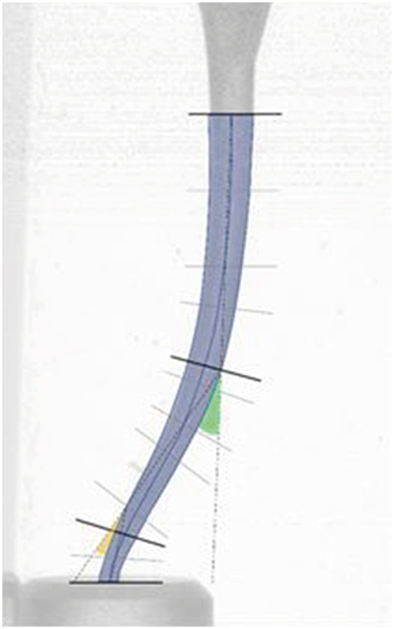
OC, One Curve; RB, Reciproc Blue; ΔA, area of the instrumentation; Δ1, angle of the first curvature after instrumentation (green angle in
Means (µm) ± standard deviation at D1, D2, D3, and D4 after preparation with RB and OC with or without glide-path. The data were analyzed using the 2-way analysis of variance test (
OC, One Curve; RB, Reciproc Blue.
Mean ± standard deviation after instrumentation with RB and OC with or without glide-path. The data were analyzed using the 2-way analysis of variance test (
OC, One Curve; RB, Reciproc Blue; ΔA, area of the instrumentation; Δ1, angle of the first curvature after instrumentation (green angle in

 KACD
KACD

 ePub Link
 Cite
Cite

