
Previous issues
- Page Path
- HOME > Browse articles > Previous issues
- Volume 45 (1); February 2020
-


Editorial
-
Appreciation to peer reviewers in 2019 for their contributions to
Restorative Dentistry and Endodontics - Kyung-San Min
- Restor Dent Endod 2020;45(1):e16. Published online February 11, 2020
- DOI: https://doi.org/10.5395/rde.2020.45.e16
- 1,160 View
- 11 Download

Research Articles
- The prevalence of radix molaris in the mandibular first molars of a Saudi subpopulation based on cone-beam computed tomography
- Hassan AL-Alawi, Saad Al-Nazhan, Nassr Al-Maflehi, Mazen A. Aldosimani, Mohammed Nabil Zahid, Ghadeer N. Shihabi
- Restor Dent Endod 2020;45(1):e1. Published online November 14, 2019
- DOI: https://doi.org/10.5395/rde.2020.45.e1
-
 Abstract
 PDF
PDF PubReader
 ePub
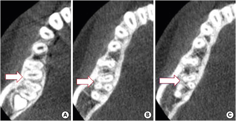
ePub Objectives The purpose of this study was to determine the incidence of radix molaris (RM) (entomolaris and paramolaris) in the mandibular first permanent molars of a sample Saudi Arabian subpopulation using cone-beam computed tomography (CBCT).
Materials and Methods A total of 884 CBCT images of 427 male and 457 female Saudi citizens (age 16 to 70 years) were collected from the radiology department archives of 4 dental centers. A total of 450 CBCT images of 741 mature mandibular first molars that met the inclusion criteria were reviewed. The images were viewed at high resolution by 3 examiners and were analyzed with Planmeca Romexis software (version 5.2).
Results Thirty-three (4.5%) mandibular first permanent molars had RM, mostly on the distal side. The incidence of radix entomolaris (EM) was 4.3%, while that of radix paramolaris was 0.3%. The RM roots had one canal and occurred more unilaterally. No significant difference in root configuration was found between males and females (
p > 0.05). Types I and III EM root canal configurations were most common, while type B was the only RP configuration observed.Conclusions The incidence of RM in the mandibular first molars of this Saudi subpopulation was 4.5%. Identification of the supernumerary root can avoid missing the canal associated with the root during root canal treatment.
-
Citations
Citations to this article as recorded by
- A comprehensive investigation into the prevalence of dental anomalies across Saudi Arabia, a systematic review
Abdulrahman K. Alshammari, Muteb A. Algharbi, Freah L. Alshammary, Nabeel S. Almotairy, Hatem D. Alshammari, Amjad M. Alsogaih, Ahmed A. Madfa
Critical Public Health.2026;[Epub] CrossRef - Anatomical Variations in Permanent Mandibular Molars in the Sharjah Population: A Cohort Study Using Cone‐Beam Computed Tomography
Shaima Dheyab, Entisar AlRasasi, Ahmed M. Aziz, Saad Albayatti, Mehmet Omer Gorduysus, Hannah Wesley
International Journal of Dentistry.2026;[Epub] CrossRef - Evaluation of the variations of mandibular molars and the distance from root apex to the inferior alveolar nerve in Saudi Sub-population: Three-dimensional radiographic evaluation
Tariq Mohammed Aqili, Esam Sami Almuzaini, Abdulbari Saleh Aljohani, Ahmed Khaled Al Saeedi, Hassan Abdulmuti Hammudah, Muath Alassaf, Muhannad M. Hakeem, Mohmed Isaqali Karobari
PLOS ONE.2025; 20(2): e0317053. CrossRef - Prevalence of radix molaris in mandibular molars of a subpopulation of Brazil’s Northeast region: a cross-sectional CBCT study
Yasmym Martins Araújo de Oliveira, Maria Clara Mendes Gomes, Maria Fernanda da Silva Nascimento, Ricardo Machado, Danna Mota Moreira, Hermano Camelo Paiva, George Táccio de Miranda Candeiro
Scientific Reports.2025;[Epub] CrossRef - Prevalence of radix entomolaris and distolingual canals and their association with the incidence of middle mesial canals in mandibular first molars of a Saudi subpopulation
Ahmed A. Madfa, Abdullah F. Alshammari, Eyad Almagadawyi, Afaf Al-Haddad, Ebtsam A. Aledaili
Scientific Reports.2025;[Epub] CrossRef - Assessment of the root and canal morphology in the permanent dentition of Saudi Arabian population using cone beam computed and micro-computed tomography – a systematic review
Mohammed Mustafa, Rumesa Batul, Mohmed Isaqali Karobari, Hadi Mohammed Alamri, Abdulaziz Abdulwahed, Ahmed A. Almokhatieb, Qamar Hashem, Abdullah Alsakaker, Mohammad Khursheed Alam, Hany Mohamed Aly Ahmed
BMC Oral Health.2024;[Epub] CrossRef - Prevalence of radix accesoria dentis in a northern Peruvian population evaluated by cone-beam tomography
Karla Renata León-Almanza, Anthony Adrián Jaramillo-Nuñez, Catherin Angélica Ruiz-Cisneros, Paul Martín Herrera-Plasencia
Heliyon.2024; 10(16): e35919. CrossRef - Radix molaris is a hidden truth of mandibular first permanent molars: A descriptive- analytic study using cone beam computed tomography
Mohammed A. Alobaid, Saurabh Chaturvedi, Ebtihal Mobarak S. Alshahrani, Ebtsam M. Alshehri, Amal S. Shaiban, Mohamed Khaled Addas, Giuseppe Minervini
Technology and Health Care.2023; 31(5): 1957. CrossRef - Prevalence of Radix Entomolaris in Mandibular Permanent Molars Analyzed by Cone-Beam CT in the Saudi Population of Ha'il Province
Moazzy I Almansour, Ahmed A Madfa, Adhwaa F Algharbi, Reem Almuslumani, Noeer K Alshammari, Ghufran M Al Hussain
Cureus.2023;[Epub] CrossRef - Prevalence of radix entomolaris in India and its comparison with the rest of the world
Sumit MOHAN, Jyoti THAKUR
Minerva Dental and Oral Science.2022;[Epub] CrossRef - Radix Paramolaris an Endodontic Challenge: A Case Report
Ashwini B Prasad, Deepak Raisingani, Ridhima Gupta, Rimjhim Jain
Journal of Mahatma Gandhi University of Medical Sciences and Technology.2022; 7(1): 32. CrossRef - Evaluation of Radix Entomolaris and Middle Mesial Canal in Mandibular Permanent First Molars in an Iraqi Subpopulation Using Cone‐Beam Computed Tomography
Ranjdar Mahmood Talabani, Kazhan Omer Abdalrahman, Rawa Jamal Abdul, Dlsoz Omer Babarasul, Sara Hilmi Kazzaz, Heng Bo Jiang
BioMed Research International.2022;[Epub] CrossRef - Evaluation of Root Canal Configuration of Maxillary and Mandibular First Molar by CBCT: A Retrospective Cross-Sectional Study
Rakan Rafdan Alhujhuj, Rizwan Jouhar, Muhammad Adeel Ahmed, Abdullatif Abdulrahman Almujhim, Mohammed Tariq Albutayh, Necdet Adanir
Diagnostics.2022; 12(9): 2121. CrossRef - Ethnical Anatomical Differences in Mandibular First Permanent Molars between Indian and Saudi Arabian Subpopulations: A Retrospective Cross-sectional Study
Abdulwahab Alamir, Mohammed Mashyakhy, Apathsakayan Renugalakshmi, Thilla S Vinothkumar, Anandhi S Arthisri, Ahmed Juraybi
The Journal of Contemporary Dental Practice.2021; 22(5): 484. CrossRef
- A comprehensive investigation into the prevalence of dental anomalies across Saudi Arabia, a systematic review
- 3,511 View
- 53 Download
- 14 Crossref
- Cytocompatibility and cell proliferation evaluation of calcium phosphate-based root canal sealers
- Letícia Boldrin Mestieri, Ivana Maria Zaccara, Lucas Siqueira Pinheiro, Fernando Branco Barletta, Patrícia Maria Polli Kopper, Fabiana Soares Grecca
- Restor Dent Endod 2020;45(1):e2. Published online November 15, 2019
- DOI: https://doi.org/10.5395/rde.2020.45.e2
-
Abstract
PDFPubReaderePub
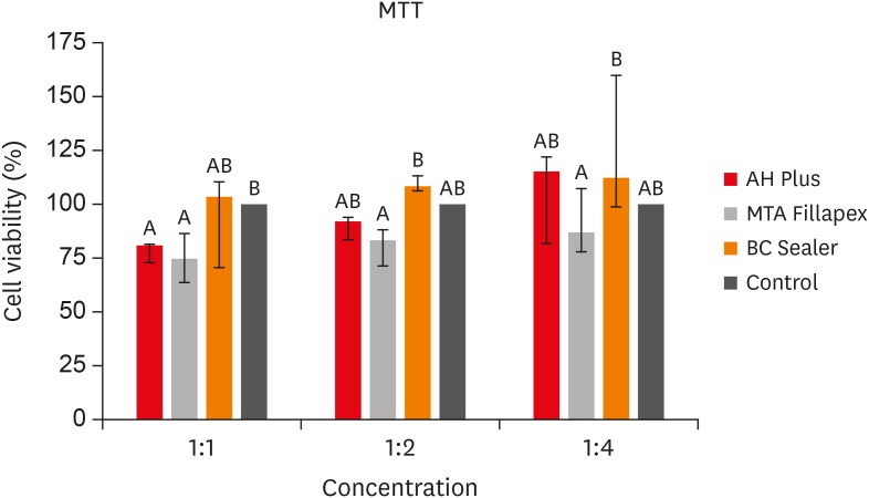
Objectives This study aimed to evaluate the cell viability and migration of Endosequence Bioceramic Root Canal Sealer (BC Sealer) compared to MTA Fillapex and AH Plus.
Materials and Methods BC Sealer, MTA Fillapex, and AH Plus were placed in contact with culture medium to obtain sealers extracts in dilution 1:1, 1:2 and 1:4. 3T3 cells were plated and exposed to the extracts. Cell viability and migration were assessed by 3-(4,5-dimethyl-thiazoyl)-2,5-diphenyl-tetrazolium bromide (MTT) and Scratch assay, respectively. Data were analyzed by Kruskal-Wallis and Dunn's test (
p < 0.05).Results The MTT assay revealed greater cytotoxicity for AH Plus and MTA Fillapex at 1:1 dilution when compared to control (
p < 0.05). At 1:2 and 1:4 dilutions, all sealers were similar to control (p > 0.05) and MTA Fillapex was more cytotoxic than BC Sealer (p < 0.05). Scratch assay demonstrated the continuous closure of the wound according to time. At 30 hours, the control group presented closure of the wound (p < 0.05). At 36 hours, only BC Sealer presented the closure when compared to AH Plus and MTA Fillapex (p < 0.05). At 42 hours, AH Plus and MTA Fillapex showed a wound healing (p > 0.05).Conclusions All tested sealers demonstrated cell viability highlighting BC Sealer, which showed increased cell migration capacity suggesting that this sealer may achieve better tissue repair when compared to other tested sealers.
-
Citations
Citations to this article as recorded by- Oxidative Stress, Pro-Inflammatory Response, Cytotoxicity and Apoptosis Induced by Contemporary Endodontic Sealers in Human Periodontal Ligament Fibroblasts
Stanisław Krokosz, Virginia Ewa Lis, Sara Zięba, Mateusz Maciejczyk, Ewa Zalewska, Maria Obrycka, Edyta Gołaś, Małgorzata Żendzian-Piotrowska, Jerzy Ładny, Anna Skutnik-Radziszewska, Karol Dąbrowski, Julia Kuźmiuk, Anna Zalewska
Journal of Functional Biomaterials.2026; 17(2): 105. CrossRef - Effect of Nano-Silica on Mechanical Properties and Cytotoxicity of Calcium-Silicate-Based Root Canal Filling Materials
Hao He, Bolang Hao, Xiang Xiong, Yi Cheng, Jia Lou, Zheyu He, Dongyang Li, Zhihuan Wang, Jian Qin
Crystals.2025; 15(1): 55. CrossRef - Premixed calcium silicate-based root canal sealers have better biological properties than AH Plus: A systematic review and meta-analysis of in vivo animal studies and in vitro laboratory studies
Cristiana Pereira Malta, Samantha Simoni Santi, Raquel Cristine Silva Barcelos, Fabrício Batistin Zanatta, Carlos Alexandre Souza Bier, Renata Dornelles Morgental
Journal of Conservative Dentistry and Endodontics.2024; 27(4): 345. CrossRef - Biological Properties of Bioceramic Sealers on Osteoblastic Cells: A Comparative Study
Angelita Piovezana Guerra, Danielle Gregorio, Gean Carlos Yamamoto, Nathalia Thalitha Bernardes dos Santos, Regina Celia Poli-Frederico, Luciana Prado Maia
Brazilian Dental Journal.2024;[Epub] CrossRef - Premixed calcium silicate‐based ceramic sealers promote osteogenic/cementogenic differentiation of human periodontal ligament stem cells: A microscopy study
Sergio López‐García, Sonia Sánchez‐Bautista, David García‐Bernal, Adrián Lozano, Leopoldo Forner, José L. Sanz, Laura Murcia, Francisco J. Rodríguez‐Lozano, Ricardo E. Oñate‐Sánchez
Microscopy Research and Technique.2024; 87(7): 1584. CrossRef - Cytotoxicity Comparison of Sure-seal root and Adseal Sealers on mouse fibroblast Cells:Invitro study
Azam haddadikohsar, Mohammad shokrzade, Marjan Fallah, Fatemeh Shakeri
journal of research in dental sciences.2024; 21(1): 46. CrossRef - Cytotoxicity and cell migration evaluation of a strontium silicate-based root canal sealer on stem cells from rat apical papilla: an in vitro study
Guanglei Zhou, Yu Zhao, Liangjing Cai, Liwei Liu, Xu Li, Lu Sun, Jiayin Deng
BMC Oral Health.2024;[Epub] CrossRef - A comparative study of biological properties of three root canal sealers
Yujia Yan, Yanyao Li, Yaqi Chi, Mengzhen Ji, Ya Shen, Ling Zou
Clinical Oral Investigations.2023;[Epub] CrossRef - Biomineralization potential and biological properties of a new tantalum oxide (Ta2O5)–containing calcium silicate cement
F. J. Rodríguez-Lozano, A. Lozano, S. López-García, D. García-Bernal, J. L. Sanz, J. Guerrero-Gironés, C. Llena, L. Forner, M. Melo
Clinical Oral Investigations.2022; 26(2): 1427. CrossRef - Cytotoxicity and Genotoxicity of Epoxy Resin-Based Root Canal Sealers before and after Setting Procedures
Mijoo Kim, Marc Hayashi, Bo Yu, Thomas K. Lee, Reuben H. Kim, Deuk-won Jo
Life.2022; 12(6): 847. CrossRef - Characterization, Antimicrobial Effects, and Cytocompatibility of a Root Canal Sealer Produced by Pozzolan Reaction between Calcium Hydroxide and Silica
Mi-Ah Kim, Vinicius Rosa, Prasanna Neelakantan, Yun-Chan Hwang, Kyung-San Min
Materials.2021; 14(11): 2863. CrossRef - Bone repair in defects filled with AH Plus sealer and different concentrations of MTA: a study in rat tibiae
Jessica Emanuella Rocha Paz, Priscila Oliveira Costa, Albert Alexandre Costa Souza, Ingrid Macedo de Oliveira, Lucas Fernandes Falcão, Carlos Alberto Monteiro Falcão, Maria Ângela Area Leão Ferraz, Lucielma Salmito Soares Pinto
Restorative Dentistry & Endodontics.2021;[Epub] CrossRef - Incorporation of amoxicillin-loaded microspheres in mineral trioxide aggregate cement: an in vitro study
Fábio Rocha Bohns, Vicente Castelo Branco Leitune, Isadora Martini Garcia, Bruna Genari, Nélio Bairros Dornelles, Silvia Stanisçuaski Guterres, Fabrício Aulo Ogliari, Mary Anne Sampaio de Melo, Fabrício Mezzomo Collares
Restorative Dentistry & Endodontics.2020;[Epub] CrossRef
- Oxidative Stress, Pro-Inflammatory Response, Cytotoxicity and Apoptosis Induced by Contemporary Endodontic Sealers in Human Periodontal Ligament Fibroblasts
- 2,412 View
- 16 Download
- 13 Crossref
- Bioactivity of endodontic biomaterials on dental pulp stem cells through dentin
- Bahar Javid, Narges Panahandeh, Hassan Torabzadeh, Hamid Nazarian, Ardavan Parhizkar, Saeed Asgary
- Restor Dent Endod 2020;45(1):e3. Published online November 4, 2019
- DOI: https://doi.org/10.5395/rde.2020.45.e3
-
Abstract
PDFPubReaderePub
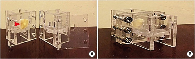
Objectives This study investigated the indirect effect of calcium-enriched mixture (CEM) cement and mineral trioxide aggregate (MTA), as 2 calcium silicate-based hydraulic cements, on human dental pulp stem cells (hDPSCs) through different dentin thicknesses.
Materials and Methods Two-chamber setups were designed to simulate indirect pulp capping (IPC). Human molars were sectioned to obtain 0.1-, 0.3-, and 0.5-mm-thick dentin discs, which were placed between the 2 chambers to simulate an IPC procedure. Then, MTA and CEM were applied on one side of the discs, while hDPSCs were cultured on the other side. After 2 weeks of incubation, the cells were removed, and cell proliferation, morphology, and attachment to the discs were evaluated under scanning electron microscopy (SEM). Energy-dispersive X-ray (EDXA) spectroscopy was performed for elemental analysis. Alkaline phosphatase (ALP) activity was assessed quantitatively. The data were analyzed using the Kruskal-Wallis and Mann-Whitney tests.
Results SEM micrographs revealed elongated cells, collagen fibers, and calcified nucleations in all samples. EDXA verified that the calcified nucleations consisted of calcium phosphate. The largest calcifications were seen in the 0.1-mm-thick dentin subgroups. There was no significant difference in ALP activity across the CEM subgroups; however, ALP activity was significantly lower in the 0.1-mm-thick dentin subgroup than in the other MTA subgroups (
p < 0.05).Conclusions The employed capping biomaterials exerted biological activity on hDPSCs, as shown by cell proliferation, morphology, and attachment and calcific precipitations, through 0.1- to 0.5-mm-thick layers of dentin. In IPC, the bioactivity of these endodontic biomaterials is probably beneficial.
-
Citations
Citations to this article as recorded by- Dental pulp capping materials: modulators of stem cell behavior and regenerative potential
Ali Cheayto, Sara Ayoub, Sarah Ayad Al-Tameemi, Mohammad Fayyad-Kazan
Biomedical Physics & Engineering Express.2025; 11(6): 062004. CrossRef - Effect of pulp capping materials on odontogenic differentiation of human dental pulp stem cells: An in vitro study
Mahmoud M. Bakr, Mohamed Shamel, Shereen N. Raafat, Robert M. Love, Mahmoud M. Al‐Ankily
Clinical and Experimental Dental Research.2024;[Epub] CrossRef - Effects of Growth Factors on the Differentiation of Dental Stem Cells: A
Systematic Review and Meta-analysis (Part I)
Sayna Shamszadeh, Armin Shirvani, Hassan Torabzadeh, Saeed Asgary
Current Stem Cell Research & Therapy.2024; 19(4): 523. CrossRef - The Role of Growth Factor Delivery Systems on Cellular Activities of Dental
Stem Cells: A Systematic Review (Part II)
Sayna Shamszadeh, Armin Shirvani, Saeed Asgary
Current Stem Cell Research & Therapy.2024; 19(4): 587. CrossRef - Comprehensive review of composition, properties, clinical applications, and future perspectives of calcium-enriched mixture (CEM) cement: a systematic analysis
Saeed Asgary, Mahtab Aram, Mahta Fazlyab
BioMedical Engineering OnLine.2024;[Epub] CrossRef - Evaluation of dental pulp stem cells behavior after odontogenic differentiation induction by three different bioactive materials on two different scaffolds
Basma Ahmed, Mai H. Ragab, Rania A. Galhom, Hayam Y. Hassan
BMC Oral Health.2023;[Epub] CrossRef - Characterization of Dental Pulp Stem Cell Responses to Functional Biomaterials Including Mineralized Trioxide Aggregates
Sejin Bae, Bueonguk Kang, Hyungbin Lee, Harrison Luu, Eric Mullins, Karl Kingsley
Journal of Functional Biomaterials.2021; 12(1): 15. CrossRef - Incorporation of amoxicillin-loaded microspheres in mineral trioxide aggregate cement: an in vitro study
Fábio Rocha Bohns, Vicente Castelo Branco Leitune, Isadora Martini Garcia, Bruna Genari, Nélio Bairros Dornelles, Silvia Stanisçuaski Guterres, Fabrício Aulo Ogliari, Mary Anne Sampaio de Melo, Fabrício Mezzomo Collares
Restorative Dentistry & Endodontics.2020;[Epub] CrossRef
- Dental pulp capping materials: modulators of stem cell behavior and regenerative potential
- 2,474 View
- 18 Download
- 8 Crossref
- A micro-computed tomography evaluation of voids using calcium silicate-based materials in teeth with simulated internal root resorption
- Vildan Tek, Sevinç Aktemur Türker
- Restor Dent Endod 2020;45(1):e5. Published online November 29, 2019
- DOI: https://doi.org/10.5395/rde.2020.45.e5
-
Abstract
PDFPubReaderePub
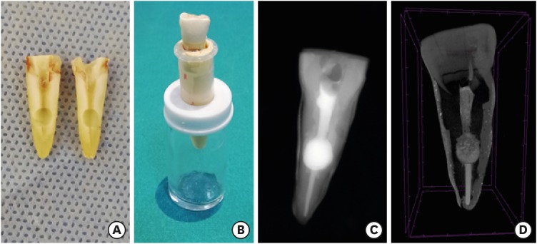
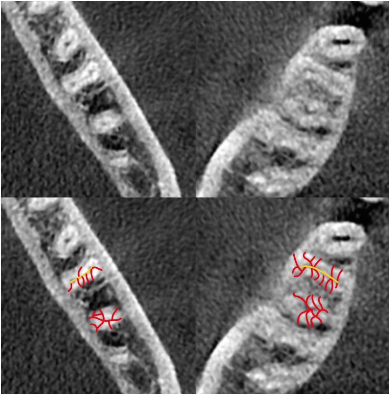
Objectives The obturation quality of MTA, Biodentine, Total Fill BC root canal sealer (RCS), and warm gutta-percha (WGP) in teeth with simulated internal root resorption (IRR) was evaluated by using micro-computed tomography.
Materials and Methods Standardized IRR cavities were created using 40 extracted maxillary central incisor teeth and randomly assigned into 4 groups (
n = 10). IRR cavities were filled with MTA, Biodentine, Total Fill BC RCS (bulk-fill form) and WGP + Total Fill BC RCS. Percentage of voids between resorptive cavity walls and obturation material (external void), and inside the filling materials (internal voids) were measured.Results Total Fill BC sealer in the bulk-fill form presented significantly highest values of external and internal void percentages (
p < 0.05). Biodentine showed a significantly lowest external void percentage (p < 0.05). WGP + Total Fill BC RCS presented significantly lower values of internal void percentages than all groups (p < 0.05), except Biodentine (p > 0.05).Conclusion None of the filling materials were created void-free obturation in resorption cavities. Biodentine may favor its application in teeth with IRR over Angelus MTA and bulk-fill form of Total Fill BC.
-
Citations
Citations to this article as recorded by- The role of calcium silicate cements in endodontics: from material science to clinical success
Takwa E. Ellakwa, Ayman Ellakwa, Doha El-Sayed Ellakwa
Discover Materials.2026;[Epub] CrossRef - Micro-CT Assessment of Hydraulic Calcium Silicate Cements for Perforating Internal Resorption in 3D-printed Tooth Replicas at Different Root Thirds: An In Vitro Study
Angelo José Sócrates Torres-Carrillo, Jardel Francisco Mazzi-Chaves, Gustavo Creazzo, Rodrigo E. Salazar-Gamarra, Helena Cristina de Assis, Ronald Ordinola-Zapata, Manoel D. Sousa-Neto, Fabiane Carneiro Lopes-Olhê
Journal of Endodontics.2026;[Epub] CrossRef - Removal of AH Plus Bioceramic Sealer from Artificial Internal Resorption Cavities Using Different Irrigation Activation Systems
Mine Büker, Meltem Sümbüllü, Emine Şimşek, Fadime Sena Sezer
Cumhuriyet Dental Journal.2025; 28(3): 383. CrossRef - Functional and Bioactive Performance of Premixed Bioceramic Sealers with Warm Obturation: A Scoping Review
Patryk Wiśniewski, Stanisław Krokosz, Małgorzata Pietruska, Anna Zalewska
Gels.2025; 11(11): 932. CrossRef - Evaluation of the effectiveness of different supplemental cleaning techniques in the retreatment of roots with small simulated internal resorption cavities: an in vitro comparative study
Sine Güngör Us, Özgür Uzun, Nazlı Merve Güngör
BMC Oral Health.2025;[Epub] CrossRef - Evaluation of Different Techniques and Materials for Filling in 3-dimensional Printed Teeth Replicas with Perforating Internal Resorption by Means of Micro–Computed Tomography
Angelo J.S. Torres-Carrillo, Helena C. Assis, Rodrigo E. Salazar-Gamarra, Leonardo Moreira Teodosio, Alice C. Silva-Sousa, Jardel F. Mazzi-Chaves, Priscila B. Ferreira-Soares, Manoel D. Sousa-Neto, Fabiane C. Lopes-Olhê
Journal of Endodontics.2024; 50(2): 205. CrossRef - Three-Dimensional Measurement of Obturation Quality of Bioceramic Materials in Filling Artificial Internal Root Resorption Cavities Using Different Obturation Techniques: An In Vitro Comparative Study
Ammar M. Sharki, Ahmed H. Ali
Journal of Endodontics.2024; 50(7): 997. CrossRef - Evaluation of calcium hydroxide root canal filling materials by cone beam computed tomography and three-dimensional modeling
Asel Usdat Ozturk, Ekin Dogan, Venus Seyedoskuyi, Berk Senguler, Asli Topaloglu-Ak
Folia Medica.2024; 66(2): 250. CrossRef - Clinical applications of calcium silicate‐based materials: a narrative review
S Küçükkaya Eren
Australian Dental Journal.2023;[Epub] CrossRef - A critical analysis of research methods and experimental models to study root canal fillings
Gustavo De‐Deus, Erick Miranda Souza, Emmanuel João Nogueira Leal Silva, Felipe Gonçalves Belladonna, Marco Simões‐Carvalho, Daniele Moreira Cavalcante, Marco Aurélio Versiani
International Endodontic Journal.2022; 55(S2): 384. CrossRef - An Updated Review on Properties and Indications of Calcium Silicate‐Based Cements in Endodontic Therapy
Fateme Eskandari, Alireza Razavian, Rozhina Hamidi, Khadije Yousefi, Susan Borzou, Zohaib Khurshid
International Journal of Dentistry.2022;[Epub] CrossRef - Efficacy Of Calcium Silicate-Based Sealers In Root Canal Treatment: A Systematic Review
Hattan Mohammed Omar Baismail, Mohammed Ghazi Moiser Albalawi, Alaa Mofareh Thoilek Alanazi, Muhannad Atallah Saleem Alatawi, Badr Soliman Alhussain
Annals of Dental Specialty.2021; 9(1): 87. CrossRef
- The role of calcium silicate cements in endodontics: from material science to clinical success
- 3,290 View
- 42 Download
- 12 Crossref
- Effect of hydrofluoric acid-based etchant at an elevated temperature on the bond strength and surface topography of Y-TZP ceramics
- Mi-Kyung Yu, Myung-Jin Lim, Noo-Ri Na, Kwang-Won Lee
- Restor Dent Endod 2020;45(1):e6. Published online December 3, 2019
- DOI: https://doi.org/10.5395/rde.2020.45.e6
-
Abstract
PDFPubReaderePub
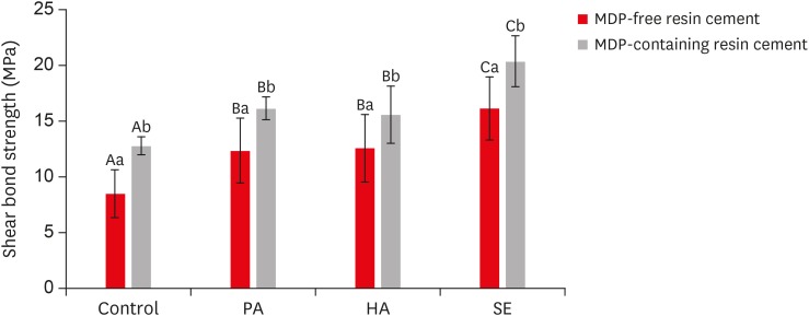
Objectives This study investigated the effects of a hydrofluoric acid (HA; solution of hydrogen fluoride [HF] in water)-based smart etching (SE) solution at an elevated temperature on yttria-stabilized tetragonal zirconia polycrystal (Y-TZP) ceramics in terms of bond strength and morphological changes.
Materials and Methods Eighty sintered Y-TZP specimens were prepared for shear bond strength (SBS) testing. The bonding surface of the Y-TZP specimens was treated with 37% phosphoric acid etching at 20°C–25°C, 4% HA etching at 20°C–25°C, or HA-based SE at 70°C–80°C. In all groups, zirconia primers were applied to the bonding surface of Y-TZP. For each group, 2 types of resin cement (with or without methacryloyloxydecyl dihydrogen phosphate [MDP]) were used. SBS testing was performed. Topographic changes of the etched Y-TZP surface were analyzed using scanning electron microscopy and atomic force microscopy. The results were analyzed and compared using 2-way analysis of variance.
Results Regardless of the type of resin cement, the highest bond strength was measured in the SE group, with significant differences compared to the other groups (
p < 0.05). In all groups, MDP-containing resin cement yielded significantly higher bond strength values than MDP-free resin cement (p < 0.05). It was also shown that the Y-TZP surface was etched by the SE solution, causing a large change in the surface topography.Conclusions Bond strength significantly improved when a heated HA-based SE solution was applied to the Y-TZP surface, and the etched Y-TZP surface was more irregular and had higher surface roughness.
-
Citations
Citations to this article as recorded by- Etchability of zirconia ceramics and its effect on adhesion: A systematic review and meta-analysis
Anina Sieber, Luiza Freitas Brum Souza, Tan Fırat Eyüboğlu, Mutlu Özcan
International Journal of Adhesion and Adhesives.2026; 148: 104303. CrossRef - Evaluation of Different Surface Roughening Techniques on Clear Aligner Attachments Bonded to Monolithic Zirconia: In Vitro Study
Nehal F Albelasy, Ahmad M Hafez, Abdullah S Alhunayni
The Journal of Contemporary Dental Practice.2025; 25(12): 1104. CrossRef - Effect of Acid Surface Treatments on the Shear Bond Strength of Metal Bracket to Zirconia Ceramics
Punchanit Wongrachit, Bancha Samruajbenjakun, Boonlert Kukiattrakoon, Tanapat Jearanai, Supontep Teerakanok, Pannapat Chanmanee
Ceramics.2024; 7(2): 689. CrossRef - Exploring Zirconia Adhesion: Pre and Postsintering Physical Surface Treatment, Chemical Treatment, and Cement Interactions
Flávia Gonçalves, Mirko Dennys Ayala-Perez, Francisco Carlos dos Santos Reis, Walter Gomes Miranda-Júnior, Letícia Cristina Cidreira Boaro, Heng Bo Jiang
BioMed Research International.2024;[Epub] CrossRef - Evaluation of zirconia surfaces and shear bond strength after acid–etching with ultrasonic vibration
Xiaozhen Zhang, Hepeng Nie, Jiaxin Lv, Shanshan Yuan, Juan Wang, Kunzhan Cai, Jin Wu, Qingqing Zhang, Chunbo Tang
Materials Research Express.2024; 11(2): 025401. CrossRef - Effects of Surface-Etching Systems on the Shear Bond Strength of Dual-Polymerized Resin Cement and Zirconia
Sang-Hyun Kim, Kyung Chul Oh, Hong-Seok Moon
Materials.2024; 17(13): 3096. CrossRef - Zirconia bond strength durability following artificial aging: A systematic review and meta-analysis of in vitro studies
Athanasios E. Rigos, Katia Sarafidou, Eleana Kontonasaki
Japanese Dental Science Review.2023; 59: 138. CrossRef - Y-TZP Physicochemical Properties Conditioned with ZrO2 and SiO2 Nanofilms and Bond Strength to Dual Resin Cement
Ricardo Faria Ribeiro, Danilo Flamini Oliveira, Camila Bussola Tovani, Ana Paula Ramos, Ana Flavia Sanches Borges, Adriana Claudia Lapria Faria, Rossana Pereira de Almeida, Renata Cristina Silveira Rodrigues
Materials.2022; 15(22): 7905. CrossRef - Effect of the nanofilm-coated zirconia ceramic on resin cement bond strength
Viviane Maria Gonçalves de Figueiredo, Alecsandro de Moura Silva, Marcos Massi, Argemiro Soares da Silva Sobrinho, José Renato Cavalcanti de Queiroz, João Paulo Barros Machado, Renata Falchete do Prado, Lafayette Nogueira Junior
Journal of Dental Research, Dental Clinics, Dental Prospects.2022; 16(3): 170. CrossRef - Change of phase transformation and bond strength of Y-TZP with various hydrofluoric acid etching
Mi-Kyung Yu, Eun-Jin Oh, Myung-Jin Lim, Kwang-Won Lee
Restorative Dentistry & Endodontics.2021;[Epub] CrossRef - Changes in Bond Strength and Topography for Y-TZP Etched with Hydrofluoric Acid Depending on Concentration and Temperature Conditions
Hyo-Eun Kim, Myung-Jin Lim, Mi-Kyung Yu, Kwang-Won Lee
Medicina.2020; 56(11): 568. CrossRef - Do different sintering conditions influence bond strength between the resin cements and a currently used esthetic zirconia?
Fatma Ayse Sanal, Hamiyet Kilinc
Journal of Adhesion Science and Technology.2020; 34(16): 1809. CrossRef
- Etchability of zirconia ceramics and its effect on adhesion: A systematic review and meta-analysis
- 3,029 View
- 17 Download
- 12 Crossref
-
Isthmuses, accessory canals, and the direction of root curvature in permanent mandibular first molars: an
in vivo computed tomography study - Aria Chuppani Dastgerdi, Manizheh Navabi, Vahid Rakhshan
- Restor Dent Endod 2020;45(1):e7. Published online December 12, 2019
- DOI: https://doi.org/10.5395/rde.2020.45.e7
-
Abstract
PDFPubReaderePub
Objectives This study was performed to assess the anatomy of mandibular first molars.
Materials and Methods In this
in vivo study, cone-beam computed tomography (CBCT) volumes of 312 bilateral intact first mandibular molars from 156 patients (79 men and 77 women; average age, 35.6 ± 11.2 years) were investigated in terms of the direction of each canal's curvature in the buccolingual and mesiodistal dimensions (direction of the position of the apex in relation to the longitudinal axis of the root), the presence of an isthmus (a narrow, ribbon-shaped communication between 2 root canals) in 3 segments (0–2, 2–4, and 4–6 mm) from the apex), and the presence and number of accessory canals (smaller canals besides the main root canals, connecting the pulp to the periodontium). Data were analyzed statistically (α = 0.05).Results Mesiolingual canals were mostly buccally and distally inclined, while mesiobuccal and distolingual canals were mostly distally curved. Isthmuses were more common in younger patients (χ2 test,
p < 0.05). The average numbers of accessory canals in the apical, middle, and coronal segments were 9.9 ± 4.2, 6.9 ± 2.9, and 9.3 ± 3.0 canals per segment, respectively (analysis of variance,p < 0.001). Age and sex were not associated with the number of accessory canals (p > 0.05).Conclusions The complex anatomy of these teeth deserves attention during non-surgical or surgical endodontic treatment. Around the apex, isthmuses might be more prevalent in younger and female individuals.
-
Citations
Citations to this article as recorded by- Micro-CT and histological examination of accessory canals in 34 equine cheek teeth
Szabolcs A. Korsós, Carsten Staszyk, Matthieu Boone, Iván Josipovic, Jörg Vogelsberg, Lieven Vlaminck
Frontiers in Veterinary Science.2024;[Epub] CrossRef - Diagnostic value of cone beam computed tomography for root canal morphology assessment – a micro-CT based comparison
Mariana Pires, Jorge N.R. Martins, Mário Rito Pereira, Isabel Vasconcelos, Rui Pereira da Costa, Isabel Duarte, António Ginjeira
Clinical Oral Investigations.2024;[Epub] CrossRef - Exploring age and gender variations in root canal morphology of maxillary premolars in Saudi sub population: a cross-sectional CBCT study
Mohmed Isaqali Karobari, Azhar Iqbal, Rumesa Batul, Abdul Habeeb Adil, Jamaluddin Syed, Hmoud Ali Algarni, Meshal Aber Alonazi, Tahir Yusuf Noorani
BMC Oral Health.2024;[Epub] CrossRef - Comprehensive analysis of root canal morphology in maxillary premolars among the Pakistani subpopulation: a CBCT-based study
Hmoud Ali Algarni, Meshal Aber Alonazi, Hamza Arshad, Fatima Zahra, Fahad Umer, Irfan Maqbool, Azhar Iqbal, Mohmed Isaqali Karobari
European Journal of Medical Research.2024;[Epub] CrossRef - The efficacy of a novel SWEEPS laser-activated irrigation compared to ultrasonic activation in the removal of pulp tissue from an isthmus area in the apical third of the root canal
Ivona Bago, Adriana Đurin, Debora Kanižaj, Lovorka Batelja Vuletić, Ivana Vidović Zdrilić, Ivica Anić
Lasers in Medical Science.2023;[Epub] CrossRef - Effect of passive ultrasonic irrigation on hard tissue debris removal: a systematic review and meta-analysis
Ana Flávia Almeida Barbosa, Carolina Oliveira de Lima, Luciana Moura Sassone, Raissa Dias Fares, Tatiana Kelly da Silva Fidalgo, Emmanuel João Nogueira Leal Silva
Brazilian Oral Research.2021;[Epub] CrossRef
- Micro-CT and histological examination of accessory canals in 34 equine cheek teeth
- 3,691 View
- 31 Download
- 6 Crossref
-
Comparative evaluation of the bond strength of self-adhering and bulk-fill flowable composites to MTA Plus, Dycal, Biodentine, and TheraCal: an
in vitro study - Aakrati Raina, Asheesh Sawhny, Saurav Paul, Sridevi Nandamuri
- Restor Dent Endod 2020;45(1):e10. Published online January 8, 2020
- DOI: https://doi.org/10.5395/rde.2020.45.e10
-
Abstract
PDFPubReaderePub
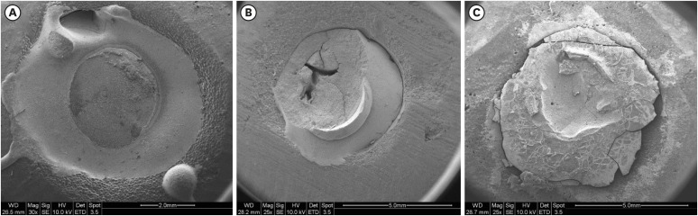
Objectives This study aimed to compare the shear bond strength (SBS) of a self-adhering flowable composite (Dyad Flow) and a bulk-fill flowable composite (Smart Dentin Replacement [SDR]) to several pulp-capping materials, including MTA Plus, Dycal, Biodentine, and TheraCal.
Materials and Methods Eighty acrylic blocks with 2-mm-deep central holes that were 4 mm in diameter were prepared and divided into 2 groups (
n = 40 each) according to the composite used (Dyad Flow or SDR). They were further divided into 4 sub-groups (n = 10 each) according to the pulp-capping agent used. SBS was tested using a universal testing machine at a crosshead speed of 1 mm/min. Data were analyzed using 2-way analysis of variance. Ap value of < 0.05 was considered to indicate statistical significance.Results A statistically significant difference (
p = 0.040) was found between Dyad Flow and SDR in terms of bond strength to MTA Plus, Dycal, Biodentine, and TheraCal.Conclusions Among the 8 sub-groups, the combination of TheraCal and SDR exhibited the highest SBS.
-
Citations
Citations to this article as recorded by- Vital pulp therapy: A three-year clinical case series on regenerative potential and adhesive restoration strategies
Vincenzo Tosco, Riccardo Monterubbianesi, Pietro Montagna, Giulia Orilisi, Flavia Vitiello, Hengyue Song, Angelo Putignano, Daniele De Santis, Giovanna Orsini
Regenesis Repair Rehabilitation.2026; 2(2): 99. CrossRef - Influence of Calcium Silicate‐Based Cement‐Repaired Root Canals on Push‐Out Bond Strength of Glass Fiber Posts: An In Vitro Study
Yasudthama Rattana‐aporn, Weeranuch Thong‐ngarm
Clinical and Experimental Dental Research.2026;[Epub] CrossRef - Shear Bond Strength of Liner Materials to Caries-Free and Caries-Affected Dentin
ZK Greene, NR Smith, T Gomes, NC Lawson
Operative Dentistry.2025; 50(3): 324. CrossRef - Shear Bond Strength of Biointeractive Restorative Materials to NeoMTA Plus and Biodentine
Zübeyde Uçar Gündoğar, Gül Keskin, Merve Yaman Küçükersen
Polymers.2025; 17(22): 3070. CrossRef - The Influence of an Adhesive Protocol on the Adhesion of Biodentine to Resin-Matrix Composites
Orlanda Torres, Nuno Caiado, Ana Sá, Rita Fidalgo-Pereira, Júlio C. M. Souza
Biomedical Materials & Devices.2025;[Epub] CrossRef - Hygroscopic bioactive light-cured composite promoting dentine bridge formation
Yunzi Long, Guibin Huang, Siyi Liu, Liju Xu, Ailing Li, Dong Qiu, Yanmei Dong
Regenerative Biomaterials.2024;[Epub] CrossRef - Comparative evaluation of shear bond strength and modes of failure of five different reinforced glass ionomer restorative cements to TheraCal LC: An in vitro study
Kalyani Gajanan Umale, Vandana Jaykumar Gade, Ambar W. Raut
Journal of Conservative Dentistry and Endodontics.2024; 27(2): 200. CrossRef - Evaluation of the Effect of Chitosan-Based Irrigation Solutions on the Bond Strength of Mineral Trioxide Aggregate to Bulk-Fill Composite
Arzu Şahin Mantı, Bağdagül Helvacıoğlu Kıvanç
Journal of Functional Biomaterials.2024; 15(12): 370. CrossRef - Radiopacity evaluations of the novel calcium-silicate and glass-Ionomer-based materials
Yeşim Şeşen Uslu, Elif Çelebi, Meriç Berkman
Journal of Health Sciences and Medicine.2024; 7(2): 192. CrossRef - Effect of Er Cr YSGG laser etching procedure on the bond strength of different calcium silicate cements
Yesim Sesen Uslu, Hakan Yasin Gönder, Pinar Sesen, Gizem Gunduz Bektaş
Lasers in Dental Science.2024;[Epub] CrossRef - The micro‐shear bond strength of new endodontic tricalcium silicate‐based putty: An in vitro study
Merve Yeniçeri Özata, Seda Falakaloğlu, Gianluca Plotino, Özkan Adıgüzel
Australian Endodontic Journal.2023; 49(1): 124. CrossRef - Analysis of the bond strength between conventional, putty or resin‐modified calcium silicate cement and bulk fill composites
İ Ipek, B Karaağaç Eskibağlar, Ş Yildiz, O Ataş, M Ünal
Australian Dental Journal.2023; 68(4): 265. CrossRef - Effect of Different Adhesive Strategies on the Microshear Bond Strength of Calcium-Silicate-Based Materials
Aliye Tuğçe Gürcan, Soner Şişmanoğlu, Görkem Sengez
Journal of Advanced Oral Research.2022; 13(2): 191. CrossRef - BULK FİLL KOMPOZİT REZİN RESTORATİF MATERYALLER
Merve NEZİR, Suat ÖZCAN
Atatürk Üniversitesi Diş Hekimliği Fakültesi Dergisi.2022; : 1. CrossRef - Effect of Bioinductive Cavity Liners on Shear Bond Strength of Dental Composite to Dentin
Saba Tohidkhah, Elham Ahmadi, Mahdi Abbasi, Reza Morvaridi Farimani, Ladan Ranjbar Omrani, Victor Feitosa
BioMed Research International.2022;[Epub] CrossRef - Bond Strength of Adhesive Systems to Calcium Silicate-Based Materials: A Systematic Review and Meta-Analysis of In Vitro Studies
Louis Hardan, Davide Mancino, Rim Bourgi, Alejandra Alvarado-Orozco, Laura Emma Rodríguez-Vilchis, Abigailt Flores-Ledesma, Carlos Enrique Cuevas-Suárez, Monika Lukomska-Szymanska, Ammar Eid, Maya-Line Danhache, Maryline Minoux, Youssef Haïkel, Naji Kharo
Gels.2022; 8(5): 311. CrossRef - How do imaging protocols affect the assessment of root-end fillings?
Fernanda Ferrari Esteves Torres, Reinhilde Jacobs, Mostafa EzEldeen, Karla de Faria-Vasconcelos, Juliane Maria Guerreiro-Tanomaru, Bernardo Camargo dos Santos, Mário Tanomaru-Filho
Restorative Dentistry & Endodontics.2022;[Epub] CrossRef - Evaluation of Shear Bond Strength of Resin‐Based Composites to Biodentine with Three Types of Seventh‐Generation Bonding Agents: An In Vitro Study
Huda Abbas Abdullah, Zahraa Abdulaali Al-Ibraheemi, Zanbaq Azeez Hanoon, Julfikar Haider, Boonlert Kukiattrakoon
International Journal of Dentistry.2022;[Epub] CrossRef - Evaluation of the Bond Strength of Different Pulp Capping Materials to Dental Adhesive Systems: An In Vitro Study
Sema Yazici Akbiyik, Elif Pınar Bakir, S¸eyhmus Bakir
Journal of Advanced Oral Research.2021; 12(2): 286. CrossRef - Differential Gene Expression Changes in Human Primary Dental Pulp Cells Treated with Biodentine and TheraCal LC Compared to MTA
Ok Hyung Nam, Ho Sun Lee, Jae-Hwan Kim, Yong Kwon Chae, Seoung-Jin Hong, Sang Wook Kang, Hyo-Seol Lee, Sung Chul Choi, Young Kim
Biomedicines.2020; 8(11): 445. CrossRef
- Vital pulp therapy: A three-year clinical case series on regenerative potential and adhesive restoration strategies
- 3,399 View
- 52 Download
- 20 Crossref
- Effect of dental bleaching on the microhardness and surface roughness of sealed composite resins
- Renan Aparecido Fernandes, Henrico Badaoui Strazzi-Sahyon, Thaís Yumi Umeda Suzuki, André Luiz Fraga Briso, Paulo Henrique dos Santos
- Restor Dent Endod 2020;45(1):e12. Published online January 10, 2020
- DOI: https://doi.org/10.5395/rde.2020.45.e12
-
Abstract
PDFPubReaderePub
Objectives The aim of this
in vitro study was to evaluate the microhardness and surface roughness of composite resins before and after tooth bleaching procedures.Materials and Methods Sixty specimens were prepared of each composite resin (Filtek Supreme XT and Opallis), and BisCover LV surface sealant was applied to half of the specimens. Thirty enamel samples were obtained from the buccal and lingual surfaces of human molars for use as the control group. The surface roughness and microhardness were measured before and after bleaching procedures with 35% hydrogen peroxide or 16% carbamide (
n = 10). Data were analyzed using 1-way analysis of variance and the Fisher test (α = 0.05).Results Neither hydrogen peroxide nor carbamide peroxide treatment significantly altered the hardness of the composite resins, regardless of surface sealant application; however, both treatments significantly decreased the hardness of the tooth samples (
p < 0.05). The bleaching did not cause any change in surface roughness, with the exception of the unsealed Opallis composite resin and dental enamel, both of which displayed an increase in surface roughness after bleaching with carbamide peroxide (p < 0.05).Conclusions The microhardness and surface roughness of enamel and Opallis composite resin were influenced by bleaching procedures.
-
Citations
Citations to this article as recorded by- Property changes in resin composite exposed to mouth rinses during 10% carbamide peroxide bleaching
Mariana Ferreira da Silva, Giovana Contin Germinari, Carolina Meneghin Barbosa, Tatiane Cristina Dotta, Renata Siqueira Scatolin, Waldemir Francisco Vieira Júnior, Laura Nobre Ferraz
Brazilian Journal of Oral Sciences.2026; 25: e260366. CrossRef - Effect of Bleaching Protocols on the Microhardness and Surface Roughness of Composite Resins with Different Filler Architectures: An In Vitro Study
Flor de Maria Celis-Jacinto , Fredy Hugo Cruzado-Oliva, Jorge Wilfredo Vera-Alvarado
Odovtos - International Journal of Dental Sciences.2026;[Epub] CrossRef - Effect of Bleaching on Surface Roughness and Color Parameters of Coffee-Stained Nanohybrid Dental Composites with Different Viscosities
Hetaf S. Redwan, Mohamed A. Hussein, Mohamed M. Abdul-Monem
European Journal of General Dentistry.2025; 14(01): 027. CrossRef - Effect of Staining and External Bleaching on the Color Stability and Surface Roughness of Universal-Shade Resin-Based Composite
AlHanouf AlHabdan, Amal Alsuhaibani, Lama Alomran, Lulwah Almutib
Clinical, Cosmetic and Investigational Dentistry.2025; Volume 17: 1. CrossRef - Comparative Analysis Between Strip and Gels Indicated for at Home Bleaching: Analysis of Color Alteration, Roughness and Microhardness of Dental Enamel
K. M. S. Aidar, L. T. A. Cintra, M. C. B. Ferreira, T. C. Fagundes, L. M. B. Esteves, J. Goto, A. Catelan, A. L. F. Briso
Journal of Esthetic and Restorative Dentistry.2025; 37(6): 1504. CrossRef - Surface properties and susceptibility to staining of a resin composite after brushing with different whitening toothpastes
Aline da Silva Barros, Carolina Meneghin Barbosa, Renata Siqueira Scatolin, Waldemir Francisco Vieira Junior, Laura Nobre Ferraz
Restorative Dentistry & Endodontics.2025; 50(1): e6. CrossRef - Degradation Resistance of Next-Generation Dental Composites Under Bleaching and Immersion: A Multiscale Investigation
Syed Zubairuddin Ahmed, Shahad Al-Qahtani, Naif H. Al-Qahtani, Hussah Al-Mulhim, Maha Al-Qahtani, Ali Albalushi, Sultan Akhtar
Prosthesis.2025; 7(3): 57. CrossRef - Effect of Over-the-Counter Whitening Dentifrices on the Color Stability and Microhardness of Composite Resins
Xinnuo Yu, Maria Pilar Melo, Sofia Folguera, Carmen Llena
Journal of Composites Science.2025; 9(7): 324. CrossRef - From Microstructure to Shade Shift: Confocal and Spectrophotometric Evaluation of Peroxide-Induced Dental Bleaching
Berivan Laura Rebeca Buzatu, Magda Mihaela Luca, Atena Galuscan, Adrian Ovidiu Vaduva, Aurora Doris Fratila, Ramona Dumitrescu, Ruxandra Sava-Rosianu, Octavia Balean, Roxana Buzatu, Daniela Jumanca
Journal of Clinical Medicine.2025; 14(13): 4642. CrossRef - In Vitro Evaluation of Chemical and Microhardness Alterations in Human Enamel Induced by Three Commercial In-Office Bleaching Agents
Berivan Laura Rebeca Buzatu, Atena Galuscan, Ramona Dumitrescu, Roxana Buzatu, Magda Mihaela Luca, Octavia Balean, Gabriela Vlase, Titus Vlase, Iasmina-Mădălina Anghel, Carmen Opris, Bianca Ioana Todor, Mihaela Adina Dumitrache, Daniela Jumanca
Dentistry Journal.2025; 13(8): 357. CrossRef - Effect of Hydrogen Peroxide Bleaching on Color Stability and Microhardness of Alkasite Restorative Materials: An In Vitro Study
Souad A Alfouzan, Rahaf A Alolayan, Asma Munir Khan
Cureus.2025;[Epub] CrossRef - Evaluation of Color Stability and Surface Roughness of Nanohybrid Resin Composites with Different Photoinitiator Systems After Staining and Home/Office Bleaching: An In Vitro Study
Fatma Yılmaz, Buse Kesgin
Meandros Medical And Dental Journal.2025; 26(3): 240. CrossRef - The Effect of Hydrogen Peroxide With Different Concentration on the Color and Surface Microhardness of the Resin Bracket
Song‐Yi Yang
Clinical and Experimental Dental Research.2025;[Epub] CrossRef - Comparative evaluation of different bleaching agents on the color stability, hardness and surface roughness of indirect esthetic restorative materials with different manufacturing methods
Ayse Atay, Defne Canpolat, Soner Sismanoglu, Aslihan Usumez
BMC Oral Health.2025;[Epub] CrossRef - Comparison of Microhardness and Surface Roughness of New Nanofiber Filled Flowable Composite
Rumeysa Hatice ENGINLER OZLEN, Zumrut Ceren OZDUMAN, Burcu OGLAKCI OZKOC, Evrim ELIGUZELOGLU DALKILIC
Bezmialem Science.2024; 12(4): 406. CrossRef - Effect of Bleaching Agents on Composite Resins with and without Bis-GMA: An In Vitro Study
María Melo, Bianca Dumitrache, James Ghilotti, José Luis Sanz, Carmen Llena
Journal of Functional Biomaterials.2024; 15(6): 144. CrossRef - Changes in physical properties of universal composites and CAD/CAM materials after bleaching and antioxidant applications: Scanning electron microscope and atomic force microscope evaluation
Oguz Kaan Tuysuz, Merve Gurses
Microscopy Research and Technique.2024; 87(5): 977. CrossRef - The Effects of Home and Over-The-Counter Whitening Agents on Surface Roughness and Microhardness of High Aesthetic Composites
Elif İpek KILIÇ DÖNMEZ, İhsan HUBBEZOĞLU
Cumhuriyet Dental Journal.2024; 27(1): 30. CrossRef - Effect of carbamide peroxide treatment on the ion release of different dental restorative materials
Merve Nur Yilmaz, Pinar Gul
BMC Oral Health.2024;[Epub] CrossRef - Inorganic Phosphate Effect in a Hydrogen Peroxide-based Bleaching Agent: Physicochemical, Mechanical, and Morphological Properties of Dental Enamel
KG Garcia, GP Nunes, ACB Delbem, PH dos Santos, GLP Fernandes, HF Robles, PBB Lemos, M Danelon
Operative Dentistry.2024; 49(4): 465. CrossRef - Effect of bleaching and repolishing on whiteness change and staining susceptibility of resin-based materials
Sultan Aktuğ Karademir, Samet Atasoy, Beyza Yılmaz
BMC Oral Health.2024;[Epub] CrossRef - Influence of Low pH on the Microhardness and Roughness Surface of Dental Composite—A Preliminary Study
Leszek Szalewski, Dorota Wójcik, Monika Sowa, Vladyslav Vivcharenko, Krzysztof Pałka
Materials.2024; 17(14): 3443. CrossRef - In Vitro Evaluation of the Effectiveness and pH Variation of Dental Bleaching Gels and Their Effect on Enamel Surface Roughness
Federica Veneri, Francesco Cavani, Giovanni Bolelli, Vittorio Checchi, Alessia Bizzi, Giacomo Setti, Luigi Generali
Dentistry Journal.2024; 12(12): 415. CrossRef - Does the combination of whitening toothpaste and hydrogen peroxide bleaching increase the surface roughness and change the morphology of a nanofilled composite?
Cecília Pereira da Silva Braga Tenório, Matheus Kury, Geyse Maria dos Santos Muniz Mota, Cecília Pedroso Turssi, Flávia Lucisano Botelho do Amaral, Vanessa Cavalli
Brazilian Journal of Oral Sciences.2024; 23: e241938. CrossRef - Effect of peroxide‐free and peroxide‐based in‐office bleaching on the surface and mechanical properties of CAD/CAM esthetic restorative materials
Majed M. Alsarani, Aftab Ahmed Khan, Leonel S. J. Bautista, Hanan Alsunbul, Jukka P. Matinlinna
European Journal of Oral Sciences.2024;[Epub] CrossRef - Effect of Repolishing on Color Stability, Translucency, and Surface Roughness of Aged Monochromatic Dental Composites
Mohamed M. Abdul-Monem, Mohamed A. Hussein, Mona G. Abdelrehim
European Journal of General Dentistry.2024; 13(03): 240. CrossRef - Color changes of nanofiller composite resin after glycerin application immersed in turmeric extract
Sukaton, Galih Sampoerno, Widyajeng Ayu Laksmi, Daradhasih Bestari Santiaji
Conservative Dentistry Journal.2023; 13(1): 37. CrossRef - Effects of Dental Bleaching Agents on the Surface Roughness of Dental Restoration Materials
Alexandru Dan Popescu, Mihaela Jana Tuculina, Oana Andreea Diaconu, Lelia Mihaela Gheorghiță, Claudiu Nicolicescu, Cristian Niky Cumpătă, Cristiana Petcu, Jaqueline Abdul-Razzak, Ana Maria Rîcă, Ruxandra Voinea-Georgescu
Medicina.2023; 59(6): 1067. CrossRef - Effect of Bleaching on the Microhardness and Modulus of Elasticity of ACTIVA BioACTIVE – RESTORATIVE: An In Vitro Study
Sushritha Sricharan, Swaroop Hegde, Narmada J., Indiresha H. Narayana, Chatura Mohan, Nithin K. Shetty
Journal of Advanced Oral Research.2023; 14(2): 190. CrossRef - The effect of bleaching on surface roughness and gloss of different CAD/CAM ceramic and hybrid ceramic materials
Ruwaida Z Alshali, Mohammed A AlQahtani, Dalea M Bukhary, Mlak A Alzahrani, Shatha S Alsoraihi, Majed A Alqahtani
Journal of Applied Biomaterials & Functional Materials.2023;[Epub] CrossRef - Effect of bleaching with 15% carbamide peroxide on color stability of microhybrid, nanohybrid, and nanofilled resin composites, each in 3 staining solutions (coffee, cola, red grape juice): A 3-phase study
Azadeh Ghaemi, Sanaz Sharifishoshtari, Mohsen Shahmoradi, Hossein Akbari, Parisa Boostanifard, Sepideh Bagheri, Mohammadreza Shokuhifar, Negin Ashoori, Vahid Rakhshan
Dental Research Journal.2023;[Epub] CrossRef - Micro-Hardness and Surface Roughness of Bulk-Fill Composite Resin: Effect of Surface Sealant Application and Two Bleaching Regimens
Reham Mohamad Attia, Eman Mohamed Sobhy, Mona El Said Abd El Hameed Essa
European Journal of General Dentistry.2023; 12(03): 169. CrossRef - Shear bond strength after using sealant before bonding: a systematic review and meta-analysis of in vitro studies
Jennifer Hoppe, Thomas Lehmann, Christoph-Ludwig Hennig, Ulrike Schulze-Späte, Collin Jacobs
Clinical Oral Investigations.2022; 26(1): 1. CrossRef - Effect of 16% Carbamide Peroxide and Activated-Charcoal-Based Whitening Toothpaste on Enamel Surface Roughness in Bovine Teeth: An In Vitro Study
Jorge Zamudio-Santiago, Marysela Ladera-Castañeda, Flor Santander-Rengifo, Carlos López-Gurreonero, Alberto Cornejo-Pinto, Ali Echavarría-Gálvez, Luis Cervantes-Ganoza, César Cayo-Rojas
Biomedicines.2022; 11(1): 22. CrossRef - Direct dentin bleaching: Would it be possible?
Camila Ferro Clemente, Sibele de Alcântara, Lívia Maria Alves Valentim da Silva, Lara Maria Bueno Esteves, Anderson Catelan, Karen Milaré Seiscento Aidar, Ticiane Cestari Fagundes, André Luiz Fraga Briso
Photodiagnosis and Photodynamic Therapy.2022; 40: 103121. CrossRef - EFFECT OF İN-OFFİCE BLEACHİNG ON THE SURFACE ROUGHNESS OF DİFFERENT COMPOSİTE RESİNS
Seher KAYA, Ozden OZEL BEKTAS
Cumhuriyet Dental Journal.2022; 25(Supplement): 78. CrossRef - Effect of Polishing on the Surface Microhardness of Nanohybrid Composite Resins Subjected to 35% Hydrogen Peroxide
Giovanna Gisella Ramírez-Vargas, Julia Elbia Medina y Mendoza, Ana Sixtina Aliaga-Mariñas, Marysela Irene Ladera-Castañeda, Luis Adolfo Cervantes-Ganoza, César Félix Cayo-Rojas
Journal of International Society of Preventive and Community Dentistry.2021; 11(2): 216. CrossRef - Intrapulpal Concentration of Hydrogen Peroxide of Teeth Restored With Bulk Fill and Conventional Bioactive Composites
DP Silva, BA Resende, M Kury, CB André, CPM Tabchoury, M Giannini, V Cavalli
Operative Dentistry.2021; 46(3): E158. CrossRef - An Environmental Scanning Electron Microscopy Evaluation on Comparison of Three Different Bleaching Agents using the Laser Activated in-Office Bleaching at Different Wavelengths
Shachi Goenka, Sushil Kumar Cirigiri, Kanika Poplai, Baig Mirza Aslam, Shalini Singh, Shweta Gangavane
Journal of Pharmacy and Bioallied Sciences.2021; 13(Suppl 2): S1478. CrossRef - Effects of Artificial Staining and Bleaching Protocols on the Surface Roughness, Color, and Whiteness Changes of an Aged Nanofilled Composite
Geyse Maria dos Santos Muniz Mota, Matheus Kury, Cecília Pereira da Silva Braga Tenório, Flávia Lucisano Botelho do Amaral, Cecília Pedroso Turssi, Vanessa Cavalli
Frontiers in Dental Medicine.2020;[Epub] CrossRef
- Property changes in resin composite exposed to mouth rinses during 10% carbamide peroxide bleaching
- 4,443 View
- 48 Download
- 40 Crossref
Case Reports
- Fiber-reinforced composite resin bridges: an alternative method to treat root-fractured teeth
- Gun Heo, Eun-Hye Lee, Jin-Woo Kim, Kyung-Mo Cho, Se-Hee Park
- Restor Dent Endod 2020;45(1):e8. Published online December 27, 2019
- DOI: https://doi.org/10.5395/rde.2020.45.e8
-
Abstract
PDFPubReaderePub
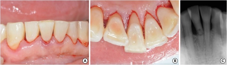
The replacement of missing teeth, especially in the anterior region, is an essential part of dental practice. Fiber-reinforced composite resin bridges are a conservative alternative to conventional fixed dental prostheses or implants. It is a minimally invasive, reversible technique that can be completed in a single visit. The two cases presented herein exemplify the treatment of root-fractured anterior teeth with a natural pontic immediately after extraction.
-
Citations
Citations to this article as recorded by- Prosthodontic Aspects of Splinting the Mandibular Anterior Teeth by Fiber Reinforced Composites
Hrelja Miroslav, Laškarin Mirko, Čimić Samir, Kraljević Sonja, Dulčić Nikša, Badel Tomislav
Journal of Dental Problems and Solutions.2025; 12(1): 004. CrossRef - Current Evidence on the Fiber-reinforced Composite Bridges
Ramesh Chowdhary, Sunil Kumar Mishra
International Journal of Prosthodontics and Restorative Dentistry.2023; 12(4): 159. CrossRef - Bridging the Gap: A Case Report of Tooth Replacement using Resin-Bonded Fiber- Reinforced Composite Resin
Vineet Sharma, Sumit Bhansali, Sonal Priya Bhansali
Journal of Pierre Fauchard Academy (India Section).2023; : 66. CrossRef - Reconstruction of Natural Smile and Splinting with Natural Tooth Pontic Fiber‐Reinforced Composite Bridge
Maryam S. Tavangar, Fatemeh Aghaei, Massoumeh Nowrouzi, Andrea Scribante
Case Reports in Dentistry.2022;[Epub] CrossRef
- Prosthodontic Aspects of Splinting the Mandibular Anterior Teeth by Fiber Reinforced Composites
- 3,027 View
- 26 Download
- 4 Crossref
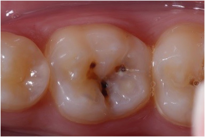
- Functional and aesthetic rehabilitation in posterior tooth with bulk-fill resin composite and occlusal matrix
- Luciana Fávaro Francisconi-dos-Rios, Johnny Alexandre Oliveira Tavares, Luanderson Oliveira, Jefferson Chaves Moreira, Flavia Pardo Salata Nahsan
- Restor Dent Endod 2020;45(1):e9. Published online January 3, 2020
- DOI: https://doi.org/10.5395/rde.2020.45.e9
-
Abstract
PDFPubReaderePub
The restorative procedure in posterior teeth involves clinical steps related to professional skill, especially when using the incremental technique, which may fail in the long term. A recent alternative is bulk-fill resins, which can reduce polymerization shrinkage, decreasing clinical problems such as marginal leakage, secondary caries, and fracture. This scientific study aims to report a clinical case using bulk-fill resin with an occlusal matrix. As determined in the treatment plan, an acrylic resin matrix was produced to establish an improved oral and aesthetic rehabilitation of the right mandibular first molar, which presented a carious lesion with dentin involvement. The occlusal matrix is a simple technique that maintains the original dental anatomy, showing satisfactory results regarding function and aesthetic rehabilitation.
-
Citations
Citations to this article as recorded by- Mastery of Aesthetic and Functional Restoration of Maxillary Molars Using the Technique of Direct Restoration (Clinical Case)
Yu. Kolenko
SUCHASNA STOMATOLOHIYA.2025; (2): 67. CrossRef - Color stability of bulk‐fill compared to conventional resin‐based composites: A scoping review
Gaetano Paolone, Mauro Mandurino, Nicola Scotti, Giuseppe Cantatore, Markus B. Blatz
Journal of Esthetic and Restorative Dentistry.2023; 35(4): 657. CrossRef - Evaluation of Abfraction Lesions Restored with Three Dental Materials: A Comparative Study
Bogdan Constantin Costăchel, Anamaria Bechir, Alexandru Burcea, Laurența Lelia Mihai, Tudor Ionescu, Olivia Andreea Marcu, Edwin Sever Bechir
Clinics and Practice.2023; 13(5): 1043. CrossRef - Aesthetic restoration of posterior teeth using different occlusal matrix techniques
Elsa Reis Carneiro, Anabela Paula, José Saraiva, Ana Coelho, Inês Amaro, Carlos Miguel Marto, Manuel Marques Ferreira, Eunice Carrilho
British Dental Journal.2021; 231(2): 88. CrossRef - The Effects of Mouthwashes on the Color Stability of Resin-Based Restorative Materials
Ayşe Tuğba Ertürk-Avunduk, Seçkin Aksu, Ebru Delikan
Odovtos - International Journal of Dental Sciences.2020; 23(1): 91. CrossRef
- Mastery of Aesthetic and Functional Restoration of Maxillary Molars Using the Technique of Direct Restoration (Clinical Case)
- 2,443 View
- 28 Download
- 5 Crossref
 First
First Prev
Prev

