Articles
- Page Path
- HOME > Restor Dent Endod > Volume 45(1); 2020 > Article
- Research Article The prevalence of radix molaris in the mandibular first molars of a Saudi subpopulation based on cone-beam computed tomography
-
Hassan AL-Alawi1
 , Saad Al-Nazhan2
 , Nassr Al-Maflehi3
 , Mazen A. Aldosimani4
 , Mohammed Nabil Zahid5
 , Ghadeer N. Shihabi6
-
Restor Dent Endod 2019;45(1):e1.
DOI: https://doi.org/10.5395/rde.2020.45.e1
Published online: November 14, 2019
1Dental Department, Ministry of Health Endodontist, Huraymala General Hospital, Riyadh, Saudi Arabia.
2Department of Restorative Dentistry-Endodontics, College of Dentistry, Riyadh Elm University, Riyadh, Saudi Arabia.
3Department of Preventive Dental Sciences-Biostatistics, College of Dentistry, King Saud University, Riyadh, Saudi Arabia.
4Department of Oral Medicine and Diagnostic Sciences, College of Dentistry, King Saud University, Riyadh, Saudi Arabia.
5Department of Preventive Dental Sciences, College of Dentistry, Prince Sattam Bin AbdulAziz University, Al Kharj, Saudi Arabia.
6General Practitioner, Riyadh, Saudi Arabia.
- Correspondence to Saad Al-Nazhan, BDS, MSD. Professor, Department of Restorative Dentistry-Endodontics, College of Dentistry, Riyadh Elm University, Riyadh 11681, Saudi Arabia. saad.alnazhan@riyadh.edu.sa
Copyright © 2020. The Korean Academy of Conservative Dentistry
This is an Open Access article distributed under the terms of the Creative Commons Attribution Non-Commercial License (https://creativecommons.org/licenses/by-nc/4.0/) which permits unrestricted non-commercial use, distribution, and reproduction in any medium, provided the original work is properly cited.
- 2,319 Views
- 36 Download
- 12 Crossref
Abstract
-
Objectives The purpose of this study was to determine the incidence of radix molaris (RM) (entomolaris and paramolaris) in the mandibular first permanent molars of a sample Saudi Arabian subpopulation using cone-beam computed tomography (CBCT).
-
Materials and Methods A total of 884 CBCT images of 427 male and 457 female Saudi citizens (age 16 to 70 years) were collected from the radiology department archives of 4 dental centers. A total of 450 CBCT images of 741 mature mandibular first molars that met the inclusion criteria were reviewed. The images were viewed at high resolution by 3 examiners and were analyzed with Planmeca Romexis software (version 5.2).
-
Results Thirty-three (4.5%) mandibular first permanent molars had RM, mostly on the distal side. The incidence of radix entomolaris (EM) was 4.3%, while that of radix paramolaris was 0.3%. The RM roots had one canal and occurred more unilaterally. No significant difference in root configuration was found between males and females (p > 0.05). Types I and III EM root canal configurations were most common, while type B was the only RP configuration observed.
-
Conclusions The incidence of RM in the mandibular first molars of this Saudi subpopulation was 4.5%. Identification of the supernumerary root can avoid missing the canal associated with the root during root canal treatment.
INTRODUCTION
Prevalence of supernumerary root in mandibular first molar
| Author/reference | Origin | Incidence (%) | Evaluation method |
|---|---|---|---|
| Curzon and Curzon [4] | Mongoloid Keewatin Eskimo (Canada) | 27 | In vitro (extracted teeth) |
| Reichart and Metah [5] | Thai (Thailand) | 19.2 | In vitro (extracted teeth) |
| Walker [6] | Chinese (Hong Kong) | 15 | In vitro (extracted teeth) |
| Younes et al. [7] | African (Egypt) | 0.7 | In vitro (extracted teeth) |
| Asian (Saudi Arabia) | 2.3 | ||
| Zaatar et al. [8] | Kuwait | 2.7 | In vivo (periapical radiographs) |
| Sperber and Moreau [9] | Senegal | 3.1 | In vitro (extracted teeth) |
| Al-Nazhan [10] | Saudi Arabia | 5.97 | In vivo (periapical radiographs) |
| Ahmed et al. [11] | Sudan | 3 | In vitro (extracted teeth) |
| Schäfer et al. [12] | Germany | 0.7 | In vivo (periapical radiographs) |
| Al-Qudah and Awawdeh [13] | Jordan | 3.9 | In vitro (extracted teeth) |
| Song et al. [14] | Korea (Mongoloid origin) | 33.1 | In vivo (periapical radiographs) |
| Zhang et al. [15] | China | 29 | In vivo (CBCT) |
| Demirbuga et al. [16] | Turkey | 2.06 | In vivo (CBCT) |
| Mukhaimer and Azizi [17] | Palestine | 3.73 | In vivo (periapical radiographs) |
| Rodrigues et al. [18] | Brazil | 2.58 | In vivo (CBCT) |
| Rahimi et al. [19] | Iran | 3.00 | In vivo (CBCT) |
| Gupta et al. [20] | Haryana (North India) | 13.00 | In vivo (periapical radiographs and CBCT) |
MATERIALS AND METHODS
Total number of evaluated cone-beam computed tomography images
RESULTS
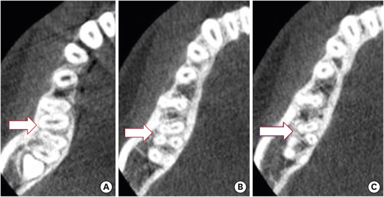
(A) Cone-beam computed tomography (CBCT) images of mandibular first molar showing 4 canals (arrow). (B) CBCT showing disto-buccal root with one canal (arrow). (C) Separated distal roots with one canal each (arrow).

Number of roots of mandibular first molar in relation to sex and jaw side
Number and percentages of patients with entomolaris (distolingual root) and paramolaris (mesiobuccal root) in mandibular first molars according to sex and jaw side (n = 741)
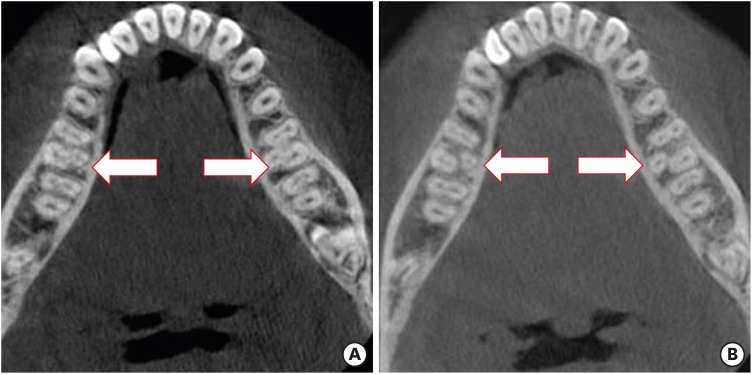
(A) Cone-beam computed tomography (CBCT) images of mandibular first molar showing disto-lingual root bilateral entomolaris (arrow). (B) Bilateral separated roots with one canal (arrow).

Morphology of the distolingual root (entomolaris) based on Song et al. [30] classification. (n = 741)
| Sex | Jaw side | Total No. of patients | Total No. of teeth | Song et al. [30] classification | ||||
|---|---|---|---|---|---|---|---|---|
| Type I | Type II | Type III | Small type | Conical type | ||||
| Male | Bilateral | 2 | 4 | 0 | 2 | 2 | 0 | 0 |
| Unilateral | 8 | 8* | 2 | 2 | 3 | 1 | 0 | |
| Total | 10 | 12 | 2 | 4 | 5 | 1 | 0 | |
| Female | Bilateral | 3 | 6 | 2 | 2 | 2 | 0 | 0 |
| Unilateral | 13 | 13 | 7 | 2 | 3 | 1 | 0 | |
| Total | 16 | 19 | 9 | 4 | 5 | 1 | 0 | |
| Grand total | 26* | 31 (4.2%) | 11 (1.5%) | 8 (1.1%) | 10 (1.3%) | 2 (0.3%) | 0 | |
DISCUSSION
CONCLUSIONS
-
Conflict of Interest: No potential conflict of interest relevant to this article was reported.
-
Author Contributions:
Conceptualization: AL-Alawi H, Aldosimani MA, Zahid MN.
Data curation: AL-Alawi H, Shihabi GN.
Formal analysis: Al-Nazhan S, AL-Alawi H.
Investigation: AL-Alawi H, Al-Nazhan S, Al-Maflehi N, Aldosimani MA, Zahid MN, Shihabi GN.
Methodology: Al-Nazhan S.
Project administration: AL-Alawi H.
Resources: AL-Alawi H, Aldosimani MA, Zahid MN.
Software: Al-Maflehi N.
Supervision: Al-Nazhan S, AL-Alawi H.
Validation: Al-Nazhan S, AL-Alawi H, Al-Maflehi N.
Visualization: Al-Nazhan S, AL-Alawi H, Aldosimani MA, Zahid MN, Shihabi GN.
Writing - original draft: Al-Nazhan S.
Writing - review & editing: Al-Nazhan S.
- 1. Segura-Egea JJ, Jiménez-Pinzón A, Ríos-Santos JV. Endodontic therapy in a 3-rooted mandibular first molar: importance of a thorough radiographic examination. J Can Dent Assoc 2002;68:541-544.PubMed
- 2. Slowey RR. Radiographic aids in the detection of extra root canals. Oral Surg Oral Med Oral Pathol 1974;37:762-772.ArticlePubMed
- 3. Vertucci FJ. Root canal anatomy of the human permanent teeth. Oral Surg Oral Med Oral Pathol 1984;58:589-599.ArticlePubMed
- 4. Curzon ME, Curzon JA. Three-rooted mandibular molars in the Keewatin Eskimo. J Can Dent Assoc (Tor) 1971;37:71-72.PubMed
- 5. Reichart PA, Metah D. Three-rooted permanent mandibular first molars in the Thai. Community Dent Oral Epidemiol 1981;9:191-192.ArticlePubMed
- 6. Walker RT. Root form and canal anatomy of mandibular first molars in a southern Chinese population. Endod Dent Traumatol 1988;4:19-22.ArticlePubMed
- 7. Younes SA, al-Shammery AR, el-Angbawi MF. Three-rooted permanent mandibular first molars of Asian and black groups in the Middle East. Oral Surg Oral Med Oral Pathol 1990;69:102-105.ArticlePubMed
- 8. Zaatar EI, al-Kandari AM, Alhomaidah S, al-Yasin IM. Frequency of endodontic treatment in Kuwait: radiographic evaluation of 846 endodontically treated teeth. J Endod 1997;23:453-456.ArticlePubMed
- 9. Sperber GH, Moreau JL. Study of the number of roots and canals in Senegalese first permanent mandibular molars. Int Endod J 1998;31:117-122.ArticlePubMed
- 10. al-Nazhan S. Incidence of four canals in root-canal-treated mandibular first molars in a Saudi Arabian sub-population. Int Endod J 1999;32:49-52.ArticlePubMed
- 11. Ahmed HA, Abu-bakr NH, Yahia NA, Ibrahim YE. Root and canal morphology of permanent mandibular molars in a Sudanese population. Int Endod J 2007;40:766-771.ArticlePubMed
- 12. Schäfer E, Breuer D, Janzen S. The prevalence of three-rooted mandibular permanent first molars in a German population. J Endod 2009;35:202-205.ArticlePubMed
- 13. Al-Qudah AA, Awawdeh LA. Root and canal morphology of mandibular first and second molar teeth in a Jordanian population. Int Endod J 2009;42:775-784.ArticlePubMed
- 14. Song JS, Kim SO, Choi BJ, Choi HJ, Son HK, Lee JH. Incidence and relationship of an additional root in the mandibular first permanent molar and primary molars. Oral Surg Oral Med Oral Pathol Oral Radiol Endod 2009;107:e56-e60.ArticlePubMed
- 15. Zhang R, Wang H, Tian YY, Yu X, Hu T, Dummer PM. Use of cone-beam computed tomography to evaluate root and canal morphology of mandibular molars in Chinese individuals. Int Endod J 2011;44:990-999.ArticlePubMed
- 16. Demirbuga S, Sekerci AE, Dinçer AN, Cayabatmaz M, Zorba YO. Use of cone-beam computed tomography to evaluate root and canal morphology of mandibular first and second molars in Turkish individuals. Med Oral Patol Oral Cir Bucal 2013;18:e737-e744.ArticlePubMedPMC
- 17. Mukhaimer R, Azizi Z. Incidence of radix entomolaris in mandibular first molars in Palestinian population: a clinical investigation. Int Sch Res Notices 2014;2014:405601.ArticlePubMedPMCPDF
- 18. Rodrigues CT, Oliveira-Santos C, Bernardineli N, Duarte MA, Bramante CM, Minotti-Bonfante PG, Ordinola-Zapata R. Prevalence and morphometric analysis of three-rooted mandibular first molars in a Brazilian subpopulation. J Appl Oral Sci 2016;24:535-542.ArticlePubMedPMC
- 19. Rahimi S, Mokhtari H, Ranjkesh B, Johari M, Frough Reyhani M, Shahi S, Seif Reyhani S. Prevalence of extra roots in permanent mandibular first molars in Iranian population: a CBCT analysis. Iran Endod J 2017;12:70-73.PubMedPMC
- 20. Gupta A, Duhan J, Wadhwa J. Prevalence of three rooted permanent mandibular first molars in Haryana (North Indian) population. Contemp Clin Dent 2017;8:38-41.ArticlePubMedPMC
- 21. Walker RT, Quackenbush LE. Three-rooted lower first permanent molars in Hong Kong Chinese. Br Dent J 1985;159:298-299.ArticlePubMedPDF
- 22. Gulabivala K, Opasanon A, Ng YL, Alavi A. Root and canal morphology of Thai mandibular molars. Int Endod J 2002;35:56-62.ArticlePubMed
- 23. Sert S, Aslanalp V, Tanalp J. Investigation of the root canal configurations of mandibular permanent teeth in the Turkish population. Int Endod J 2004;37:494-499.ArticlePubMed
- 24. Bahammam LA, Bahammam HA. The incidence of radix entomolaris in mandibular first permanent molars in a Saudi Arabian sub-population. JKAU Med Sci 2011;18:83-90.Article
- 25. Wu DM, Wu YN, Guo W, Sameer S. Accuracy of direct digital radiography in the study of the root canal type. Dentomaxillofac Radiol 2006;35:263-265.ArticlePubMed
- 26. Omer OE, Al Shalabi RM, Jennings M, Glennon J, Claffey NM. A comparison between clearing and radiographic techniques in the study of the root-canal anatomy of maxillary first and second molars. Int Endod J 2004;37:291-296.ArticlePubMed
- 27. Matherne RP, Angelopoulos C, Kulild JC, Tira D. Use of cone-beam computed tomography to identify root canal systems in vitro . J Endod 2008;34:87-89.ArticlePubMed
- 28. Al-Shehri S, Al-Nazhan S, Shoukry S, Al-Shwaimi E, Al-Sadhan R, Al-Shemmery B. Root and canal configuration of the maxillary first molar in a Saudi subpopulation: a cone-beam computed tomography study. Saudi Endod J 2017;2:69-76.
- 29. Carlsen O, Alexandersen V. Radix paramolaris in permanent mandibular molars: identification and morphology. Scand J Dent Res 1991;99:189-195.ArticlePubMed
- 30. Song JS, Choi HJ, Jung IY, Jung HS, Kim SO. The prevalence and morphologic classification of distolingual roots in the mandibular molars in a Korean population. J Endod 2010;36:653-657.ArticlePubMed
- 31. Wang Y, Zheng QH, Zhou XD, Tang L, Wang Q, Zheng GN, Huang DM. Evaluation of the root and canal morphology of mandibular first permanent molars in a western Chinese population by cone-beam computed tomography. J Endod 2010;36:1786-1789.ArticlePubMed
- 32. Quackenbush LE. Mandibular molar with three distal root canals. Endod Dent Traumatol 1986;2:48-49.ArticlePubMed
- 33. Loh HS. Incidence and features of three-rooted permanent mandibular molars. Aust Dent J 1990;35:434-437.ArticlePubMed
- 34. Salarpour M, Farhad Mollashahi N, Mousavi E, Salarpour E. Evaluation of the effect of tooth type and canal configuration on crown size in mandibular premolars by cone-beam computed tomography. Iran Endod J 2013;8:153-156.PubMedPMC
- 35. Patel S, Dawood A, Ford TP, Whaites E. The potential applications of cone beam computed tomography in the management of endodontic problems. Int Endod J 2007;40:818-830.ArticlePubMed
- 36. Cotton TP, Geisler TM, Holden DT, Schwartz SA, Schindler WG. Endodontic applications of cone-beam volumetric tomography. J Endod 2007;33:1121-1132.ArticlePubMed
- 37. Neelakantan P, Subbarao C, Subbarao CV. Comparative evaluation of modified canal staining and clearing technique, cone-beam computed tomography, peripheral quantitative computed tomography, spiral computed tomography, and plain and contrast medium-enhanced digital radiography in studying root canal morphology. J Endod 2010;36:1547-1551.ArticlePubMed
- 38. Gu Y, Lu Q, Wang H, Ding Y, Wang P, Ni L. Root canal morphology of permanent three-rooted mandibular first molars--part I: pulp floor and root canal system. J Endod 2010;36:990-994.ArticlePubMed
- 39. Fabra-Campos H. Unusual root anatomy of mandibular first molars. J Endod 1985;11:568-572.ArticlePubMed
- 40. Wasti F, Shearer AC, Wilson NH. Root canal systems of the mandibular and maxillary first permanent molar teeth of south Asian Pakistanis. Int Endod J 2001;34:263-266.ArticlePubMedPDF
- 41. Rwenyonyi CM, Kutesa A, Muwazi LM, Buwembo W. Root and canal morphology of mandibular first and second permanent molar teeth in a Ugandan population. Odontology 2009;97:92-96.ArticlePubMedPDF
- 42. Scott GR, Turner CG. The anthropology of modern human teeth: dental morphology and its variation in recent human populations. Cambridge, NY: Cambridge University Press; 1997. p. 74-130.
- 43. De Moor RJ, Deroose CA, Calberson FL. The radix entomolaris in mandibular first molars: an endodontic challenge. Int Endod J 2004;37:789-799.ArticlePubMed
- 44. Tinelli ME. Ethnic variations in the topography of the root canals. Electronic J Endod Rosario 2011;2:558-562.
- 45. Kim KR, Song JS, Kim SO, Kim SH, Park W, Son HK. Morphological changes in the crown of mandibular molars with an additional distolingual root. Arch Oral Biol 2013;58:248-253.ArticlePubMed
- 46. Kim HH, Jo HH, Min JB, Hwang HK. CBCT study of mandibular first molars with a distolingual root in Koreans. Restor Dent Endod 2018;43:e33.ArticlePubMedPMCPDF
REFERENCES
Tables & Figures
REFERENCES
Citations

- Evaluation of the variations of mandibular molars and the distance from root apex to the inferior alveolar nerve in Saudi Sub-population: Three-dimensional radiographic evaluation
Tariq Mohammed Aqili, Esam Sami Almuzaini, Abdulbari Saleh Aljohani, Ahmed Khaled Al Saeedi, Hassan Abdulmuti Hammudah, Muath Alassaf, Muhannad M. Hakeem, Mohmed Isaqali Karobari
PLOS ONE.2025; 20(2): e0317053. CrossRef - Prevalence of radix molaris in mandibular molars of a subpopulation of Brazil’s Northeast region: a cross-sectional CBCT study
Yasmym Martins Araújo de Oliveira, Maria Clara Mendes Gomes, Maria Fernanda da Silva Nascimento, Ricardo Machado, Danna Mota Moreira, Hermano Camelo Paiva, George Táccio de Miranda Candeiro
Scientific Reports.2025;[Epub] CrossRef - Prevalence of radix entomolaris and distolingual canals and their association with the incidence of middle mesial canals in mandibular first molars of a Saudi subpopulation
Ahmed A. Madfa, Abdullah F. Alshammari, Eyad Almagadawyi, Afaf Al-Haddad, Ebtsam A. Aledaili
Scientific Reports.2025;[Epub] CrossRef - Assessment of the root and canal morphology in the permanent dentition of Saudi Arabian population using cone beam computed and micro-computed tomography – a systematic review
Mohammed Mustafa, Rumesa Batul, Mohmed Isaqali Karobari, Hadi Mohammed Alamri, Abdulaziz Abdulwahed, Ahmed A. Almokhatieb, Qamar Hashem, Abdullah Alsakaker, Mohammad Khursheed Alam, Hany Mohamed Aly Ahmed
BMC Oral Health.2024;[Epub] CrossRef - Prevalence of radix accesoria dentis in a northern Peruvian population evaluated by cone-beam tomography
Karla Renata León-Almanza, Anthony Adrián Jaramillo-Nuñez, Catherin Angélica Ruiz-Cisneros, Paul Martín Herrera-Plasencia
Heliyon.2024; 10(16): e35919. CrossRef - Radix molaris is a hidden truth of mandibular first permanent molars: A descriptive- analytic study using cone beam computed tomography
Mohammed A. Alobaid, Saurabh Chaturvedi, Ebtihal Mobarak S. Alshahrani, Ebtsam M. Alshehri, Amal S. Shaiban, Mohamed Khaled Addas, Giuseppe Minervini
Technology and Health Care.2023; 31(5): 1957. CrossRef - Prevalence of Radix Entomolaris in Mandibular Permanent Molars Analyzed by Cone-Beam CT in the Saudi Population of Ha'il Province
Moazzy I Almansour, Ahmed A Madfa, Adhwaa F Algharbi, Reem Almuslumani, Noeer K Alshammari, Ghufran M Al Hussain
Cureus.2023;[Epub] CrossRef - Prevalence of radix entomolaris in India and its comparison with the rest of the world
Sumit MOHAN, Jyoti THAKUR
Minerva Dental and Oral Science.2022;[Epub] CrossRef - Radix Paramolaris an Endodontic Challenge: A Case Report
Ashwini B Prasad, Deepak Raisingani, Ridhima Gupta, Rimjhim Jain
Journal of Mahatma Gandhi University of Medical Sciences and Technology.2022; 7(1): 32. CrossRef - Evaluation of Radix Entomolaris and Middle Mesial Canal in Mandibular Permanent First Molars in an Iraqi Subpopulation Using Cone‐Beam Computed Tomography
Ranjdar Mahmood Talabani, Kazhan Omer Abdalrahman, Rawa Jamal Abdul, Dlsoz Omer Babarasul, Sara Hilmi Kazzaz, Heng Bo Jiang
BioMed Research International.2022;[Epub] CrossRef - Evaluation of Root Canal Configuration of Maxillary and Mandibular First Molar by CBCT: A Retrospective Cross-Sectional Study
Rakan Rafdan Alhujhuj, Rizwan Jouhar, Muhammad Adeel Ahmed, Abdullatif Abdulrahman Almujhim, Mohammed Tariq Albutayh, Necdet Adanir
Diagnostics.2022; 12(9): 2121. CrossRef - Ethnical Anatomical Differences in Mandibular First Permanent Molars between Indian and Saudi Arabian Subpopulations: A Retrospective Cross-sectional Study
Abdulwahab Alamir, Mohammed Mashyakhy, Apathsakayan Renugalakshmi, Thilla S Vinothkumar, Anandhi S Arthisri, Ahmed Juraybi
The Journal of Contemporary Dental Practice.2021; 22(5): 484. CrossRef


Figure 1
Figure 2
Prevalence of supernumerary root in mandibular first molar
| Author/reference | Origin | Incidence (%) | Evaluation method |
|---|---|---|---|
| Curzon and Curzon [ | Mongoloid Keewatin Eskimo (Canada) | 27 | In vitro (extracted teeth) |
| Reichart and Metah [ | Thai (Thailand) | 19.2 | In vitro (extracted teeth) |
| Walker [ | Chinese (Hong Kong) | 15 | In vitro (extracted teeth) |
| Younes et al. [ | African (Egypt) | 0.7 | In vitro (extracted teeth) |
| Asian (Saudi Arabia) | 2.3 | ||
| Zaatar et al. [ | Kuwait | 2.7 | In vivo (periapical radiographs) |
| Sperber and Moreau [ | Senegal | 3.1 | In vitro (extracted teeth) |
| Al-Nazhan [ | Saudi Arabia | 5.97 | In vivo (periapical radiographs) |
| Ahmed et al. [ | Sudan | 3 | In vitro (extracted teeth) |
| Schäfer et al. [ | Germany | 0.7 | In vivo (periapical radiographs) |
| Al-Qudah and Awawdeh [ | Jordan | 3.9 | In vitro (extracted teeth) |
| Song et al. [ | Korea (Mongoloid origin) | 33.1 | In vivo (periapical radiographs) |
| Zhang et al. [ | China | 29 | In vivo (CBCT) |
| Demirbuga et al. [ | Turkey | 2.06 | In vivo (CBCT) |
| Mukhaimer and Azizi [ | Palestine | 3.73 | In vivo (periapical radiographs) |
| Rodrigues et al. [ | Brazil | 2.58 | In vivo (CBCT) |
| Rahimi et al. [ | Iran | 3.00 | In vivo (CBCT) |
| Gupta et al. [ | Haryana (North India) | 13.00 | In vivo (periapical radiographs and CBCT) |
CBCT, cone-beam computed tomography.
Total number of evaluated cone-beam computed tomography images
| Dental center | Grand total | Total fit criteria | ||
|---|---|---|---|---|
| Male | Female | Total | ||
| Riyadh Elm University | 105 | 160 | 265 | 82 |
| King Saud University | 134 | 135 | 269 | 193 |
| Prince Sattam Bin Abdulaziz University | 144 | 81 | 225 | 116 |
| Uranus Dental Center | 44 | 81 | 125 | 59 |
| Total | 427 | 457 | 884 | 450 |
Number of roots of mandibular first molar in relation to sex and jaw side
| Sex | Jaw side | Total No. of patients | Total No. of teeth | No. of roots | ||
|---|---|---|---|---|---|---|
| 1 | 2 | 3 | ||||
| Male | Bilateral | 165 | 330 | 0 | 326 (44.0%) | 4 (0.5%) |
| Unilateral | 87 | 87 | 0 | 77 (10.4%) | 10 (1.4%) | |
| Female | Bilateral | 126 | 252 | 1 (0.1%) | 245 (33.1%) | 6 (0.8%) |
| Unilateral | 72 | 72 | 0 | 59 (8.0%) | 13 (1.8%) | |
| Total | 450 | 741 | 1 (0.1%) | 707 (95.4%) | 33 (4.5%) | |
Number and percentages of patients with entomolaris (distolingual root) and paramolaris (mesiobuccal root) in mandibular first molars according to sex and jaw side (n = 741)
| Sex | Jaw side | Total No. of patients | Total No. of teeth | Radix molaris | |
|---|---|---|---|---|---|
| Entomolaris | Paramolaris | ||||
| Male | Bilateral | 2 | 4 | 4 (0.5%) | 0 |
| Unilateral | 10 | 10 | 8 (1.1%) | 2 (0.3%) | |
| Total | 12 | 14 | 12 (1.6%) | 2 (0.3%) | |
| Female | Bilateral | 3 | 6 | 6 (0.8%) | 0 |
| Unilateral | 13 | 13 | 13 (1.8%) | 0 | |
| Total | 16 | 19 | 19 (2.6%) | 0 | |
| Grand total | 28 | 33 (4.5%) | 31 (4.2%) | 2 (0.3%) | |
Morphology of the distolingual root (entomolaris) based on Song et al. [30] classification. (n = 741)
| Sex | Jaw side | Total No. of patients | Total No. of teeth | Song et al. [ | ||||
|---|---|---|---|---|---|---|---|---|
| Type I | Type II | Type III | Small type | Conical type | ||||
| Male | Bilateral | 2 | 4 | 0 | 2 | 2 | 0 | 0 |
| Unilateral | 8 | 8* | 2 | 2 | 3 | 1 | 0 | |
| Total | 10 | 12 | 2 | 4 | 5 | 1 | 0 | |
| Female | Bilateral | 3 | 6 | 2 | 2 | 2 | 0 | 0 |
| Unilateral | 13 | 13 | 7 | 2 | 3 | 1 | 0 | |
| Total | 16 | 19 | 9 | 4 | 5 | 1 | 0 | |
| Grand total | 26* | 31 (4.2%) | 11 (1.5%) | 8 (1.1%) | 10 (1.3%) | 2 (0.3%) | 0 | |
*The other 2 teeth of 2 patients were type B of Carlsen and Alexandersen [
CBCT, cone-beam computed tomography.
*The other 2 teeth of 2 patients were type B of Carlsen and Alexandersen [

 KACD
KACD
 ePub Link
 Cite
Cite

