-
Nanoleakage of apical sealing using a calcium silicate-based sealer according to canal drying methods
-
Yoon-Joo Lee, Kyung-Mo Cho, Se-Hee Park, Yoon Lee, Jin-Woo Kim
-
Restor Dent Endod 2024;49(2):e20. Published online April 19, 2024
-
DOI: https://doi.org/10.5395/rde.2024.49.e20
-
-
 Abstract Abstract
 PDF PDF PubReader PubReader ePub ePub
- Objectives
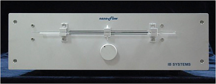
This study investigated the nanoleakage of root canal obturations using calcium silicate-based sealer according to different drying methods. Materials and MethodsFifty-two extracted mandibular premolars with a single root canal and straight root were selected for this study. After canal preparation with a nickel-titanium rotary file system, the specimens were randomly divided into 4 groups according to canal drying methods (1: complete drying, 2: blot drying/distilled water, 3: blot drying/NaOCl, 4: aspiration only). The root canals were obturated using a single-cone filling technique with a calcium silicate–based sealer. Nanoleakage was evaluated using a nanoflow device after 24 hours, 1 week, and 1 month. Data were collected twice per second at the nanoscale and measured in nanoliters per second. Data were statistically analyzed using the Kruskal-Wallis and Mann–Whitney U-tests (p < 0.05). ResultsThe mean flow rate measured after 24 hours showed the highest value among the time periods in all groups. However, the difference in the flow rate between 1 week and 1 month was not significant. The mean flow rate of the complete drying group was the highest at all time points. After 1 month, the mean flow rate in the blot drying group and the aspiration group was not significantly different. ConclusionsWithin the limitations of this study, the canal drying method had a significant effect on leakage and sealing ability in root canal obturations using a calcium silicate-based sealer. Thus, a proper drying procedure is critical in endodontic treatment.
-
Citations
Citations to this article as recorded by  - Effects of interactions between sealers and irrigants on the physicochemical and surface characteristics of endodontic sealers
Hye-In Kim, Yeon-Ju You, Yemi Kim, Jin-Woo Kim, Young-Eun Jang
Clinical Oral Investigations.2026;[Epub] CrossRef - Assessment of Shear Bond Strength of MTA and ApaCal ART Liners to a Universal Bonding Agent: An In Vitro Study
Angel Kurian, Banibrata Lahiri, Archana A Thomas, Gautham Anilkumar, Fawaz Pullishery, Reshma Raju
The Journal of Contemporary Dental Practice.2026; 27(3): 254. CrossRef
-
3,260
View
-
123
Download
-
1
Web of Science
-
2
Crossref
-
Effect of acidic solutions on the microhardness of dentin and set OrthoMTA and their cytotoxicity on murine macrophage
-
Soram Oh, Hiran Perinpanayagam, Yoon Lee, Jae-Won Kum, Yeon-Jee Yoo, Sang-Min Lim, Seok Woo Chang, Won-Jun Shon, Woocheol Lee, Seung-Ho Baek, Kee-Yeon Kum
-
Restor Dent Endod 2016;41(1):12-21. Published online December 1, 2015
-
DOI: https://doi.org/10.5395/rde.2016.41.1.12
-
-
Abstract
PDFPubReaderePub
- Objectives
To evaluate the effects of three acids on the microhardness of set mineral trioxide aggregate (MTA) and root dentin, and cytotoxicity on murine macrophage. Materials and MethodsOrthoMTA (BioMTA) was mixed and packed into the human root dentin blocks of 1.5 mm diameter and 5 mm height. Four groups, each of ten roots, were exposed to 10% citric acid (CA), 5% glycolic acid (GA), 17% ethylenediaminetetraacetic acid (EDTA), and saline for five minutes after setting of the OrthoMTA. Vickers surface microhardness of set MTA and dentin was measured before and after exposure to solutions, and compared between groups using one-way ANOVA with Tukey test. The microhardness value of each group was analyzed using student t test. Acid-treated OrthoMTA and dentin was examined by scanning electron microscope (SEM). Cell viability of tested solutions was assessed using WST-8 assay and murine macrophage. ResultsThree test solutions reduced microhardness of dentin. 17% EDTA demonstrated severe dentinal erosion, significantly reduced the dentinal microhardness compared to 10% CA (p = 0.034) or 5% GA (p = 0.006). 10% CA or 5% GA significantly reduced the surface microhardness of set MTA compared to 17% EDTA and saline (p < 0.001). Acid-treated OrthoMTA demonstrated microporous structure with destruction of globular crystal. EDTA exhibited significantly more cellular toxicity than the other acidic solutions at diluted concentrations (0.2, 0.5, 1.0%). ConclusionsTested acidic solutions reduced microhardness of root dentin. Five minute's application of 10% CA and 5% GA significantly reduced the microhardness of set OrthoMTA with lower cellular cytotoxicity compared to 17% EDTA.
-
Citations
Citations to this article as recorded by - The efficacy of acetic acid and citric acid on calcium silicate based cement removal from root canals and their effect on dentine structure – an in vitro study
Hanan Alzraikat, Ahmad S. Al-Hiyasat, Estabraq Sarya Jamous, Mohammad A. Alebrahim
Biomaterial Investigations in Dentistry.2026; 13: 154. CrossRef - Removal Efficiency of Mineral Trioxide Aggregate: A narrative Review of Retrieval Techniques
Mohammed A. Hussein, Rasha H. Jehad
Journal of Medical and Oral Biosciences.2025; : 36. CrossRef - Impact of calcium hydroxide and 2-hydroxyisocaproic acid on the microhardness of root dentine: an in vitro study
Nandini T. Niranjan, Protim Ghosh Dastidar, Raghavendra Penukonda, Galvin Sim Siang Lin, Roopa Babannavar, Arun Jaysheel, Harshada Pattar
Odontology.2024; 112(3): 711. CrossRef - Evaluation of the Effect of Chitosan-Based Irrigation Solutions on the Bond Strength of Mineral Trioxide Aggregate to Bulk-Fill Composite
Arzu Şahin Mantı, Bağdagül Helvacıoğlu Kıvanç
Journal of Functional Biomaterials.2024; 15(12): 370. CrossRef - Effect of Various Acid Solutions as an Aid in Removing the OrthoMTA-Based Root Canal Filling
Naveen Chhabra, Abhishek Parolia
Materials.2023; 16(13): 4535. CrossRef - Effect of Glycolic Acid, Maleic Acid, and EDTA in the Removal of Smear Layer from Root Canal Dentin
Tarini Mullick, Nidambur Vasudev Ballal
Pesquisa Brasileira em Odontopediatria e Clínica Integrada.2023;[Epub] CrossRef - A comparative evaluation of the effect of various chelating agents on the microhardness of root canal dentin: An in vitro study
Mineet Kaul, Zinnie Nanda, Kranthikumar Reddy, Rahul Deore, Divya Mandlecha, Esha Jaiswal
Endodontology.2023; 35(3): 234. CrossRef - Calcium hydroxide and niobium pentoxide treatment effects before MTA placement
Kolli Sankeerthana, Kittappa Karthikeyan, Sekar Mahalaxmi
Australian Endodontic Journal.2023; 49(1): 48. CrossRef - Calcium silicate and calcium aluminate cements for dentistry reviewed
Carolyn Primus, James L. Gutmann, Franklin R. Tay, Anna B. Fuks
Journal of the American Ceramic Society.2022; 105(3): 1841. CrossRef - Influence of Acidic Environmental Conditions on Push‐Out Bonding Strength of Four Calcium Silicate‐Based Materials to Root Dentin
Beliz Özel, Raif Erişen, Boonlert Kukiattrakoon
International Journal of Dentistry.2022;[Epub] CrossRef - The effects of sodium hypochlorite and ethylenediaminetetraacetic acid on the microhardness of Mineral Trioxide Aggregate and TotalFill Bioceramic Putty
Jacklyn H.R. Chu, Kalie Y. Chia, Alexander L. Qui, Alex Moule, William N. Ha
Australian Endodontic Journal.2020; 46(1): 33. CrossRef - Pre-application of dentin bonding agent prevents discoloration caused by mineral trioxide aggregate
Yoo-Lim Choi, Young-Eun Jang, Bom Sahn Kim, Jin-Woo Kim, Yemi Kim
BMC Oral Health.2020;[Epub] CrossRef - Glycolic acid as the final irrigant in endodontics: Mechanical and cytotoxic effects
Yuri Dal Bello, Hisadora Fracaro Porsch, Ana Paula Farina, Matheus Albino Souza, Emmanuel João Nogueira Leal Silva, Ana Karina Bedran-Russo, Doglas Cecchin
Materials Science and Engineering: C.2019; 100: 323. CrossRef - Carbohydrate-electrolyte drinks exhibit risks for human enamel surface loss
Mary Anne Sampaio de Melo, Vanara Florêncio Passos, Juliana Paiva Marques Lima, Sérgio Lima Santiago, Lidiany Karla Azevedo Rodrigues
Restorative Dentistry & Endodontics.2016; 41(4): 246. CrossRef
-
3,229
View
-
20
Download
-
14
Crossref
-
Synergistic effect of xylitol and ursolic acid combination on oral biofilms
-
Yunyun Zou, Yoon Lee, Jinyoung Huh, Jeong-Won Park
-
Restor Dent Endod 2014;39(4):288-295. Published online August 27, 2014
-
DOI: https://doi.org/10.5395/rde.2014.39.4.288
-
-
Abstract
PDFPubReaderePub
- Objectives
This study was designed to evaluate the synergistic antibacterial effect of xylitol and ursolic acid (UA) against oral biofilms in vitro. Materials and MethodsS. mutans UA 159 (wild type), S. mutans KCOM 1207, KCOM 1128 and S. sobrinus ATCC 33478 were used. The susceptibility of S. mutans to UA and xylitol was evaluated using a broth microdilution method. Based on the results, combined susceptibility was evaluated using optimal inhibitory combinations (OIC), optimal bactericidal combinations (OBC), and fractional inhibitory concentrations (FIC). The anti-biofilm activity of xylitol and UA on Streptococcus spp. was evaluated by growing cells in 24-well polystyrene microtiter plates for the biofilm assay. Significant mean differences among experimental groups were determined by Fisher's Least Significant Difference (p < 0.05). ResultsThe synergistic interactions between xylitol and UA were observed against all tested strains, showing the FICs < 1. The combined treatment of xylitol and UA inhibited the biofilm formation significantly and also prevented pH decline to critical value of 5.5 effectively. The biofilm disassembly was substantially influenced by different age of biofilm when exposed to the combined treatment of xylitol and UA. Comparing to the single strain, relatively higher concentration of xylitol and UA was needed for inhibiting and disassembling biofilm formed by a mixed culture of S. mutans 159 and S. sobrinus 33478. ConclusionsThis study demonstrated that xylitol and UA, synergistic inhibitors, can be a potential agent for enhancing the antimicrobial and anti-biofilm efficacy against S. mutans and S. sobrinus in the oral environment.
-
Citations
Citations to this article as recorded by - Therapeutic potential of ursolic acid (UA) and their derivatives with nanoformulations to combat nosocomial pathogens
Umesh Chand, Pramod Kumar Kushawaha
Future Journal of Pharmaceutical Sciences.2025;[Epub] CrossRef - Anti-cariogenic effect of experimental resin cement containing ursolic acid using dental microcosm biofilm
Jonghyun Jo, Mi-Jeong Jeon, Sun Kyu Park, Su-Jung Shin, Baek-il Kim, Jeong-Won Park
Journal of Dentistry.2024; 151: 105447. CrossRef - Alteration of oral microbial biofilms by sweeteners
Geum-Jae Jeong, Fazlurrahman Khan, Nazia Tabassum, Young-Mog Kim
Biofilm.2024; 7: 100171. CrossRef - Synergistic inhibitory activity of Glycyrrhizae Radix and Rubi Fructus extracts on biofilm formation of Streptococcus mutans
Youngseok Ham, Tae-Jong Kim
BMC Complementary Medicine and Therapies.2023;[Epub] CrossRef - Anti-Planktonic and Anti-Biofilm Properties of Pentacyclic Triterpenes—Asiatic Acid and Ursolic Acid as Promising Antibacterial Future Pharmaceuticals
Zuzanna Sycz, Dorota Tichaczek-Goska, Dorota Wojnicz
Biomolecules.2022; 12(1): 98. CrossRef - Does Secondary Plant Metabolite Ursolic Acid Exhibit Antibacterial Activity against Uropathogenic Escherichia coli Living in Single- and Multispecies Biofilms?
Zuzanna Sycz, Dorota Wojnicz, Dorota Tichaczek-Goska
Pharmaceutics.2022; 14(8): 1691. CrossRef - The physical properties and anticariogenic effect of experimental resin cement containing ursolic acid
Hyunkyung Yoo, So Youn Kim, Su-Jung Shin, Jeong-Won Park
Odontology.2021; 109(3): 641. CrossRef - Exploration of singular and synergistic effect of xylitol and erythritol on causative agents of dental caries
Siiri Kõljalg, Imbi Smidt, Anirikh Chakrabarti, Douwina Bosscher, Reet Mändar
Scientific Reports.2020;[Epub] CrossRef - The investigation of synergistic activity of protamine with conventional antimicrobial agents against oral bacteria
Masashi Fujiki, Michiyo Honda
Biochemical and Biophysical Research Communications.2020; 523(3): 561. CrossRef - Concentration in Saliva and Antibacterial Effect of Xylitol Chewing Gum: In Vivo and In Vitro Study
Fabio Cocco, Maria Grazia Cagetti, Osama Majdub, Guglielmo Campus
Applied Sciences.2020; 10(8): 2900. CrossRef - Enhanced synergistic effects of xylitol and isothiazolones for inhibition of initial biofilm formation by Pseudomonas aeruginosa ATCC 9027 and Staphylococcus aureus ATCC 6538
Gang Zhou, Hong Peng, Ying-si Wang, Xiao-mo Huang, Xiao-bao Xie, Qing-shan Shi
Journal of Oral Science.2019; 61(2): 255. CrossRef - Alkyl rhamnosides, a series of amphiphilic materials exerting broad-spectrum anti-biofilm activity against pathogenic bacteria via multiple mechanisms
Guanghua Peng, Xucheng Hou, Wenxi Zhang, Maoyuan Song, Mengya Yin, Jiaxing Wang, Jiajia Li, Yajie Liu, Yuanyuan Zhang, Wenkai Zhou, Xinru Li, Guiling Li
Artificial Cells, Nanomedicine, and Biotechnology.2018; 46(sup3): 217. CrossRef - Ursolic acid from apple pomace and traditional plants: A valuable triterpenoid with functional properties
Simone Tasca Cargnin, Simone Baggio Gnoatto
Food Chemistry.2017; 220: 477. CrossRef - Phytochemicals for human disease: An update on plant-derived compounds antibacterial activity
Ramona Barbieri, Erika Coppo, Anna Marchese, Maria Daglia, Eduardo Sobarzo-Sánchez, Seyed Fazel Nabavi, Seyed Mohammad Nabavi
Microbiological Research.2017; 196: 44. CrossRef - Ursolic acid (UA): A metabolite with promising therapeutic potential
Dharambir Kashyap, Hardeep Singh Tuli, Anil K. Sharma
Life Sciences.2016; 146: 201. CrossRef - Experimental Models of Oral Biofilms Developed on Inert Substrates: A Review of the Literature
Lopez-Nguyen Darrene, Badet Cecile
BioMed Research International.2016; 2016: 1. CrossRef - Multi-functional Liposomes Enhancing Target and Antibacterial Immunity for Antimicrobial and Anti-Biofilm Against Methicillin-Resistant Staphylococcus aureus
Yansha Meng, Xucheng Hou, Jiongxi Lei, Mengmeng Chen, Shuangchen Cong, Yuanyuan Zhang, Weiming Ding, Guiling Li, Xinru Li
Pharmaceutical Research.2016; 33(3): 763. CrossRef - Antibacterial activity of constituents from Salvia buchananii Hedge (Lamiaceae)
Angela Bisio, Anna Maria Schito, Anita Parricchi, Giacomo Mele, Giovanni Romussi, Nicola Malafronte, Patrizia Oliva, Nunziatina De Tommasi
Phytochemistry Letters.2015; 14: 170. CrossRef
-
2,973
View
-
7
Download
-
18
Crossref
-
Esthetic rehabilitation of single anterior edentulous space using fiber-reinforced composite
-
Hyeon Kim, Min-Ju Song, Su-Jung Shin, Yoon Lee, Jeong-Won Park
-
Restor Dent Endod 2014;39(3):220-225. Published online May 19, 2014
-
DOI: https://doi.org/10.5395/rde.2014.39.3.220
-
-
Abstract
PDFPubReaderePub
A fiber-reinforced composite (FRC) fixed prosthesis is an innovative alternative to a traditional metal restoration, as it is a conservative treatment method. This case report demonstrates a detailed procedure for restoring a missing anterior tooth with an FRC. A 44-year-old woman visited our department with an avulsed tooth that had fallen out on the previous day and was completely dry. This tooth was replanted, but it failed after one year. A semi-direct technique was used to fabricate a FRC fixed partial prosthesis for its replacement. The FRC framework and the pontic were fabricated using a duplicated cast model and nanofilled composite resin. Later on, interproximal contact, tooth shape, and shade were adjusted at chairside. This technique not only enables the clinician to replace a missing tooth immediately after extraction for minimizing esthetic problems, but it also decreases both tooth reduction and cost. -
Citations
Citations to this article as recorded by - Anterior provisional fixed partial dentures: A finite element analysis
Nouf Almeganni, Rotana Abulaban, Ghada Naguib, Mohamed Tharwat, Hani M. Nassar
Journal of Prosthodontics.2024; 33(4): 367. CrossRef - FİBERLE GÜÇLENDİRİLMİŞ ADEZİV KÖPRÜLER VE UYGULAMA YÖNTEMLERİ
Gözde YALÇIN, Asude Dilek NALBANT
Atatürk Üniversitesi Diş Hekimliği Fakültesi Dergisi.2022; : 1. CrossRef - Fiber-reinforced composite resin bridges: an alternative method to treat root-fractured teeth
Gun Heo, Eun-Hye Lee, Jin-Woo Kim, Kyung-Mo Cho, Se-Hee Park
Restorative Dentistry & Endodontics.2020;[Epub] CrossRef - A New Technique for Direct Fabrication of Fiber-Reinforced Composite Bridge: A Long-Term Clinical Observation
Matías Ferrán Escobedo Martínez, Samuel Rodríguez López, Jairo Valdés Fontela, Sonsoles Olay García, Mario Mauvezín Quevedo
Dentistry Journal.2020; 8(2): 48. CrossRef - Customized Treatment Option for Malpositioned Dental Implant Placed in Aesthetic Zone
Priyanka N. Khungar, Trupti M. Dahane, Ramnath P. Revankar, Rupali Patel
Journal of Evolution of Medical and Dental Sciences.2020; 9(39): 2930. CrossRef - Fiber reinforced composite bridge as a replacement for missing upper permanent lateral incisor – a case report
Ana Todorović, Danica Popović, Igor Djordjević, Vojkan Lazić
Stomatoloski glasnik Srbije.2016; 63(3): 133. CrossRef - Evaluation of the Viability of Rat Periodontal Ligament Cells after Storing at 0℃/2 MPa Condition up to One Week: In Vivo MTT Method
Sun Mi Jang, Sin-Yeon Cho, Eui-Seong Kim, Il-Young Jung, Seung Jong Lee
Journal of Korean Dental Science.2016; 9(1): 1. CrossRef - Semidirect Restorations in Multidisciplinary Treatment: Viable Option for Children and Teenagers
Mateus Rodrigues Tonetto, Milton Carlos Kuga, Fausto Frizzera, Matheus Coelho Bandeca, Shilpa H Bhandi, Célia Regina Maio Pinzan-Vercelino, Monica Barros da Silva, Kamila Figueiredo Pereira
The Journal of Contemporary Dental Practice.2015; 16(4): 280. CrossRef
-
2,353
View
-
10
Download
-
8
Crossref
-
Effect of calcium hydroxide application time on dentin
-
Yoon Lee
-
Restor Dent Endod 2013;38(3):186-186. Published online August 23, 2013
-
DOI: https://doi.org/10.5395/rde.2013.38.3.186
-
-
PDFPubReaderePub
-
Citations
Citations to this article as recorded by - Investigating the strengthening effect of Nigella sativa seed extract and Er,Cr:YSGG laser on artificially demineralized dentin: In Vitro study
Sherif Elsayed, Doaa M. Sadony, Ramadan N. Elshaer, Mohamed M. Kandil, Hadeel Farouk
Lasers in Dental Science.2026;[Epub] CrossRef - Retrievability of triple antibiotic paste from simulated grooves in molars: Impact of access design and agitation technique
Oorja Nanda, Vineeta Nikhil
Endodontology.2026; 38(2): 193. CrossRef - Surgical and Nonsurgical Endodontic Management of Horizontal Root Fracture and Cervical Root Perforation
Sweta Rastogi, Shahnaz Nabi, Sanjay Miglani, Priyanshu Kumar Shrivastava
Current Trends in Dentistry.2025; 2(1): 55. CrossRef - Effect of niobium pentoxide incorporated calcium hydroxide as an intracanal medicament on fracture resistance of root canal dentin
Nadimpalli Teja Varma, Venkatappan Sujatha, Kittappa Karthikeyan, Sekar Mahalaxmi
Journal of Conservative Dentistry and Endodontics.2025; 28(12): 1211. CrossRef - The effectiveness of single antibiotic paste nitrofurantoin V/S double antibiotic paste in alleviation of post-operative pain of patients suffering from symptomatic irreversible pulpitis—A randomized controlled trial
Hira Abbasi, Muhammad Saqib, Afsheen Maqsood, Rizwan Jouhar, Haroon Rashid, Naseer Ahmed, Mohmed Isaqali Karobari, Artak Heboyan
SAGE Open Medicine.2024;[Epub] CrossRef - Management of Maxillary Anterior Tooth with Open Apex by MTA Apexification
Surya K R, Radhakrishnan Nair K, Nisha B Kurup, Xavier Jithu A A
International Journal of Innovative Science and Research Technology (IJISRT).2024; : 2725. CrossRef - Effect of different intracanal medicaments on the fracture resistance of the human root
Saeed Rahimi, Negin Ghasemi, Golchin Jabbari, Zahra Zaheri, ZahraSadat Torabi, NaghmehRahimi Darehchi
Dental Research Journal.2022; 19(1): 9. CrossRef - The effect of calcium hydroxide on dentine composition and root fracture resistance of human teeth: An in vitro study
Ahmad S. Al‐Hiyasat, Hamza S. El‐Farraj, Mohammad A. Alebrahim
European Journal of Oral Sciences.2021;[Epub] CrossRef - Fracture behaviour of teeth with conventional and mini-invasive access cavity designs
Zdeněk Chlup, Radovan Žižka, Jakub Kania, Michal Přibyl
Journal of the European Ceramic Society.2017; 37(14): 4423. CrossRef - Chronic maxillary sinusitis caused by root canal overfilling of Calcipex II
Jin-Woo Kim, Kyung-Mo Cho, Se-Hee Park, Soh-Ra Park, Sang-Shin Lee, Suk-Keun Lee
Restorative Dentistry & Endodontics.2014; 39(1): 63. CrossRef - Adult stem cell-based apexogenesis
Yao Li
World Journal of Methodology.2014; 4(2): 99. CrossRef
-
5,480
View
-
59
Download
-
11
Crossref
-
In-depth morphological study of mesiobuccal root canal systems in maxillary first molars: review
-
Seok-Woo Chang, Jong-Ki Lee, Yoon Lee, Kee-Yeon Kum
-
Restor Dent Endod 2013;38(1):2-10. Published online February 26, 2013
-
DOI: https://doi.org/10.5395/rde.2013.38.1.2
-
-
Abstract
PDFPubReaderePub
A common failure in endodontic treatment of the permanent maxillary first molars is likely to be caused by an inability to locate, clean, and obturate the second mesiobuccal (MB) canals. Because of the importance of knowledge on these additional canals, there have been numerous studies which investigated the maxillary first molar MB root canal morphology using in vivo and laboratory methods. In this article, the protocols, advantages and disadvantages of various methodologies for in-depth study of maxillary first molar MB root canal morphology were discussed. Furthermore, newly identified configuration types for the establishment of new classification system were suggested based on two image reformatting techniques of micro-computed tomography, which can be useful as a further 'Gold Standard' method for in-depth morphological study of complex root canal systems. -
Citations
Citations to this article as recorded by - An epidemiological study of extracted mandibular premolars from adolescent patients in Damascus using two classification system analyzed with CBCT and digital periapical radiographs
Yasser Alsayed Tolibah, Mohammed N. Al-Shiekh, Mohammad Tamer Abbara, Marwan Alhaji, Osama Aljabban, Nada Bshara
BMC Oral Health.2025;[Epub] CrossRef - Cone beam computed tomography analysis of the root and canal morphology of the maxillary second molars in a Hail province of the Saudi population
Ahmed A. Madfa, Moazzy I. Almansour, Saad M. Al-Zubaidi, Albandari H. Alghurayes, Safanah D. AlDAkhayel, Fatemah I. Alzoori, Taif F. Alshammari, Abrar M. Aldakhil
Heliyon.2023; 9(9): e19477. CrossRef - Signs of a missed root canal
M. Yu. Pokrovsky, O. A. Aleshina, T. P. Goryacheva, A. M. Pokrovskiy
Endodontics Today.2023; 21(3): 205. CrossRef - Root Canal Morphology of Maxillary First and Second Molars in a Qatari Population: A Cone-Beam Computed Tomography Study
Maryam Mohammed Al-Obaid, Fatima Abdullah Al-Sheeb
European Dental Research and Biomaterials Journal.2021; 2(01): 34. CrossRef - A Study Comparing the Characteristics of Zinc Oxide Eugenol-Based and Mineral Trioxide Aggregate-Based Root Canal Sealers
Seok-Eun Lee, Ja-Won Cho, Hyun-Jun Yoo, Myung-Gu Lee, Yeol-Mae Jeon, Da-Hui Kim, Hye-Won Park
International Journal of Clinical Preventive Dentistry.2021; 17(3): 117. CrossRef - Root Canal Configuration of Burmese (Myanmar) Maxillary First Molar: A Micro-Computed Tomography Study
M. M. Kyaw Moe, H. J. Jo, J. H. Ha, S. K. Kim, Antonino Lo Giudice
International Journal of Dentistry.2021; 2021: 1. CrossRef - Three-Dimensional Analysis of Root Anatomy and Root Canal Curvature in Mandibular Incisors Using Micro-Computed Tomography with Novel Software
JongKi Lee, Shin-Hoon Lee, Jong-Rak Hong, Kee-Yeon Kum, Soram Oh, Adel Saeed Al-Ghamdi, Fawzi Ali Al-Ghamdi, Ayman Omar Mandorah, Ji-Hyun Jang, Seok Woo Chang
Applied Sciences.2020; 10(12): 4385. CrossRef - An investigation into dose optimisation for imaging root canal anatomy using cone beam CT
Margarete B McGuigan, Christie Theodorakou, Henry F Duncan, Jonathan Davies, Anita Sengupta, Keith Horner
Dentomaxillofacial Radiology.2020; 49(7): 20200072. CrossRef - Analysis of Root Canal Anatomy and Variation in Morphology of Maxillary First Molar Using Various Methods: An In Vitro Study
Youssef A Algarni
World Journal of Dentistry.2019; 10(4): 291. CrossRef - Root Canal Morphology of Mandibular Primary Molars: A Micro-CT Study
Meryem ZİYA, Burcu Nihan YÜKSEL, Şaziye SARI
Cumhuriyet Dental Journal.2019; 22(4): 382. CrossRef - Comparison of the implementation of extra root canal treatment before and after fee schedule change in the Taiwan National Health Insurance System
Nien-Chieh Lee, Yen-Hsiang Chang, Hui-Tzu Tu, Chang-Fu Kuo, Kuang-Hui Yu, Lai-Chu See
Journal of Dental Sciences.2018; 13(2): 145. CrossRef - Influence of environment on testing of hydraulic sealers
Mira Kebudi Benezra, Pierre Schembri Wismayer, Josette Camilleri
Scientific Reports.2017;[Epub] CrossRef - CBCT uses in clinical endodontics: the effect of CBCT on the ability to locate MB2 canals in maxillary molars
J. Parker, A. Mol, E. M. Rivera, P. Tawil
International Endodontic Journal.2017; 50(12): 1109. CrossRef - Comparison of Alternative Image Reformatting Techniques in Micro–Computed Tomography and Tooth Clearing for Detailed Canal Morphology
Ki-Wook Lee, Yeun Kim, Hiran Perinpanayagam, Jong-Ki Lee, Yeon-Jee Yoo, Sang-Min Lim, Seok Woo Chang, Byung-Hyun Ha, Qiang Zhu, Kee-Yeon Kum
Journal of Endodontics.2014; 40(3): 417. CrossRef - In Vitro Biocompatibility, Inflammatory Response, and Osteogenic Potential of 4 Root Canal Sealers: Sealapex, Sankin Apatite Root Sealer, MTA Fillapex, and iRoot SP Root Canal Sealer
Seok-Woo Chang, So-Youn Lee, Soo-Kyung Kang, Kee-Yeon Kum, Eun-Cheol Kim
Journal of Endodontics.2014; 40(10): 1642. CrossRef - Análise do preparo de canais radiculares utilizando-se a diafanização
Georje de Martin, Rogério Albuquerque Azeredo
Revista de Odontologia da UNESP.2014; 43(2): 111. CrossRef
-
2,949
View
-
20
Download
-
16
Crossref
-
Considerations during crown reattachment procedure over the pulpal exposure: case report
-
Bona Kim, Yoon Lee, Min-Ju Song, Su-Jung Shin, Jeong-Won Park
-
Restor Dent Endod 2012;37(4):240-244. Published online November 21, 2012
-
DOI: https://doi.org/10.5395/rde.2012.37.4.240
-
-
Abstract
PDFPubReaderePub
Crown reattachment is the most conservative treatment which can be used to restore fractured tooth, presumably with sufficient strength, while maintaining original contour, incisal translucency, and reducing chair time and cost. However, in case of crown fracture with pin-point pulp exposure, we should cautiously minimize the irritation to the pulp and consider pre-treatment pulpal status, choice of pulp capping materials, choice of bonding system and treatment sequence during crown reattachment procedures. This case reports the considerations while crown reattachment with direct pulp capping using calcium hydroxide (Dycal, Dentsply Caulk). -
Citations
Citations to this article as recorded by - A Conservative Approach to the Management of a Dental Trauma for Immediate Natural Esthetics
Pallav Mahesh Patni, Pradeep Jain, Mona Jain Patni
Archives of Trauma Research.2016;[Epub] CrossRef
-
1,972
View
-
9
Download
-
1
Crossref
-
Effect of moisture and drying time on the bond strength of the one-step self-etching adhesive system
-
Yoon Lee, Jeong-Won Park
-
Restor Dent Endod 2012;37(3):155-159. Published online August 29, 2012
-
DOI: https://doi.org/10.5395/rde.2012.37.3.155
-
-
Abstract
PDFPubReaderePub
- Objectives
To investigate the effect of dentin moisture degree and air-drying time on dentin-bond strength of two different one-step self-etching adhesive systems. Materials and MethodsTwenty-four human third molars were used for microtensile bond strength testing of G-Bond and Clearfil S3 Bond. The dentin surface was either blot-dried or air-dried before applying these adhesive agents. After application of the adhesive agent, three different air drying times were evaluated: 1, 5, and 10 sec. Composite resin was build up to 4 mm thickness and light cured for 40 sec with 2 separate layers. Then the tooth was sectioned and trimmed to measure the microtensile bond strength using a universal testing machine. The measured bond strengths were analyzed with three-way ANOVA and regression analysis was done (p = 0.05). ResultsAll three factors, materials, dentin wetness and air drying time, showed significant effect on the microtensile bond strength. Clearfil S3 Bond, dry dentin surface and 10 sec air drying time showed higher bond strength. ConclusionsWithin the limitation of this experiment, air drying time after the application of the one-step self-etching adhesive agent was the most significant factor affecting the bond strength, followed by the material difference and dentin moisture before applying the adhesive agent.
-
Citations
Citations to this article as recorded by - An in vitro study on comparative evaluation of shear bond strength of bioactive composite to tooth structure with various dentin conditioning agents
Priyanka Pokkula, Shaik Mohammed Asif, Abdullah Alqarni, Shahabe Saquib Abullais, Shaik Mohamed Shamsudeen, Syed M Yassin, Abosofyan S. Atta, Wahaj Ahmad Khan
AIP Advances.2025;[Epub] CrossRef - The Influence of Drying Time, Application Mode, and Agitation on the Dentin Bond Strength of a Novel Mesoporous Bioactive Glass-Containing Universal Dentin Adhesive
Jiyoung Kwon, Jungwon Kim, Dongseok Choi, Duck-Su Kim
Journal of Functional Biomaterials.2025; 16(7): 247. CrossRef - The occluding effects of layered calcium phosphate and cyanoacrylate on dentinal tubules: a SEM study
Özge Uzuner Bilgiç, Sühan Gürbüz, Altan Dogan
BMC Oral Health.2025;[Epub] CrossRef - Shear bond strengths of two newly marketed self‐adhesive resin cements to different substrates: A light and scanning electron microscopy evaluation
Cansu Atalay, Uzay Koc Vural, Ivana Miletic, Sevil Gurgan
Microscopy Research and Technique.2022; 85(5): 1694. CrossRef - The effect of curing mode of dual-cure resin cements on bonding performance of universal adhesives to enamel, dentin and various restorative materials
Erick LUZ MADRIGAL, Antonin TICHY, Keiichi HOSAKA, Masaomi IKEDA, Masatoshi NAKAJIMA, Junji TAGAMI
Dental Materials Journal.2021; 40(2): 446. CrossRef - Effect of adhesive air-drying time on bond strength to dentin: A systematic review and meta-analysis
Mohamed M. Awad, Ali Alrahlah, Jukka P. Matinlinna, Hamdi Hosni Hamama
International Journal of Adhesion and Adhesives.2019; 90: 154. CrossRef - Effect of pre-curing of two universal adhesives on the shear bond strength of resin cement to zirconia
Ga-Eun Son, Tae-Yub Kwon, Young Kyung Kim
Korean Journal of Dental Materials.2019; 46(1): 21. CrossRef - Bonding effectiveness of different dentin conditions on etch-and-rinse mode of two universal adhesives: the confocal laser scanning and shear bond strength
Jounghyun Lee, Ka-Young Cho, Jin-Young Kim, Sungho Park, Byoung-Duck Roh, Yooseok shin
Journal of Adhesion Science and Technology.2017; 31(9): 933. CrossRef - Effect of dentin dehydration and composite resin polymerization mode on bond strength of two self-etch adhesives
Pooran Samimi, Mehdi Alizadeh, Farinaz Shirban, Amin Davoodi, Maryam Khoroushi
Contemporary Clinical Dentistry.2016; 7(1): 16. CrossRef - Effect of different air-drying time on the microleakage of single-step self-etch adhesives
Horieh Moosavi, Maryam Forghani, Esmatsadat Managhebi
Restorative Dentistry & Endodontics.2013; 38(2): 73. CrossRef
-
2,065
View
-
11
Download
-
10
Crossref
-
Microbial profile of asymptomatic and symptomatic teeth with primary endodontic infections by pyrosequencing
-
Sang-Min Lim, Tae-Kwon Lee, Eun-Jeong Kim, Jun-Hong Park, Yoon Lee, Kwang-Shik Bae, Kee-Yeon Kum
-
J Korean Acad Conserv Dent 2011;36(6):498-505. Published online November 30, 2011
-
DOI: https://doi.org/10.5395/JKACD.2011.36.6.498
-
-
Abstract
PDFPubReaderePub
-
Objectives
The purpose of this in vivo study was to investigate the microbial diversity in symptomatic and asymptomatic canals with primary endodontic infections by using GS FLX Titanium pyrosequencing.
Materials and Methods
Sequencing was performed on 6 teeth (symptomatic, n = 3; asymptomatic, n = 3) with primary endodontic infections. Amplicons from hypervariable region of the small-subunit ribosomal RNA gene were generated by polymerized chain reaction (PCR), and sequenced by means of the GS FLX Titanium pyrosequencing.
Results
On average, 10,639 and 45,455 16S rRNA sequences for asymptomatic and symptomatic teeth were obtained, respectively. Based on Ribosomal Database Project Classifier analysis, pyrosequencing identified the 141 bacterial genera in 13 phyla. The vast majority of sequences belonged to one of the seven phyla: Actinobacteria, Bacteroidetes, Firmicutes, Fusobacteria, Proteobacteria, Spirochetes, and Synergistetes. In genus level, Pyramidobacter, Streptococcus, and Leptotrichia constituted about 50% of microbial profile in asymptomatic teeth, whereas Neisseria, Propionibacterium, and Tessaracoccus were frequently found in symptomatic teeth (69%). Grouping the sequences in operational taxonomic units (3%) yielded 450 and 1,997 species level phylotypes in asymptomatic and symptomatic teeth, respectively. The total bacteria counts were significantly higher in symptomatic teeth than that of asymptomatic teeth (p < 0.05).
Conclusions
GS FLX Titanium pyrosequencing could reveal a previously unidentified high bacterial diversity in primary endodontic infections.
-
Citations
Citations to this article as recorded by - Root Canal Microbiome Associated With Asymptomatic Apical Periodontitis as Determined by High-Throughput Sequencing
Rodrigo Rodrigues Amaral, Tiago Braga, José F. Siqueira, Isabela N. Rôças, Caio Tavora Coelho da Costa Rachid, Anna Gabriella Guimarães Oliveira, Maria Ilma de Souza Côrtes, Robert Mattew Love
Journal of Endodontics.2022; 48(4): 487. CrossRef - A critical analysis of research methods and experimental models to study the root canal microbiome
José F. Siqueira, Isabela N. Rôças
International Endodontic Journal.2022; 55(S1): 46. CrossRef - Oral microbiomes in children with asthma and dental caries
Sergey V. Cherkasov, Larisa Yu. Popova, Tatyana V. Vivtanenko, Rimma R. Demina, Yuri A. Khlopko, Alexander S. Balkin, Andrey O. Plotnikov
Oral Diseases.2019; 25(3): 898. CrossRef - Insights into the human oral microbiome
Digvijay Verma, Pankaj Kumar Garg, Ashok Kumar Dubey
Archives of Microbiology.2018; 200(4): 525. CrossRef - Solving the etiology of dental caries
Aurea Simón-Soro, Alex Mira
Trends in Microbiology.2015; 23(2): 76. CrossRef - Present status and future directions in endodontic microbiology
José F. Siqueira, Isabela N. Rôças
Endodontic Topics.2014; 30(1): 3. CrossRef - Application of high-throughput sequencing in understanding human oral microbiome related with health and disease
Hui Chen, Wen Jiang
Frontiers in Microbiology.2014;[Epub] CrossRef
-
1,986
View
-
8
Download
-
7
Crossref
-
Effect of cross-sectional area of 6 nickel-titanium rotary instruments on the fatigue fracture under cyclic flexural stress: A fractographic analysis
-
Soo-Youn Hwang, So-Ram Oh, Yoon Lee, Sang-Min Lim, Kee-Yeon Kum
-
J Korean Acad Conserv Dent 2009;34(5):424-429. Published online September 30, 2009
-
DOI: https://doi.org/10.5395/JKACD.2009.34.5.424
-
-
Abstract
PDFPubReaderePub
This study aimed to assess the influence of different cross-sectional area on the cyclic fatigue fracture of Ni-Ti rotary files using a fatigue tester incorporating cyclical axial movement. Six brands of Ni-Ti rotary files (ISO 30 size with .04 taper) of 10 each were tested: Alpha system (KOMET), HeroShaper (MicroMega), K3 (SybronEndo), Mtwo (VDW), NRT (Mani), and ProFile (Dentsply). A fatigue-tester (Denbotix) was designed to allow cyclic tension and compressive stress on the tip of the instrument. Each file was mounted on a torque controlled motor (Aseptico) using a 1:20 reduction contra-angle and was rotated at 300 rpm with a continuous, 6 mm axial oscillating motion inside an artificial steel canal. The canal had a 60° angle and a 5 mm radius of curvature. Instrument fracture was visually detected and the time until fracture was recorded by a digital stop watch. The data were analyzed statistically. Fractographic analysis of all fractured surfaces was performed to determine the fracture modes using a scanning electron microscope. Cross-sectional area at 3 mm from the tip of 3 unused Ni-Ti instruments for each group was calculated using Image-Pro Plus (Imagej 1.34n, NIH). Results showed that NRT and ProFile had significantly longer time to fracture compared to the other groups (p < .05). The cross-sectional area was not significantly associated with fatigue resistance. Fractographycally, all fractured surfaces demonstrated a combination of ductile and brittle fracture. In conclusion, there was no significant relationship between fatigue resistance and the cross-sectional area of Ni-Ti instruments under experimental conditions.
-
The role of Type 2 Diabetes as a predisposing risk factor on the pulpo-periapical pathogenesis: review article
-
Jin-Hee Kim, Kwang-shik Bae, Deog-Gyu Seo, Sung-Tae Hong, Yoon Lee, Sam-Pyo Hong, Kee-Yeon Kum
-
J Korean Acad Conserv Dent 2009;34(3):169-176. Published online May 31, 2009
-
DOI: https://doi.org/10.5395/JKACD.2009.34.3.169
-
-
Abstract
PDFPubReaderePub
Diabetes Mellitus (DM) is a syndrome accompanied with the abnormal secretion or function of insulin, a hormone that plays a vital role in controlling the blood glucose level (BGL). Type 1and 2 DM are most common form and the prevalence of the latter is recently increasing. The aim of this article was to assess whether Type 2 DM could act as a predisposing risk factor on the pulpo-periapical pathogenesis. Previous literature on the pathologic changes of blood vessels in DM was thoroughly reviewed. Furthermore, a histopathologic analysis of artificially-induced periapical specimens obtained from Type 2 diabetic and DM-resistant rats was compared. Histopathologic results demonstrate that the size of periapical bone destruction was larger and the degree of pulpal inflammation was more severe in diabetic rats, indicating that Type 2 DM itself can be a predisposing risk factor that makes the host more susceptible to pulpal infection. The possible reasons may be that in diabetic state the lumen of pulpal blood vessels are thickened by atheromatous deposits, and microcirculation is hindered. The function of polymorphonuclear leukocyte is also impaired and the migration of immune cells is blocked, leading to increased chance of pulpal infection. Also, lack of collateral circulation of pulpal blood vessels makes the pulp more susceptible to infection. These decrease the regeneration capacity of pulpal cells or tissues, delaying the healing process. Therefore, when restorative treatment is needed in Type 2 DM patients, dentists should minimize irritation to the pulpal tissue un der control of BGL. -
Citations
Citations to this article as recorded by - Pulp necrosis following luxated injury to teeth in a patient with uncontrolled type II diabetes mellitus: a case report
Haneol Shin, Seung-Jong Lee, Il-Young Jung, Chan-Young Lee
Restorative Dentistry & Endodontics.2012; 37(1): 61. CrossRef
-
1,674
View
-
12
Download
-
1
Crossref
-
Influence of three different preparation designs on the marginal and internal gaps of CEREC3 CAD/CAM inlays
-
Deog-Gyu Seo, Young-Ah Yi, Yoon Lee, Byoung-Duck Roh
-
J Korean Acad Conserv Dent 2009;34(3):177-183. Published online May 31, 2009
-
DOI: https://doi.org/10.5395/JKACD.2009.34.3.177
-
-
Abstract
PDFPubReaderePub
The aim of this study was to evaluate the marginal and internal gaps in CEREC3 CAD/CAM inlays of three different preparation designs. CEREC3 Inlays of three different preparation designs (n = 10) were fabricated according to Group I-conventional functional cusp capping/shoulder preparation, Group II-horizontal reduction of cusps and Group III-complete reduction of cusps/shoulder preparation. After cementation of inlays, the bucco-lingual cross section was performed through the center of tooth. Cross section images of 20 magnifications were obtained through the stereomicroscope. The gaps were measured using the Leica application suite software at each reference point. Statistical analysis was performed using one-way ANOVA and Tukey's test (α<0.05).
The marginal gaps ranged from 80.0 to 97.8 µm for Group I, 42.0 to 194.8 µm for Group II, 51.0 to 80.2 µm for Group III. The internal gaps ranged from 90.5 to 304.1 µm for Group I, 80.0 to 274.8 µm for Group II, 79.7 to 296.7 µm for Group III. The gaps of each group were the smallest on the margin and the largest on the horizontal wall. For the CEREC3 CAD/CAM inlays, the simplified designs (groups II and III) did not demonstrate superior results compared to the traditional cusp capping design (group I). -
Citations
Citations to this article as recorded by - Effect of axiopulpal line angle design and cement space settings on seating accuracy of CAD/CAM ceramic inlays: an in vitro study
Donghwan Kim, Mi-Jeong Jeon, Young Suk Kang, Yu-Sung Choi, Jeong-Kil Park, Deog-Gyu Seo
Journal of Dentistry.2026; 170: 106677. CrossRef - Influence of preparation design on fit and ceramic thickness of CEREC 3 partial ceramic crowns after cementation
Jae-Hoon Kim, Byeong-Hoon Cho, Jin-Hee Lee, Soo-Jung Kwon, Young-Ah Yi, Yooseok Shin, Byoung-Duck Roh, Deog-Gyu Seo
Acta Odontologica Scandinavica.2015; 73(2): 107. CrossRef
-
1,798
View
-
5
Download
-
2
Crossref
-
THE DYNAMIC CHANGE OF ARTIFICIALLY DEMINERALIZED ENAMEL BY DEGREE OF SATURATION OF REMINERALIZATION SOLUTION AT pH 4.3
-
Ji-Sook Yi, Bung-Duk Roh, Su-Jung Shin, Yoon Lee, Hyung-Kyu Gong, Chan-Young Lee
-
J Korean Acad Conserv Dent 2009;34(1):20-29. Published online January 14, 2009
-
DOI: https://doi.org/10.5395/JKACD.2009.34.1.020
-
-
Abstract
PDFPubReaderePub
- Abstract
The purpose of this study is to observe and compare the dynamic change of artificially demineralized enamel by remineralization solutions of different degrees of saturation at pH 4.3.
In this study, 30 enamel specimens were demineralized artificially by lactic acid buffered solution. Each of 10 specimens was immersed in pH 4.3 remineralization solution of three different degrees of saturation (0.22, 0.30, 0.35) for 10 days. After demineralization and remineralization, images were taken by a polarizing microscope (× 100). The density of lesion were determined from images taken after demineralization and remineralization.
During remineralization process, mineral deposition and mineral loss occurred at the same time. After remineralization, total mineral amount and width of surface lesion increased in all groups. The higher degree of saturation was, the more mineral deposition occurred in surface lesion and the amount of mineral deposition was not much in subsurface lesion. Total demineralized depth increased in all groups.
-
Citations
Citations to this article as recorded by - Effect of fluoride concentration in pH 4.3 and pH 7.0 supersaturated solutions on the crystal growth of hydroxyapatite
Haneol Shin, Sung-Ho Park, Jeong-Won Park, Chan-Young Lee
Restorative Dentistry & Endodontics.2012; 37(1): 16. CrossRef
-
1,748
View
-
3
Download
-
1
Crossref
-
Clinical diagnosis of herpes zoster presenting as odontogenic pain
-
Seong-Hak Yang, Dong-Ho Jung, Hae-Doo Lee, Yoon Lee, Hoon-Sang Chang, Kyung-San Min
-
J Korean Acad Conserv Dent 2008;33(5):452-456. Published online September 30, 2008
-
DOI: https://doi.org/10.5395/JKACD.2008.33.5.452
-
-
Abstract
PDFPubReaderePub
Herpes zoster, an acute viral infection produced by the varicella zoster virus, may affect any of the trigeminal branches. This case report presents a patient with symptoms mimicking odontogenic pain. No obvious cause of the symptoms could be found based on clinical and radiographic examinations. After a dermatologist made a diagnosis of herpes zoster involving the third trigeminal branch, the patient was given antiviral therapy. Two months later, the facial lesions and pain had almost disappeared, and residual pigmented scars were present. During the diagnostic process, clinicians should keep in mind the possibility that orofacial pain might be related to herpes zoster. -
Citations
Citations to this article as recorded by - Delayed Apical Pathology during Trigeminal Postherpetic Neuralgia Managed with Antiviral-Antibiotic Therapy: A Case Report
William W. Ja
Journal of Endodontics.2026;[Epub] CrossRef - Herpes Zoster Accompanying Odontogenic Inflammation: A Case Report with Literature Review
Soyeon Lee, Minsik Kim, Jong-Ki Huh, Jae-Young Kim
Journal of Oral Medicine and Pain.2021; 46(1): 9. CrossRef - Recurrent Herpetic Stomatitis Mimicking Post-Root Resection Complication
Sung-Ok Hong, Jae-Kwan Lee, Hoon-Sang Chang
Journal of Dental Rehabilitation and Applied Science.2013; 29(4): 418. CrossRef - Diagnostic challenges of nonodontogenic toothache
Hyung-Ok Park, Jung-Hong Ha, Myoung-Uk Jin, Young-Kyung Kim, Sung-Kyo Kim
Restorative Dentistry & Endodontics.2012; 37(3): 170. CrossRef
-
4,264
View
-
20
Download
-
4
Crossref
-
The effectiveness of sealing technique on in-office bleaching
-
Yoon Lee, So-Ran Kwon, Jeong-Won Park
-
J Korean Acad Conserv Dent 2008;33(5):463-471. Published online September 30, 2008
-
DOI: https://doi.org/10.5395/JKACD.2008.33.5.463
-
-
Abstract
PDFPubReaderePub
This study investigated the clinical effectiveness and safety of sealed bleaching compared to conventional in-office bleaching using a randomized clinical trial of split arch design. Ten participants received a chairside bleaching treatment on the upper anterior teeth, and each side was randomly designated as sealed or control side. A mixture of Brite powder (PacDent, Walnut, USA), 3% hydrogen peroxide and carbamide peroxide (KoolWhite, PacDent, Walnut, USA) were used as bleaching agent. The control side was unwrapped and the experimental side was covered with a linear low density polyethylene (LLDPE) wrap for sealed bleaching. The bleaching gel was light activated for 1 hour. The tooth shades were evaluated before treatment, after treatment, and at one week check up by means of a visual shade (VS) assessment using a value oriented shade guide and a computer assisted shade assessment using a spectrophotometer (SP). The data were analyzed by paired t-test.
In the control and sealed groups, the visual shade scores after bleaching treatment and at check up showed statistically significant difference from the preoperative shade scores (p < .05). The shade scores of the sealed group were significantly lighter than the control immediately after bleaching and at the check-up appointment (p < 0.05). Compared to prebleaching status, the ΔE values at post-bleaching condition were 4.35 ± 1.38 and 5.08 ± 1.34 for the control and sealed groups, respectively. The ΔE values at check up were 3.73 ± 1.95 and 4.38 ± 2.08 for the control and sealed groups. ΔE values were greater for the sealed group both after bleaching (p < .05) and at check up (p < .05).
In conclusion, both ΔE and shade score changes were greater for the sealed bleaching group than the conventional bleaching group, effectively demonstrating the improvement of effectiveness through sealing.
-
The remineralizing features of pH 5.5 solutions of different degree of saturations on artificially demineralized enamel
-
Young-Jun Kwak, Eui-seoug Kim, Sung-Ho Park, Hyung Kyu Gong, Yoon Lee, Chan-Young Lee
-
J Korean Acad Conserv Dent 2008;33(5):481-492. Published online September 30, 2008
-
DOI: https://doi.org/10.5395/JKACD.2008.33.5.481
-
-
Abstract
PDFPubReaderePub
The purpose of this study is to observe and compare the remineralization tendencies of artificially demineralized enamel by remineralization solutions of different degree of saturations at pH 5.5, using a polarizing microscope and computer programs (Photoshop, Image pro plus, Scion Image, Excel).
For this study, 36 sound permanent teeth with no signs of demineralization, cracks, or dental restorations were used. The specimens were immersed in lactic acid demineralization solution for 3 days in order to produce dental caries artificially that consist of surface and subsurface lesions. Each of 9 or 10 specimens was immersed in pH 5.5 lactic acid buffered remineralization solution of three different degrees of saturation (0.25, 0.30, 0.35) for 12 days. After the demineralization and remineralization, images were taken by a polarizing microscope (× 100). The results were obtained by observing images of the specimens, and using computer programs, the density of caries lesions were determined.
In conclusion, in the group with the lowest degree of saturation, remineralization occurred thoroughly from the surface to the subsurface lesion, whereas in the groups with greater degree of saturation showed no significant change in the subsurface lesion, although there was corresponding increase in the remineralization width on the surface zones. -
Citations
Citations to this article as recorded by - Effect of fluoride concentration in pH 4.3 and pH 7.0 supersaturated solutions on the crystal growth of hydroxyapatite
Haneol Shin, Sung-Ho Park, Jeong-Won Park, Chan-Young Lee
Restorative Dentistry & Endodontics.2012; 37(1): 16. CrossRef
-
1,820
View
-
3
Download
-
1
Crossref
-
Effect of intermittent polymerization on the rate of polymerization shrinkage and cuspal deflection in composite resin
-
Min Kyung Kim, Sung Ho Park, Deog Gyu Seo, Yun Jung Song, Yoon Lee, Chan Young Lee
-
J Korean Acad Conserv Dent 2008;33(4):341-351. Published online July 31, 2008
-
DOI: https://doi.org/10.5395/JKACD.2008.33.4.341
-
-
Abstract
PDFPubReaderePub
This study investigated the effect of intermittent polymerization on the rate of polymerization shrinkage and cuspal deflection in composite resins.
The linear polymerization shrinkage of each composite was measured using the custom-made linometer along with the light shutter specially devised to block the light at the previously determined interval. Samples were divided into 4 groups by light curing method; Group 1) continuous light (60s with light on); Group 2) intermittent light (cycles of 3s with 2s light on & 1s with light off for 90s); Group 3) intermittent light (cycles of 2s with 1s light on & 1s with light off for 120s); Group 4) intermittent light (cycles of 3s with 1s light on & 2s with light off for 180s). The amount of linear polymerization shrinkage was measured and its maximum rate (Rmax) and peak time (PT) in the first 15 seconds were calculated. For the measurement of cuspal deflection of teeth, MOD cavities were prepared in 10 extracted maxillary premolars. Reduction in the intercuspal distance was measured by the custom-made cuspal deflection measuring machine. ANOVA analysis was used for the comparison of the light curing groups and t-test was used to determine significant difference between the composite resins.
Pyramid showed the greater amount of polymerization shrinkage than Heliomolar (p < 0.05). There was no significant difference in the linear polymerization shrinkage among the groups. The Rmax was group 4 < 3, 2 < 1 in Heliomolar and group 3 < 4 < 2, 1 in Pyramid (p < 0.05). Pyramid demonstrated greater cuspal deflection than Heliomolar. The cuspal deflection in Heliomolar was group 4 < 3 < 2, 1 and group 4, 3 < 2, 1 in Pyramid (p < 0.05).
It was concluded that the reduced rate of polymerization shrinkage by intermittent polymerization can help to decrease the cuspal deflection.
-
Evaluation of retrievability using a new soft resin based root canal filling material
-
Su-Jung Shin, Yoon Lee, Jeong-Won Park
-
J Korean Acad Conserv Dent 2006;31(4):323-329. Published online July 31, 2006
-
DOI: https://doi.org/10.5395/JKACD.2006.31.4.323
-
-
Abstract
PDFPubReaderePub
The aim of this study was to evaluate the retrievability of Resilon as a root canal filling material. Twenty-seven human single-rooted extracted teeth were instrumented utilizing a crown down technique with Gates-Glidden burs and ProFile system. In group1 (n = 12) canals were obturated with gutta percha and AH-26 plus sealer using a continuous wave technique and backfilled. In group 2 (n = 15) Resilon was used as a filling material. Then teeth were sealed and kept in 37℃ and 100% humidity for 7 days. For retreatment, the samples were re-accessed and filling material was removed using Gates-Glidden burs and ProFiles. Teeth were sectioned longitudinally to compare the general cleanliness and amount of debris (× 75) using SEM. Chi-square test was used (α = 0.05) to analyze the data. The total time required for removal of filling materials was expressed as mean ± SD (min) and analyzed by the Student t-test (α = 0.05). Required time for retreatment was 3.25 ± 0.32 minutes for gutta percha/AH 26 plus sealer and 3.05 ± 0.34 minutes for Resilon. There was no statistically significant difference between the two experimental groups. There was no significant difference between the groups in the cleanliness of the root canal wall. This study showed that Resilon was effectively removed by Gates-Glidden burs and ProFiles. -
Citations
Citations to this article as recorded by - An in vitro evaluation of effectiveness of Xylene, Thyme oil and Orange oil in dissolving three different endodontic sealers
N Aiswarya, TN Girish, KC Ponnappa
Journal of Conservative Dentistry.2023; 26(3): 305. CrossRef - A Comparative Evaluation of Two Commonly Used GP Solvents on Different Epoxy Resin-based Sealers: An In Vitro Study
Sakshi Tyagi, Ekta Choudhary, Rajat Chauhan, Ashish Choudhary
International Journal of Clinical Pediatric Dentistry.2020; 13(1): 35. CrossRef - Evaluation of softening ability of Xylene & Endosolv-R on three different epoxy resin based sealers within 1 to 2 minutes - anin vitrostudy
Pratima Ramakrishna Shenoi, Gautam Pyarelal Badole, Rajiv Tarachand Khode
Restorative Dentistry & Endodontics.2014; 39(1): 17. CrossRef - Microleakage of resilon by methacrylate-based sealer and self-adhesive resin cement
Sun-Young Ham, Jin-Woo Kim, Hye-Jin Shin, Kyung-Mo Cho, Se-Hee Park
Journal of Korean Academy of Conservative Dentistry.2008; 33(3): 204. CrossRef - Microleakage of resilon: Effects of several self-etching primer
Jong-Hyeon O, Se-Hee Park, Hye-Jin Shin, Kyung-Mo Cho, Jin-Woo Kim
Journal of Korean Academy of Conservative Dentistry.2008; 33(2): 133. CrossRef
-
1,973
View
-
3
Download
-
5
Crossref
|