Articles
- Page Path
- HOME > Restor Dent Endod > Volume 33(5); 2008 > Article
- Original Article Clinical diagnosis of herpes zoster presenting as odontogenic pain
- Seong-Hak Yang, Dong-Ho Jung, Hae-Doo Lee, Yoon Lee, Hoon-Sang Chang, Kyung-San Min
-
2008;33(5):-456.
DOI: https://doi.org/10.5395/JKACD.2008.33.5.452
Published online: September 30, 2008
Department of Conservative Dentistry, College of Dentistry, Wonkwang University, Korea.
- Corresponding Author: Kyung-San Min. Department of Conservative Dentistry, College of Denstistry, Wonkwang University, 344-2 Shinyong, Iksan, 570-749, Republic of Korea. Tel: +82-63-850-6930, Fax: +82-63-859-2932, mksdd@wonkwang.ac.kr
• Received: July 2, 2008 • Revised: August 4, 2008 • Accepted: August 25, 2008
Copyright © 2008 The Korean Academy of Conservative Dentistry
- 2,737 Views
- 12 Download
- 3 Crossref
Abstract
- Herpes zoster, an acute viral infection produced by the varicella zoster virus, may affect any of the trigeminal branches. This case report presents a patient with symptoms mimicking odontogenic pain. No obvious cause of the symptoms could be found based on clinical and radiographic examinations. After a dermatologist made a diagnosis of herpes zoster involving the third trigeminal branch, the patient was given antiviral therapy. Two months later, the facial lesions and pain had almost disappeared, and residual pigmented scars were present. During the diagnostic process, clinicians should keep in mind the possibility that orofacial pain might be related to herpes zoster.
I. INTRODUCTION
Diagnostic assessment in patients with orofacial pain may be challenging due to the close proximity between the teeth and other orofacial tissues, and symptoms associated with neurological disorders. Herpes zoster (shingles) is caused by the reactivation of the latent varicella-zoster virus from a chickenpox infection1). Factors associated with recurrence are older age, immunocompromisation, stress, and tumors affecting the brain or spinal cord2,3).
Herpes zoster may affect any sensory ganglia and its cutaneous nerve, including cranial nerves. Among the cranial nerves, the trigeminal nerve is affected by the reactivation of the latent herpes zoster virus the most. The first division of the trigeminal nerve is commonly affected, whereas the second and third divisions are rarely involved4). If the third division of the trigeminal nerve is affected, it may be characterized by pulpitis in the mandibular molars and vesicular skin eruptions in the affected sensory nerve area.
During the prodromal stage of herpes zoster in particular, the only presenting symptoms may be similar to pulpitis; this may be a diagnostic challenge to the clinician who is not familiar with herpes zoster of the trigeminal nerve5). However, to our knowledge, there have been few case reports on herpes zoster infection involving the third branch of the trigeminal nerve and presenting as odontogenic pain. Therefore, the objective of this report is to present a brief review of herpes zoster infection involving the third branch of the trigeminal nerve, along with a treatment modality and diagnostic considerations.
II. CASE REPORT
A 43-year-old man presented with severe pain and a swelling sensation in the right mandibular molar area. He had been experiencing a severe toothache for two days in the right mandibular area. He reported a history of gold inlay restoration on the right mandibular first premolar, first molar and second molar, which were treated a few years ago.
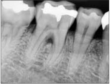
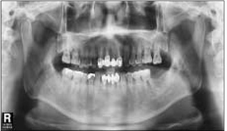
A clinical examination did not reveal a sinus tract or swelling in the patient's face. There were moderate calculus and 4-mm periodontal pockets in the lower molar. No intraoral lesions were observed, and all of the teeth in the lower and upper right quadrant responded normally to cold stimuli, with the exception of the mandibular second premolar, which had little response. Radiographically, no periapical pathosis was apparent (Figure 1 and 2).
Initially, we decided to treat the second premolar due to a tentative diagnosis of apical periodontitis for that tooth. However, no periapical pathosis was seen radographically. Pulpal and periapical diagnostic testing, in conjunction with a radiographic examination, revealed that the second premolar responded normally to all tests, including a percussion test, electrical pulp test, and cold/hot test, and that no periradicular involvement was noted. Furthermore, anesthetic test did not relieve the pain. Therefore it was decided to control the pain with analgesics and to re-evaluate the symptoms in a few days. We also considered the possibility of non-odontogenic pain such as trigeminal neuralgia, migraine, temporomandibular joint disorder, etc.
Three days later, the patient returned to the clinic complaining of intense pain and a rash on the right side of the face. The rash and blisters were localized to the right mandible and chin (Figure 3A). Therefore, we suspected that it might be non-odontogenic pain related to the trigeminal nerve, specifically related to herpes zoster. But it was difficult to accurately diagnose it as herpes zoster. Therefore, we referred the patient to a dermatologist for an accurate diagnosis. The dermatologist reported that it was diagnosed as herpes zoster involving the mandibular branch of the trigeminal nerve. The patient was given antiviral therapy by the dermatologist. It was found that the patient had been given prescriptions for betamethasone, acetaminophen, hydroxyzine, famciclovir, gabapentin, antidepressant, etc.
Two months later, the patient returned to our clinic for a follow-up evaluation of his condition. The facial lesions, rash, and blisters, had almost disappeared but the patient still noted a little pain in the right jaw area, and residual pigmented scars were present (Figure 3B). The dermatologist informed the patient that drugs might be necessary for an extended period of time.
III. DISCUSSION
Herpes zoster occurs when the varicella zoster virus that has remained latent is reactivated6). Herpes Zoster is a less common disease and the factors causing reactivation are still not well known, but it occurs more often in older and/or immunocompromised individuals7).
Clinicians should understand that when herpes zoster involves the mandibular branch, it can mimic a toothache. Because it can appear in the presence or absence of skin lesions, its diagnosis might be difficult for clinicians.
Patients with a herpes zoster infection usually progress through three stages: a prodromal stage, active stage (also called acute stage), and chronic stage8,9). The pain of the prodromal stage, 3-5 days before vesicular eruption2,10), can simulate odontogenic pain. Therefore if there is no convincing evidence of disease of the pulp, unnecessary treatment must be avoided.
In this case report, when the patient visited our clinic presenting odontogenic pain on the lower right mandible, it may have been the prodromal stage with no skin lesions. He complained of pain and a swelling sensation on the lower right mandible. It is believed that these sensory changes are the result of degeneration of nerve fibrils from viral infection activity. This usually precedes the rash of the active stage by a few hours to several days5,8,9).
Ragozziuo et al.10,11) reported that the incidence of trigeminal herpes zoster virus is relatively low, especially in the second and third branch; it only accounts for about 1.7% of all reported cases. In this report, the herpes zoster was involved with the 3rd branch, the mandibular nerve, of the trigeminal nerve. Symptoms mimic odontogenic pain, especially pulpitis in the lower right molars.
For the treatment of herpes zoster, (i) patients with herpes zoster infection should be isolated due to the contagious nature of the infection, (ii) pain should be reduced by analgesics, such as acetaminophen, codeine, and nonsteroidal anti-inflammatory agents, and (iii) antiviral therapy must be swift and precise. Acyclovir has been the drug of choice for a number of years and other antiviral agents, such as famciclovir and valacyclovir, can also be used, (iv) the treatment of post-herpetic neuralgia includes the topical use of capsaicin cream, transcutaneous nerve stimulation, topical anesthetics, injected local anesthetics, and low dose amitriptyline12). Strommen et al.9) offered an in-depth review of the use of antidepressants and neurolyptics in the management of post-herpetic neuralgia.
This case showed that the signs and symptoms of a herpes zoster infection in the mandibular branch can be misdiagnosed. During the prodromal stage, the presenting symptoms may include odontalgia, which may prove to be a diagnostic challenge for the dentist, since many diseases can cause orofacial pain that is similar to pulpitis. Therefore the diagnosis must be established before any invasive treatment, such as endodontic treatment or extraction.
- 1. Baron R. Post-herpetic neuralgia case study: optimizing pain control. Eur J Neurol. 2004;11: Suppl 1. 3-11.ArticlePubMed
- 2. Gross G, Schofer H, Wassilew S, Friese K, Timm A, Guthoff R, Pau HW, Malin JP, Wutzler P, Doerr HW. Herpes zoster guidelines of the German Dermatology Society (DDG). J Clin Virol. 2003;26: 277-289.PubMed
- 3. Ragozzino MW, Melton LJ 3rd, Kudand LT, Chu CP, Perry HO. Population-based study of herpes zoster and its sequelae. Medicine (Baltimore). 1982;61: 310-316.ArticlePubMed
- 4. Carbone V, Leonardi A, Pavese M, Raviola E, Giordano M. Herpes zoster of the trigeminal nerve: a case report and review of the literature. Minerva Stomatol. 2004;53: 49-59.PubMed
- 5. Millar EP, Troulis MJ. Herpes zoster of the trigeminal nerve: The dentists' role in diagnosis and management. J Can Dent Assoc. 1994;60: 450-453.PubMed
- 6. Gelb LD. In: Watson CPN, editor. The varicella-zoster virus. Pain research and clinical management. Vol. 8: herpes zoster and postherpetic neuralgia. 1993;Amsterdam: Elsevier; 7-25.
- 7. Ragozzino MW, Melton LJ 3rd, Kurland LT, Chu CP, Perry HO. Population-based study of herpes zoster and its sequelae. Medicine (Baltimore). 1982;61: 310-316.ArticlePubMed
- 8. Carmichael JK. Treatment of herpes Zoster and postherpetic neuralgia. Am Fam Physician. 1991;44: 203-210.PubMed
- 9. Strommen GL, Pucino F, Tight RR, Beck CL. Human infection with herpes zoster: etiology, pathophysiology, diagnosis, clinical course, and treatment. Pharmacotherapy. 1988;8: 52-68.ArticlePubMedPDF
- 10. Sigurdsson A, Jacoway JR. Herpes zoster infection presenting as an acute pulpitis. Oral Surg Oral Med Oral Pathol Oral Radiol Endod. 1995;80: 92-95.ArticlePubMed
- 11. Ragozzino MW, Melton LJ 3rd, Kudand LT, Chu CP, Perry HO. Population-based study of herpes zoster and its sequelae. Medicine (Baltimore). 1982;61: 310-316.ArticlePubMed
- 12. Tidwell E, Huston B, Burkhart N, Gutmann JL, Ellis CD. Herepes zoster of the trigeminal nerve third branch: a case report and review of the literature. Int Endod J. 1999;32: 61-66.PubMed
REFERENCES
Tables & Figures
REFERENCES
Citations
Citations to this article as recorded by 

- Herpes Zoster Accompanying Odontogenic Inflammation: A Case Report with Literature Review
Soyeon Lee, Minsik Kim, Jong-Ki Huh, Jae-Young Kim
Journal of Oral Medicine and Pain.2021; 46(1): 9. CrossRef - Recurrent Herpetic Stomatitis Mimicking Post-Root Resection Complication
Sung-Ok Hong, Jae-Kwan Lee, Hoon-Sang Chang
Journal of Dental Rehabilitation and Applied Science.2013; 29(4): 418. CrossRef - Diagnostic challenges of nonodontogenic toothache
Hyung-Ok Park, Jung-Hong Ha, Myoung-Uk Jin, Young-Kyung Kim, Sung-Kyo Kim
Restorative Dentistry & Endodontics.2012; 37(3): 170. CrossRef
Clinical diagnosis of herpes zoster presenting as odontogenic pain


Figure 1
Normal periapical radiograph of lower right first premolar and first and second molars. First and second molar have gold restorations.
Figure 2
Panoramic radiograph showing no specific pathosis on the lower left molar area.
Figure 3
(A) Three days after the initial visit: extraoral lesions or rash show the distribution of the involved nerve. (B) Two months after the dermatological referral, no rash is present but pigmented scars can be seen on the lower right facial area.
Figure 1
Figure 2
Figure 3
Clinical diagnosis of herpes zoster presenting as odontogenic pain

 KACD
KACD


 ePub Link
 Cite
Cite

