
Search
- Page Path
- HOME > Search
Research Articles
- In vitro experimental study comparing continuous and intermittent irrigation protocols: influence of sodium hypochlorite volume and contact time on tissue dissolution
- Alfredo Iandolo, Dina Abdellatif, Davide Mancino, Gwenael Rolin, Camille Coussens, Aurelian Louvrier, Felipe G Belladonna, Edouard Euvrard, Emmanuel João Nogueira Leal da Silva
- Restor Dent Endod 2025;50(4):e36. Published online October 15, 2025
- DOI: https://doi.org/10.5395/rde.2025.50.e36
-
 Abstract
 PDF
PDF PubReader
 ePub
ePub - Objectives
This study aimed to evaluate whether continuous irrigation with larger volumes or allowing sodium hypochlorite (NaOCl) resting time is more critical for pulp tissue dissolution using a controlled artificial root canal system.
Methods
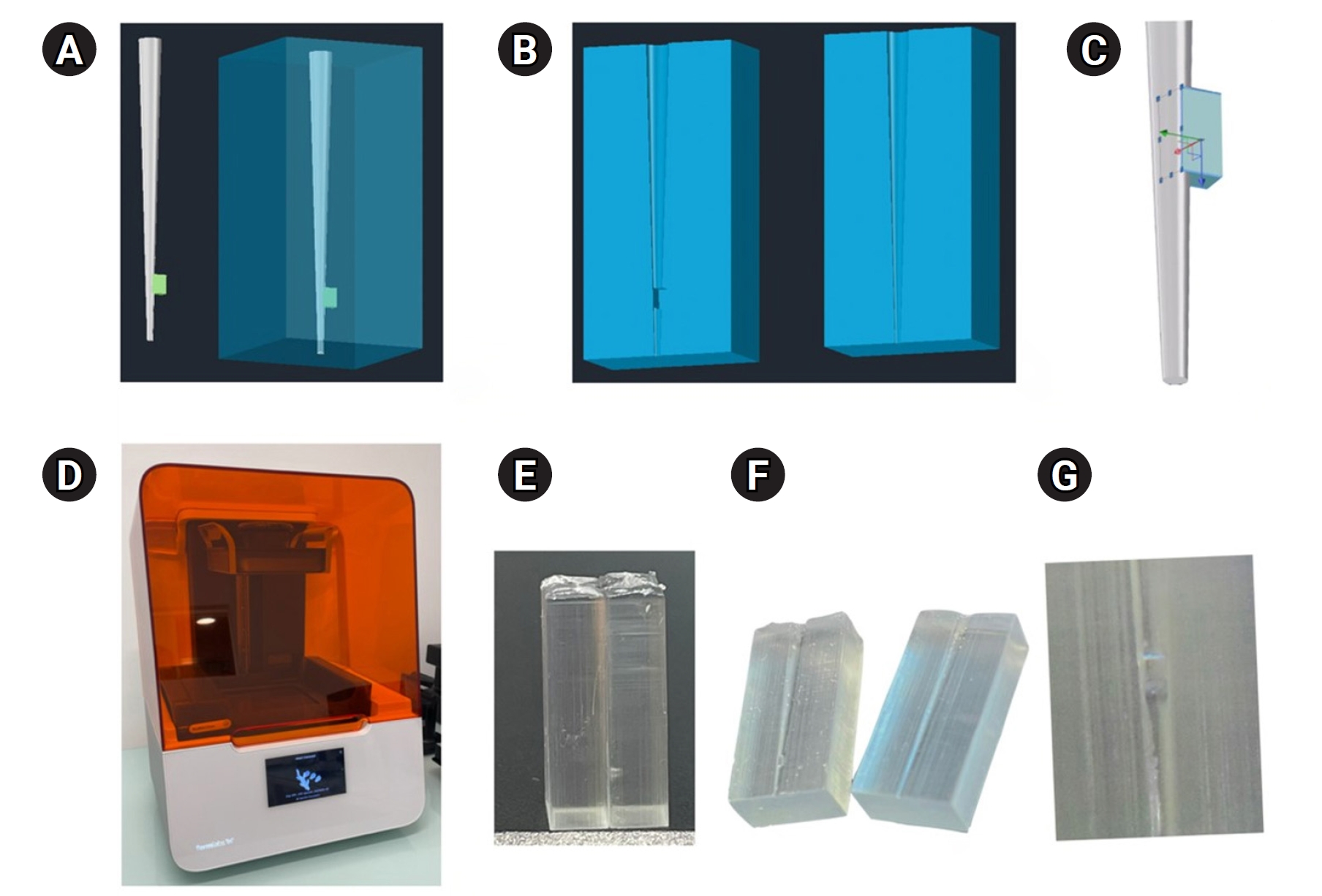
A three-dimensional printed artificial root canal with a lateral canal in the apical third was fabricated. Standardized bovine pulp tissue specimens were inserted, and three irrigation protocols were tested: group A (continuous NaOCl irrigation at 1 mL/min via syringe pump), group B (intermittent NaOCl irrigation with 0.1 mL and a 3-minute resting period), and group C (control, saline irrigation). The time for complete dissolution and the total NaOCl volume were recorded.
Results
Complete dissolution occurred in groups A and B, with significant differences in NaOCl volume and time (p < 0.05). In group A, complete dissolution was consistently observed after the 6th irrigation cycle, corresponding to a total NaOCl volume of 6.0 ± 0.66 mL per test. The average time required for complete dissolution in this group was 6 ± 0.66 minutes. In group B, complete dissolution occurred after the 4th cycle, with a total NaOCl volume of 0.4 ± 0.06 mL per test and a mean dissolution time of 12.6 ± 1.8 minutes.
Conclusions
NaOCl volume and exposure time significantly influence pulp tissue dissolution.
- 2,086 View
- 178 Download

- Epigallocatechin-3-gallate prior to composite resin in abfraction lesions: a split-mouth randomized clinical trial
- Luísa Valente Gotardo Lara Alves, Lisiane Martins Fracasso, Thiago Vinicius Cortez, Aline Evangelista Souza-Gabriel, Silmara Aparecida Milori Corona
- Restor Dent Endod 2023;48(2):e13. Published online March 20, 2023
- DOI: https://doi.org/10.5395/rde.2023.48.e13
-
Abstract
PDFPubReaderePub
Objectives Natural extracts have been investigated as a biomimetic strategy to mechanically strengthen the collagen network and control the biodegradation of extracellular matrix. This study evaluated the effect of epigallocatechin-3-gallate (EGCG) on abfraction lesions prior to the composite resin.
Materials and Methods The sample consisted of 30 patients (aged between 28 and 60 years) with abfraction lesions located in 2 homologous premolars. The teeth were randomly assigned according to dentin treatment: 0.02% EGCG solution or distilled water (control). After enamel acid etching, the solutions were applied immediately for 1 minute. The teeth were restored with Universal Adhesive (3M) and Filtek Z350 XT (3M). Analyzes were done by 2 independent examiners using modified USPHS (retention, secondary caries, marginal adaptation, and postoperative sensitivity) and photographic (color, marginal pigmentation, and anatomical form) criteria at baseline (7 days) and final (18 months). The data analysis used Friedman and Wilcoxon signed-rank tests (α = 0.05).
Results At baseline, all restorations were evaluated as alpha for all criteria. After 18 months, restorations were evaluated as alpha for secondary caries, color, and marginal pigmentation. There was significant difference between baseline and 18 months (
p = 0.009) for marginal adaptation and postoperative sensitivity (p = 0.029), but no significant difference were verified between treatments (p = 0.433). The EGCG group had a restoration retention rate of 93.3%, while the control group had 96.7%.Conclusions The application of EGCG solution on abfraction lesions did not significantly influence the survival of the restorations based on clinical and photographic criteria.
-
Citations
Citations to this article as recorded by
- Therapeutic potential of flavonoids in erosive tooth wear management: a scoping review
Gabriel Pereira Nunes, Renata de Oliveira Alves, Geórgia Rondó Peres, Priscila Toninatto Alves de Toledo, Aline Rogéria Freire de Castilho
Clinical Oral Investigations.2025;[Epub] CrossRef
- Therapeutic potential of flavonoids in erosive tooth wear management: a scoping review
- 2,212 View
- 48 Download
- 1 Web of Science
- 1 Crossref
- A new phantom to evaluate the tissue dissolution ability of endodontic irrigants and activating devices
- Kimia Khoshroo, Brinda Shah, Alexander Johnson, John Baeten, Katherine Barry, Mohammadreza Tahriri, Mohamed S. Ibrahim, Lobat Tayebi
- Restor Dent Endod 2020;45(4):e45. Published online August 24, 2020
- DOI: https://doi.org/10.5395/rde.2020.45.e45
-
Abstract
PDFPubReaderePub
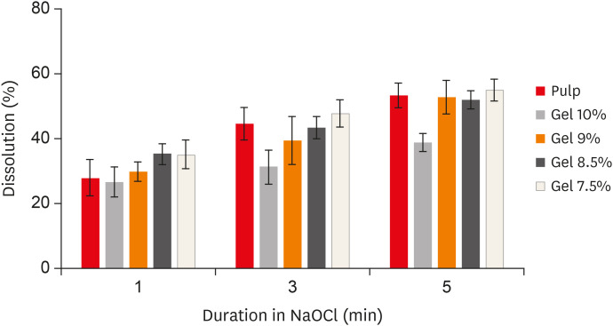
Objective The aim of this study was to introduce a gelatin/bovine serum albumin (BSA) tissue standard, which provides dissolution properties identical to those of biological tissues. Further, the study evaluated whether the utilization of endodontic activating devices led to enhanced phantom dissolution rates.
Materials and Methods Bovine pulp tissue was obtained to determine a benchmark of tissue dissolution. The surface area and mass of samples were held constant while the ratio of gelatin and BSA were varied, ranging from 7.5% to 10% gelatin and 5% BSA. Each sample was placed in an individual test tube that was filled with an appropriate sodium hypochlorite solution for 1, 3, and 5 minutes, and then removed from the solution, blotted dry, and weighed again. The remaining tissue was calculated as the percent of initial tissue to determine the tissue dissolution rate. A radiopaque agent (sodium diatrizoate) and a fluorescent dye (methylene blue) were added to the phantom to allow easy quantification of phantom dissolution in a canal block model when activated using ultrasonic (EndoUltra) or sonic (EndoActivator) energy.
Results The 9% gelatin + 5% BSA phantom showed statistically equivalent dissolution to bovine pulp tissue at all time intervals. Furthermore, the EndoUltra yielded significantly more phantom dissolution in the canal block than the EndoActivator or syringe irrigation.
Conclusions Our phantom is comparable to biological tissue in terms of tissue dissolution and could be utilized for
in vitro tests due to its injectability and detectability.-
Citations
Citations to this article as recorded by- Evaluation of pulp tissue dissolving efficiency of sodium and calcium hypochlorite solutions activated by ultrasonics and laser: an in vitro study
Oznur Ozturk, Ozgur Genc Sen
BMC Oral Health.2024;[Epub] CrossRef
- Evaluation of pulp tissue dissolving efficiency of sodium and calcium hypochlorite solutions activated by ultrasonics and laser: an in vitro study
- 2,336 View
- 12 Download
- 1 Crossref
- Smear layer removal by different chemical solutions used with or without ultrasonic activation after post preparation
- Daniel Poletto, Ana Claudia Poletto, Andressa Cavalaro, Ricardo Machado, Leopoldo Cosme-Silva, Cássia Cilene Dezan Garbelini, Márcio Grama Hoeppner
- Restor Dent Endod 2017;42(4):324-331. Published online November 1, 2017
- DOI: https://doi.org/10.5395/rde.2017.42.4.324
-
Abstract
PDFPubReaderePub
Objectives This study evaluated smear layer removal by different chemical solutions used with or without ultrasonic activation after post preparation.
Materials and Methods Forty-five extracted uniradicular human mandibular premolars with single canals were treated endodontically. The cervical and middle thirds of the fillings were then removed, and the specimens were divided into 9 groups: G1, saline solution (NaCl); G2, 2.5% sodium hypochlorite (NaOCl); G3, 2% chlorhexidine (CHX); G4, 11.5% polyacrylic acid (PAA); G5, 17% ethylenediaminetetraacetic acid (EDTA). For the groups 6, 7, 8, and 9, the same solutions used in the groups 2, 3, 4, and 5 were used, respectively, but activated with ultrasonic activation. Afterwards, the roots were analyzed by a score considering the images obtained from a scanning electron microscope.
Results EDTA achieved the best performance compared with the other solutions evaluated regardless of the irrigation method (
p < 0.05).Conclusions Ultrasonic activation did not significantly influence smear layer removal.
-
Citations
Citations to this article as recorded by- Cerium Oxide Nanoparticle Loaded with Toluidine Blue as Cavity Disinfectant Activated via Light-Emitting Diode on the Shear Bond Strength and Resin Tag Length of Universal Adhesive: A Scanning Electron Microscope-EDX Study
Amer M. Alanazi, Syed Hussain Askary, Ibrahim Warsi, Aamir Afzal, Muhammad Omar Niaz, Ambrina Qureshi
Photobiomodulation, Photomedicine, and Laser Surgery.2026;[Epub] CrossRef - O papel do ultrassom no tratamento e retratamento de canais radiculares: Revisão de literatura
Carlos Roberto Souza Hipp, Joaquim Carlos Fest da Silveira, Luiz Felipe Gilson de Oliveira Rangel, Tatiana Federici de Souza Fest da Silveira, Carla Minozzo Mello, Rodrigo Simões de Oliveira
Research, Society and Development.2025; 14(8): e1314849323. CrossRef - Effect of sodium hypochlorite, ethylenediaminetetraacetic acid, and dual-rinse irrigation on dentin adhesion using an etch-and-rinse or self-etch approach
Matej Par, Tobias Steffen, Selinay Dogan, Noah Walser, Tobias T. Tauböck
Scientific Reports.2024;[Epub] CrossRef - Evaluation of Effect of Poloxamer on Smear Layer Removal Using Apical Negative Pressure: An In Vitro Scanning Electron Microscopy Study
Chandra Prabha, Chitharanjan Shetty, Aditya Shetty
Journal of International Oral Health.2024; 16(6): 498. CrossRef - Laboratory Assessment of Antibacterial Efficacy of Five Different Herbal-based Potential Endodontic Irrigants
Anjali A Oak, Kailash Attur, Kamal Bagda, Nitish Mathur, Lubna Mohammad, Nikhat M Attar
Advances in Human Biology.2023; 13(4): 350. CrossRef - Dental Surface Conditioning Techniques to Increase the Micromechanical Retention to Fiberglass Posts: A Literature Review
Paulina Leticia Moreno-Sánchez, Maricela Ramírez-Álvarez, Alfredo del Rosario Ayala-Ham, Erika de Lourdes Silva-Benítez, Miguel Ángel Casillas-Santana, Diana Leyva del Rio, León Francisco Espinosa-Cristóbal, Erik Lizárraga-Verdugo, Mariana Melisa Avendaño
Applied Sciences.2023; 13(14): 8083. CrossRef - Effect of irrigation protocols on smear layer removal, bond strength and nanoleakage of fiber posts using a self-adhesive resin cement
Rodrigo Stadler Alessi, Renata Terumi Jitumori, Bruna Fortes Bittencourt, Giovana Mongruel Gomes, João Carlos Gomes
Restorative Dentistry & Endodontics.2023;[Epub] CrossRef - Effects of using different root canal sealers and protocols for cleaning intraradicular dentin on the bond strength of a composite resin used to reinforce weakened roots
Luiz Pascoal Vansan, Ricardo Machado, Celso Bernardes de Souza, Ricardo Gariba, Antônio Miranda da Cruz, Cinara Muniz, Jardel FranciscoX Jardel Francisco Mazzi-Chaves, Lucas da Fonseca Roberti Garcia
Journal of Oral Research.2022; 11(6): 1. CrossRef - Influence of the use of chelating agents as final irrigant on the push‐out bond strength of epoxy resin‐based root canal sealers: A systematic review
Carla M. Augusto, Miguel A. Cunha Neto, Karem P. Pinto, Ana Flavia A. Barbosa, Emmanuel J. N. L. Silva, Ana Paula P. dos Santos, Luciana M. Sassone
Australian Endodontic Journal.2022; 48(2): 347. CrossRef - Adhesion and whitening efficacy of P11-4 self-assembling peptide and HAP suspension after using NaOCl as a pre-treatment agent
Niloofar Hojabri, Karl-Heinz Kunzelmann
BMC Oral Health.2022;[Epub] CrossRef - Influence of resin cements and root canal disinfection techniques on the adhesive bond strength of fibre reinforced composite post to radicular dentin
Zaid A. Al Jeaidi
Photodiagnosis and Photodynamic Therapy.2021; 33: 102108. CrossRef - The Antibacterial Efficacy and In Vivo Toxicity of Sodium Hypochlorite and Electrolyzed Oxidizing (EO) Water-Based Endodontic Irrigating Solutions
Sung-Chih Hsieh, Nai-Chia Teng, Chia Chun Chu, You-Tai Chu, Chung-He Chen, Liang-Yu Chang, Chieh-Yun Hsu, Ching-Shuan Huang, Grace Ying-Wen Hsiao, Jen-Chang Yang
Materials.2020; 13(2): 260. CrossRef
- Cerium Oxide Nanoparticle Loaded with Toluidine Blue as Cavity Disinfectant Activated via Light-Emitting Diode on the Shear Bond Strength and Resin Tag Length of Universal Adhesive: A Scanning Electron Microscope-EDX Study
- 3,220 View
- 19 Download
- 12 Crossref
- Evaluation of internal adaptation of dental adhesive restorations using micro-CT
- Oh-Hyun Kwon, Sung-Ho Park
- Restor Dent Endod 2012;37(1):41-49. Published online March 2, 2012
- DOI: https://doi.org/10.5395/rde.2012.37.1.41
-
Abstract
PDFPubReaderePub
Objectives The internal adaptation of composite restorations with or without resin modified glass ionomer cement (RMGIC) was analyzed non-destructively using Microcomputed tomography (micro-CT).
Materials and Methods Thirty intact human teeth were used. The specimens were divided into 3 groups. In the control group, the cavities were etched with 10% phosphoric acid for 15 sec. Composite resin was filled into the cavity without adhesive. In group 1, light cured glass ionomer cement (GIC, Fuji II LC, GC) was applied as a base. The cavities were then etched, bonded, light cured and filled with composites. In group 2, the cavities were then etched, bonded, light cured and filled with composites without base application. They were immersed in a 25% silver nitrate solution. Micro-CT was performed before and after mechanical loading. One-way ANOVA with Duncan analysis was used to compare the internal adaptation between the groups before or after loading. A paired
t -test was used to compare internal adaptation before and after mechanical loading. All statistical inferences were made within the 95% confidence interval.Results The silver nitrate solution successfully penetrated into the dentinal tubules from the pulp spaces, and infiltrated into the gap between restoration and pulpal floor. Group 2 showed a lower adaptation than the control group and group 1 (
p < 0.05). There was no significant difference between the control group and group 1. For all groups, there was a significant difference between before and after mechanical loading (p < 0.05).Conclusions The internal adaptation before and after loading was better when composites were bonded to tooth using adhesive than composites based with RMGIC.
-
Citations
Citations to this article as recorded by- Micro-computed tomography in preventive and restorative dental research: A review
Mehrsima Ghavami-Lahiji, Reza Tayefeh Davalloo, Gelareh Tajziehchi, Paria Shams
Imaging Science in Dentistry.2021; 51(4): 341. CrossRef - Gaps at the interface between dentine and self‐adhesive resin cement in post‐endodontic restorations quantified in 3D by phase contrast‐enhanced micro‐CT
A. P. Soares, K. Bitter, A. Lagrange, A. Rack, H. Shemesh, P. Zaslansky
International Endodontic Journal.2020; 53(3): 392. CrossRef - Hard X-ray phase-contrast-enhanced micro-CT for quantifying interfaces within brittle dense root-filling-restored human teeth
Ana Prates Soares, Uwe Blunck, Kerstin Bitter, Sebastian Paris, Alexander Rack, Paul Zaslansky
Journal of Synchrotron Radiation.2020; 27(4): 1015. CrossRef - Comparison of micro-CT and conventional dye penetration for microleakage assessment after different aging conditions
Rayssa Ferreira Zanatta, Annette Wiegand, Christian Dullin, Alessandra Bühler Borges, Carlos Rocha Gomes Torres, Marta Rizk
International Journal of Adhesion and Adhesives.2019; 89: 161. CrossRef - Comparison of Internal Adaptation of Bulk-fill and Increment-fill Resin Composite Materials
FS Alqudaihi, NB Cook, KE Diefenderfer, MC Bottino, JA Platt
Operative Dentistry.2019; 44(1): E32. CrossRef - Polymerization shrinkage assessment of dental resin composites: a literature review
Dalia Kaisarly, Moataz El Gezawi
Odontology.2016; 104(3): 257. CrossRef - Non-destructive evaluation of an internal adaptation of resin composite restoration with swept-source optical coherence tomography and micro-CT
Seung-Hoon Han, Alireza Sadr, Junji Tagami, Sung-Ho Park
Dental Materials.2016; 32(1): e1. CrossRef - Micro-CT evaluation of internal adaptation in resin fillings with different dentin adhesives
Seung-Hoon Han, Sung-Ho Park
Restorative Dentistry & Endodontics.2014; 39(1): 24. CrossRef
- Micro-computed tomography in preventive and restorative dental research: A review
- 2,201 View
- 9 Download
- 8 Crossref
Original Articles
- Effect of soft chelating irrigation on the sealing ability of GP/AH Plus root fillings
- Yi-Suk Yu, Tae-Gun Kim, Kwang-Won Lee, Mi-Kyung Yu
- J Korean Acad Conserv Dent 2009;34(6):484-490. Published online November 30, 2009
- DOI: https://doi.org/10.5395/JKACD.2009.34.6.484
-
Abstract
PDFPubReaderePub
The purpose of this study was to evaluate the effect of soft chelating irrigant on the sealing ability of root fillings by using a glucose leakage test.
A total of 45 single-rooted teeth were selected for the study. The teeth were decoronated leaving a total length of 13mm. The root canals prepared using K3 NiTi rotary instruments to an apical dimension of size 45(0.06 taper). The specimens were then randomly divided into 3 experimental groups of 13 roots each and 2 control groups of 3 roots each. Specimen in each group were prepared with different irrigation protocols : group 1, 2.5% NaOCl; group 2, 2.5% NaOCl and 17% EDTA; group 3, 2.5% NaOCl and 15% HEBP. The root canals were filled with gutta-percha and AH Plus sealer using lateral condensation. After 7 days in 37℃, 100% humidity, the coronal-to-apical microleakage was evaluated quantitatively using a glucose leakage model. The leaked glucose concentration was measured with spectrophotometry at 1, 4, 7, 14, 21 and 28 days.
There was a tendency of increase in leakage in all experimental groups during experimental period. HEBP-treated dentin showed no significant difference with EDTA-treated dentin during experimental period. From the 21th day onward, HEBP-treated dentin showed significantly lower leakage than smear-covered dentin. HEBP-treated dentin displayed a similar sealing pattern to EDTA-treated dentin and a better sealing ability than smear-covered dentin. Consequently, a soft chelator(HEBP) could be considered as the possible alternative to EDTA.
-
Citations
Citations to this article as recorded by- Effect of moisture on sealing ability of root canal filling with different types of sealer through the glucose penetration model
Jin-Ah Jang, Hee-Lyang Kim, Mi-Ja Her, Kwang-Won Lee, Mi-Kyung Yu
Journal of Korean Academy of Conservative Dentistry.2010; 35(5): 335. CrossRef
- Effect of moisture on sealing ability of root canal filling with different types of sealer through the glucose penetration model
- 1,564 View
- 3 Download
- 1 Crossref
- CYCLIC FATIGUE OF THE SODIUM HYPOCHLORITE TREATED AND /OR STEAM AUTOCLAVED NICKEL-TITANIUM ENDODONTIC FILES
- Hye-Young Cho, Il-Young Jung, Chan-Young Lee, Euiseong Kim
- J Korean Acad Conserv Dent 2008;33(1):54-65. Published online January 14, 2008
- DOI: https://doi.org/10.5395/JKACD.2008.33.1.054
-
Abstract
PDFPubReaderePub
Abstract The purpose of this study was to determine the effect of sodium hypochlorite and steam autoclaving on the cyclic fatigue of nickel-titanium endodontic files.
Two types of files with a .06 taper and #30 were used, K3® (SybronEndo, Glendora, California, USA) and Hero642®(Micro-Mega, Besançon, France).
The files were divided into 6 experimental groups containing 10 files each group depending the soaking time in 6% sodium hypochlorite solution and number of cycles of steam autoclave. After sterilization, a cyclic fatigue test was performed on each file, and the fracture time was recorded in seconds. The control group underwent the cyclic fatigue test only. After the test, the surface characteristics of the files were observed using scanning electron microscopy (SEM).
All groups containing the Hero 642® files showed a similar cyclic fatigue fracture time. However, the cyclic fatigue fracture time with the K3® files was significantly shorter in groups which were treated with sodium hypochlorite than in the control group (P < 0.05). SEM revealed both Hero642® and K3® files to have significant corrosion on the file surface in groups treated with sodium hypochlorite, compared with the sharp and regular blades of the control group. K3® files showed more corrosion than the Hero642® files. Bluntness of the blades of the K3® file was observed in groups treated with steam autoclave. Although there was no obvious destruction on the surface of steam autoclaved Hero642® files, slight bluntness was observed.
Sterilizing with a steam autoclave is much less destructive to K3® files than sodium hypochlorite. The longer time exposed to sodium hypochlorite, the more destructive pattern was shown on the blades of the files. Therefore, when using sodium hypochlorite solution, the exposure time should be as short as possible in order to prevent corrosion and increase the cyclic fatigue fracture time.
- 1,200 View
- 1 Download
- The influence of pH and lactic acid concentration on the formation of artificial root caries in acid buffer solution
- Hyun-Suk Oh, Byoung-Duck Roh, Chan-Young Lee
- J Korean Acad Conserv Dent 2007;32(1):47-60. Published online January 31, 2007
- DOI: https://doi.org/10.5395/JKACD.2007.32.1.047
-
Abstract
PDFPubReaderePub
The purpose of this study is to compare and to evaluate the effect of pH and lactic acid concentration on the progression of artificial root caries lesion using polarizing microscope, and to evaluate the morphological changes of hydroxyapatite crystals of the demineralized area and to investigate the process of demineralization using scanning electron microscope.
Artificial root caries lesion was created by dividing specimens into 3 pH groups (pH 4.3, 5.0, 5.5), and each pH group was divided into 3 lactic acid concentration groups (25 mM, 50 mM, 100 mM). Each group was immersed in acid buffer solution for 5 days and examined. The results were as follows:
1. Under polarized microscope, the depth of lesion was more effected by the lactic acid concentration rather than the pH.
2. Under scanning electron microscope, dissolution of hydroxyapatite crystals were increased as the lactic acid concentration increased and the pH decreased.
3. Demineralized hydroxyapatite crystals showed peripheral dissolution and decreased size and number within cluster of hydroxyapatite crystals and widening of intercluster and intercrystal spaces as the pH decreased and the lactic acid concentration increased.
4. Under scanning electron microscope evaluation of the surface zone, clusters of hydroxyapatite crystals were dissolved, and dissolution and reattachment of crystals on the surface of collagen fibrils were observed as the lactic acid concentration increased.
5. Under scanning electron microscope, demineralization of dentin occurred not only independently but also with remineralization simultaneously.
In conclusion, the study showed that pH and lactic acid concentration influenced the rate of progression of the lesion in artificial root caries. Demineralization process was progressed from the surface of the cluster of hydroxyapatite crystals and the morphology of hydroxyapatite crystals changed from round or elliptical shape into irregular shape as time elapsed.
- 1,311 View
- 7 Download
- The influence of the degree of saturation of acidulated buffer solutions in the root dentin demineralization
- Hye-Sil Kang, Chan-Young Lee
- J Korean Acad Conserv Dent 2004;29(5):454-461. Published online September 30, 2004
- DOI: https://doi.org/10.5395/JKACD.2004.29.5.454
-
Abstract
PDFPubReaderePub
The purpose of this study is to compare and to evaluate the effects of the degree of saturation on the progression of artificial root caries lesion.
A total of 8 human premolars without any defects and cracks selected and the cementum were removed and the teeth were cleaned with ultrasonic device and pumice without fluoride.
Each tooth was sectioned into 6 pieces and they were ground with #800 sandpaper until they had a thickness of 200µm. Specimens were applied with nail vanish except for the 2-3 mm window area after application of bonding agent. Under the constant pH, the specimens were divided into 6 groups (degree of saturation; 0.1415, 0.1503, 0.1597, 0.1676, 0.1771, 0.1977). Each group was immersed in acid buffer solution for 1, 2, 3, 5 days under controlled temperature (25℃) and imbibed in water and examined using the polarizing microscope.
The results were as follows
1. Although the degree of saturation of demineralization solution decreased, the depth of penetration in the dentin was constant.
2. Erosion was observed on the surface of all the teeth in the group I, II. In the group III, IV, V, surfaces were not changed. The teeth in the group VI showed the more mineralized surface but not the shape of the dentinal tubules distinctively.
3. In all groups, the lesion progressed rapidly at the first day of the experiment, but increased gradually as time elapsed.
-
Citations
Citations to this article as recorded by- Management of white spots: resin infiltration technique and microabrasion
Jeong-Hye Son, Bock Hur, Hyeon-Cheol Kim, Jeong-Kil Park
Journal of Korean Academy of Conservative Dentistry.2011; 36(1): 66. CrossRef
- Management of white spots: resin infiltration technique and microabrasion
- 1,352 View
- 1 Download
- 1 Crossref
-
In vivo quantitative analysis of remineralization effect of remineralization solution "R" of incipient enamel dental caries - Myung-Eun Kim, Il-young Jung, Kee-Yeon Kum, Chang-young Lee, Byoung-Duck Roh
- J Korean Acad Conserv Dent 2002;27(2):175-182. Published online March 31, 2002
- DOI: https://doi.org/10.5395/JKACD.2002.27.2.175
-
Abstract
PDFPubReaderePub
Dental caries is a chronic disease that causes the destruction of tooth structure by the interaction of plaque bacteria, food debris, and saliva.
There has been attempts to induce remineralization by supersaturating the intra-oral environment around the surface enamel, where there is incipient caries.
In this study, supersaturated remineralized solution "R" was applied to specimens with incipient enamel caries, and the quantitative ananlysis of remineralization was evaluated using microradiography. Thirty subjects volunteered to participate in this study. Removable appliances were constructed for the subjects, and the enamel specimen with incipient caries were embedded in the appliances. The subjects wore the intra-oral appliance for 15 days except while eating and sleeping.
The removable appliance were soaked in supersaturated solution "R", saline, or Senstime® to expose the specimen to those solutions three times a day, 5 minutes each time. After 15 days, microradiography was retaken to compare and evaluate remineralization.
The results were as the following:
1. The ratio of remineralized area to demineralized area was significantly higher in the supersaturated solution "R" and Senstime® than in the saline. (p<0.05)
2. Remineralization in the supersaturated buffer solution "R" occurred in the significantly deeper parts of the tooth, compared to the Senstime® group containing high concentration of fluoride.(p<0.05)
As in the above results, the remineralization effect of remineralized buffer solution "R" on incipient enamel caries has been proven. For clinical utilization, further studies on soft tissue reaction and the effect on dentin and cementum are necessary.
In conclusion compared to commercially available fluoride solution, remineralization solution "R" showed better remineralization effect on early enamel caries lesion, so it is considered as effecient solution for clinical application.
-
Citations
Citations to this article as recorded by- Color and hardness changes in artificial white spot lesions after resin infiltration
Ji-Hoon Kim, Ho-Hyun Son, Juhea Chang
Restorative Dentistry & Endodontics.2012; 37(2): 90. CrossRef - Changes in surface content and crystal structure after fluoride gel or hydroxyapatite paste application on stripped enamel
Sang-Cheol Kim, Hyun-Sil Hong, Young-Cheol Hwang
The Korean Journal of Orthodontics.2008; 38(6): 407. CrossRef
- Color and hardness changes in artificial white spot lesions after resin infiltration
- 1,645 View
- 10 Download
- 2 Crossref
 First
First Prev
Prev

