Search
- Page Path
- HOME > Search
- Analysis of the reciprocating kinematics of the VDW Silver Reciproc, E-Connect Pro, Ecom, and Endopen endodontic motors: an in vitro experimental study
- Cristielly França, Juliana D. Bronzato, Dieimes Braambati, Adriana de-Jesus-Soares, Carla C. R. B. Félix, Michelle A. N. S. Ferreira, Marcos Frozoni
- Received August 18, 2025 Accepted October 12, 2025 Published online January 20, 2026
- DOI: https://doi.org/10.5395/rde.2026.51.e5 [Epub ahead of print]
-
 Abstract
 PDF
PDF PubReader
 ePub
ePub - Objectives
This study aimed to evaluate the actual parameters of four endodontic motors, each adjusted for reciprocating motion, and compare them to the manufacturers’ declared values.
Methods
The motors used were the VDW Silver Reciproc (VDW GmbH), E-Connect Pro (MK Life), Ecom (Woodpecker), and Endopen (Schuster Woodpecker). A custom optical target was attached to the motor contra-angle, the movements were recorded with a high-resolution camera, and the images were analyzed. Engagement, disengagement, net angles, and speed for each operation cycle, duration of clockwise (CW) and counter-clockwise (CCW) movement, duration of standstill after CW and CCW movement, and the number of cycles to complete a full rotation were analyzed. The data were statistically analyzed at a significance level of 5%. The replicability of all reciprocal parameters analyzed was statistically different from that reported by the manufacturers.
Results
There was no statistically significant difference between the VDW Silver Reciproc, Ecom, and Endopen for the engagement angle. The E-Connect Pro was the least reliable at the 150°/30° settings for both angle parameters. There was no significant difference between the set and actual cycle net angles for the VDW Silver Reciproc (p = 0.493). While the actual values for the Ecom and E-Connect Pro were significantly higher than the set (p < 0.001), the actual values for the Endopen were significantly lower than the set (p < 0.001).
Conclusions
Experiments on four commercially available reciprocating endodontic motors revealed that the actual motor values differed significantly from the set values.
- 57 View
- 3 Download

- Shaping ability and cyclic fatigue resistance between Genius ProFlex, ZenFlex, and TruNatomy rotary systems: an experimental study
- Raimundo Sales de Oliveira Neto, Murilo Priori Alcalde, Pedro Cesar Gomes Titato, Pedro Henrique Souza Calefi, Carlos Alberto Spironelli Ramos, Guilherme Ferreira da Silva, Rodrigo Ricci Vivan, Marco Antonio Hungaro Duarte
- Restor Dent Endod 2025;50(1):e9. Published online February 13, 2025
- DOI: https://doi.org/10.5395/rde.2025.50.e9

-
 Abstract
 PDF
PDF PubReader
 ePub
ePub - Objectives
The aim of this study was to investigate the efficacy of three newly introduced rotary endodontic systems: Genius ProFlex (Medidenta), TruNatomy (Dentsply Maillefer), and ZenFlex (Kerr).
Methods
Forty-five mandibular molars with root canal curvatures <5° were utilized. Micro-computed tomography scans were performed pre- and post-preparation to assess apical transportation, centralization, percentage of dentin wear, and canal volume alterations. Eight instruments of each diameter underwent cyclic fatigue testing.
Results
The percentage of dentin wear on mesial and distal walls showed no significant differences among ZenFlex, TruNatomy, and Genius ProFlex at 1, 2, 3, and 4 mm from the apical foramen and root canal orifice (p > 0.05). Centering ability varied in the mesiolingual canal (p < 0.05). No notable differences were observed in transportation (p > 0.05). Genius ProFlex demonstrated lower volumetric changes (p < 0.05). There were significant differences in cyclic fatigue, with higher values for Genius ProFlex and lower values for TruNatomy (p < 0.05).
Conclusions
The three nickel-titanium rotary instruments are safe and efficient for root canal preparation, with Genius ProFlex exhibiting superior cyclic fatigue resistance.
- 2,895 View
- 159 Download

-
Influence of disinfecting solutions on the surface topography of gutta-percha cones: a systematic review of
in vitro studies - Lora Mishra, Gathani Dash, Naomi Ranjan Singh, Manoj Kumar, Saurav Panda, Franck Diemer, Monika Lukomska-Szymanska, Barbara Lapinska, Abdul Samad Khan
- Restor Dent Endod 2024;49(4):e42. Published online November 1, 2024
- DOI: https://doi.org/10.5395/rde.2024.49.e42
-
 Abstract
 PDF
PDF Supplementary Material
 PubReader
 ePub
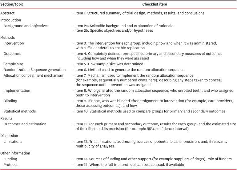
ePub The surface integrity of gutta-percha cones is a crucial factor in the success of endodontic procedures. Disinfecting solutions play a pivotal role in sterilizing gutta-percha cones, but their influence on gutta-percha surface topography remains a subject of concern. This systematic review aimed to present a qualitative synthesis of available laboratory studies assessing the influence of disinfecting solutions on the surface topography of gutta-percha and offers insights into the implications for clinical practice. The present review followed PRISMA 2020 guidelines. An advanced database search was performed in PubMed, Google Scholar, Embase, Scopus, LILAC, non-indexed citations and reference lists of eligible studies in May 2024. Laboratory studies, in English language, were considered for inclusion. The quality (risk of bias) of the included studies was assessed using parameters for
in vitro studies. A total of 28 studies were included in the qualitative synthesis. Based on the included in vitro studies, surface deposits and alterations in the physical properties of gutta-percha cones were observed after the disinfection protocol. A comprehensive review of the available literature indicates that the choice of disinfecting solution, its concentration, and immersion time significantly affect the surface topography of gutta-percha cones.-
Citations
Citations to this article as recorded by- In Vitro Evaluation of Disinfectants on Gutta-Percha Cones: Antimicrobial Efficacy Against Enterococcus faecalis and Candida albicans
Tringa Kelmendi, Donika Bajrami Shabani, Aida Meto, Hani Ounsi
Journal of Clinical Medicine.2025; 14(19): 6846. CrossRef
- In Vitro Evaluation of Disinfectants on Gutta-Percha Cones: Antimicrobial Efficacy Against Enterococcus faecalis and Candida albicans
- 3,601 View
- 189 Download
- 1 Web of Science
- 1 Crossref

-
Fracture resistance after root canal filling removal using ProTaper Next, ProTaper Universal Retreatment or hybrid instrumentation: an
ex vivo study - Hadeel Hassan Hanafy, Marwa Mahmoud Bedier, Suzan Abdul Wanees Amin
- Restor Dent Endod 2024;49(4):e38. Published online October 11, 2024
- DOI: https://doi.org/10.5395/rde.2024.49.e38
-
 Abstract
 PDF
PDF PubReader
 ePub
ePub Objectives This study evaluated the effect of ProTaper Next (PTN), ProTaper Universal Retreatment (PTR) and hybrid instrumentation (HI) for canal filling removal on the fracture resistance (FR), mode of failure (MoF), and filling removal time.
Materials and Methods Ninety-six, mandibular premolars were decoronated and randomly divided into 6 groups (
n = 16), as follows: sound (S), untreated canals; prepared teeth (P), canals only prepared to ProTaper Universal finishing instrument (F4); endodontically-treated (ET), prepared and obturated canals using the single-cone technique; and groups PTN, PTR, and HI where filling was removed using PTN, PTR, or HI respectively. FR under vertical loading; MoF and time were assessed. Data were analyzed (Significance level [α] = 0.05).Results There was a significant difference in FR among all groups (
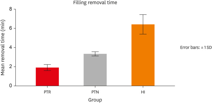
p < 0.001) (HI < P < PTN < S < ET < PTR). HI showed lower FR than S, ET and PTR, and P showed lower FR than PTR (p < 0.05). For experimental groups, there was a significant difference between every group pair (p < 0.05) No significant difference was found regarding MoF distribution (p > 0.05). HI required the highest filling removal time, while PTR required the least (p < 0.05 between every group pair).Conclusions The effect of filling removal on FR may depend on the filling removal technique/system used. PTR could be faster and protect against fracture followed by PTN; HI could adversely affect FR. FR may be associated with filling removal time.
- 2,816 View
- 122 Download

- Comparison of shaping ability of the Reciproc Blue and One Curve with or without glide path in simulated S-shaped root canals
- Vincenzo Biasillo, Raffaella Castagnola, Mauro Colangeli, Claudia Panzetta, Irene Minciacchi, Gianluca Plotino, Simone Staffoli, Luca Marigo, Nicola Maria Grande
- Restor Dent Endod 2022;47(1):e3. Published online December 28, 2022
- DOI: https://doi.org/10.5395/rde.2022.47.e3
-
 Abstract
 PDF
PDF PubReader
 ePub
ePub Objectives This study aimed to assess the impact of a glide-path on the shaping ability of 2 single-file instruments and to compare the centering ability, maintenance of original canal curvatures and area of instrumentation in simulated S-shaped root canals.
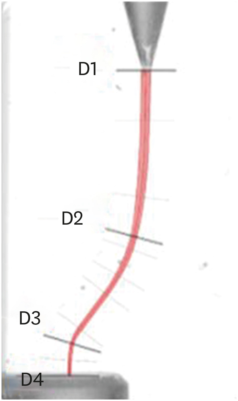
Materials and Methods Forty simulated S-shaped root canals were used and were prepared with One Curve (group OC), One G and OC (group GOC), Reciproc Blue (group RB) and R-Pilot and RB (group PRB) and scanned before and after instrumentation. The images were analyzed using AutoCAD. After superimposing the samples, 4 levels (D1, D2, D3, and D4) and 2 angles (Δ1 and Δ2) were established to evaluate the centering ability and modification of the canal curvatures. Then, the area of instrumentation (ΔA) was measured. The data were analyzed using 2-way analysis of variance and Tukey's test for multiple comparisons (
p < 0.05).Results Regarding the centering ability in the apical part (D3, D4), the use of the glide-path yielded better results than the single-file groups. Among the groups at D4, OC showed the worst results (
p < 0.05). The OC system removed less material (ΔA) than the RB system, and for Δ1, OC yielded a worse result than RB (p < 0.05).Conclusions The glide-path improved the centering ability in the apical part of the simulated S-shaped canals. The RB system showed a better centering ability in the apical part and major respect of the canal curvatures compared with OC system.
-
Citations
Citations to this article as recorded by- Evaluation of Apical Debris Extrusion and the Remaining Canal Material during Retreatment of a Bioceramic Sealer by the XP-endo Finisher File System, Followed by Various Supplementary Methods: An in Vitro Study
Paras Mull Gehlot, Parvathi Sudeep, Annapoorna B Mariswamy
World Journal of Dentistry.2025; 15(10): 837. CrossRef - Shaping Ability of Rotary NiTi Systems in S‐Shaped Root Canals of Mandibular Molars
Renata M. S. Leal, Emmanuel J. N. L. Silva, Maria C. B. P. Campos, Clarissa T. Rodrigues, Marco A. H. Duarte, Bruno C. Cavenago
Australian Endodontic Journal.2025; 51(1): 133. CrossRef - Comparison of Debris Extrusion and Preparation Time by Traverse, R‐Motion Glider C, and Other Glide Path Systems in Severely Curved Canals
Taher Al Omari, Layla Hassouneh, Khawlah Albashaireh, Alaa Dkmak, Rami Albanna, Ali Al-Mohammed, Ahmed Jamleh, Lucas da Fonseca Roberti Garcia
International Journal of Dentistry.2025;[Epub] CrossRef - Glide Path – An Ineluctable Route for Successful Endodontic Mechanics: A Literature Review
Mahima Bharat Mehta, Anupam Sharma, Aniket Jadhav, Aishwarya Handa, Abhijit Bajirao Jadhav, Ashwini A. Narayanan
Journal of the International Clinical Dental Research Organization.2024; 16(2): 101. CrossRef - Screw-in force, torque generation, and performance of glide-path files with three rotation kinetics
Jee-Yeon Woo, Ji-Hyun Jang, Seok Woo Chang, Soram Oh
Odontology.2024; 112(3): 761. CrossRef - Glide Path in Endodontics: A Literature Review of Current Knowledge
Vlad Mircea Lup, Giulia Malvicini, Carlo Gaeta, Simone Grandini, Gabriela Ciavoi
Dentistry Journal.2024; 12(8): 257. CrossRef - In Vitro Research Methods Used to Evaluate Shaping Ability of Rotary Endodontic Files—A Literature Review
Ranya F. Elemam, Ana Mano Azul, João Dias, Khaled El Sahli, Renato de Toledo Leonardo
Dentistry Journal.2024; 12(10): 334. CrossRef - Endodontic glide path - importance and performance techniques
Milica Jovanovic-Medojevic, Мiljan Stosic, Vanja Opacic-Galic, Violeta Petrovic
Srpski arhiv za celokupno lekarstvo.2023; 151(5-6): 380. CrossRef
- Evaluation of Apical Debris Extrusion and the Remaining Canal Material during Retreatment of a Bioceramic Sealer by the XP-endo Finisher File System, Followed by Various Supplementary Methods: An in Vitro Study
- 2,836 View
- 41 Download
- 8 Web of Science
- 8 Crossref

- Effect of intracanal cryotherapy on postoperative pain after endodontic treatment: systematic review with meta-analysis
- Fernanda Garcias Hespanhol, Ludmila Silva Guimarães, Lívia Azeredo Alves Antunes, Leonardo Santos Antunes
- Restor Dent Endod 2022;47(3):e30. Published online July 4, 2022
- DOI: https://doi.org/10.5395/rde.2022.47.e30
-
 Abstract
 PDF
PDF PubReader
 ePub
ePub Objectives This study aimed to evaluate the effectiveness of final irrigation with cold saline solution after endodontic treatment compared with saline solution at room temperature against postoperative pain following endodontic treatment.
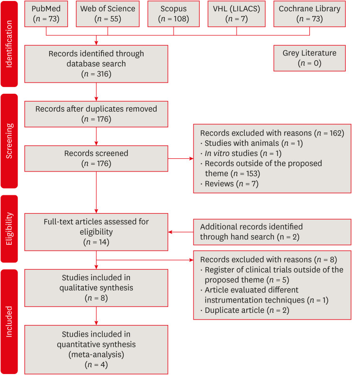
Materials and Methods A broad search was performed in the PubMed, Web of Science, Scopus, Cochrane Library, Virtual Health Library (LILACS), and Grey Literature databases. Two independent reviewers performed data extraction, risk of bias using the Cochrane methodology, and certainty of evidence using the Grading of Recommendations, Assessment, Development and Evaluations (GRADE) approach.
Results Eight studies were included in qualitative synthesis. Intracanal cryotherapy favored the reduction of postoperative pain in the systematic review. Four studies were included in meta-analyses. The meta-analysis showed that intracanal cryotherapy reduced postoperative pain in teeth with symptomatic apical periodontitis (SAP) at 24 hours. There was no association between intracanal cryotherapy and control (room temperature) groups in teeth with normal periapical tissue with respect to postoperative pain at 24 hours and 48 hours.
Conclusions Intracanal cryotherapy was effective in reducing postoperative pain after endodontic treatment in teeth with SAP.
-
Citations
Citations to this article as recorded by- Impact of intracanal cryotherapy on postoperative pain in symptomatic apical periodontitis: A systematic review and meta-analysis of randomized clinical trials
Nishtha K. Patel, Prerak Doshi, Shaily R. Dalal, Pooja R. Kesharani, Shilpa S. Shah, Mohil H. Kale
Endodontology.2025; 37(2): 101. CrossRef - Evaluation of Post‐Endodontic Pain Reduction Using Intracanal Cryotherapy in Symptomatic Apical Periodontitis
Anam Fayyaz Bashir, Ussamah Waheed Jatala, Muhammad Amber Fareed, Sheryar Sheryar, Saadia Ahmad Chattha, Saima Razaq Khan, Shahzad Ahmad, Shazia Iqbal, Muhammad Sohail Zafar, Shahzad Ali
Australian Endodontic Journal.2025; 51(3): 677. CrossRef - Comparing cryotherapy and ketorolac tromethamine against room-temperature saline irrigation using interleukin-8 levels and post-operative pain within single-visit endodontic treatment of symptomatic irreversible pulpitis superimposed by apical periodontit
Yousra Khaled Ezzat, Alaa Diab, Olfat Shaker, Sarah Abouelenien
BMC Oral Health.2025;[Epub] CrossRef - Determining Efficacy of Intracanal Cryotherapy on Post Endodontic Pain in Irreversible Pulpitis
Anam Fayyaz Bashir, Ussamah Waheed Jatala, Moeen ud din Ahmad, Muhammad Talha Khan, Saima Razzaq Khan, Aisha Arshad Butt
Pakistan Journal of Health Sciences.2024; : 68. CrossRef - The effect of intracanal cryotherapy with and without foraminal enlargement on pain prevention after endodontic treatment: a randomized clinical trial
Marcos Felipe Iparraguirre Nuñovero, Marco Antonio Hungaro Duarte, André Vinícius Kaled Segato, Ulisses Xavier da Silva Neto, Vania Portela Ditzel Westphalen, Everdan Carneiro
Scientific Reports.2024;[Epub] CrossRef - Effect of cryotherapy duration on experimentally induced connective tissue inflammationin vivo
Jorge Vera, Mayra Alejandra Castro-Nuñez, María Fernanda Troncoso-Cibrian, Ana Gabriela Carrillo-Varguez, Edgar Ramiro Méndez Sánchez, Viviana Sarmiento, Lourdes Lanzagorta-Rebollo, Prasanna Neelakantan, Monica Romero, Ana Arias
Restorative Dentistry & Endodontics.2023;[Epub] CrossRef - Evaluation of knowledge and awareness of pediatric oral health among school teachers of Hazaribag before and after oral health education.
Vipin Ahuja, Annapurna Ahuja, Nilima Thosar
F1000Research.2023; 12: 1292. CrossRef
- Impact of intracanal cryotherapy on postoperative pain in symptomatic apical periodontitis: A systematic review and meta-analysis of randomized clinical trials
- 3,393 View
- 71 Download
- 6 Web of Science
- 7 Crossref

- Combination of a new ultrasonic tip with rotary systems for the preparation of flattened root canals
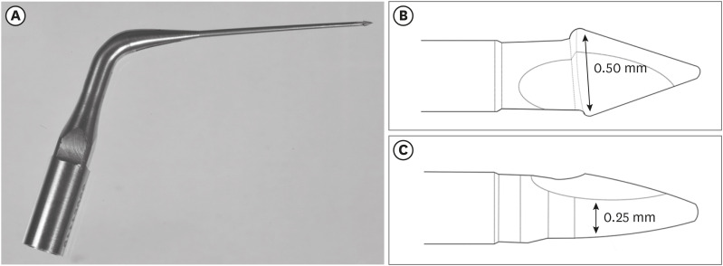
- Karina Ines Medina Carita Tavares, Jáder Camilo Pinto, Airton Oliveira Santos-Junior, Fernanda Ferrari Esteves Torres, Juliane Maria Guerreiro-Tanomaru, Mario Tanomaru-Filho
- Restor Dent Endod 2021;46(4):e56. Published online October 27, 2021
- DOI: https://doi.org/10.5395/rde.2021.46.e56
-
 Abstract
 PDF
PDF PubReader
 ePub
ePub Objectives This study evaluated 2 nickel-titanium rotary systems and a complementary protocol with an ultrasonic tip and a small-diameter instrument in flattened root canals.
Materials and Methods Thirty-two human maxillary second premolars with flattened canals (buccolingual diameter ≥4 times larger than the mesiodistal diameter) at 9 mm from the radiographic apex were selected. The root canals were prepared by ProDesign Logic (PDL) 30/0.01 and 30/0.05 or Hyflex EDM (HEDM) 10/0.05 and 25/0.08 (
n = 16), followed by application of the Flatsonic ultrasonic tip in the cervical and middle thirds and a PDL 25/0.03 file in the apical third (FPDL). The teeth were scanned using micro-computed tomography before and after the procedures. The percentage of volume increase, debris, and uninstrumented surface area were analyzed using the Kruskal-Wallis, Dunn, Wilcoxon, analysis of variance/Tukey, and paired and unpairedt -tests (α = 0.05).Results No significant difference was found in the volume increase and uninstrumented surface area between PDL and HEDM (
p > 0.05). PDL had a higher percentage of debris than HEDM in the middle and apical thirds (p < 0.05). The FPDL protocol resulted in less debris and uninstrumented surface area for PDL and HEDM (p < 0.05). This protocol, with HEDM, reduced debris in the middle and apical thirds and uninstrumented surface area in the apical third (p < 0.05).Conclusions High percentages of debris and uninstrumented surface area were observed after preparation of flattened root canals. The HEDM, Flatsonic tip, and 25/0.03 instrument protocol enhanced cleaning in flattened root canals.
-
Citations
Citations to this article as recorded by- Kök Kanal Tedavisi Yenilemelerinde Ultrasonik Uç Kullanımı
Ayşenur Kızıltaş Gül, Turan Mert Hisar, Seniha Miçooğulları
Selcuk Dental Journal.2025; 12(1): 157. CrossRef - Flatsonic Ultrasonic Tip Optimizes the Removal of Remaining Filling Material in Flattened Root Canals: A Micro–computed Tomographic Analysis
Airton Oliveira Santos-Junior, Karina Ines Medina Carita Tavares, Jáder Camilo Pinto, Fernanda Ferrari Esteves Torres, Juliane Maria Guerreiro-Tanomaru, Mário Tanomaru-Filho
Journal of Endodontics.2024; 50(5): 612. CrossRef - The Effect of Combined Ultrasonic Tip and Mechanized Instrumentation on the Reduction of the Percentage of Non-Instrumented Surfaces in Oval/Flat Root Canals: A Systematic Review and Meta-Analysis
Marcella Dewes Cassal, Pedro Cardoso Soares, Marcelo dos Santos
Cureus.2023;[Epub] CrossRef - Heat-treated NiTi instruments and final irrigation protocols for biomechanical preparation of flattened canals
Kleber Kildare Teodoro CARVALHO, Igor Bassi Ferreira PETEAN, Alice Corrêa SILVA-SOUSA, Rafael Verardino CAMARGO, Jardel Francisco MAZZI-CHAVES, Yara Terezinha Corrêa SILVA-SOUSA, Manoel Damião SOUSA-NETO
Brazilian Oral Research.2022;[Epub] CrossRef
- Kök Kanal Tedavisi Yenilemelerinde Ultrasonik Uç Kullanımı
- 1,644 View
- 26 Download
- 3 Web of Science
- 4 Crossref

- Shaping ability and apical debris extrusion after root canal preparation with rotary or reciprocating instruments: a micro-CT study
- Emmanuel João Nogueira Leal da Silva, Sara Gomes de Moura, Carolina Oliveira de Lima, Ana Flávia Almeida Barbosa, Waleska Florentino Misael, Mariane Floriano Lopes Santos Lacerda, Luciana Moura Sassone
- Restor Dent Endod 2021;46(2):e16. Published online February 25, 2021
- DOI: https://doi.org/10.5395/rde.2021.46.e16
-
 Abstract
 PDF
PDF PubReader
 ePub
ePub Objectives The aim of this study was to evaluate the shaping ability of the TruShape and Reciproc Blue systems and the apical extrusion of debris after root canal instrumentation. The ProTaper Universal system was used as a reference for comparison.
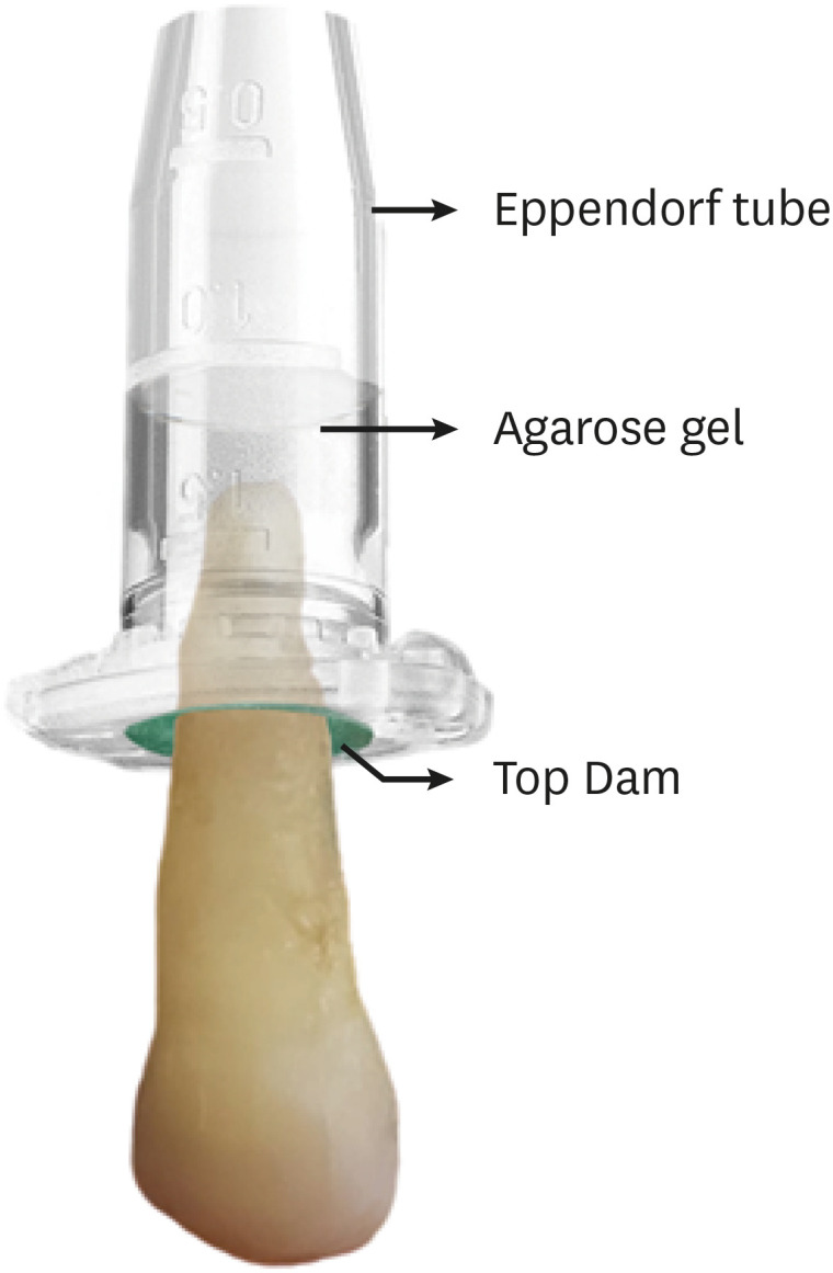
Materials and Methods Thirty-three mandibular premolars with a single canal were scanned using micro-computed tomography and were matched into 3 groups (
n = 11) according to the instrumentation system: TruShape, Reciproc Blue and ProTaper Universal. The teeth were accessed and mounted in an apparatus with agarose gel, which simulated apical resistance provided by the periapical tissue and enabled the collection of apically extruded debris. During root canal preparation, 2.5% sodium hypochlorite was used as an irrigant. The samples were scanned again after instrumentation. The percentage of unprepared area, removed dentin, and volume of apically extruded debris were analyzed. The data were analyzed using 1-way analysis of variance and the Tukey test for multiple comparisons at a 5% significance level.Results No significant differences in the percentage of unprepared area were observed among the systems (
p > 0.05). ProTaper Universal presented a higher percentage of dentin removal than the TruShape and Reciproc Blue systems (p < 0.05). The systems produced similar volumes of apically extruded debris (p > 0.05).Conclusions All systems caused apically extruded debris, without any significant differences among them. TruShape, Reciproc Blue, and ProTaper Universal presented similar percentages of unprepared area after root canal instrumentation; however, ProTaper Universal was associated with higher dentin removal than the other systems.
-
Citations
Citations to this article as recorded by- Evaluation of Silver-Ion-Coated Rotary Nickel Titanium Files - An In Vitro Study
Jhanvi H. Sadaria, Kondas V. Venkatesh, Dhanasekaran Sihivahanan
Indian Journal of Dental Research.2026;[Epub] CrossRef - Comparison of post-operative pain prevalence after single visit endodontic treatment with two NiTi rotary files - a randomized clinical trial
M. E. Khallaf, Yousra Aly, Amira Ibrahim Mohamed
Scientific Reports.2025;[Epub] CrossRef - A quantitative comparison of apically extruded debris during root canal preparation using NiTi full-sequence rotary and single-file rotary systems: An in vitro study
Pallavi Goel, R. Vikram, R. Anithakumari, M. S. Adarsha, M. E. Sudhanva
Endodontology.2024; 36(3): 235. CrossRef - Extrusion of Sodium Hypochlorite in Oval-Shaped Canals: A Comparative Study of the Potential of Four Final Agitation Approaches Employing Agarose-Embedded Mandibular First Premolars
Aalisha Parkar, Kulvinder Singh Banga, Ajinkya M. Pawar, Alexander Maniangat Luke
Journal of Clinical Medicine.2024; 13(10): 2748. CrossRef - Shaping Efficiency of Rotary and Reciprocating Kinematics of Engine-driven Nickel-Titanium Instruments in Moderate and Severely curved Root Canals Using Microcomputed Tomography: A Systematic Review of Ex Vivo Studies
Claudiu Călin, Ana-Maria Focșăneanu, Friedrich Paulsen, Andreea C. Didilescu, Tiberiu Niță
Journal of Endodontics.2024; 50(7): 907. CrossRef - Intracanal removal and apical extrusion of filling material after retreatment using rotary or reciprocating instruments: A new approach using human cadavers
Thamyres M. Monteiro, Victor O. Cortes‐Cid, Marilia F. V. Marceliano‐Alves, Andrea F. Campello, Luan F. Bastos, Ricardo T. Lopes, José F. Siqueira, Flávio R. F. Alves
International Endodontic Journal.2024; 57(1): 100. CrossRef - Assessment of debris extrusion on using automated irrigation device with conventional needle irrigation – An ex vivo study
Sahil Choudhari, Kavalipurapu Venkata Teja, Raja Kumar, Sindhu Ramesh
Saudi Endodontic Journal.2023; 13(3): 263. CrossRef - Postoperative pain perception and associated risk factors in children after continuous rotation versus reciprocating kinematics: A randomised prospective clinical trial
Ahmad Abdel Hamid Elheeny, Dania Ibrahem Sermani, Mahmoud Ahmed Abdelmotelb
Australian Endodontic Journal.2023; 49(S1): 345. CrossRef - A critical analysis of research methods and experimental models to study apical extrusion of debris and irrigants
Jale Tanalp
International Endodontic Journal.2022; 55(S1): 153. CrossRef - Quantitative evaluation of apically extruded debris using TRUShape, TruNatomy, and WaveOne Gold in curved canals
Nehal Nabil Roshdy, Reham Hassan
BDJ Open.2022;[Epub] CrossRef - Shaping ability of new reciprocating or rotary instruments with two cross‐sectional designs: An ex vivo study
Isabela G. Guedes, Renata C. V. Rodrigues, Marília F. Marceliano‐Alves, Flávio R. F. Alves, Isabela N. Rôças, José F. Siqueira
International Endodontic Journal.2022; 55(12): 1385. CrossRef
- Evaluation of Silver-Ion-Coated Rotary Nickel Titanium Files - An In Vitro Study
- 2,521 View
- 49 Download
- 8 Web of Science
- 11 Crossref

- YouTube as an information source for instrument separation in root canal treatment
- Yağız Özbay, Neslihan Yılmaz Çırakoğlu
- Restor Dent Endod 2021;46(1):e8. Published online January 12, 2021
- DOI: https://doi.org/10.5395/rde.2021.46.e8
-
 Abstract
 PDF
PDF PubReader
 ePub
ePub Objectives The reliability and educational quality of videos on YouTube for patients seeking information regarding instrument separation in root canal treatment were evaluated.
Materials and Methods YouTube was searched for videos on instrument separation in root canal treatment. Video content was scored based on reliability in terms of 3 categories (etiology, procedure, and prognosis) and based on video flow, quality, and educational usefulness using the Global Quality Score (GQS). Descriptive statistics were obtained and the data were analyzed using analysis of variance and the Kruskal-Wallis test.
Results The highest mean completeness scores were obtained for videos published by dentists or specialists (1.48 ± 1.06). There was no statistically significant difference among sources of upload in terms of content completeness. The highest mean GQS was found for videos published by dentists or specialists (1.82 ± 0.96), although there was no statistically significant correlation between GQS and the source of upload.
Conclusions Videos on YouTube have incomplete and low-quality content for patients who are concerned about instrument separation during endodontic treatment, or who experience this complication during endodontic treatment.
-
Citations
Citations to this article as recorded by- Quality of information in #brokenfileremoval Reels videos on Instagram: a cross-sectional study
Dilek Hancerliogullari, Eray Ceylanoglu
Journal of Public Health.2025; 33(8): 1617. CrossRef - Evaluation of the Educational Value, Reliability, and Quality of Vaginally-Assisted Natural Orifice Transluminal Endoscopic Surgery (v-NOTES) Videos on YouTube
Isa Temur
Cureus.2025;[Epub] CrossRef - Evaluating the Quality and Reliability of YouTube Videos Providing Nutritional Recommendations for Irritable Bowel Syndrome
Eda Başmısırlı, Merve Kip, Hande Altun, Neriman İnanç
Journal of Human Nutrition and Dietetics.2025;[Epub] CrossRef - Comparison of YouTube, TikTok, and Instagram as digital sources for obtaining information about pulp therapy in primary and permanent teeth
Hüseyin Gürkan Güneç, Emine Kaya, Dila Nur Okumuş, Merve Gül Erence
Restorative Dentistry & Endodontics.2025; 50(3): e26. CrossRef - Is YouTube a reliable source for learning pre-endodontic build-up? A cross-sectional study
Merve Gökyar, İdil Özden, Hesna Sazak Öveçoğlu
Restorative Dentistry & Endodontics.2025; 50(3): e27. CrossRef - Youtube As Sources of Information About Fiber-Reinforcement Composite Resin Bridge: Accuracy and Reliability Assessment
Merve Arslan, Zeyneb Merve Ozdemır
Akdeniz Diş Hekimliği Dergisi.2025; 4(2): 89. CrossRef - Evaluation of the Quality and Reliability of YouTubeTM Videos Created by Orthodontists as an Information Source for Clear Aligners
Emre Cesur, Koray Tuncer, Duygu Sevgi, Barkın Cem Balaban, Can Arslan
Turkish Journal of Orthodontics.2024; 37(1): 44. CrossRef - Is it safe to learn about vital pulp capping from YouTube™ videos? A content and quality analysis
Celalettin Topbaş, Tuğçe Paksoy, Ayşe Gülnihal İslamoğlu, Kemal Çağlar, Abdurrahman Kerim Kul
International Journal of Medical Informatics.2024; 185: 105409. CrossRef - Quality of Patient-Centered eHealth Information on Erosive Tooth Wear: Systematic Search and Evaluation of Websites and YouTube Videos
Lena Holland, Amelie Friederike Kanzow, Annette Wiegand, Philipp Kanzow
Journal of Medical Internet Research.2024; 26: e49514. CrossRef - Evaluation of YouTubeTM as an Information Source for Indirect Restorations: Cross-Sectional Evaluation
Işıl Doğruer, Merve Kütük Ömeroğlu
European Annals of Dental Sciences.2024; 51(3): 102. CrossRef - Evaluating YouTube as a Patient Information Source for the Risks of Root Canal Treatment
Stewart McLean, Neil Cook, Alexander Rovira-Wilde, Shanon Patel, Shalini Kanagasingam
Journal of Endodontics.2023; 49(2): 155. CrossRef - Evaluation of the quality of YouTube™ videos about pit and fissure sealant applications
Ayse Tugba Erturk‐Avunduk, Ebru Delikan
International Journal of Dental Hygiene.2023; 21(3): 590. CrossRef - Avülsiyon Yaralanmalarının Acil Müdahalesinde Hasta Bilgi Kaynağı Olarak Türkçe YouTube™ Videolarının Güvenilirliği: Kesitsel İçerik Analizi
Gülçin CAGAY SEVENCAN, Zeynep Şeyda YAVŞAN
Selcuk Dental Journal.2023; 10(3): 583. CrossRef - Analyzing Content and Quality of YouTube™ Videos on Removal of Amalgam Fillings
Mehmet BULDUR, Fatma AYTAÇ BAL
Clinical and Experimental Health Sciences.2022; 12(2): 423. CrossRef - Assessment of reliability and information quality of YouTube videos about root canal treatment after 2016
Myoung-jun Jung, Min-Seock Seo
BMC Oral Health.2022;[Epub] CrossRef - Are YouTube Videos Reliable Sources of Information About Devital Bleaching?
Gülbahar ERDİNÇ, Yağız ÖZBAY, Neslihan YILMAZ ÇIRAKOĞLU
Mersin Üniversitesi Tıp Fakültesi Lokman Hekim Tıp Tarihi ve Folklorik Tıp Dergisi.2022; 12(3): 637. CrossRef - Assessment of the educational value of endodontic access cavity preparation YouTube video as a learning resource for students
Ahmed Jamleh, Shouq Mohammed Aljohani, Faisal Fahad Alzamil, Shahad Muhammad Aljuhayyim, Modhi Nasser Alsubaei, Showq Raad Alali, Nawaf Munawir Alotaibi, Mohannad Nassar, MariKannan Maharajan
PLOS ONE.2022; 17(8): e0272765. CrossRef - Evaluation of YouTube videos for patients’ education on periradicular surgery
Ahmed Jamleh, Mohannad Nassar, Hamad Alissa, Abdulmohsen Alfadley, Tanay Chaubal
PLOS ONE.2021; 16(12): e0261309. CrossRef
- Quality of information in #brokenfileremoval Reels videos on Instagram: a cross-sectional study
- 2,326 View
- 14 Download
- 14 Web of Science
- 18 Crossref

- Micro-computed tomographic assessment of the shaping ability of the One Curve, One Shape, and ProTaper Next nickel-titanium rotary systems
- Pelin Tufenkci, Kaan Orhan, Berkan Celikten, Burak Bilecenoglu, Gurkan Gur, Semra Sevimay
- Restor Dent Endod 2020;45(3):e30. Published online May 22, 2020
- DOI: https://doi.org/10.5395/rde.2020.45.e30
-
 Abstract
 PDF
PDF PubReader
 ePub
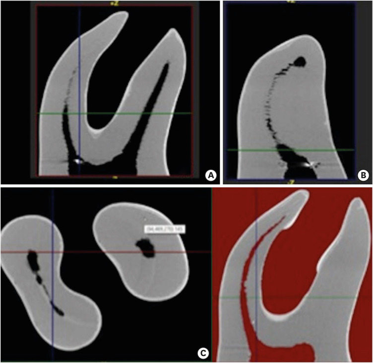
ePub Objectives This micro-computed tomographic (CT) study aimed to compare the shaping abilities of ProTaper Next (PTN), One Shape (OS), and One Curve (OC) files in 3-dimensionally (3D)-printed mandibular molars.
Materials and Methods In order to ensure standardization, 3D-printed mandibular molars with a consistent mesiobuccal canal curvature (45°) were used in the present study (
n = 18). Specimens were instrumented with the OC, OS, or PTN files. The teeth were scanned pre- and post-instrumentation using micro-CT to detect changes of the canal volume and surface area, as well as to quantify transportation of the canals after instrumentation. Two-way analysis of variance was used for statistical comparisons.Results No statistically significant differences were found between the OC and OS groups in the changes of the canal volume and surface area before and after instrumentation (
p > 0.05). The OC files showed significantly less transportation than the OS or PTN systems for the apical section (p < 0.05). In a comparison of the systems, similar values were found at the coronal and middle levels, without any significant differences (p > 0.05).Conclusions These 3 instrumentation systems showed similar shaping abilities, although the OC file achieved a lesser extent of transportation in the apical zone than the OS and PTN files. All 3 file systems were confirmed to be safe for use in mandibular mesial canals.
-
Citations
Citations to this article as recorded by- Effect of different kinematics and perforation diameter on integrated electronic apex locator accuracy in detecting root canal perforations
Ecenur Tuzcu, Safa Kurnaz
European Journal of Oral Sciences.2025;[Epub] CrossRef - Micro‐CT Evaluation of the Shaping Outcomes of Different Instruments in Oval‐Shaped Maxillary Premolar Canals
Merve Yeniçeri Özata, Seda Falakaloğlu, Ali Keleş, Özkan Adıgüzel, Sadullah Kaya
Australian Endodontic Journal.2025;[Epub] CrossRef - A Comparative Evaluation of the Efficiencies of Different Rotary File Systems in Terms of Remaining Dentin Thickness Using Cone Beam Computed Tomography: An In Vitro Study
Vivek P Vadera , Sandhya K Punia, Saleem D Makandar, Rahul Bhargava, Pradeep Bapna
Cureus.2024;[Epub] CrossRef - Comparison of Different Rotary Nickel–titanium Systems to Evaluate Coronal Leakage of Root Canals: An in Vitro Study
Rasha M. Al-Shamaa
Dental Hypotheses.2023; 14(3): 81. CrossRef - Comparative evaluation of canal transportation and canal centering ability in oval canals with newer nickel–titanium rotary single file systems – A cone-beam computed tomography study
SimarKaur Manocha, SuparnaGanguly Saha, RollyS Agarwal, Neelam Vijaywargiya, MainakKanti Saha, Anjali Surana
Journal of Conservative Dentistry.2023; 26(3): 326. CrossRef - Accumulated Hard Tissue Debris and Root Canal Shaping Profiles Following Instrumentation with Gentlefile, One Curve, and Reciproc Blue
Chi Wai Chan, Virginia Rosy Romeo, Angeline Lee, Chengfei Zhang, Prasanna Neelakantan, Eugenio Pedullà
Journal of Endodontics.2023; 49(10): 1344. CrossRef - Comparative evaluation of canal transportation and centering ability of rotary and reciprocating file systems using cone-beam computed tomography: An in vitro study
Tanisha Singh, Manju Kumari, Rohit Kochhar
Journal of Conservative Dentistry.2023; 26(3): 332. CrossRef - Retreatability of Bioceramic Sealer Using One Curve Rotary File Assessed by Microcomputed Tomography
Dina G Mufti, Saad A Al-Nazhan
The Journal of Contemporary Dental Practice.2022; 22(10): 1175. CrossRef - Micro-computed tomography in preventive and restorative dental research: A review
Mehrsima Ghavami-Lahiji, Reza Tayefeh Davalloo, Gelareh Tajziehchi, Paria Shams
Imaging Science in Dentistry.2021; 51(4): 341. CrossRef
- Effect of different kinematics and perforation diameter on integrated electronic apex locator accuracy in detecting root canal perforations
- 1,648 View
- 12 Download
- 9 Crossref

- The top 10 most-cited articles on the management of fractured instruments: a bibliometric analysis
- Lora Mishra, Hyeon-Cheol Kim, Naomi Ranjan Singh, Priti Pragati Rath
- Restor Dent Endod 2019;44(1):e2. Published online December 26, 2018
- DOI: https://doi.org/10.5395/rde.2019.44.e2
-
 Abstract
 PDF
PDF PubReader
 ePub
ePub Objectives The purpose of this research was to identify the top 10 most-cited articles on the management of fractured or broken instruments and to perform a bibliometric analysis thereof.
Materials and Methods Published articles related to fractured instruments were screened from online databases, such as Web of Science, Scopus, PubMed, and ScienceDirect, and highly cited papers, with at least 50 citations since publication, were identified. The most-cited articles were selected and analysed with regard to publication title, authorship, the journal of publication, year, institution, country of origin, article type, and number of citations.
Results The top 10 most-cited articles were from various journals. Most were published in the
Journal of Endodontics , followed by theInternational Endodontic Journal , andDental Traumatology . The leading countries were Australia, Israel, Switzerland, the USA, and Germany, and the leading institution was the University of Melbourne. The majority of articles among the top 10 articles were clinical research studies (n = 8), followed by a basic research article and a non-systematic review article.Conclusions This bibliometric analysis revealed interesting information about scientific progress in endodontics regarding fractured instruments. Overall, clinical research studies and basic research articles published in high-impact endodontic journals had the highest citation rates.
-
Citations
Citations to this article as recorded by- Bibliometric analysis of the publications that list the most-cited articles in endodontics
Oscar Alejandro Gutiérrez-Alvarez, Luis Alberto Pantoja-Villa, Benigno Miguel Calderón-Rojas
Endodontology.2025; 37(2): 128. CrossRef - A Bibliometric Analysis of the 100 Top-Cited Articles on Vertical Root Fractures
Pillai Arun Gopinathan , Ikram UI Haq, Nawaf Alfahad, Saleh Alwatban, Abdullah Alghamdi, Amal Alamri, Kiran Iyer
Cureus.2024;[Epub] CrossRef - Predictive factors in the retrieval of endodontic instruments: the relationship between the fragment length and location
Ricardo Portigliatti, Eugenia Pilar Consoli Lizzi, Pablo Alejandro Rodríguez
Restorative Dentistry & Endodontics.2024;[Epub] CrossRef - A bibliometric analysis of the top 100 most‐cited case reports and case series in Endodontic journals
Venkateshbabu Nagendrababu, Jelena Jacimovic, Aleksandar Jakovljevic, Giampiero Rossi‐Fedele, Paul M. H. Dummer
International Endodontic Journal.2022; 55(3): 185. CrossRef - The Most Highly Cited Publications on Basketball Originate From English-Speaking Countries, Are Published After 2000, Are Focused on Medicine-Related Topics, and Are Level III Evidence
Zachary D. Griffin, Jordan R. Pollock, M. Lane Moore, Kade S. McQuivey, Jaymeson R. Arthur, Anikar Chhabra
Arthroscopy, Sports Medicine, and Rehabilitation.2022; 4(3): e891. CrossRef - Ten years of minimally invasive access cavities in Endodontics: a bibliometric analysis of the 25 most-cited studies
Emmanuel João Nogueira Leal Silva, Karem Paula Pinto, Natasha C. Ajuz, Luciana Moura Sassone
Restorative Dentistry & Endodontics.2021;[Epub] CrossRef - Publication trends in micro‐CT endodontic research: a bibliometric analysis over a 25‐year period
U. Aksoy, M. Küçük, M. A. Versiani, K. Orhan
International Endodontic Journal.2021; 54(3): 343. CrossRef
- Bibliometric analysis of the publications that list the most-cited articles in endodontics
- 1,417 View
- 10 Download
- 7 Crossref

- Incidence of apical crack formation and propagation during removal of root canal filling materials with different engine driven nickel-titanium instruments
- Taha Özyürek, Vildan Tek, Koray Yılmaz, Gülşah Uslu
- Restor Dent Endod 2017;42(4):332-341. Published online November 4, 2017
- DOI: https://doi.org/10.5395/rde.2017.42.4.332
-
 Abstract
 PDF
PDF PubReader
 ePub
ePub Objectives To determine the incidence of crack formation and propagation in apical root dentin after retreatment procedures performed using ProTaper Universal Retreatment (PTR), Mtwo-R, ProTaper Next (PTN), and Twisted File Adaptive (TFA) systems.
Materials and Methods The study consisted of 120 extracted mandibular premolars. One millimeter from the apex of each tooth was ground perpendicular to the long axis of the tooth, and the apical surface was polished. Twenty teeth served as the negative control group. One hundred teeth were prepared, obturated, and then divided into 5 retreatment groups. The retreatment procedures were performed using the following files: PTR, Mtwo-R, PTN, TFA, and hand files. After filling material removal, apical enlargement was done using apical size 0.50 mm ProTaper Universal (PTU), Mtwo, PTN, TFA, and hand files. Digital images of the apical root surfaces were recorded before preparation, after preparation, after obturation, after filling removal, and after apical enlargement using a stereomicroscope. The images were then inspected for the presence of new apical cracks and crack propagation. Data were analyzed with χ2 tests using SPSS 21.0 software.
Results New cracks and crack propagation occurred in all the experimental groups during the retreatment process. Nickel-titanium rotary file systems caused significantly more apical crack formation and propagation than the hand files. The PTU system caused significantly more apical cracks than the other groups after the apical enlargement stage.
Conclusions This study showed that retreatment procedures and apical enlargement after the use of retreatment files can cause crack formation and propagation in apical dentin.
-
Citations
Citations to this article as recorded by- Microcracks induced by XP-endo retreatment system in root canals filled with bioceramic sealer: A micro-computed tomographic analysis
Sarah M. Alkahtany
The Saudi Dental Journal.2025;[Epub] CrossRef - A comparative evaluation of different retreatment methods on apical root microcracks initiation and propagation: An in vitro study
Shweta Lodha, Zinnie Nanda
Endodontology.2025; 37(2): 175. CrossRef - Efficacy of Endodontic Files in Root Canal Retreatment: A Systematic Review of In Vitro Studies
Anna Soler-Doria, José Luis Sanz, Marcello Maddalone, Leopoldo Forner
Journal of Functional Biomaterials.2025; 16(8): 293. CrossRef - Efficacy of Various Heat-treated Retreatment File Systems on Dentin Removal and Crack Analysis: An in vitro Study
Swathi Suresh, Pradeep Solete, Delphine Priscilla Antony, Kavalipurapu Venkata Teja, Adimulapu Hima Sandeep, Sruthi Sairaman, Marco Cicciù, Giuseppe Minervini
Pesquisa Brasileira em Odontopediatria e Clínica Integrada.2024;[Epub] CrossRef - A comparative evaluation of the dentinal microcracks formed and propagated during the removal of gutta-percha using hand and three rotary retreatment file systems: A micro-computed tomography study
Srivastava Sanjeev, Rita Gupta, Dubey Sandeep, Tewari Tanu, Shukla Namita, Singh Arohan
Endodontology.2023; 35(2): 155. CrossRef - Comparative evaluation of incidence of dentinal defects after root canal preparation using hand, rotary, and reciprocating files: An ex vivo study
Debanjan Das, Sudipto Barai, Rohit Kumar, Sourav Bhattacharyya, AsimB Maity, Pushpa Shankarappa
Journal of International Oral Health.2022; 14(1): 78. CrossRef - Critical analysis of research methods and experimental models to study removal of root filling materials
Mahdi A. Ajina, Pratik K. Shah, Bun San Chong
International Endodontic Journal.2022; 55(S1): 119. CrossRef - Evaluation of Dentinal Crack Propagation, Amount of Gutta Percha Remaining and Time Required During Removal of Gutta Percha Using Two Different Rotary Instruments and Hand Instruments - An In vitro Study
S Tejaswi, A Singh, S Manglekar, UK Ambikathanaya, S Shetty
Nigerian Journal of Clinical Practice.2022; 25(4): 524. CrossRef - The Influence of Root Canal Preparation with ProTaper Next, WaveOne Gold, and Twisted Files on Dentine Crack Formation
Wojciech Eliasz, Beata Czarnecka, Anna Surdacka
Machines.2021; 9(12): 332. CrossRef - The potential effect of instrumentation with different nickel titanium rotary systems on dentinal crack formation—An in vitro study
Márk Fráter, András Jakab, Gábor Braunitzer, Zsolt Tóth, Katalin Nagy, Andrej M. Kielbassa
PLOS ONE.2020; 15(9): e0238790. CrossRef - Micro–computed Tomographic Assessment of the Residual Filling Volume, Apical Transportation, and Crack Formation after Retreatment with Reciproc and Reciproc Blue Systems in Curved Root Canals
Damla Kırıcı, Sezer Demirbuga, Ertuğrul Karataş
Journal of Endodontics.2020; 46(2): 238. CrossRef - Force and vibration generated in apical direction by three endodontic files of different kinematics during simulated canal preparation: An in vitro analytical study
Ankit Nayak, PK Kankar, Prashant K Jain, Niharika Jain
Proceedings of the Institution of Mechanical Engineers, Part H: Journal of Engineering in Medicine.2019; 233(8): 839. CrossRef - Effect of Aging on Dentinal Crack Formation after Treatment and Retreatment Procedures: a Micro-CT Study
Lilian Rachel de Lima Aboud, Bernardo Camargo dos Santos, Ricardo Tadeu Lopes, Leonardo Aboud Costa Viana, Miriam Fátima Zaccaro Scelza
Brazilian Dental Journal.2018; 29(6): 530. CrossRef
- Microcracks induced by XP-endo retreatment system in root canals filled with bioceramic sealer: A micro-computed tomographic analysis
- 1,653 View
- 12 Download
- 13 Crossref

- Post space preparation timing of root canals sealed with AH Plus sealer
- Hae-Ri Kim, Young Kyung Kim, Tae-Yub Kwon
- Restor Dent Endod 2017;42(1):27-33. Published online December 19, 2016
- DOI: https://doi.org/10.5395/rde.2017.42.1.27

-
 Abstract
 PDF
PDF PubReader
 ePub
ePub Objectives To determine the optimal timing for post space preparation of root canals sealed with epoxy resin-based AH Plus sealer in terms of its polymerization and influence on apical leakage.
Materials and Methods The epoxy polymerization of AH Plus (Dentsply DeTrey) as a function of time after mixing (8, 24, and 72 hours, and 1 week) was evaluated using Fourier transform infrared (FTIR) spectroscopy and microhardness measurements. The change in the glass transition temperature (
Tg ) of the material with time was also investigated using differential scanning calorimetry (DSC). Fifty extracted human single-rooted premolars were filled with gutta-percha and AH Plus, and randomly separated into five groups (n = 10) based on post space preparation timing (immediately after root canal obturation and 8, 24, and 72 hours, and 1 week after root canal obturation). The extent of apical leakage (mm) of the five groups was compared using a dye leakage test. Each dataset was statistically analyzed by one-way analysis of variance and Tukey'spost hoc test (α = 0.05).Results Continuous epoxy polymerization of the material with time was observed. Although the
Tg values of the material gradually increased with time, the specimens presented no clearTg value at 1 week after mixing. When the post space was prepared 1 week after root canal obturation, the leakage was significantly higher than in the other groups (p < 0.05), among which there was no significant difference in leakage.Conclusions Poor apical seal was detected when post space preparation was delayed until 1 week after root canal obturation.
-
Citations
Citations to this article as recorded by- Bacterial microleakage in endodontically treated teeth following two methods of postspace preparation at two-time intervals: An in vitro study
AzamS Mostafavi, Mahsa Rasoulzadehsheikh, Naghmeh Meraji, Maryam Pourhajibagher
The Journal of Indian Prosthodontic Society.2022; 22(3): 233. CrossRef - Comparison of the effect of post space preparation time on the apical seal of two different sealers
Neda Hajihassani, Navid Mohammadi, Ahmad Karimi Kelayeh, Shima Aalaei
BMC Oral Health.2022;[Epub] CrossRef - Immediate and Delayed Post Space Preparations in Endodontically Treated Teeth: A Scoping Review
Sadaf Mahmoudi, Pedram Iranmanesh, Saber Khazaei, Maryam Zare Jahromi
BMC Oral Health.2022;[Epub] CrossRef - Physicochemical properties of a novel bioceramic silicone-based root canal sealer
Wei-Jia Lyu, Wei Bai, Xiao-Yan Wang, Yu-Hong Liang
Journal of Dental Sciences.2022; 17(2): 831. CrossRef - Impact of Immersion Media on Physical Properties and Bioactivity of Epoxy Resin-Based and Bioceramic Endodontic Sealers
Thais Gomes de Moraes, Alan Silva de Menezes, Renata Grazziotin-Soares, Rafael Ubaldo Moreira e Moraes, Paulo Vitor Campos Ferreira, Ceci Nunes Carvalho, Jose Bauer, Edilausson Moreno Carvalho
Polymers.2022; 14(4): 729. CrossRef - The effect of two endodontic sealers and interval before post-preparation and cementation on the bond strength of fiber posts
He Yuanli, Wu Juan, Ji Mengzhen, Chen Xuan, Xiong Kaixin, Yang Xueqin, Qiao Xin, Hu Hantao, Gao Yuan, Zou Ling
Clinical Oral Investigations.2021; 25(11): 6211. CrossRef - Sealing Ability of Various Types of Root Canal Sealers at Different Levels of Remaining Gutta Percha After Post Space Preparation at Two Time Intervals
Wiaam M O Al-Ashou, Rasha M Al-Shamaa, Shaymaa S Hassan
Journal of International Society of Preventive and Community Dentistry.2021; 11(6): 721. CrossRef - Comparison between immediate and delayed post space preparations: a systematic review and meta-analysis
Alexandre Henrique dos Reis-Prado, Lucas Guimarães Abreu, Warley Luciano Fonseca Tavares, Isabella Faria da Cunha Peixoto, Ana Cecília Diniz Viana, Elen Marise Castro de Oliveira, Juliana Vilela Bastos, Antônio Paulino Ribeiro-Sobrinho, Francine Benetti
Clinical Oral Investigations.2021; 25(2): 417. CrossRef - Apical Displacement and Residual Root Canal Filling with Single-Cone After Post Space Preparation: A Micro-CT Analysis
Camila Maria Peres de Rosatto, Lilian Vieira Oliveira, Danilo Cassiano Ferraz, Priscilla Barbosa Ferreira Soares, Carlos José Soares, Camilla Christian Gomes Moura
Brazilian Dental Journal.2020; 31(1): 25. CrossRef - Do Contaminating Substances Influence the Rheological Properties of Root Canal Sealers?
Jéssica Vavassori de Freitas, Johannes Ebert, Jardel Francisco Mazzi-Chaves, Manoel Damião de Sousa-Neto, Ulrich Lohbauer, Flares Baratto-Filho
Journal of Endodontics.2020; 46(2): 258. CrossRef
- Bacterial microleakage in endodontically treated teeth following two methods of postspace preparation at two-time intervals: An in vitro study
- 1,654 View
- 10 Download
- 10 Crossref

- Shaping ability of four rotary nickel-titanium instruments to prepare root canal at danger zone
- Seok-Dong Choi, Myoung-Uk Jin, Ki-Ok Kim, Sung-Kyo Kim
- J Korean Acad Conserv Dent 2004;29(5):446-453. Published online September 30, 2004
- DOI: https://doi.org/10.5395/JKACD.2004.29.5.446
-
 Abstract
 PDF
PDF PubReader
 ePub
ePub The aim of this study was to evaluate the shaping abilities of four different rotary nickel-titanium instruments with anticurvature motion to prepare root canal at danger zone by measuring the change of dentin thickness in order to have techniques of safe preparation of canals with nickel-titanium files.
Mesiobuccal and mesiolingual canals of forty mesial roots of extracted human lower molars were instrumented using the crown-down technique with ProFile, GT™ Rotary file, Quantec file and ProTaper™. In each root, one canal was prepared with a straight up-and-down motion and the other canal was with an anticurvature motion. Canals were instrumented until apical foramens were up to size of 30 by one operator. The muffle system was used to evaluate the root canal preparation. After superimposing the pre- and post-instrumentation canal, change in root dentin thickness was measured at the inner and outer sides of the canal at 1, 3, and 5 mm levels from the furcation. Data were analyzed using two-way ANOVA.
Root dentin thickness at danger zone was significantly thinner than that at safe zone at all levels (
p < 0.05).There was no significant difference in the change of root dentin thickness between the straight up-and-down and the anticurvature motions at both danger and safe zones in all groups (
p > 0.05).ProTaper removed significantly more dentin than other files especially at furcal 3 mm level of danger and safe zones (
p < 0.05)Therefore, it was concluded that anticurvature motion with nickel-titanium rotary instruments does not seem to be effective in danger zone of lower molars.
-
Citations
Citations to this article as recorded by- Conservation of dentin thickness in the root canals orifice following two preparation techniques
Ranjdar Talabani, Shawbo Ahmad, Arass Noori
Sulaimani Dental Journal.2014; 1(2): 6. CrossRef - Change of working length in curved canals by various instrumentation techniques
Jeong-Im Jo, Myoung-Uk Jin, Young Kyung Kim, Sung Kyo Kim
Journal of Korean Academy of Conservative Dentistry.2006; 31(1): 30. CrossRef - Effect of anticurvature filing method on preparation of the curved root canal using ProFile
Hyun-Ji Song, Juhea Chang, Kyung-Mo Cho, Jin-Woo Kim
Journal of Korean Academy of Conservative Dentistry.2005; 30(4): 327. CrossRef
- Conservation of dentin thickness in the root canals orifice following two preparation techniques
- 1,325 View
- 1 Download
- 3 Crossref


 KACD
KACD

 First
First Prev
Prev


