
Search
- Page Path
- HOME > Search
Research Articles
- Calcium silicate-based sealers remnants in isthmuses of mesial roots of mandibular molars: an in vitro evaluation
- David Saldanha de Brito Alencar, Ana Cristina Padilha Janini, Lauter Eston Pelepenko, Brenda Fornazaro Moraes, Francisco Haiter Neto, Marco Antonio Hungaro Duarte, Marina Angélica Marciano
- Restor Dent Endod 2025;50(3):e25. Published online July 15, 2025
- DOI: https://doi.org/10.5395/rde.2025.50.e25
-
 Abstract
 PDF
PDF PubReader
 ePub
ePub - Objectives
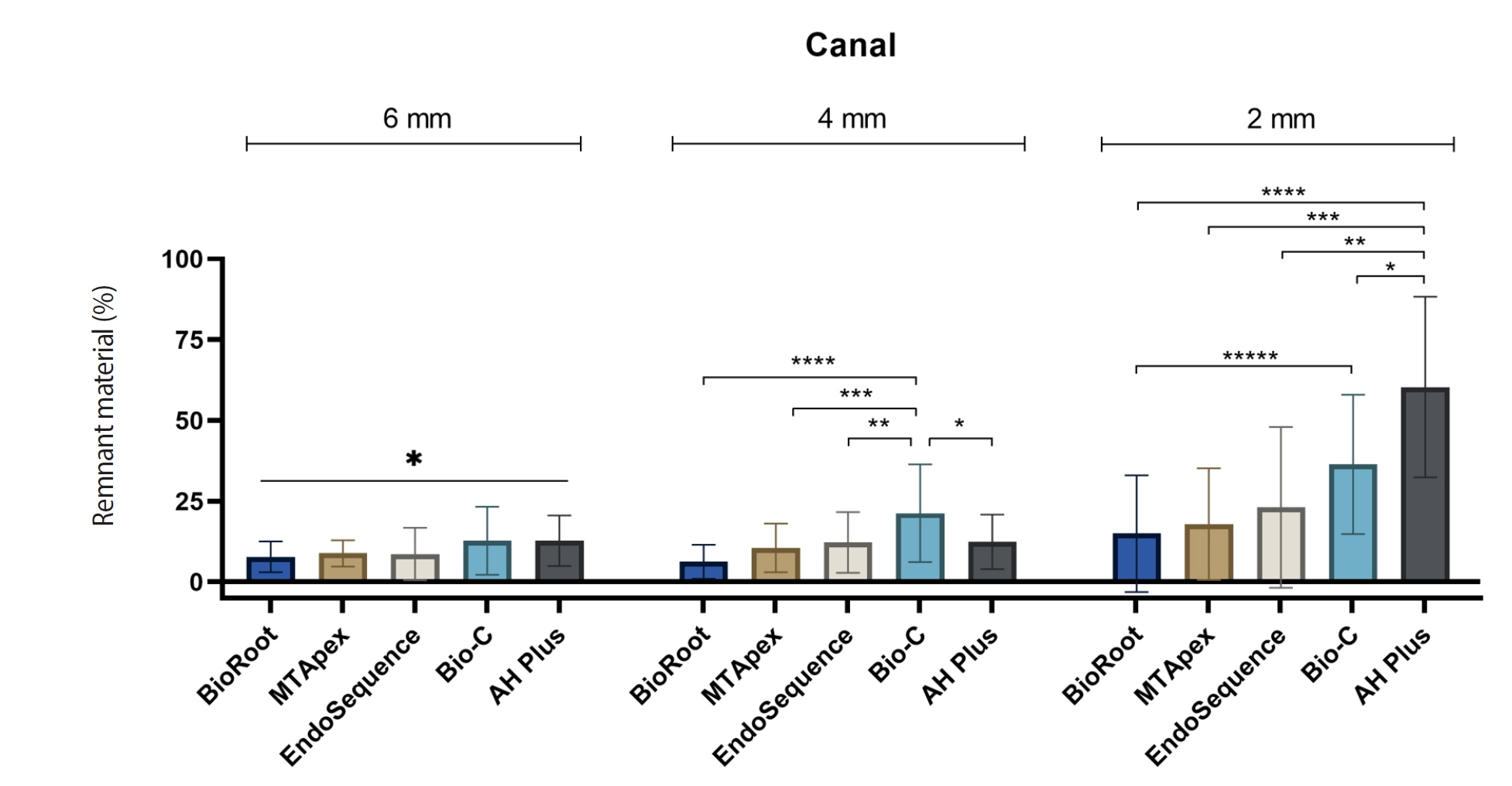
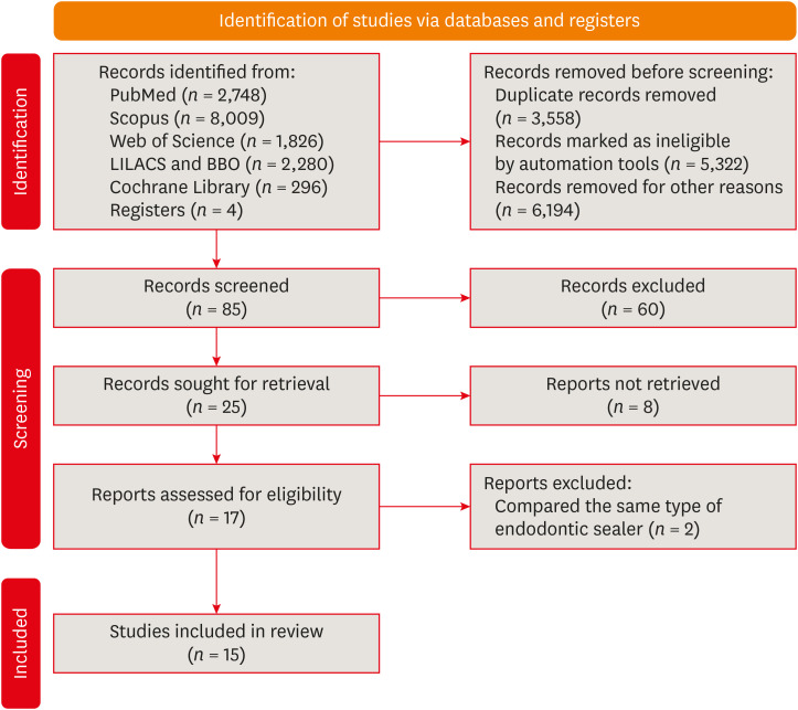
Endodontic retreatment aims to address treatment failure through the removal of root canal filling materials. This in vitro study evaluated the presence of filling material remnants in the mesial root canals, specifically focusing on the isthmuses, of mandibular molars after retreatment.
Methods
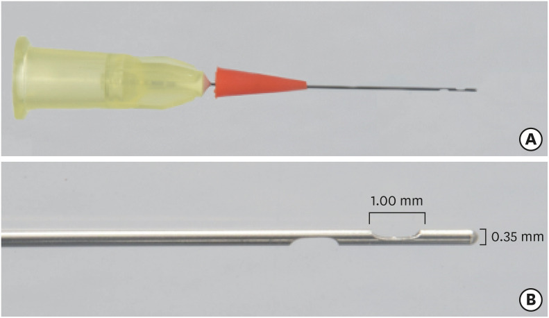
One hundred extracted mandibular molar mesial roots with isthmuses were prepared with an R25 file, obturated with one of five calcium silicate-based sealers (BioRoot RCS [Septodont], MTApex [Ultradent Products Inc.], EndoSequence BC Sealer HiFlow [Brasseler USA], Bio-C Sealer [Angelus]) or an epoxy resin-based sealer (AH Plus Jet [Dentsply Maillefer]), all stained with rhodamine B, and stored at 37ºC for 30 days to allow for setting. Retreatment was subsequently performed using R40 and XP-endo Finisher R instruments (FKG Dentaire) with 2.5% sodium hypochlorite irrigation. The presence of remaining filling material was then assessed using confocal microscopy, and setting times were tested per ISO 6876:2012.
Results
AH Plus Jet showed the most remnants at 2 mm and the longest retreatment time. Calcium silicate-based sealers exhibited prolonged setting times under dry conditions, with EndoSequence BC Sealer HiFlow showing a particularly extended setting period.
Conclusions
Despite retreatment, residues remained in all canals and isthmus regions, particularly Bio-C Sealer and AH Plus Jet in apical areas, emphasizing the difficulty of complete removal and the persistence of filling material. -
Citations
Citations to this article as recorded by
- Bonding effects of mechanical removal of bioceramic sealer residues using glycine or glass microparticles abrasion
Jesus Aranda, Julia de Freitas Ceccato, Eduardo Fernández Godoy, João Felipe Besegato, Joissi Ferrari Zaniboni, Regina Guenka Palma-Dibb, Milton Carlos Kuga
International Journal of Adhesion and Adhesives.2026; 148: 104289. CrossRef
- Bonding effects of mechanical removal of bioceramic sealer residues using glycine or glass microparticles abrasion
- 2,841 View
- 130 Download
- 1 Web of Science
- 1 Crossref

- Cleaning protocols to enhance bond strength of fiberglass posts on root canals filled with bioceramic sealer: an in vitro comparative study
- Thiago Bessa Marconato Antunes, Juliana Delatorre Bronzato, Joice Graciani, Ana Cristina Padilha Janini, Rocharles Cavalcante Fontenele, Francisco Haiter Neto, Brenda Paula Figueiredo de Almeida Gomes, Marina Angélica Marciano da Silva
- Restor Dent Endod 2025;50(2):e20. Published online May 21, 2025
- DOI: https://doi.org/10.5395/rde.2025.50.e20
-
Abstract
PDFPubReaderePub
- Objectives
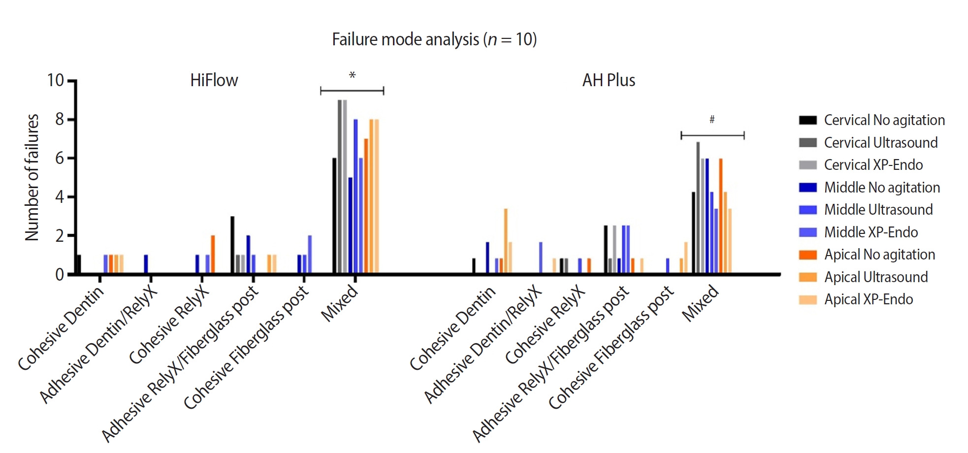
This study aimed to evaluate whether the agitation protocols using ultrasonic inserts or the XP-endo Finisher R file improved the removal of two different endodontic sealer remnants and the bond strength of fiberglass posts to dentin.
Methods
Seventy-two human teeth were selected. The canals were prepared with Reciproc 50 and Easy ProDesign 30/.10 and root filled according to the endodontic sealer groups: AH Plus or EndoSequence BC Sealer HiFlow. The samples were kept at 37ºC and 95% humidity for 28 days. During the post space preparation, the obturation was removed with Largo burs, and the groups were divided according to the irrigant agitation protocols (n = 12): no agitation, agitation with R1-Clearsonic associated with E1-Irrisonic ultrasonic inserts, or agitation with XP-endo Finisher R file. The fiberglass posts were cemented with RelyX ARC. The roots were sectioned into slices and submitted to the push-out test. Micro-computed tomography analysis was used to check the effectiveness of irrigating solution agitation in the elimination of remnants.
Results
The cleaning protocols with agitation were more effective in increasing the bond strength of posts to dentin for both sealer groups compared to non-agitation (p < 0.05). There was no difference between the same cleaning protocols for the different sealers. Among the different thirds, there was no statistical difference for the same sealer in the different cleaning protocols (p > 0.05).
Conclusions
Both agitation protocols effectively clean root-filled canals sealed with resin-based and calcium silicate-based sealers during fiberglass post space preparation. These protocols result in improved bond strength compared to non-agitation methods. -
Citations
Citations to this article as recorded by- Cleaning efficacy and bond interaction of glycine-based air polishing and glass microparticles abrasion on dentin impregnated with premixed bioceramic sealer
Ândresson Aurélio Fernandes Martins, Maria Carolina Sidonio Alves, Bruno Martins Maciel, José Rodolfo Estruc Verbicário, João Felipe Besegato, Wilfredo Gustavo Escalante-Otárola, Milton Carlos Kuga
International Journal of Adhesion and Adhesives.2026; 147: 104277. CrossRef - Effect of Endodontic Sealers on the Bond Strength of Glass Fibre Posts: A Systematic Review
Thiago Bessa Marconato Antunes, Juliana D. Bronzato, Vanessa Gallego Arias Pecorari, Jennifer Santos Pereira, Talita Tartari, Adriana de Jesus Soares, Brenda P. F. A. Gomes, Marina Angélica Marciano
Australian Endodontic Journal.2026;[Epub] CrossRef - Effects of Calcium Silicate-Based Sealer Residues on Adhesive Bonding to Coronal Dentin: An in Vitro Study
Mariana Bena Gelio, Thais Piragine Leandrin, Ana Lídia Pinheiro Silva Sato, Milton Carlos Kuga, Joissi Ferrari Zaniboni
Journal of Dental Research, Dental Clinics, Dental Prospects.2026; 20(1): 25. CrossRef
- Cleaning efficacy and bond interaction of glycine-based air polishing and glass microparticles abrasion on dentin impregnated with premixed bioceramic sealer
- 5,344 View
- 248 Download
- 2 Web of Science
- 3 Crossref
- A scientometric, bibliometric, and thematic map analysis of hydraulic calcium silicate root canal sealers
- Anastasios Katakidis, Konstantinos Kodonas, Anastasia Fardi, Christos Gogos
- Restor Dent Endod 2023;48(4):e41. Published online November 13, 2023
- DOI: https://doi.org/10.5395/rde.2023.48.e41
-
Abstract
PDFPubReaderePub
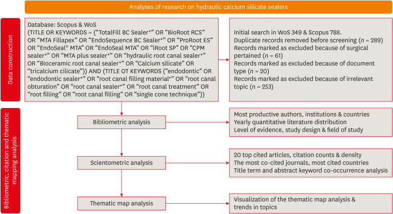
Objectives This scientometric and bibliometric analysis explored scientific publications related to hydraulic calcium silicate-based (HCSB) sealers used in endodontology, aiming to describe basic bibliometric indicators and analyze current research trends.
Materials and Methods A comprehensive search was conducted in Web of Science and Scopus using specific HCSB sealer and general endodontic-related terms. Basic research parameters were collected, including publication year, authorship, countries, institutions, journals, level of evidence, study design and topic of interest, title terms, author keywords, citation counts, and density.
Results In total, 498 articles published in 136 journals were retrieved for the period 2008–2023. Brazil was the leading country, and the universities of Bologna in Italy and Sao Paolo in Brazil were represented equally as leading institutions. The most frequently occurring keywords were “calcium silicate,” “root canal sealer MTA-Fillapex,” and “biocompatibility,” while title terms such as “calcium,” “sealers,” “root,” “canal,” “silicate based,” and “endodontic” occurred most often. According to the thematic map analysis, “solubility” appeared as a basic theme of concentrated research interest, and “single-cone technique” was identified as an emerging, inadequately developed theme. The co-occurrence analysis revealed 4 major clusters centered on sealers’ biological and physicochemical properties, obturation techniques, retreatability, and adhesion.
Conclusions This analysis presents bibliographic features and outlines changing trends in HCSB sealer research. The research output is dominated by basic science articles scrutinizing the biological and specific physicochemical properties of commonly used HCSB sealers. Future research needs to be guided by studies with a high level of evidence that utilize innovative, sophisticated technologies.
-
Citations
Citations to this article as recorded by- Top 100 Most Cited Articles on Antibiotics in Endodontics: A Bibliometric Analysis
Hajar Albanyan, Mohammed Asseery, Haitham Alahmari, Ikram Ul Haq, Ali Alaqla
Journal of Endodontics.2026; 52(3): 345. CrossRef - Top-cited articles on vital pulp therapy for irreversible pulpitis in permanent teeth: a bibliometric analysis
İkbal Sena Çelebi Keskin, Ferda Karabay
Odontology.2026;[Epub] CrossRef - Agri-Food Sector: Contemporary Trends, Possible Gaps, and Prospective Directions
José Roberto Herrera Cantorani, Meire Ramalho de Oliveira, Luiz Alberto Pilatti, Thales Botelho de Sousa
Metrics.2025; 2(1): 3. CrossRef - Scientific mapping of experimental research on solar cookers: Global trends, evolution, and future directions
Flavio Odoi-Yorke, Bismark Baah, Richard Opoku
Solar Energy Advances.2025; 5: 100093. CrossRef - Bibliometric analysis of the GentleWave system: trends, collaborations, and research gaps
Raimundo Sales de Oliveira Neto, Thais de Moraes Souza, João Vitor Oliveira de Amorim, Thaine Oliveira Lima, Guilherme Ferreira da Silva, Rodrigo Ricci Vivan, Murilo Priori Alcalde, Marco Antonio Hungaro Duarte
Restorative Dentistry & Endodontics.2025; 50(2): e17. CrossRef - A Scientometric Review of Practical Applications in Quantum Natural Language Processing (QNLP): Trends, Gaps, and Research Opportunities
Victor R. Silva, Fábio R. Barbosa, Jasson C. Silva, Francisco J. Santos, Ricardo A. L. Rabelo, Joel J. P. C. Rodrigues
IEEE Access.2025; 13: 210169. CrossRef - A bibliometric analysis of global research trend and progress on Dy doped materials
Sangeeta Kadyan, Manju Nain, Ashima Makhija, Poonam Punia, Anil Ohlan, Sajjan Dahiya, R. Punia, A.S. Maan
Journal of Alloys and Compounds Communications.2024; 3: 100006. CrossRef - Comparative bioactivity and immunomodulatory potential of the new Bioroot Flow and AH Plus Bioceramic sealer: An in vitro study on hPDLSCs
José Luis Sanz, Sergio López-García, David García-Bernal, Francisco Javier Rodríguez-Lozano, Leopoldo Forner, Adrián Lozano, Laura Murcia
Clinical Oral Investigations.2024;[Epub] CrossRef - Analyzing collaboration and impact: A bibliometric review of four highly published authors’ research profiles on collaborative maps
Willy Chou, Julie Chi Chow
Medicine.2024; 103(28): e38686. CrossRef
- Top 100 Most Cited Articles on Antibiotics in Endodontics: A Bibliometric Analysis
- 3,903 View
- 55 Download
- 7 Web of Science
- 9 Crossref
- Tip and taper compatibility of accessory gutta-percha points with rotary and reciprocating instruments
- Júlia Niero Zanatta Streck, Sabrina Arcaro, Renan Antônio Ceretta, Eduardo Antunes Bortoluzzi, Lucas da Fonseca Roberti Garcia, Josiane de Almeida, Patrícia Maria Poli Kopper, Anarela Vassen Bernardi
- Restor Dent Endod 2023;48(3):e22. Published online June 8, 2023
- DOI: https://doi.org/10.5395/rde.2023.48.e22
-
Abstract
PDFPubReaderePub
Objectives This study was conducted to evaluate and compare the tip and taper compatibility of accessory gutta-percha points (AGPs) with various rotary and reciprocating instruments.
Materials and Methods Using a profile analyzer, tip and taper measurements were taken of 10 AGPs of each of the 14 models available from Odous de Deus and the 4 models available from Dentsply-Maillefer. Diameter measurements were taken at 1-mm intervals, from 3 mm from the tip (D3) to 16 mm.
Results Based on the mean values obtained, 3-dimensional (3D) models of the AGPs were drawn in Autodesk Fusion 360 and superimposed on 3D models of each instrument selected (Mtwo, Reciproc, RaCe, K3, and ProDesign Logic) to determine the compatibility between the instrument and the AGP. Data corresponding to the tips and tapers of the various AGPs, as well as the tip and taper differences between the AGPs and the instruments, were analyzed using descriptive statistics. The tapers of the AGPs were subject to the American National Standards Institute/American Dental Association No. 57 standard. The Odous de Deus extra-long medium and extra-long extra-medium AGPs were shown to be compatible with Mtwo, K3, and ProDesign Logic instruments with taper 0.06 and tip sizes 25 and 30, while the Dentsply fine and fine medium cones were compatible with Mtwo, RaCe, and K3 instruments with conicity of 0.04 and tip sizes 35 and 40.
Conclusions Both the Odous de Deus and Dentsply commercial brands included 2 AGP models with tip (D3) and taper compatibility with Mtwo, RaCe, K3, and/or Prodesign Logic instruments.
- 3,762 View
- 53 Download
Review Articles
- Effect of endodontic sealer on postoperative pain: a network meta-analysis
- Cynthia Maria Chaves Monteiro, Ana Cristina Rodrigues Martins, Alessandra Reis, Juliana Larocca de Geus
- Restor Dent Endod 2023;48(1):e5. Published online December 29, 2022
- DOI: https://doi.org/10.5395/rde.2023.48.e5
-
Abstract
PDFPubReaderePub
This systematic review and network meta-analysis aimed to answer the following focused research question: “Does the type of endodontic sealer affect the postoperative pain in patients who received endodontic treatment?” Different databases and grey literature were surveyed. Only one randomized controlled trial were included. The risk of bias in the studies was evaluated by using the Cochrane Collaboration’s tool. A random-effects meta-analysis was conducted to compare the risk and intensity of postoperative pain. The quality of the body of evidence was assessed using the Grading of Recommendations Assessment, Development, and Evaluation approach. Out of 11,601 studies, 15 remained for qualitative analyses and 12 for meta-analysis. Seven studies were classified at high risk of bias, and 8 studies raised some concerns. No significant differences between the endodontic materials were observed in the direct comparisons, both in risk and in intensity of postoperative pain (pairwise comparisons with 2 studies: I2 = 0%;
p > 0.05 and 8 studies: I2 = 23%;p > 0.05, respectively). The certainty of the evidence was graded as low or moderate. There was no difference in the risk and intensity of postoperative pain after filling with different endodontic sealers. Further systematic reviews should be conducted.Trial Registration PROSPERO Identifier:
CRD42020215314 -
Citations
Citations to this article as recorded by- Does the Use of a Bioceramic Sealer Reduce Postoperative Pain Compared With an Epoxy Resin‐Based Sealer After Primary Root Canal Treatment and Retreatment?—An Umbrella Review
Lokhasudhan Govindaraju, Rajeswari Kalaiselvam, Mathan Rajan Rajendran, Aleksandar Jakovljevic, Jelena Jacimovic, Henry F. Duncan, Venkateshbabu Nagendrababu
International Endodontic Journal.2026; 59(3): 341. CrossRef - Evidence synthesis of postoperative pain with bioceramic vs. epoxy resin sealers: umbrella review of randomized trials within existing systematic reviews
Mrunali Dahikar, Ashish Mandwe, Kulvinder Singh Banga, Alexander Maniangat Luke, Suraj Arora, Unmesh Khanvilkar, Ajinkya M. Pawar
Frontiers in Dental Medicine.2026;[Epub] CrossRef - Factors Influencing Apical Extrusion of 2 Types of Endodontic Sealers with Different Delivery Systems
Shumaila Iqbal, Nicholas S. Adams, Josette Camilleri
Journal of Endodontics.2026; 52(5): 806. CrossRef - Evaluation of Postoperative Pain Frequency in Single‐Session Endodontic Treatments With Patency and Foraminal Enlargement
Viviane Barbosa Godoy, Ana Grasiela Limoeiro, Vanessa Sandini, Vini Mehta, Wayne Martins Nascimento, Marilia Fagury Videira Marceliano‐Alves, Marcos Frozoni
Clinical and Experimental Dental Research.2026;[Epub] CrossRef - Silicone vs. Silicon/Silica in Intraoral Healing: A Systematic Review
David Parker, Aditi Bopardikar, Georgios E. Romanos
Materials.2026; 19(7): 1425. CrossRef - Post-Operative Pain After Endodontic Instrumentation, Irrigation and Obturation: An Umbrella Review of Systematic Reviews Published from 2016 to 2025
Fausto Zamparini, Andrea Spinelli, Gioia Quadrini, Maria Giovanna Gandolfi, Carlo Prati
Journal of Clinical Medicine.2026; 15(12): 4775. CrossRef - Comparative Evaluation of Postoperative Pain Following Nonsurgical Endodontic Therapy with Calcium Silicate-Based Sealer and Traditional Sealers: A Systematic Review and Meta-Analysis
Guha Poulomi, Solete Pradeep, Antony Delphine, Arun Nishitha, Surendar Ramamoorthi, Choudhari Sahil, Hima Sandeep Adimulapu
Pesquisa Brasileira em Odontopediatria e Clínica Integrada.2026;[Epub] CrossRef - Effect of occlusal reduction on post-operative pain of symptomatic and asymptomatic molar teeth
Aysenur Kamacı Esen, Fatma Furuncuoğlu, Fatima Betul Basturk, Muhammet Nuri Taşcıoğlu, Masoud Parirokh
Acta Odontologica Scandinavica.2025; 84: 371. CrossRef - An Observational Study on Pain Occurrence After Root Canal Treatment: Role of Operator Experience When Using a Bioceramic Sealer
Mihai Merfea, Ioana Sofia Pop-Ciutrila, Mindra Eugenia Badea, Ada Gabriela Delean, Oana Cimponeriu, Razvan Corneliu Pop, Maria Peter, Iulia Clara Badea, Sanda Ileana Cimpean
Journal of Clinical Medicine.2025; 14(13): 4558. CrossRef - Assessment of Postoperative Pain After Single‐ or Multiple‐Visit Endodontic Therapy and Its Molecular Aspects: A Randomised Controlled Study
Larissa Nunes Rosa Bedene, Denise Piotto Leonardi, Joana Santana Couto, Bruno Marques‐da‐Silva, Marilisa Carneiro Leão Gabardo, João Arnando Brancher, Flávia Sens Fagundes Tomazinho
Australian Endodontic Journal.2025; 51(3): 668. CrossRef - Clinical and Radiographic Outcomes of Root Canal Obturation with Hydraulic Condensation and Tricalcium Silicate Bioceramic Sealer: A 12-Month Observational Study on Periapical Healing
Kostadin Zhekov, Vesela Stefanova
Journal of Functional Biomaterials.2025; 16(11): 412. CrossRef - Comparative evaluation of postoperative pain and periapical healing after root canal treatment using three different endodontic sealers: A randomized controlled clinical trial
Ruchika Pandey, Nitin Kararia, Deepak Kumar Sharma, Vishal Rathod, Anand Vilas Bansod, Dhaval Desai
Journal of Conservative Dentistry and Endodontics.2024; 27(9): 962. CrossRef - Effect of bioceramic-based and resin-based sealers on postoperative discomfort following root canal therapy: a systematic review and meta-analysis
Mansi Supare, Ajinkya M. Pawar, Kashmira Sawant, Dian Agustin Wahjuningrum, Suraj Arora, Firas Elmsmari, Mohmed Isaqali Karobari, Bhagyashree Thakur
PeerJ.2024; 12: e18198. CrossRef - Comparative Evaluation of Incidences of Post Operative Pain in Patient Treated in Single Visit Root Canal Treatment by Using Different Sealers: - An in-Vivo Study
Sadashiv Daokar, Aishwarya Ranjalkar, Kalpana Pawar, Komal Potfode, Dhanashri Padwal, Sana Khan
International Journal of Innovative Science and Research Technology (IJISRT).2024; : 2743. CrossRef
- Does the Use of a Bioceramic Sealer Reduce Postoperative Pain Compared With an Epoxy Resin‐Based Sealer After Primary Root Canal Treatment and Retreatment?—An Umbrella Review
- 8,404 View
- 163 Download
- 12 Web of Science
- 14 Crossref
- Influence of the root canal filling technique on the success rate of primary endodontic treatments: a systematic review
- Daniel Feijolo Marconi, Giovana Siocheta da Silva, Theodoro Weissheimer, Isadora Ames Silva, Gabriel Barcelos Só, Leonardo Thomasi Jahnke, Jovito Adiel Skupien, Marcus Vinicius Reis Só, Ricardo Abreu da Rosa
- Restor Dent Endod 2022;47(4):e40. Published online October 11, 2022
- DOI: https://doi.org/10.5395/rde.2022.47.e40
-
Abstract
PDF
 Supplementary MaterialPubReaderePub
 Objectives This study aimed to investigate the influence of different obturation techniques compared to cold lateral compaction on the success rate of primary non-surgical endodontic treatments.
Materials and Methods Systematic searches were performed for studies published up to May 17th, 2022 in MEDLINE/PubMed, Cochrane Library, Web of Science, Scopus, EMBASE, and Grey Literature Reports. Randomized clinical trials and nonrandomized (nonrandomized clinical trials, prospective or retrospective) studies that evaluated the success rate of primary non-surgical endodontic treatments obturated with the cold lateral compaction (control) and other obturation techniques were included. The revised Cochrane risk of bias tools for randomized trials (RoB 2) and nonrandomized studies of interventions (ROBINS-I) were used to evaluate the risk of bias. The Grading of Recommendations Assessment, Development, and Evaluation (GRADE) tool was used to evaluate the certainty of evidence.
Results Eleven studies (4 randomized clinical trials (RCTs), 4 prospective, and 3 retrospectives) were included. Two RCTs were classified as having some concerns risk of bias and 2 as a low risk of bias. Two nonrandomized studies were classified as having a critical risk of bias and 5 as having a moderate risk of bias. The GRADE analysis demonstrated a very low to moderate certainty of evidence.
Conclusions This systematic review generally evidenced no differences in the success rate of primary non-surgical endodontic treatments when the cold lateral compaction technique and other obturation techniques are performed. Further well-designed studies are still necessary.
-
Citations
Citations to this article as recorded by- Assessing Sealing Ability of C-Root SP Strontium Silicate Sealer With Different Obturation Techniques: An in vitro Study
Suixin Hu, Jianshe Li, Meng Xu, Laiqing Xu, Yangming Yin, Peng Xue, Liping Dong, Lin Wang, Huixia He, Ying Liu, Qiang Luo, Fei Chen
International Dental Journal.2026; 76(1): 109283. CrossRef - Effect of root canal filling techniques and materials on endodontic treatment outcomes: a systematic review and meta-analysis
Ahmed Mushtaq, Sura Alsanafi, Firas Elmsmari, José A. González, Marc Garcia-Font, Francesc Abella Sans, Kelvin I. Afrashtehfar, Paul V. Abbott
Scientific Reports.2026;[Epub] CrossRef - Dimensional and cross-sectional compatibility of contemporary nickel–titanium files and their corresponding gutta-percha cones: A micro-computed tomography study
Cem Sarkan, Faruk Haznedaroglu
The Saudi Dental Journal.2026;[Epub] CrossRef - Comparative Analysis Of Obturation Techniques In Endodontics: Lateral Vs. Thermoplasticized. Thermoplasticized
Juan Esteban Díaz Pacheco , Rómulo Guillermo López Torres , Verónica Alejandra Salame Ortíz
Salud, Ciencia y Tecnología.2025; 5: 1626. CrossRef - Effect of ultrasonic activation of endodontic sealers on root canal filling quality during the single-cone obturation procedure: a systematic review and meta-analysis of laboratory-based studies
Shuting Feng, Weiqing Zhou, Xiaojun Chu, Shuaimei Xu, Xiongqun Zeng
Odontology.2025; 113(4): 1380. CrossRef - In Vitro and In Vivo Evaluation of a New Experimental Polydimethylsiloxane-Based Endodontic Sealer
Fabiola Cardoso Maldonado, Cesar Gaitan Fonseca, Carlos Bermudez Jimenez, Luis Alejandro Aguilera Galaviz, Margarita L. Martinez-Fierro, Lorena Troncoso Vazquez, Martha Eugenia Reyes Ortiz
Journal of Functional Biomaterials.2025; 16(11): 402. CrossRef - Evaluation of three obturation techniques in 3D-printed models of oval canals with standardized prepared morphology: a micro-CT study
Wenjun Xia, Qisheng Gu, Yingshuang Song, Yunjia Liu, Xuetao Deng, Wenhao Qian
BMC Oral Health.2025;[Epub] CrossRef - Clinical and Radiographic Failure of Nonsurgical Endodontic Treatment and Retreatment Using Single-cone Technique With Calcium Silicate-based Sealers: A Systematic Review and Meta-analysis
Mohammad A. Sabeti, Negah Karimpourtalebi, Arash Shahravan, Omid Dianat
Journal of Endodontics.2024; 50(6): 735. CrossRef - Method of microbial decontamination of endodontic absorbent paper points: a randomised experimental study
O. A. Pavlovskaya, O. A. Kachanova, V. V. Volobuev, M. N. Mitropanova, A. R. Gazarova, V. Y. Zobenko, A. G. Uvarova
Pediatric dentistry and dental prophylaxis.2024; 24(2): 157. CrossRef - The Push-Out Bond Strength, Surface Roughness, and Antimicrobial Properties of Endodontic Bioceramic Sealers Supplemented with Silver Nanoparticles
Karla Navarrete-Olvera, Nereyda Niño-Martínez, Idania De Alba-Montero, Nuria Patiño-Marín, Facundo Ruiz, Horacio Bach, Gabriel-Alejandro Martínez-Castañón
Molecules.2024; 29(18): 4422. CrossRef - Clinical outcome of non-surgical root canal treatment using different sealers and techniques of obturation in 237 patients: A retrospective study
Mateusz Radwanski, Krystyna Pietrzycka, Tan Fırat Eyüboğlu, Mutlu Özcan, Monika Lukomska-Szymanska
Clinical Oral Investigations.2024;[Epub] CrossRef
- Assessing Sealing Ability of C-Root SP Strontium Silicate Sealer With Different Obturation Techniques: An in vitro Study
- 8,965 View
- 145 Download
- 10 Web of Science
- 11 Crossref
Case Report
- The fate of overfilling in root canal treatments with long-term follow-up: a case series
- Vito Antonio Malagnino, Alfio Pappalardo, Gianluca Plotino, Teocrito Carlesi
- Restor Dent Endod 2021;46(2):e27. Published online April 29, 2021
- DOI: https://doi.org/10.5395/rde.2021.46.e27
-
Abstract
PDFPubReaderePub
This study describes 6 cases of endodontic overfilling with successful clinical outcomes during long-term (up to 35 years) radiographic follow-up. Successful endodontic treatment depends on proper shaping, disinfection, and obturation of root canals. Filling materials should completely fill the root canal space without exceeding the anatomical apex. Overfilling may occur when the filling material extrudes into the periapical tissues beyond the apex. The present case series describes 6 root canal treatments in which overfilling of root canal sealer and gutta-percha accidentally occurred. Patients’ teeth were periodically checked with periapical radiographs in order to evaluate the outcomes during long-term follow-up. All cases showed healing and progressive resorption of the extruded materials in the periapex. The present cases showed that if a 3-dimensional seal was present at the apical level, overfilling did not negatively affect the long-term outcomes of root canal treatment.
-
Citations
Citations to this article as recorded by- Cytotoxicity and Bone Biocompatibility of the C‐Root SP Experimental Root Canal Sealer
Xiliang Yang, Lingyun Xia, Yongji Chen, Lei Jiang, Tianxia Zheng, Yuhong Bai
Australian Endodontic Journal.2025; 51(1): 124. CrossRef - Comparative Analysis Of Obturation Techniques In Endodontics: Lateral Vs. Thermoplasticized. Thermoplasticized
Juan Esteban Díaz Pacheco , Rómulo Guillermo López Torres , Verónica Alejandra Salame Ortíz
Salud, Ciencia y Tecnología.2025; 5: 1626. CrossRef - Frecuencia de complicaciones endodónticas en la práctica clínica de estudiantes de pregrado. Revisión de literatura
Lourdes Katherine Poma Carchi, Priscila Alexandra León Castro
Anatomía Digital.2025; 8(3.2): 91. CrossRef - Clinical implications of apically extruded resin-based sealers: A long-term observation
Mukesh Kumar Hasija, Babita Meena, Lubna Ahmad, K. K. Wadhwani
Journal of Conservative Dentistry and Endodontics.2025; 28(10): 1062. CrossRef - Endodontic outcomes of traumatised permanent incisors in paediatric patients: a service evaluation
Nicola Storch, Jasmine Cachia Mintoff, Thayalan Kandiah, Sophie Marshall
Faculty Dental Journal.2025; 16(4): 136. CrossRef - The association of endodontic prognostic factors with periapical lesions and cortical bone destruction: a cross-sectional study
Nazanin Zargar, Negar Dehghani, Hoda Alimadadi, Yaser Safi, Seyedeh Yasaman Bathaei
British Dental Journal.2025;[Epub] CrossRef - Comparison between AH plus sealer and total fill bioceramic sealer performance in previously untreated and retreatment cases of maxillary incisors with large-sized periapical lesion: a randomized controlled trial
Eisa Wahbi, Hassan Achour, Yasser Alsayed Tolibah
BDJ Open.2024;[Epub] CrossRef - Comparison of Easydo Activator, ultrasonic and needle irrigation techniques on sealer penetration and smear layer removal in vitro
Shao-Hui Zhang, Zheng-Rong Gao, Ying-Hui Zhou, Li Tan, Yao Feng, Qin Ye, Jie Zhao, Ya-Qiong Zhao, Jing Hu, Yun Chen, Qiong Liu, Dusenge Marie Aimee, Yue Guo, Yun-Zhi Feng
BMC Oral Health.2024;[Epub] CrossRef - The effectiveness of single antibiotic paste nitrofurantoin V/S double antibiotic paste in alleviation of post-operative pain of patients suffering from symptomatic irreversible pulpitis—A randomized controlled trial
Hira Abbasi, Muhammad Saqib, Afsheen Maqsood, Rizwan Jouhar, Haroon Rashid, Naseer Ahmed, Mohmed Isaqali Karobari, Artak Heboyan
SAGE Open Medicine.2024;[Epub] CrossRef - Assessment of dental students’ knowledge and performance of master gutta-percha cone selection and fitting during root canal treatment: a pilot study
Sarah M. Alkahtany, Shaima E. Alabdulkareem, Wajd H. Alharbi, Norah F. Alrebdi, Tomather Sultan Askar, Sundus M. Bukhary, Amal A. Almohaimede, Kholod Khalil Al-Manei
BMC Medical Education.2024;[Epub] CrossRef - Evaluación de lesiones apicales en dientes con obturación endodóntica a partir de tomografías computarizadas de haz cónico (CBCT), usando el índice periapical (PAI) modificado
Didier Rodríguez-Lezama, Julián Camilo Mena-Falla, Luz Eugenia Duque-Gómez, Yolanda Grajales-Garay, Estephania Castro-Castaño
Revista Facultad de Odontología.2024;[Epub] CrossRef - A Review of the research methods and progress of biocompatibility evaluation of root canal sealers
Xiliang Yang, Tianxia Zheng, Nuoya Yang, Zihan Yin, Wuliang Wang, Yuhong Bai
Australian Endodontic Journal.2023; 49(S1): 508. CrossRef - The Prevalence Of Cases With Apical Sealer Extrusion Published In Recent Articles Of The Endodontic Literature
Ayman Abulhamael, Doo-Yong Lim, Kevin Chiang, Faisal Alghamdi, Rafael Roges
Annals of Dental Specialty.2022; 10(1): 62. CrossRef - The Assessment of Quality of the Root Canal Filling and the Number of Visits Needed for Completing Primary Root Canal Treatment by Operators with Different Experience
Krystyna Pietrzycka, Mateusz Radwanski, Louis Hardan, Rim Bourgi, Davide Mancino, Youssef Haikel, Monika Lukomska-Szymanska
Bioengineering.2022; 9(9): 468. CrossRef
- Cytotoxicity and Bone Biocompatibility of the C‐Root SP Experimental Root Canal Sealer
- 9,497 View
- 195 Download
- 10 Web of Science
- 14 Crossref
Research Articles
- The effect of different confluence confirmation strategies on the obturation of Vertucci type II canal: micro-CT analysis
- Seungjae Do, Min-Seock Seo
- Restor Dent Endod 2021;46(1):e12. Published online January 26, 2021
- DOI: https://doi.org/10.5395/rde.2021.46.e12
-
Abstract
PDFPubReaderePub
Objectives The present study aims to compare the obturation quality of 2 confluence confirmation techniques in artificial maxillary first premolars showing Vertucci type II root canal configuration.
Materials and Methods Thirty artificial maxillary premolars having Vertucci type II root canal configuration were made. They were divided into 3 groups according to the confluence confirmation technique as follows. Gutta-percha indentation (GPI) group (confluence confirmation using a gutta-percha cone and a K file); electronic apex locator (EAL) group (confluence confirmation using K files and EAL); and no confluence detection (NCD) group. In the GPI group and the EAL group, shaping and obturation were performed with the modified working length (WL). In the NCD group, shaping was performed without WL adjustment and obturation was carried out with an adjusted master cone. Micro-computed tomography was used before preparation and after obturation to calculate the percentage of gutta-percha occupied volume (%GPv) and the volume increase in the apical 4 mm. Data were analyzed using 1-way analysis of variance and
post hoc Tukey's test.Results Statistically significant difference was not found in terms of the %GPv from the apex to apical 4 mm. However, the NCD group showed a statistically significant volume increase compared with the EAL group (
p < 0.05).Conclusions In terms of gutta-percha occupied volume, no significant difference was observed among the 3 groups. Confluence confirmation using an EAL in teeth with Vertucci type II configuration showed less volume increase during canal shaping compared with no confluence confirmation.
-
Citations
Citations to this article as recorded by- ANALISE TOMOGRÁFICA DAS TERMINAÇÕES APICAIS DOS CONDUTOS MV1 e MV2 EM MOLARES SUPERIORES
Milena Matos Borges, Giulio César Moreira Manzi, Michel Sena Fernandes Faria Lima, Izabella Lucas de Abreu Lima, Diogo de Azevedo Miranda, Flávio Ricardo Manzi
ARACÊ .2026; 8(1): e11921. CrossRef - Root and root canal morphology of mandibular first and second molars in a Jordanian subpopulation: a cross-sectional cone-beam computed tomography study
Rawan Abu Zaghlan, Laith Abu Qdais, Farouq Mansour, Faisal Mansour, Faleh Sawair
Scientific Reports.2025;[Epub] CrossRef - Can the addition of surfactants to NaOCl irrigation impact on the percentage of voids of root canal filling?
Laise Pena Braga Monteiro, Marcella Yasmin Reis Guerreiro, Felipe Gonçalves Belladonna, Carolina Oliveira de Lima, Emmanuel João Nogueira Leal da Silva, Juliana Melo da Silva Brandão
Australian Endodontic Journal.2024; 50(2): 260. CrossRef
- ANALISE TOMOGRÁFICA DAS TERMINAÇÕES APICAIS DOS CONDUTOS MV1 e MV2 EM MOLARES SUPERIORES
- 3,147 View
- 71 Download
- 2 Web of Science
- 3 Crossref
- Micro-computed tomographic evaluation of a new system for root canal filling using calcium silicate-based root canal sealers
- Mario Tanomaru-Filho, Fernanda Ferrari Esteves Torres, Jader Camilo Pinto, Airton Oliveira Santos-Junior, Karina Ines Medina Carita Tavares, Juliane Maria Guerreiro-Tanomaru
- Restor Dent Endod 2020;45(3):e34. Published online June 9, 2020
- DOI: https://doi.org/10.5395/rde.2020.45.e34
-
Abstract
PDFPubReaderePub
Objectives This study evaluated by using micro-computed tomography (micro-CT) the filling ability and sealer apical extrusion promoted by a new Sealer Injection System (SIS; Angelus) with side openings needle, in comparison with the conventional injection system, associated with a new ready-to-use calcium silicate-based sealer (Bio-C Sealer).
Materials and Methods Acrylic resin models containing a main curved artificial canal and 3 simulated lateral canals in apical, middle and cervical thirds were used. The main root canals were prepared using a rotary system up to size 35.05. The canals were filled with Bio-C sealer by using a single cone technique and the conventional delivery system or SIS. Samples were scanned in micro-CT. The percentage of voids throughout the entire extension of the main root canal and in each third of the lateral canals, besides the apical extrusion of the sealer was calculated. Data were submitted to
t -test (p < 0.05).Results There was no difference between both systems in the main root canals filling. Although the volume percentage of voids was similar in the apical and middle thirds of lateral canals, SIS had the greatest filling ability of the cervical third lateral canal. Moreover, the conventional system showed the highest apical extrusion of the sealer.
Conclusions The conventional and SIS obturation systems had an appropriate filling ability of the main root canal. SIS had the best filling of the cervical third of the lateral canals, besides lower sealer apical extrusion, suggesting its clinical indication.
-
Citations
Citations to this article as recorded by- Comparative analysis between resin-based root canal sealer and recent bioceramic-based root canal sealers using MicroCT, film thickness, and solubility
Amira Galal Ismail, Manar M. Galal, Tamer M. Hamdy
Journal of Oral Biology and Craniofacial Research.2026; 16(2): 101400. CrossRef - Remineralizing capacity of zinc oxide eugenol sealer following the addition of nanohydroxyapatite-tyrosine amino acid: An in vivo animal study
Rasha M. Al-Shamaa, Raghad A. Al-Askary
Journal of Oral Biosciences.2025; 67(1): 100567. CrossRef - Advanced analytical tests and acellular bioactivity of zinc oxide eugenol sealer following the addition of nanohydroxyapatite-tyrosine amino acid: An in vitro study
Rasha M. Al-Shamaa, Raghad A. Al-Askary
Endodontology.2025; 37(2): 149. CrossRef - Tissues response and bone-forming capacity of zinc oxide–eugenol sealer following the addition of nanohydroxyapatite-tyrosine amino acid: An in vivo animal study
Rasha M. Al-Shamaa, Raghad A. Al-Askary
Saudi Endodontic Journal.2024; 14(3): 322. CrossRef - Filling ability of ready-to-use or powder-liquid calcium silicate-based sealers after ultrasonic agitation
Mário Tanomaru-Filho, Maíra Bonassi Lucchesi, Airton Oliveira Santos-Junior, Karina Ines Medina Carita Tavares, Jáder Camilo Pinto, Juliane Maria Guerreiro-Tanomaru
Brazilian Dental Journal.2024;[Epub] CrossRef - Assessment the bioactivity of zinc oxid eugenol sealer after the addition of different concentrations of nano hydroxyapatite-tyrosine amino acid
Rasha M. Al-Shamaa, Raghad A. Al-Askary
Brazilian Journal of Oral Sciences.2024; 23: e243733. CrossRef - Comparative evaluation of dentinal tubule penetration and push-out bond strength of new injectable hydraulic calcium disilicate based root canal sealer: A single blinded in vitro study
Aman Verma, Anshul Arora, Sonali Taneja
Journal of Oral Biology and Craniofacial Research.2024; 14(2): 143. CrossRef - A critical analysis of research methods and experimental models to study root canal fillings
Gustavo De‐Deus, Erick Miranda Souza, Emmanuel João Nogueira Leal Silva, Felipe Gonçalves Belladonna, Marco Simões‐Carvalho, Daniele Moreira Cavalcante, Marco Aurélio Versiani
International Endodontic Journal.2022; 55(S2): 384. CrossRef - Micro-computed tomography in preventive and restorative dental research: A review
Mehrsima Ghavami-Lahiji, Reza Tayefeh Davalloo, Gelareh Tajziehchi, Paria Shams
Imaging Science in Dentistry.2021; 51(4): 341. CrossRef - Contribution of XP‐Endo files to the root canal filling removal: A systematic review and meta‐analysis ofin vitrostudies
Emel Uzunoglu‐Özyürek, Selen Küçükkaya Eren, Sevilay Karahan
Australian Endodontic Journal.2021; 47(3): 703. CrossRef - Micro‐CT evaluation of filling of flattened root canals using a new premixed ready‐to‐use calcium silicate sealer by single‐cone technique
Karina I. M. C. Tavares, Jáder C. Pinto, Airton O. Santos‐Junior, Fernanda F. E. Torres, Juliane M. Guerreiro‐Tanomaru, Mário Tanomaru‐Filho
Microscopy Research and Technique.2021; 84(5): 976. CrossRef - Development of A Nano-Apatite Based Composite Sealer for Endodontic Root Canal Filling
Angelica Bertacci, Daniele Moro, Gianfranco Ulian, Giovanni Valdrè
Journal of Composites Science.2021; 5(1): 30. CrossRef
- Comparative analysis between resin-based root canal sealer and recent bioceramic-based root canal sealers using MicroCT, film thickness, and solubility
- 2,621 View
- 18 Download
- 12 Crossref
- A micro-computed tomographic study of remaining filling materials of two bioceramic sealers and epoxy resin sealer after retreatment
- KyungJae Kim, Da Vin Kim, Sin-Young Kim, SungEun Yang
- Restor Dent Endod 2019;44(2):e18. Published online April 26, 2019
- DOI: https://doi.org/10.5395/rde.2019.44.e18
-
Abstract
PDFPubReaderePub
Objective This study evaluated the presence of residual root canal filling material after retreatment using micro-computed tomography (micro-CT).
Materials and Methods Extracted human teeth (single- and double-rooted,
n = 21/each; C-shaped,n = 15) were prepared with ProFile and randomly assigned to three subgroups for obturation with gutta-percha and three different sealers (EndoSeal MTA, EndoSequence BC sealer, and AH Plus). After 10 days, the filling material was removed and the root canals were instrumented one size up from the previous master apical file size. The teeth were scanned using micro-CT before and after retreatment. The percentage of remaining filling material after retreatment was calculated at the coronal, middle, and apical thirds. Data were analyzed using the Kruskal-Wallis test and Mann-WhitneyU test with Bonferronipost hoc correction.Results The tested sealers showed no significant differences in the percentage of remaining filling material in single- and double-rooted teeth, although EndoSeal MTA showed the highest value in C-shaped roots (
p < 0.05). The percentage of remaining filling material of AH Plus and EndoSeal MTA was significantly higher in C-shaped roots than in single- or double-roots (p < 0.05), while that of BC sealer was similar across all root types. EndoSeal MTA showed the highest values at the apical thirds of single- and double-roots (p < 0.05); otherwise, no significant differences were observed among the coronal, middle, and apical thirds.Conclusions Within the limitations of this study, a large amount of EndoSeal MTA remained after retreatment, especially in C-shaped root canals.
-
Citations
Citations to this article as recorded by- A laboratory study comparing two methods for removing plastic carrier obturators from severely curved root canals
Tania Gancedo-Gancedo, Patricia Pereira-Lores, Venkateshbabu Nagendrababu, Paul MH Dummer, Jenifer Martín-González, Alba Bello-Castro, Inmaculada Tomás, Benjamín Martín-Biedma, Pablo Castelo-Baz
BMC Oral Health.2026;[Epub] CrossRef - Comparative evaluation of the retreatability of bioceramic-based root canal sealers: an in vitro study
Sinem Esen Mutlu, Tamer Taşdemir, Tugba Kosar
Journal of the Australian Ceramic Society.2026;[Epub] CrossRef - Development of a deep neural network and empirical model for predicting local gas holdup profiles in bubble columns
Sebastián Uribe, Ahmed Alalou, Mario E. Cordero, Muthanna Al‐Dahhan
The Canadian Journal of Chemical Engineering.2025; 103(6): 2918. CrossRef - An In Vitro Comparison of Epoxy Resin Sealer Removal During Endodontic Retreatment
Prashant A Bondarde, Aditi S Patkar, Aishwarya R Pawar, Rukmini Pande, Akshata Deshpande, Rachana S Agrawal, Seema Gupta
Cureus.2025;[Epub] CrossRef - Calcium silicate-based sealers remnants in isthmuses of mesial roots of mandibular molars: an in vitro evaluation
David Saldanha de Brito Alencar, Ana Cristina Padilha Janini, Lauter Eston Pelepenko, Brenda Fornazaro Moraes, Francisco Haiter Neto, Marco Antonio Hungaro Duarte, Marina Angélica Marciano
Restorative Dentistry & Endodontics.2025; 50(3): e25. CrossRef - Push-out bond strength of two endodontic sealers in retreated canals using different solvents
Sara Gamal Ghanem, Walaa M. Ghoneim, Ahmed H. Labib
Tanta Dental Journal.2025; 22(3): 504. CrossRef - Assessing Volume of Two Sealers’ Remnants after Reinstrumentation Using 3D Imaging Technology: An In Vitro Comparative Study
Khalel Mutaz Dawod, Raghad Abdulrazzaq Al-Hashimi
The Journal of Contemporary Dental Practice.2025; 26(8): 743. CrossRef - Removal efficacy of two different root canal sealers in retrograde cavities: a micro-CT study
Özge Başar, Ahter Şanal Çıkman, Cangül Keskin
BMC Oral Health.2025;[Epub] CrossRef - Evaluation of the retreatability of bioceramic root canal sealers with various formulations in simulated grooves
Meltem Sümbüllü, Afzal Ali, Abdulaziz Bakhsh, Hakan Arslan
PeerJ.2025; 13: e20398. CrossRef - Correlation of Bond Strength and Dentinal Tubule Penetration Evaluation of Four Different Endodontic Sealers: AH Plus, MTA Fillapex, Endoseal MTA, and Endoseal TCS (Maruchi): An In Vitro Study
Arezoo Mirzaei Sadeghloo, Seyedali Seyedmajidi, Akam Saeidi, Elham Mahmoudi, Murilo Baena Lopes
International Journal of Dentistry.2025;[Epub] CrossRef - Root canal cleanliness and debris extrusion following retreatment of thermoplastic injection technique and bioceramic-based root canal sealer
Deniz Bender, Mert Ocak, Emel Uzunoğlu Özyürek
Clinical Oral Investigations.2024;[Epub] CrossRef - The Effect of Different Obturation Techniques Using Different Root Canal Sealers on the Residual Filling Material After Retreatment Procedures
M Sarı, K Yılmaz
Nigerian Journal of Clinical Practice.2024; 27(2): 174. CrossRef - Effect of Different Obturation Techniques on the Amount of Debris Extrusion During Endodontic Retreatment Using XP Endo Retreatment Set Files (In vitro Study)
Pawan Mohamad Amin, Hawzhen Mohammed Saeed
Sulaimani Dental Journal.2023; 10: 49. CrossRef - The efficiency of different irrigation activation techniques in the removal of calcium silicate‐based endodontic sealer from artificially created groove
Meltem Sümbüllü, Afzal Ali, Mine Büker, Hakan Arslan
Australian Endodontic Journal.2023; 49(S1): 238. CrossRef - Efficiency of diode laser and ultrasonic‐activated irrigation in retreatment of gutta percha and bioceramic sealer: An in vitro study
Rahaf A. Almohareb, Reem M. Barakat, Noor Aljarallah, Halah Mudhish, Amjaad Almutairi, Fahda N. Algahtani
Australian Endodontic Journal.2023; 49(2): 318. CrossRef - Efficiency of the new reciprocating and rotary systems with or without ultrasonics in removing root-canals filling with calcium silicate-based sealer (MTA)
Ahmad A. Madarati, Aya M. N. Sammani, Ahmad A. Alnazzawi, Ali Alrahlah
BMC Oral Health.2023;[Epub] CrossRef - Retreatability of calcium silicate‐based root canal sealer using reciprocating instrumentation with different irrigation activation techniques in single‐rooted canals
Daniele Angerame, Matteo De Biasi, Davide Porrelli, Lorenzo Bevilacqua, Riccardo Zanin, Matteo Olivi, Vassilios Kaitsas, Giovanni Olivi
Australian Endodontic Journal.2022; 48(3): 415. CrossRef - Critical analysis of research methods and experimental models to study removal of root filling materials
Mahdi A. Ajina, Pratik K. Shah, Bun San Chong
International Endodontic Journal.2022; 55(S1): 119. CrossRef - An Updated Review on Properties and Indications of Calcium Silicate‐Based Cements in Endodontic Therapy
Fateme Eskandari, Alireza Razavian, Rozhina Hamidi, Khadije Yousefi, Susan Borzou, Zohaib Khurshid
International Journal of Dentistry.2022;[Epub] CrossRef - How do imaging protocols affect the assessment of root-end fillings?
Fernanda Ferrari Esteves Torres, Reinhilde Jacobs, Mostafa EzEldeen, Karla de Faria-Vasconcelos, Juliane Maria Guerreiro-Tanomaru, Bernardo Camargo dos Santos, Mário Tanomaru-Filho
Restorative Dentistry & Endodontics.2022;[Epub] CrossRef - The Efficacy of Er:YAG Laser-Activated Shock Wave-Enhanced Emission Photoacoustic Streaming Compared to Ultrasonically Activated Irrigation and Needle Irrigation in the Removal of Bioceramic Filling Remnants from Oval Root Canals—An Ex Vivo Study
Gabrijela Kapetanović Petričević, Marko Katić, Valentina Brzović Rajić, Ivica Anić, Ivona Bago
Bioengineering.2022; 9(12): 820. CrossRef - An in vitro comparative evaluation of retreatability of a bioceramic and resin sealer using cone-beam computed tomography analysis
Sumit Sharma, Ramya Raghu, Ashish Shetty, Subhashini Rajasekhara, Harika Lakshmisetty, G. Bharath
Endodontology.2022; 34(3): 173. CrossRef - Positive and negative properties of four endodontic sealant groups: a systematic review
E. V. Chestnyh, I. O. Larichkin, M. V. Iusufova, D. I. Oreshkina, E. I. Oreshkina, V. S. Minakova, S. V. Plekhanova
Kuban Scientific Medical Bulletin.2021; 28(3): 130. CrossRef - Retrievability of bioceramic-based sealers in comparison with epoxy resin-based sealer assessed using microcomputed tomography: A systematic review of laboratory-based studies
Buvaneshwari Arul, Aswathi Varghese, Anisha Mishra, Subashini Elango, Sairathna Padmanaban, Velmurugan Natanasabapathy
Journal of Conservative Dentistry.2021; 24(5): 421. CrossRef - Micro CT pilot evaluation of removability of two endodontic sealers
David Colmenar, Tenzin Tamula, Qiang Zhu, Chul Ahn, Carolyn Primus, Takashi Komabayashi
Journal of Oral Science.2021; 63(4): 306. CrossRef - Comparison of Obturation Quality between Calcium Silicate-Based Sealers and Resin-Based Sealers for Endodontic Re-treatment
Hye-Ryeon Jin, Young-Eun Jang, Yemi Kim
Materials.2021; 15(1): 72. CrossRef - Micro-computed tomographic evaluation of a new system for root canal filling using calcium silicate-based root canal sealers
Mario Tanomaru-Filho, Fernanda Ferrari Esteves Torres, Jader Camilo Pinto, Airton Oliveira Santos-Junior, Karina Ines Medina Carita Tavares, Juliane Maria Guerreiro-Tanomaru
Restorative Dentistry & Endodontics.2020;[Epub] CrossRef - Micro-computed tomographic evaluation of the flow and filling ability of endodontic materials using different test models
Fernanda Ferrari Esteves Torres, Juliane Maria Guerreiro-Tanomaru, Gisselle Moraima Chavez-Andrade, Jader Camilo Pinto, Fábio Luiz Camargo Villela Berbert, Mario Tanomaru-Filho
Restorative Dentistry & Endodontics.2020;[Epub] CrossRef - Retreatment efficacy of hydraulic calcium silicate sealers used in single cone obturation
M. Garrib, J. Camilleri
Journal of Dentistry.2020; 98: 103370. CrossRef
- A laboratory study comparing two methods for removing plastic carrier obturators from severely curved root canals
- 3,241 View
- 38 Download
- 29 Crossref
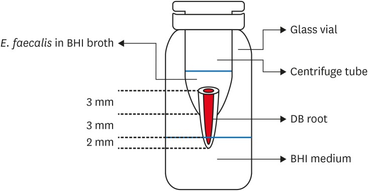
- Bacterial leakage and micro-computed tomography evaluation in round-shaped canals obturated with bioceramic cone and sealer using matched single cone technique
- Kallaya Yanpiset, Danuchit Banomyong, Kanet Chotvorrarak, Ratchapin Laovanitch Srisatjaluk
- Restor Dent Endod 2018;43(3):e30. Published online July 5, 2018
- DOI: https://doi.org/10.5395/rde.2018.43.e30
-
Abstract
PDFPubReaderePub
Objectives To evaluate sealing ability of root canals obturated with bioceramic-impregnated gutta percha cone (BCC) or gutta percha (GP), with bioceramic sealer (BCS) or AH Plus (AH; Dentsply-Maillefer), in roundly-prepared canals using matched single-cone technique, based on bacterial leakage test, and to analyze obturation quality using micro-computed tomography (CT) analysis.
Materials and Methods Ninety-two distobuccal roots of maxillary molars were prepared using nickel-titanium files to apical size 40/0.06. The roots were divided into 4 groups (
n = 20) that were obturated with a master cone and sealer: GP/AH, BCC/AH, GP/BCS, and BCC/BCS. Bacterial leakage model usingEnterococcus faecalis was used to evaluate sealing ability for 60-day period. Obturated samples from each group (n = 4) were analyzed using micro-CT.Results All groups showed bacterial leakage at 20%–45% of samples with mean leakage times of 42–52 days. There were no significant differences in bacterial leakage among the groups. Micro-CT showed minimal gaps and voids in all groups at less than 1%.
Conclusions In roundly-prepared canals, the single cone obturation with BCC/BCS was comparable to GP/AH for bacterial leakage at 60 days.
-
Citations
Citations to this article as recorded by- Time-Dependent Volumetric and Porosity Changes of Bioceramic, Silicone Bioactive Glass-Based, and Epoxy Resin-Based Root Canal Sealers: A Micro-CT Analysis
Thanh Quang Nguyen, Chantida Pawaputanon Na Mahasarakham, Pinpana Thaweesit, Kanet Chotvorrarak, Angsana Jainaen
European Journal of Dentistry.2026;[Epub] CrossRef - Assessment of Sealing Ability and Degradation Resistance of a Hydrogel‐Based Root Canal Filling Material Using a Bacterial Leakage Model and SEM Analysis
Else Ellermann, Daniel Richter, Ignasi Belda Punzano, Andreas Schmocker, Mark Bispinghoff, Tan Fırat Eyüboğlu, Mutlu Özcan, Yoel A. Klug
BioMed Research International.2026;[Epub] CrossRef - Marginal Adaptation of GuttaFlow 2, GuttaFlow Bioseal, and AH Plus in the Apical Third of Human Teeth Obturated by the Single-Cone Technique
Sayeh Abbasnejad, Faezeh Abbasi, Solmaz Araghi, Sohrab Tour Savadkouhi
Journal of Research in Dental and Maxillofacial Sciences.2026; 11(2): 107. CrossRef - Influence of Delivery Method of Hydraulic Calcium Silicate Cement‐Based Sealer on Void Formation: A Micro‐Computed Tomography Study
Yasmina Andreani, Ryan David Butler, Diana Patalwala, Paul V. Abbott, Mostafa M. A. Elkholy
Australian Endodontic Journal.2026;[Epub] CrossRef - Impact of Thermal Cycling on Volumetric Stability of Endodontic Filling Materials
Petra Dijanic, Marko Katic, Ivan Tomasic, Ana Ivanisevic, Jurica Matijevic
Clinical and Experimental Dental Research.2026;[Epub] CrossRef - Effect of Root Dentin Moisture on the Apical Sealing Ability of Root Canal Sealers: In vitro Study
Zahraa Khalil Alani, Manal Hussain Abd-alla
Al-Rafidain Journal of Medical Sciences ( ISSN 2789-3219 ).2025; 8(2): 122. CrossRef - Synthesis, physical properties, and root canal sealing of experimental MTA- and salicylate-based root canal sealers
Rafael Pino Vitti, Kusai Baroudi, Tarun Walia, Raghavandra M. Shetty, Flávia Goulart da Rosa Cardoso, Flávia de Moura Pereira, Evandro Piva, Cesar Henrique Zanchi, Gabriel Flores Abuna, Carolina Oliveira de Lima, Emmanuel João Nogueira Leal Silva, Flávio
PLOS One.2025; 20(7): e0329476. CrossRef - Impact of cone system compatibility on single cone bioceramic obturation in canals prepared with variable taper NiTi rotary files
Reem M. Barakat, Rahaf A. Almohareb, Njoom Aleid, Hoor Almowais, Aljawhara Alharbi, Meshal Al-Sharafa, Ali Alrahlah
Scientific Reports.2025;[Epub] CrossRef - Estudio de la obturación con selladores biocerámicos de conductos radiculares de premolares inferiores
Alicia Beatriz Bonafé, Cecilia Inés Rourera, Carla Pedraza, Yamila Victoria Zanoni, Soledad Salduna, Cecilia Noemi De Caso, Gabriela Martín
Methodo Investigación Aplicada a las Ciencias Biológicas.2025; 10(3): 31. CrossRef - Sealing ability of mineral trioxide aggregate: A scoping review of laboratory assessment methods
Kenta Tsuchiya, Salvatore Sauro, Jukka P. Matinlinna, Hidehiko Sano, Monica Yamauti, Deepak Mehta, Kyung‐San Min, Atsushi Tomokiyo
European Journal of Oral Sciences.2025;[Epub] CrossRef - Bacterial Leakage Testing in Dentistry: A Comprehensive Review on Methods, Models, and Clinical Relevance
Niher Tabassum Snigdha, Mohmed Isaqali Karobari, Sukhamoy Gorai
Scientifica.2025;[Epub] CrossRef - In vitro comparative evaluation of apical leakage using a bioceramic sealer with three different obturating techniques: A glucose leakage model
Tanvi S Agrawal, Shishir Singh, Rajesh S Podar, Gaurav Kulkarni, Anuprita Gadkari, Navin Agarwal
Journal of Conservative Dentistry and Endodontics.2024; 27(1): 76. CrossRef - In Vitro Microscopical and Microbiological Assessment of the Sealing Ability of Calcium Silicate-Based Root Canal Sealers
Karin Christine Huth, Sabina Noreen Wuersching, Leander Benz, Stefan Kist, Maximilian Kollmuss
Journal of Functional Biomaterials.2024; 15(11): 341. CrossRef - Comparison between AH plus sealer and total fill bioceramic sealer performance in previously untreated and retreatment cases of maxillary incisors with large-sized periapical lesion: a randomized controlled trial
Eisa Wahbi, Hassan Achour, Yasser Alsayed Tolibah
BDJ Open.2024;[Epub] CrossRef - Bacterial sealing ability of calcium silicate-based sealer for endodontic surgery: an in-vitro study
Mai M. Mansour, Sybel M. Moussa, Marwa A. Meheissen, Mahmoud R. Aboelseoud
BMC Oral Health.2024;[Epub] CrossRef - Assessment the bioactivity of zinc oxid eugenol sealer after the addition of different concentrations of nano hydroxyapatite-tyrosine amino acid
Rasha M. Al-Shamaa, Raghad A. Al-Askary
Brazilian Journal of Oral Sciences.2024; 23: e243733. CrossRef - Assessment of Bacterial Sealing Ability of Two Different Bio-Ceramic Sealers in Single-Rooted Teeth Using Single Cone Obturation Technique: An In Vitro Study
Doaa M. AlEraky, Ahmed M. Rahoma, Hatem M. Abuohashish, Abdullh AlQasser, Abbas AlHamali, Hussain M. AlHussain, Hussain M. AlShoalah, Zakrya AlSaghah, Abdulrahman Khattar, Shimaa Rifaat
Applied Sciences.2023; 13(5): 2906. CrossRef - How do imaging protocols affect the assessment of root-end fillings?
Fernanda Ferrari Esteves Torres, Reinhilde Jacobs, Mostafa EzEldeen, Karla de Faria-Vasconcelos, Juliane Maria Guerreiro-Tanomaru, Bernardo Camargo dos Santos, Mário Tanomaru-Filho
Restorative Dentistry & Endodontics.2022;[Epub] CrossRef - The impact of Morse taper implant design on microleakage at implant-healing abutment interface
Soyeon KIM, Joo Won LEE, Jae-Heon KIM, Van Mai TRUONG, Young-Seok PARK
Dental Materials Journal.2022; 41(5): 767. CrossRef - A critical analysis of research methods and experimental models to study root canal fillings
Gustavo De‐Deus, Erick Miranda Souza, Emmanuel João Nogueira Leal Silva, Felipe Gonçalves Belladonna, Marco Simões‐Carvalho, Daniele Moreira Cavalcante, Marco Aurélio Versiani
International Endodontic Journal.2022; 55(S2): 384. CrossRef - Micro‐CT assessment of gap‐containing areas along the gutta‐percha‐sealer interface in oval‐shaped canals
Gustavo De‐Deus, Gustavo O. Santos, Iara Zamboni Monteiro, Daniele M. Cavalcante, Marco Simões‐Carvalho, Felipe G. Belladonna, Emmanuel J. N. L. Silva, Erick M. Souza, Raphael Licha, Carla Zogheib, Marco A. Versiani
International Endodontic Journal.2022; 55(7): 795. CrossRef - Comparison of Sealing Ability of Bioceramic Sealer, AH Plus, and GuttaFlow in Conservatively Prepared Curved Root Canals Obturated with Single-Cone Technique: An In vitro Study
Shalan Kaul, Ajay Kumar, Bhumika Kamal Badiyani, Laxmi Sukhtankar, M. Madhumitha, Amit Kumar
Journal of Pharmacy and Bioallied Sciences.2021; 13(Suppl 1): S857. CrossRef - Micro-CT Evaluation of Four Root Canal Obturation Techniques
Mahmood Reza Kalantar Motamedi, Amin Mortaheb, Maryam Zare Jahromi, Brett E. Gilbert, Marilena Vivona
Scanning.2021; 2021: 1. CrossRef - Effects of Both Fiber Post/Core Resin Construction System and Root Canal Sealer on the Material Interface in Deep Areas of Root Canal
Hiroki Miura, Shinji Yoshii, Masataka Fujimoto, Ayako Washio, Takahiko Morotomi, Hiroshi Ikeda, Chiaki Kitamura
Materials.2021; 14(4): 982. CrossRef - Sealing ability and microbial leakage of root-end filling materials: MTA versus epoxy resin: A systematic review and meta-analysis
Mario Dioguardi, Mario Alovisi, Diego Sovereto, Giuseppe Troiano, Giancarlo Malagnino, Michele Di Cosola, Angela Pia Cazzolla, Luigi Laino, Lorenzo Lo Muzio
Heliyon.2021; 7(7): e07494. CrossRef - Development of A Nano-Apatite Based Composite Sealer for Endodontic Root Canal Filling
Angelica Bertacci, Daniele Moro, Gianfranco Ulian, Giovanni Valdrè
Journal of Composites Science.2021; 5(1): 30. CrossRef - BIOCERAMIC-BASED ROOT CANAL SEALERS
L Somolová, Z Zapletalová, M Rosa, B Novotná, I Voborná, Y Morozova
Česká stomatologie a praktické zubní lékařství.2021; 121(4): 116. CrossRef - Calcium Silicate-Based Root Canal Sealers: A Narrative Review and Clinical Perspectives
Germain Sfeir, Carla Zogheib, Shanon Patel, Thomas Giraud, Venkateshbabu Nagendrababu, Frédéric Bukiet
Materials.2021; 14(14): 3965. CrossRef - Physico-Chemical Properties of Calcium-Silicate vs. Resin Based Sealers—A Systematic Review and Meta-Analysis of Laboratory-Based Studies
Viresh Chopra, Graham Davis, Aylin Baysan
Materials.2021; 15(1): 229. CrossRef - Comparison of apical sealing ability of bioceramic sealer and epoxy resin-based sealer using the fluid filtration technique and scanning electron microscopy
Widcha Asawaworarit, Thitapa Pinyosopon, Kanittha Kijsamanmith
Journal of Dental Sciences.2020; 15(2): 186. CrossRef - Micro-computed tomographic evaluation of a new system for root canal filling using calcium silicate-based root canal sealers
Mario Tanomaru-Filho, Fernanda Ferrari Esteves Torres, Jader Camilo Pinto, Airton Oliveira Santos-Junior, Karina Ines Medina Carita Tavares, Juliane Maria Guerreiro-Tanomaru
Restorative Dentistry & Endodontics.2020;[Epub] CrossRef - A micro-computed tomographic evaluation of root canal filling with a single gutta-percha cone and calcium silicate sealer
Jong Cheon Kim, Maung Maung Kyaw Moe, Sung Kyo Kim
Restorative Dentistry & Endodontics.2020;[Epub] CrossRef - Comparative evaluation of sealing ability of gutta percha and resilon as root canal filling materials- a systematic review
Pragya Pandey, Himanshi Aggarwal, A.P. Tikku, Arpit Singh, Rhythm Bains, Shambhavi Mishra
Journal of Oral Biology and Craniofacial Research.2020; 10(2): 220. CrossRef - Micro-computed tomographic evaluation of the flow and filling ability of endodontic materials using different test models
Fernanda Ferrari Esteves Torres, Juliane Maria Guerreiro-Tanomaru, Gisselle Moraima Chavez-Andrade, Jader Camilo Pinto, Fábio Luiz Camargo Villela Berbert, Mario Tanomaru-Filho
Restorative Dentistry & Endodontics.2020;[Epub] CrossRef - Root fillings with a matched-taper single cone and two calcium silicate–based sealers: an analysis of voids using micro-computed tomography
Eugenio Pedullà, Roula S. Abiad, Gianluca Conte, Giusy R. M. La Rosa, Ernesto Rapisarda, Prasanna Neelakantan
Clinical Oral Investigations.2020; 24(12): 4487. CrossRef - Influence of different disinfection protocols on gutta-percha cones surface roughness assessed by two different methods
A.M. Nunes, J.P. Gouvea, L. da Silva
Journal of Materials Research and Technology.2019; 8(6): 5464. CrossRef - Endodontic sealers based on calcium silicates: a systematic review
David Donnermeyer, Sebastian Bürklein, Till Dammaschke, Edgar Schäfer
Odontology.2019; 107(4): 421. CrossRef
- Time-Dependent Volumetric and Porosity Changes of Bioceramic, Silicone Bioactive Glass-Based, and Epoxy Resin-Based Root Canal Sealers: A Micro-CT Analysis
- 3,740 View
- 57 Download
- 37 Crossref
- Effect of ultrasonic agitation on push-out bond strength and adaptation of root-end filling materials
- Murilo Priori Alcalde, Rodrigo Ricci Vivan, Marina Angélica Marciano, Jussaro Alves Duque, Samuel Lucas Fernandes, Mariana Bailo Rosseto, Marco Antonio Hungaro Duarte
- Restor Dent Endod 2018;43(2):e23. Published online April 27, 2018
- DOI: https://doi.org/10.5395/rde.2018.43.e23
-
Abstract
PDFPubReaderePub
Objectives This study evaluated the effect of ultrasonic agitation of mineral trioxide aggregate (MTA), calcium silicate-based cement (CSC), and Sealer 26 (S26) on adaptation at the cement/dentin interface and push-out bond strength.
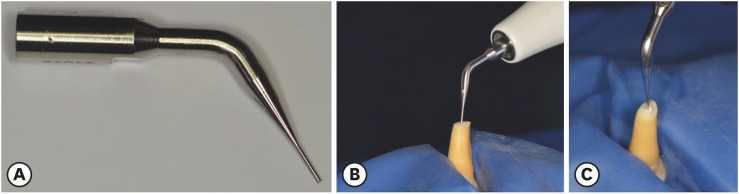
Materials and Methods Sixty maxillary canines were divided into 6 groups (
n = 10): MTA, S26, and CSC, with or without ultrasonic activation (US). After obturation, the apical portions of the teeth were sectioned, and retrograde cavities were prepared and filled with cement by hand condensation. In the US groups, the cement was activated for 60 seconds: 30 seconds in the mesio-distal direction and 30 seconds in the buccal-lingual direction, using a mini Irrisonic insert coupled with the ultrasound transducer. After the materials set, 1.5-mm thick sections were obtained from the apexes. The presence of gaps and the bond between cement and dentin were analyzed using low-vacuum scanning electron microscopy. Push-out bond strength was measured using a universal testing machine.Results Ultrasonic agitation increased the interfacial adaptation of the cements. The S26 US group showed a higher adaptation value than MTA (
p < 0.05). US improved the push-out bond strength for all the cements (p < 0.05).Conclusions The US of retrograde filling cements enhanced the bond to the dentin wall of the root-end filling materials tested.
-
Citations
Citations to this article as recorded by- Effect of ultrasonic activation on setting time, pH and calcium ion release, solubility, and chemical structure of calcium silicate sealers
Simone Argenta Scalabrin, Lina Naomi Hashizume, Theodoro Weissheimer, Gabriel Barcelos Só, Jefferson Ricardo Pereira, Milton Carlos Kuga, Ricardo Abreu da Rosa, Marcus Vinicius Reis Só
Brazilian Dental Journal.2024;[Epub] CrossRef - Impact of different disinfection protocols on the bond strength of NeoMTA 2 bioceramic sealer used as a root canal apical plug (in vitro study)
Nada Omar, Nihal Refaat Kabel, Muhammad Abbass Masoud, Tamer M. Hamdy
BDJ Open.2024;[Epub] CrossRef - Effect of Endo-Z bur or Bladesonic ultrasonic tip on the adaptation of filling material. A micro-CT study
Pedro Henrique Fiorin de Souza, Airton Oliveira Santos-Junior, Jáder Camilo Pinto, Karina Ines Medina Carita Tavares, Juliane Maria Guerreiro-Tanomaru, Mário Tanomaru-Filho
Brazilian Dental Journal.2023; 34(5): 29. CrossRef - Effect of Different Mixing Methods on Physicochemical Properties of Mineral Trioxide Aggregate: A Systematic Review
Amin Salem Milani, Faraz Radmand, Behrad Rahbani, Mahdi Hadilou, Farnaz Haji Abbas Oghli, Fatemeh Salehnia, Milad Baseri, Stefano Pagano
International Journal of Dentistry.2023; 2023: 1. CrossRef - Micro-CT comparative evaluation of porosity and dentin adaptation of root end filling materials applied with incremental, bulk, and ultrasonic activation techniques
Berkan Celikten, Aysenur Oncu, Mehrdad Koohnavard, Mert Ocak, Kaan Orhan
Proceedings of the Institution of Mechanical Engineers, Part H: Journal of Engineering in Medicine.2022; 236(8): 1209. CrossRef - Effect of ultrasonic activation of the adhesive system on dentin tubule penetration and the pushout bond strength of fiber posts
Isabel Verdum, Igor Abreu de Bem, Pedro Henrique Marks Duarte, Lucas Silveira Machado, Jefferson Ricardo Pereira, Marcus Vinícius Reis Só, Ricardo Abreu da Rosa
The Journal of Prosthetic Dentistry.2022; 127(2): 295. CrossRef - Influence of Ultrasonic Activation on the Physicochemical Properties of Calcium Silicate-Based Cements
Fredson Márcio Acris De Carvalho, Yara Teresinha Corrêa Silva-Sousa, Carlos Eduardo Saraiva Miranda, Paulo Henrique Miller Calderon, Ana Flávia Simões Barbosa, Luciana Martins Domingues De Macedo, Fuad Jacob Abi Rached-Junior, Boonlert Kukiattrakoon
International Journal of Dentistry.2021; 2021: 1. CrossRef - Micro-computed tomographic evaluation of the flow and filling ability of endodontic materials using different test models
Fernanda Ferrari Esteves Torres, Juliane Maria Guerreiro-Tanomaru, Gisselle Moraima Chavez-Andrade, Jader Camilo Pinto, Fábio Luiz Camargo Villela Berbert, Mario Tanomaru-Filho
Restorative Dentistry & Endodontics.2020;[Epub] CrossRef - Dental discoloration caused by Grey-MTAFlow cement: analysis of its physicochemical, biological and antimicrobial properties
Lauter Eston PELEPENKO, Flávia SAAVEDRA, Gabriela Fernanda BOMBARDA, Brenda Paula Figueiredo de Almeida GOMES, Adriana DE-JESUS-SOARES, Alexandre Augusto ZAIA, Marco Antonio Hungaro DUARTE, Mario TANOMARU-FILHO, Marina Angélica MARCIANO
Journal of Applied Oral Science.2020;[Epub] CrossRef - Effect of Ultrasonic Activation of Endodontic Sealers on Intratubular Penetration and Bond Strength to Root Dentin
Igor Abreu De Bem, Renata Aqel de Oliveira, Theodoro Weissheimer, Carlos Alexandre Souza Bier, Marcus Vinícius Reis Só, Ricardo Abreu da Rosa
Journal of Endodontics.2020; 46(9): 1302. CrossRef
- Effect of ultrasonic activation on setting time, pH and calcium ion release, solubility, and chemical structure of calcium silicate sealers
- 2,413 View
- 16 Download
- 10 Crossref
- Quantification of the tug-back by measuring the pulling force and micro computed tomographic evaluation
- Su-Jin Jeon, Young-Mi Moon, Min-Seock Seo
- Restor Dent Endod 2017;42(4):273-281. Published online September 4, 2017
- DOI: https://doi.org/10.5395/rde.2017.42.4.273
-
Abstract
PDFPubReaderePub
Objectives The aims of this study were to quantify tug-back by measuring the pulling force and investigate the correlation of clinical tug-back pulling force with
in vitro gutta-percha (GP) cone adaptation score using micro-computed tomography (µCT).Materials and Methods Twenty-eight roots from human single-rooted teeth were divided into 2 groups. In the ProTaper Next (PTN) group, root canals were prepared with PTN, and in the ProFile (PF) group, root canals were prepared using PF (
n = 14). The degree of tug-back was scored after selecting taper-matched GP cones. A novel method using a spring balance was designed to quantify the tug-back by measuring the pulling force. The correlation between tug-back scores, pulling force, and percentage of the gutta-percha occupied area (pGPOA) within apical 3 mm was investigated using µCT. The data were analyzed using Pearson's correlation analysis, one-way analysis of variance (ANOVA) and Tukey's test.Results Specimens with a strong tug-back had a mean pulling force of 1.24 N (range, 0.15–1.70 N). This study showed a positive correlation between tug-back score, pulling force, and pGPOA. However, there was no significant difference in these factors between the PTN and PF groups. Regardless of the groups, pGPOA and pulling force were significantly higher in the specimens with a higher tug-back score (
p < 0.05).Conclusions The degree of subjective tug-back was a definitive determinant for master cone adaptation in the root canal. The use of the tug-back scoring system and pulling force allows the interpretation of subjective tug-back in a more objective and quantitative manner.
-
Citations
Citations to this article as recorded by- Dimensional and cross-sectional compatibility of contemporary nickel–titanium files and their corresponding gutta-percha cones: A micro-computed tomography study
Cem Sarkan, Faruk Haznedaroglu
The Saudi Dental Journal.2026;[Epub] CrossRef
- Dimensional and cross-sectional compatibility of contemporary nickel–titanium files and their corresponding gutta-percha cones: A micro-computed tomography study
- 2,131 View
- 23 Download
- 1 Crossref
- Quality of root canal fillings using three gutta-percha obturation techniques
- Edith Siu Shan Ho, Jeffrey Wen Wei Chang, Gary Shun Pan Cheung
- Restor Dent Endod 2016;41(1):22-28. Published online January 4, 2016
- DOI: https://doi.org/10.5395/rde.2016.41.1.22

-
Abstract
PDFPubReaderePub
Objectives The goal of this study was to compare the density of gutta-percha root fillings obturated with the following techniques: cold lateral (CL) compaction, ultrasonic lateral (UL) compaction, and warm vertical (WV) compaction.
Materials and Methods Thirty-three extracted mandibular first molars, with two separate mesial canals in each, were selected. After instrumentation, the canals were stratified into three groups based on canal length and curvature, and underwent obturation with one of the techniques. No sealer was used in order to avoid masking any voids. The teeth were imaged pre- and post-obturation using micro-computed tomography. The reconstructed three-dimensional images were analyzed volumetrically to determine the amount of gutta-percha present in every 2 mm segment of the canal.
P values < 0.05 were considered to indicate statistical significance.Results The overall mean volume fraction of gutta-percha was 68.51 ± 6.75% for CL, 86.56 ± 5.00% for UL, and 88.91 ± 5.16% for WV. Significant differences were found between CL and UL and between CL and WV (
p < 0.05), but not between UL and WV (p = 0.526). The gutta-percha density of the roots treated with WV and UL increased towards the coronal aspect, but this trend was not noted in the CL group.Conclusions WV compaction and UL compaction produced a significantly denser gutta-percha root filling than CL compaction. The density of gutta-percha was observed to increase towards the coronal aspect when the former two techniques were used.
-
Citations
Citations to this article as recorded by- Management of internal root resorption in permanent tooth with dens invaginatus Type II: A clinical approach
Chanchal Meena, Ashwini B. Prasad, Deepak Raisingani, Harshit Srivastava, Charu Thanvi, Mohit Kumar Jyotiyana
Endodontology.2026; 38(1): 99. CrossRef - Evaluation of Postoperative Pain and Apical Outcomes Following Three Distinct Obturation Techniques: A Randomized Controlled Trial
Pragati Agarwal, Pravin Kumar, Vinay Kumar Chugh, Sharmila Shanmugam, Soundharrajan P, Karishma Pathak
Cureus.2026;[Epub] CrossRef - Effectiveness of propolis nanoparticles as endodontic therapeutic agents: a systematic review
Golnoosh Golestane, Reza Fallah Tafti, Zahra Khiali, Hadi Shakerin
Journal of Herbmed Pharmacology.2026; 15(3): 311. CrossRef - Effect of quality of radiographs taken during root canal treatment on technical quality of root canal fillings and endodontic outcome
Jia Min Ng, Yan Yee Lee, Prashanti Chippagiri, Elaheh Ahanin, Abhishek Parolia
Restorative Dentistry & Endodontics.2025; 50(1): e3. CrossRef - Restorative and endodontic clinical strategies during COVID-19 (SARS-CoV-2) pandemic: a revision of the literature
Manuele MANCINI, Flavio PALAZZI, Francesco IACONO
Minerva Dental and Oral Science.2025;[Epub] CrossRef - Evaluation of the tubular penetration of two different types of nanoparticle root canal sealers over apically separated files: a scanning electron microscopic study (in vitro study)
Alaa H. Nagdi, Nayera A. Mokhless, Mahmoud R. Aboelseoud
BMC Oral Health.2025;[Epub] CrossRef - From hopeless to regenerated: successful preservation of a tooth with massive periapical lesion in an adolescent
Adna Begović, Lajla Hasić-Branković, Samra Korać, Faruk Ljaljević, Mirna Pašić, Madžida Halilović Mehinović
Stomatological Review.2025; : 34. CrossRef - Different strategies for treating intracanal fractured instruments in a single tooth: A case report
Rong Chai, Xinpei Jiang, Ruixia Ma, Qiang Zhang, E Yang, Ansheng Zhang
Experimental and Therapeutic Medicine.2024;[Epub] CrossRef - An Experimental Anatomic CBCT Study on the Correlations Between MB1 and MB2 of the Mesio-Vestibular Root of the Upper First Molars
Luca Fiorillo, Cesare D’Amico, Giusy Rita Maria La Rosa, Francesco Calanna, Alfio Pappalardo, Eugenio Pedullà
Journal of Craniofacial Surgery.2024; 35(2): 672. CrossRef - Comparative Evaluation of Different Obturation Techniques for Root Canal Filling of Permanent Teeth: An In-Vitro Study
Adhishree S Chib, Neeta S Padmawar, Sonali Waghmare, Durgesh A Tiwari, Shahinwaz Mulani, Megna Bhatt
Cureus.2024;[Epub] CrossRef - Root canal treatment of a rhizomegaly tooth 36 mm long right permanent maxillary canine – A case report
Anita Kapri, Kiran Reddy, Varun Rana, Oliver Jacob, Pushpa Kumari
IP Annals of Prosthodontics and Restorative Dentistry.2024; 10(1): 59. CrossRef - Thermal and volumetric assessment of endodontic filling techniques using infrared thermography and micro-CT
Fernanda Clotilde M. Suassuna, Débora Ketley M. de Araújo, Ana Marly A. M. Amorim, Saulo Leonardo S. Melo, Richard J. Heck, Antonio Celso D. Antonino, Patrícia M. Bento, Diego Filipe B. Silva, Daniela P. de Melo
Journal of Oral Science.2023; 65(1): 34. CrossRef - A Comparative Evaluation of Efficacy of Various Obturating Techniques for the Presence of Voids
Rehan Ahmad Khan, Shailja Singh, Shazia Siddiqui, Mariyam Khan, Arfat Ahmad, Parul Shakarwal
Journal of Pharmacy and Bioallied Sciences.2023; 15(Suppl 2): S895. CrossRef - Continuous Wave of Condensation Improves the Filling of Curved Canals: a Micro-CT Study
Jader Camilo Pinto, Mariana Mena Barreto Pivoto-João, Juliane Maria Guerreiro-Tanomaru, Jessie Fabiola Reyes-Carmona, Mario Tanomaru-Filho
Odovtos - International Journal of Dental Sciences.2023; 25(3): 32. CrossRef - Influence of the root canal filling technique on the success rate of primary endodontic treatments: a systematic review
Daniel Feijolo Marconi, Giovana Siocheta da Silva, Theodoro Weissheimer, Isadora Ames Silva, Gabriel Barcelos Só, Leonardo Thomasi Jahnke, Jovito Adiel Skupien, Marcus Vinicius Reis Só, Ricardo Abreu da Rosa
Restorative Dentistry & Endodontics.2022;[Epub] CrossRef - Current trends in bio‐based elastomer materials
Shuai Tang, Jiao Li, Runguo Wang, Jichuan Zhang, Yonglai Lu, Guo‐Hua Hu, Zhao Wang, Liqun Zhang
SusMat.2022; 2(1): 2. CrossRef - Carrier-Based Obturation: Effect of Sonication Technique on Sealer Penetration in Dentinal Tubules: A Confocal Laser Scanning Microscope Study
Riccardo Tonini, Matteo Salvadori, Marco Bartoli, Jacopo Francinelli, Paolo Bertoletti, Maria Luisa Garo, Stefano Salgarello
Applied Sciences.2022; 12(17): 8877. CrossRef - A critical analysis of research methods and experimental models to study root canal fillings
Gustavo De‐Deus, Erick Miranda Souza, Emmanuel João Nogueira Leal Silva, Felipe Gonçalves Belladonna, Marco Simões‐Carvalho, Daniele Moreira Cavalcante, Marco Aurélio Versiani
International Endodontic Journal.2022; 55(S2): 384. CrossRef - The effect of two endodontic sealers and interval before post-preparation and cementation on the bond strength of fiber posts
He Yuanli, Wu Juan, Ji Mengzhen, Chen Xuan, Xiong Kaixin, Yang Xueqin, Qiao Xin, Hu Hantao, Gao Yuan, Zou Ling
Clinical Oral Investigations.2021; 25(11): 6211. CrossRef - Complete Obturation—Cold Lateral Condensation vs. Thermoplastic Techniques: A Systematic Review of Micro-CT Studies
Shilpa Bhandi, Mohammed Mashyakhy, Abdulaziz S. Abumelha, Mazen F. Alkahtany, Mohamed Jamal, Hitesh Chohan, A. Thirumal Raj, Luca Testarelli, Rodolfo Reda, Shankargouda Patil
Materials.2021; 14(14): 4013. CrossRef - Root canal filling quality of mandibular molars with EndoSequence BC and AH Plus sealers: A micro‐CT study
Rafael Nigri Roizenblit, Fabiola Ormiga Soares, Ricardo Tadeu Lopes, Bernardo Camargo dos Santos, Heloisa Gusman
Australian Endodontic Journal.2020; 46(1): 82. CrossRef - Effect of four different root canal obturation techniques on marginal adaptation of bioceramic sealer: An in vitro scanning electron microscopic study
NawalA Al-Sabawi, MahaM Yahya, NjwanF Shehab
Journal of International Oral Health.2020; 12(5): 455. CrossRef - Micro-computed tomographic evaluation of a new system for root canal filling using calcium silicate-based root canal sealers
Mario Tanomaru-Filho, Fernanda Ferrari Esteves Torres, Jader Camilo Pinto, Airton Oliveira Santos-Junior, Karina Ines Medina Carita Tavares, Juliane Maria Guerreiro-Tanomaru
Restorative Dentistry & Endodontics.2020;[Epub] CrossRef - Quantification of the tug-back by measuring the pulling force and micro computed tomographic evaluation
Su-Jin Jeon, Young-Mi Moon, Min-Seock Seo
Restorative Dentistry & Endodontics.2017; 42(4): 273. CrossRef
- Management of internal root resorption in permanent tooth with dens invaginatus Type II: A clinical approach
- 3,829 View
- 38 Download
- 24 Crossref
Review Article
- Thermal irritation of teeth during dental treatment procedures
- Su-Jung Kwon, Yoon-Jung Park, Sang-Ho Jun, Jin-Soo Ahn, In-Bog Lee, Byeong-Hoon Cho, Ho-Hyun Son, Deog-Gyu Seo
- Restor Dent Endod 2013;38(3):105-112. Published online August 23, 2013
- DOI: https://doi.org/10.5395/rde.2013.38.3.105
-
Abstract
PDFPubReaderePub
While it is reasonably well known that certain dental procedures increase the temperature of the tooth's surface, of greater interest is their potential damaging effect on the pulp and tooth-supporting tissues. Previous studies have investigated the responses of the pulp, periodontal ligament, and alveolar bone to thermal irritation and the temperature at which thermal damage is initiated. There are also many
in vitro studies that have measured the temperature increase of the pulp and tooth-supporting tissues during restorative and endodontic procedures. This review article provides an overview of studies measuring temperature increases in tooth structures during several restorative and endodontic procedures, and proposes clinical guidelines for reducing potential thermal hazards to the pulp and supporting tissues.-
Citations
Citations to this article as recorded by- Impact of Various Sleeve Materials on Temperature Variations During Guided Endodontic Access Cavity Preparation Utilizing Finite‐Element Analysis
Anna Muryani, Wandi Prasetia, Dudi Aripin, Hendra Dian Adhita Dharsono, Zainul Ahmad Rajion, Satrio Wicaksono
Clinical and Experimental Dental Research.2026;[Epub] CrossRef - Comparative evaluation of microcrack formation and fracture resistance of root dentin with the use of three different rotary files: An in vitro study
Paridhi Maheshwari, Sayantan Mukherjee, Ipsita Maity, Paromita Mazumdar
Journal of Oral Research and Review.2026; 18(1): 59. CrossRef - Infrared thermographic evaluation of root surface during warm vertical compaction technique
Aysenur Oncu, Ecem Ozgur, Merve Sarı, Pelin Tufenkci, Berkan Celikten
European Journal of Oral Sciences.2026;[Epub] CrossRef - Effects of voltage reduction on changes of shear bond strength and fracture mode of resin-modified glass-ionomer cements after current application
Ryouhei WADA, Emi TAKEGAWA-UYAMA, Shinya HORIUCHI, Kazumitsu SEKINE, Eiji TANAKA, Kenichi HAMADA
Dental Materials Journal.2026; 45(3): 284. CrossRef - Inverse Heat Conduction Estimation of Heat Flux in Human Dentin from Dental Curing Lights Using the Conjugate Gradient Method
Ahmad Soori, Farshad Kowsary, Shadab Safarzadeh Khosroshahi, Mohammad Vahedi
International Journal of Thermophysics.2026;[Epub] CrossRef - Temperature Variations on the External Root Surface During Warm Injectable Gutta-Percha Obturation at Different Thermo-Plasticization Settings: An In Vitro Study
Mihai Paven, Adrian-George Marinescu, Osama Abuabboud, Laura-Elena Cîrligeriu, Luminița-Maria Nica, Bianca Georgiana Cărăbiș, Izabella Maria Kovacs, Oana-Alexandra Velea-Barta, Roxana Oancea
Medicina.2026; 62(6): 1159. CrossRef - Deviation analysis of guided fiber post removal using an assembled sleeveless guide system: A case series
Jingqi Zhu, Siyi Mo, Yuan Li, Yaojun Zhang, Xutong Song, Jingwen Liu, Ye Cao, Xiaoxiang Xu
The Journal of Prosthetic Dentistry.2026;[Epub] CrossRef - External Root Temperature and Its Relationship With Dentin Thickness During Gutta-Percha Removal Procedures With Ultrasound. An Ex Vivo Study
Juan Ramon Salazar-Silva, Carlos Emilio Paschoal, Daniela de Fatima Teixeira da Silva, Denise Maria Zezell, Fábio Luiz Cunha D'Assuncao, Celso Luiz Caldeira
Journal of Endodontics.2025; 51(3): 340. CrossRef - Temperature Changes of NaOCl after Irrigation Using Passive Ultrasonic Irrigation, Easy Clean, and XP-Endo Finisher: A Randomized Crossover Clinical Trial
Geraldo Edson Freitas Athayde de Moraes, Daniel Guimarães Pedro Rocha, Carlos Eduardo Fontana, Rina Andréa Pelegrine, Alexandre Sigrist de Martin, Índia Olinta De Azevedo Queiroz, Carlos Eduardo da Silveira Bueno
European Journal of Dentistry.2025; 19(03): 660. CrossRef - Real‐Time Analysis of Changes in Internal and External Root Temperatures Using Different Systems for Activating the Irrigation Solution
Maria Eduarda Paz Dotto, Julia Menezes Savaris, Luiz Carlos de Lima Dias-Junior, Tamer Ferreira Schmidt, Lucas da Fonseca Roberti Garcia, Cleonice da Silveira Teixeira, Eduardo Antunes Bortoluzzi, Paolo Francesco Manicone
International Journal of Dentistry.2025;[Epub] CrossRef - Thermal Behaviour of Teeth With Internal Root Resorption During Obturation and Enhancing Thermal Simulations: A Finite-Element Analysis
Alper Kabakci, Ayca Yilmaz, Dilek Helvacioglu-Yigit, Nawar Naguib Nawar, Hyeon-Cheol Kim
International Dental Journal.2025; 75(6): 103903. CrossRef - Determination of a safe protocol for using laser ablation with indocyanine green dye in endodontic treatment. In vitro, in vivo and human study
Renato de Toledo Leonardo, Mirtha Perdomo, Marcelo Costa Perdomo, María Betania Acevedo Giménez, Celso Kenji Nishiyama, Fernando Accorsi Orosco, Arturo Javier Aranda Garcia, Carolina Sayuri Wajima, Cristiane Cantiga-Silva, Ana Maria Veiga Vasques, Flávio
Lasers in Medical Science.2025;[Epub] CrossRef - Antibacterial PEEK-Ag Surfaces: Development and In Vitro Evaluation Against Staphylococcus aureus and Pseudomonas aeruginosa
Flávio Rodrigues, Mariana Fernandes, Filipe Samuel Silva, Óscar Carvalho, Sara Madeira
Journal of Functional Biomaterials.2025; 16(10): 388. CrossRef - The effect of di̇fferent preheati̇ng methods on the intrapulpal temperature of bulk-fi̇ll composi̇te resi̇ns
Hilal Ateş, Merve İşcan Yapar
BMC Oral Health.2025;[Epub] CrossRef - Comparative evaluation of increase in temperature on the external root surface of teeth during retrieval of broken NiTi instrument using two ultrasonic tips and two power settings: An in vitro study
Ashish K. Jain, Rishabhkumar Jain, Rahul Rao, Prajakta Rao, Pooja Yadav, Vinayak Thorat
Journal of Conservative Dentistry and Endodontics.2024; 27(6): 634. CrossRef - Dental health concerns for patients suffering from facial, peri-oral burns, and inhalation injury: A persistent yet underappreciated challenge
Hans-Oliver Rennekampff, Isabelle Rennekampff, Mayer Tenenhaus
Burns.2024; 50(9): 107224. CrossRef - Recent advances in the pathogenesis and prevention strategies of dental calculus
Yu Wei, Gao-peng Dang, Zhao-yang Ren, Mei-chen Wan, Chen-yu Wang, Hong-bo Li, Tong Zhang, Franklin R. Tay, Li-na Niu
npj Biofilms and Microbiomes.2024;[Epub] CrossRef - In Vitro Evaluation of Root Surface Temperature Using Different Endodontic Filling Techniques
Lea Külzer, Theresia Saban, Andreas Braun, Johannes-S. Wenzler
Applied Sciences.2024; 14(21): 9830. CrossRef - Accuracy comparison of single- and double-sleeve endodontic guides for fiber post removal
Omid Dianat, Mandana Naseri, Yaser Safi, Ali Modaberi, Nazanin Zargar, Ove A. Peters, Mehran Farajollahi
BMC Oral Health.2024;[Epub] CrossRef - The Effect of Water Coolant and Bur Type on Pulp Temperature When Removing Tooth Structure and Restorative Dental Materials
C Mafrici, M Kingston, R Grice, PV Abbott
Operative Dentistry.2024; 49(1): 91. CrossRef - A Comparative Study of Temperature Variations in Incisor Root Surfaces During Root Canal Preparation Using Various Rotary Systems and Irrigation Protocols
Mihai Paven, Adrian-George Marinescu, Osama Abuabboud, Laura-Elena Cirligeriu, Luminita Maria Nica, Vlad Tiberiu Alexa, Ruxandra Sava Rosianu, Atena Galuscan, Roxana Oancea
Journal of Clinical Medicine.2024; 13(23): 7484. CrossRef - The Effect of Restoration Polymerization and Residual Dentine Thickness on Thermal Changes of Pulp Chamber of Immature Permanent Teeth
Kevser Kolçakoğlu, Merve Aksoy, Cenkhan Bal, Akif Demirel, Firdevs Tulga Öz
Pesquisa Brasileira em Odontopediatria e Clínica Integrada.2024;[Epub] CrossRef - Efficacy of pulpotomy in managing irreversible pulpitis in mature permanent teeth: A systematic review and meta-analysis
Yuanyuan Li, Wenying Wang, Qian Zeng, Michelle Tang, Joshua Massey, Brian E. Bergeron, Lisha Gu, Franklin R. Tay
Journal of Dentistry.2024; 144: 104923. CrossRef - Evaluation of diamond rotary instruments marketed for removing zirconia restorations
Severin Hunziker, Lea Thorpe, Nicola U. Zitzmann, Nadja Rohr
The Journal of Prosthetic Dentistry.2024; 131(5): 895. CrossRef - Influence of different cutting instruments and rotational speeds on heat generation and cutting efficiency when sectioning different types of zirconia
Lisa Türp, Frank Lehmann, Sebastian Wille, Matthias Kern
Journal of the Mechanical Behavior of Biomedical Materials.2024; 160: 106715. CrossRef - Loss of pulp vitality correlated with the duration of the interim restoration and the experience of the dentist: A retrospective study
Göran Nilsson, Stefan Ellner, Liselott Arnebrant, Lars Brudin, Christel Larsson
The Journal of Prosthetic Dentistry.2023; 130(6): 833. CrossRef - Thermal Sensing of Photo-Activated Dental Resin Composites Using Infrared Thermography
Turki A. Bakhsh, Abdulaziz Alfaifi, Yousef Alghamdi, Mohannad Nassar, Roaa A. Abuljadyel
Polymers.2023; 15(20): 4117. CrossRef - Effect of irrigation acid solutions on cleaning and bond strength to post‐space dentin
C. de Melo Alencar, J. Ferrari Zaniboni, J. Felipe Besegato, A. Patricia Oliveira Barros, M. Bena Gélio, L Garcia Belizário, E. Maximiliano Fernandez Godoy, M. Carlos Kuga
European Journal of Oral Sciences.2023;[Epub] CrossRef - Effect of Intermediate Irrigation on Temperature Rise during Broken NiTi File Removal Using Ultrasonic Device
László Pintér, Károly Krajczár, Fanni Őry, József Szalma, Edina Lempel
Applied Sciences.2023; 13(17): 9761. CrossRef - Top tips for improving crown preparations
James Baker, Ewen McColl, Christopher Tredwin
British Dental Journal.2023; 234(1): 16. CrossRef - Evaluation of knowledge and awareness of pediatric oral health among school teachers of Hazaribag before and after oral health education.
Vipin Ahuja, Annapurna Ahuja, Nilima Thosar
F1000Research.2023; 12: 1292. CrossRef - Applications of single laser pulse from Nd:doped lasers for cleaning of small diameter carious lesions. Modelling and analytical study
T Uzunov, M Deneva, P Uzunova, M Nenchev
Journal of Physics: Conference Series.2023; 2487(1): 012022. CrossRef - Accuracy of a 3D printed sleeveless guide system used for fiber post removal: An in vitro study
Siyi Mo, Yongwei Xu, Lei Zhang, Ye Cao, Yongsheng Zhou, Xiaoxiang Xu
Journal of Dentistry.2023; 128: 104367. CrossRef - Heat generated during dental treatments affecting intrapulpal temperature: a review
Xin Er Lau, Xiaoyun Liu, Helene Chua, Wendy Jingwen Wang, Maykon Dias, Joanne Jung Eun Choi
Clinical Oral Investigations.2023; 27(5): 2277. CrossRef - Pulp chamber temperature rise in light-cure bonding of brackets with and without primer, in intact versus restored teeth
Gabriela Cenci SCHMITZ, Fernanda de Souza HENKIN, Mauricio MEZOMO, Mariana MARQUEZAN, Gabriela BONACINA, Maximiliano Schünke GOMES, Eduardo Martinelli Santayana de LIMA
Dental Press Journal of Orthodontics.2023;[Epub] CrossRef - Temperature Rise in Curing Modes of Two Different Dental Light-Curing Units: The Importance of Heating Rate
Ahmad Soori, Faezeh Soori, Farshad Kowsary, Shahin Kasraei
International Journal of Thermophysics.2023;[Epub] CrossRef - Effective application of suitable single pulse of Nd:doped lasers for cleaning of initial carious lesions of human teeth. Experimental study
T Uzunov, M Deneva, V Kazakov, P Uzunova, N Kaimakanova, M Nenchev
Journal of Physics: Conference Series.2023; 2487(1): 012021. CrossRef - Comparative Evaluation of Intrapulpal Thermal Changes during the Polymerization of Different Adhesive Resin Materials: An In Vitro Study
Pavithra K Ramanna, Suneel V Vadavadagi, Konsam Bidya Devi, Pawankumar Kamalapurkar, Shreeshail Indi, Vineetha Chakravarthy Srinivas
The Journal of Contemporary Dental Practice.2022; 23(5): 539. CrossRef - Anesthetic-, irrigation- and pain-free dentistry? The case for a femtosecond laser enabled intraoral robotic device
Ludovic Rapp, Steve Madden, Andrei V. Rode, Laurence J. Walsh, Heiko Spallek, Quan Nguyen, Van Dau, Peter Woodfield, Dzung Dao, Omar Zuaiter, Alaa Habeb, Timothy R. Hirst
Frontiers in Dental Medicine.2022;[Epub] CrossRef - 4D Printing of Shape‐Memory Semi‐Interpenetrating Polymer Networks Based On Aromatic Heterochain Polymers
Kseniia N. Bardakova, Bato Ch. Kholkhoev, Ivan A. Farion, Evgenii O. Epifanov, Olga S. Korkunova, Yuri M. Efremov, Nikita V. Minaev, Anna B. Solovieva, Peter S. Timashev, Vitaliy F. Burdukovskii
Advanced Materials Technologies.2022;[Epub] CrossRef - How does indirect air-cooling influence pulp chamber temperature in different volume teeth and absence/presence of resin-based composite during light curing?
Mathieu Mouhat, Lina Stangvaltaite-Mouhat, Emil Finnäs, Amani Andersen, Anneli Lirhus Evertsen, Bo W. Nilsen
BMC Oral Health.2022;[Epub] CrossRef - Intrapulpal temperature changes during the cementation of ceramic veneers
Edina Lempel, Dóra Kincses, Donát Szebeni, Dóra Jordáki, Bálint Viktor Lovász, József Szalma
Scientific Reports.2022;[Epub] CrossRef - Femtosecond laser dentistry for precise and efficient cavity preparation in teeth
Ludovic Rapp, Steve Madden, Julia Brand, Laurence J. Walsh, Heiko Spallek, Omar Zuaiter, Alaa Habeb, Timothy R. Hirst, Andrei V. Rode
Biomedical Optics Express.2022; 13(9): 4559. CrossRef - Three Dimensional mapping of the root apex: distances between apexes and anatomical structures and external cortical plates
Carlos Henrique FERRARI, Amjad ABU HASNA, Frederico Canato MARTINHO
Brazilian Oral Research.2021;[Epub] CrossRef - Effects of 9,300 nm Carbon Dioxide Laser on Dental Hard Tissue: A Concise Review
Vicky Wenqing Xue, Irene Shuping Zhao, Iris Xiaoxue Yin, John Yun Niu, Edward Chin Man Lo, Chun Hung Chu
Clinical, Cosmetic and Investigational Dentistry.2021; Volume 13: 155. CrossRef - PHOTOPOLYMERIZED COMPOSITIONS AND LIGHT SOURCES FOR DENTAL PRACTICE (REVIEW)
A. M. Lalatovich, M. A. Vaniev, N. V. Sidorenko, Y. A. Makedonova, D. Yu. Dyachenko, S. V. Dyachenko
IZVESTIA VOLGOGRAD STATE TECHNICAL UNIVERSITY.2021; (12(259)): 7. CrossRef - Spray mist reduction by means of a high-volume evacuation system—Results of an experimental study
Martin Koch, Christian Graetz, Essam Al-Moraissi
PLOS ONE.2021; 16(9): e0257137. CrossRef - Pulp chamber temperature changes during orthodontic bonding – an in vitro study
Aysegul Ayhan Bani, Burcu Balos Tuncer, Cumhur Tuncer
Australasian Orthodontic Journal.2021; 37(2): 157. CrossRef - Thermal Behavior of Teeth During Restoration Procedure With Composite: Experimental Tests and Numerical Simulation
M. Potenza, P. Coppa, L. Cerroni, G. Bovesecchi
Journal of Heat Transfer.2021;[Epub] CrossRef - Comparative Evaluation of Thermal Alterations on External Root Surface during Mechanical Instrumentation and Thermoplasticized Gutta-percha Obturation: An Ex Vivo Study
Rohit Sharma, Atul Jain, Madhurima Sharma, Shivani Chauhan, Abhinay Agarwal
World Journal of Dentistry.2021; 12(5): 367. CrossRef - Degree of conversion and in vitro temperature rise of pulp chamber during polymerization of flowable and sculptable conventional, bulk-fill and short-fibre reinforced resin composites
Edina Lempel, Zsuzsanna Őri, Dóra Kincses, Bálint Viktor Lovász, Sándor Kunsági-Máté, József Szalma
Dental Materials.2021; 37(6): 983. CrossRef - Pulp chamber temperature changes during orthodontic bonding – an in vitro study
Aysegul Ayhan Bani, Burcu Balos Tuncer, Cumhur Tuncer
Australasian Orthodontic Journal.2021; 37(2): 157. CrossRef - Shot peening increases resistance to cyclic fatigue fracture of endodontic files
Javier Nino-Barrera, Jose Sanchez-Aleman, Manuel Acosta-Humanez, Luis Gamboa-Martinez, Carlos Cortes-Rodriguez
Scientific Reports.2021;[Epub] CrossRef - Temperature Dependence of Specific Heat of Human Enamel and Dentin: An Experimental Study
Ahmad Soori, Farshad Kowsary, Shahin Kasraei
International Journal of Thermophysics.2021;[Epub] CrossRef - Prosthodontic Applications of Polymethyl Methacrylate (PMMA): An Update
Muhammad Sohail Zafar
Polymers.2020; 12(10): 2299. CrossRef - The effect of halogen bulb and light-emitting diode light curing units on temperature increase and fibroblast viability
Georgia Memari Trava, Juliane Almeida Santos, Lucas Paula Ramos, Pamela Beatriz Rosário Estevam dos Santos, Amjad Abu Hasna, Karen Cristina Yui, Adriano Bressane, Luciane Dias de Oliveira, Marianne Spalding
F1000Research.2020; 9: 1369. CrossRef - A Study on Temperature Changes during Bone Scaling and Cutting of Dental Ultrasonic Scaling/Surgery System
Min-Woo Sa, Tae-Jo Ko, Jong Young Kim
Journal of the Korean Society of Manufacturing Process Engineers.2020; 19(2): 1. CrossRef - Controlling In Vivo, Human Pulp Temperature Rise Caused by LED Curing Light Exposure
DC Zarpellon, P Runnacles, C Maucoski, U Coelho, FA Rueggeberg, CAG Arrais
Operative Dentistry.2019; 44(3): 235. CrossRef - Pulp Temperature Rise Induced by Light-Emitting Diode Light-Curing Units Using an Ex Vivo Model
Alexandra Vinagre, João Ramos, Clara Rebelo, José Basto, Ana Messias, Nélia Alberto, Rogério Nogueira
Materials.2019; 12(3): 411. CrossRef - The cooling efficiency of different dental high-speed handpiece coolant port designs
Helene Chua, Joanne Jung Eun Choi, Rishi Sanjay Ramani, Ritu Ganjigatti, John Neil Waddell
Heliyon.2019; 5(8): e02185. CrossRef - Polymerisation Shrinkage Profiling of Dental Composites using Optical Fibre Sensing and their Correlation with Degree of Conversion and Curing Rate
Ginu Rajan, Raju Raju, Sagar Jinachandran, Paul Farrar, Jiangtao Xi, B. Gangadhara Prusty
Scientific Reports.2019;[Epub] CrossRef - Tooth sectioning for coronectomy: how to perform?
József Szalma, László Vajta, Lajos Olasz, Edina Lempel
Clinical Oral Investigations.2019; 23(2): 519. CrossRef - Dentistry Applications of Fiber Bragg Gratings: Irradiation Protocols for Bulk Fill Flow Dental Composites
Ana Paula Gebert de Oliveira Franco, Manoella Maria Machado Costa, Leandro Zen Karam, Osnara Maria Mongruel Gomes, Hypolito Jose Kalinowski
Journal of Lightwave Technology.2019; 37(18): 4881. CrossRef - In Vitro Analysis of Techniques that Alter the Surface Hardness of a Glass Ionomer Restorative Material
Riaan Mulder, Naeemah Noordien, Shaun Rossouw, Luzaan van Zyl
The Journal of Contemporary Dental Practice.2019; 20(12): 1362. CrossRef - Changes in the radicular pulp-dentine complex in healthy intact teeth and in response to deep caries or restorations: A histological and histobacteriological study
Domenico Ricucci, Simona Loghin, Li-na Niu, Franklin R. Tay
Journal of Dentistry.2018; 73: 76. CrossRef - Effect of Irradiance and Exposure Duration on Temperature and Degree of Conversion of Dual-Cure Resin Cement for Ceramic Restorations
JS Shim, SH Han, N Jha, ST Hwang, W Ahn, JY Lee, JJ Ryu
Operative Dentistry.2018; 43(6): E280. CrossRef - Protective Effects of Base Cements against Intrapulpal Temperature Rise during Curing of Composite Resins: An In Vitro Study by Pulpal Blood Microcirculation Model
Ihsan F Ertugrul, Basak Yazkan, Ceylan Ç Ertugrul
International Journal of Experimental Dental Science.2018; 7(2): 85. CrossRef - Thermal imaging of the pulp during residual adhesive removal
Gökmen Kurt, Nisa Gül, Özgür Er, Gülşen Çakmak, Emre Bendeş, Veysel Aslantaş
Journal of Orofacial Orthopedics / Fortschritte der Kieferorthopädie.2017; 78(4): 330. CrossRef - Influence of the material for preformed moulds on the polymerization temperature of resin materials for temporary FPDs
Philipp-Cornelius Pott, Hans Schmitz-Wätjen, Meike Stiesch, Michael Eisenburger
The Journal of Advanced Prosthodontics.2017; 9(4): 294. CrossRef - Light curing in dentistry and clinical implications: a literature review
Frederick Allen RUEGGEBERG, Marcelo GIANNINI, Cesar Augusto Galvão ARRAIS, Richard Bengt Thomas PRICE
Brazilian Oral Research.2017;[Epub] CrossRef - Intrapulpal temperature changes during curing of different bulk-fill restorative materials
Elif YASA, Cigdem ATALAYIN, Gamze KARACOLAK, Tugrul SARI, L. Sebnem TURKUN
Dental Materials Journal.2017; 36(5): 566. CrossRef - Can Mineral Trioxide Aggregate and Nanoparticulate EndoSequence Root Repair Material Produce Injurious Effects to Rat Subcutaneous Tissues?
Wafaa A. Khalil, Siham K. Abunasef
Journal of Endodontics.2015; 41(7): 1151. CrossRef - Comparison of photopolymerization temperature increases in internal and external positions of composite and tooth cavities in real time: Incremental fillings of microhybrid composite vs. bulk filling of bulk fill composite
Ryan Jin-Young Kim, Sung-Ae Son, Ji-Yun Hwang, In-Bog Lee, Deog-Gyu Seo
Journal of Dentistry.2015; 43(9): 1093. CrossRef - Real-Time Analysis of Temperature Changes in Composite Increments and Pulp Chamber during Photopolymerization
Ryan Jin-Young Kim, In-Bog Lee, Jin-Young Yoo, Su-Jung Park, Sin-Young Kim, Young-Ah Yi, Ji-Yun Hwang, Deog-Gyu Seo
BioMed Research International.2015; 2015: 1. CrossRef - Temperature changes under demineralized dentin during polymerization of three resin-based restorative materials using QTH and LED units
Sayed-Mostafa Mousavinasab, Maryam Khoroushi, Mohammadreza Moharreri, Mohammad Atai
Restorative Dentistry & Endodontics.2014; 39(3): 155. CrossRef - Comparison of Exothermic Release during the Polymerization of Four Materials used to fabricate Provisional Restorations
Minu Raju
International Journal of Prosthodontics and Restorative Dentistry.2014; 4(1): 1. CrossRef
- Impact of Various Sleeve Materials on Temperature Variations During Guided Endodontic Access Cavity Preparation Utilizing Finite‐Element Analysis
- 7,586 View
- 51 Download
- 76 Crossref
Original Articles
- The Effect of canal obturation according to the depth of the System B Plugger tip in the Type IV canal
- Hee-Won Choi, Soo-Mee Kim, Ho-Keel Hwang
- J Korean Acad Conserv Dent 2008;33(6):518-525. Published online November 30, 2008
- DOI: https://doi.org/10.5395/JKACD.2008.33.6.518
-
Abstract
PDFPubReaderePub
The purpose of this study was to evaluate the effect of the apical sealing according to the depth of the System B Plugger tip when root canal was filled with gutta-percha and sealer by Continuous Wave of Condensation technique in the Type IV canal.
50 simulated resin blocks with J-shaped curvature canals were instrumented by ProTaper (Dentsply Maillefer, Ballagiues, Switzerland) Ni-Ti files using the crown-down technique. Type IV canals were made using a broken ProTaper F3 Ni-Ti file for making a ledge at 3mm short from the working length. And ProTaper F1 Ni-Ti file was used for perforating resin block.
The prepared Type IV canals were randomly divided into three experimental groups of 15 each according to the depth of System B Plugger tip.
All of experimental groups were obturated with Continuous Wave of Condensation technique. The length of gutta-percha and sealer in lingual of the Type IV canals was measured with a measuring digital calliper under magnifying glass (× 2.3).
The results are as follows :
In control group, there was no gutta-percha and sealer in lingual canal.
3 mm group showed relatively more gutta-percha than 5mm or 7 mm group (p<0.05).
7 mm group did not showed gutta-percha and relatively more void were observed than 3mm or 5 mm group. (p<0.05)
In conclusion, within the limits of the results of this experiment, the 3 mm depth of System B Plugger tip was acceptable for obturating the Type IV canal.
- 1,500 View
- 2 Download
- Effect of irrigation methods on the adhesion of Resilon/Epiphany sealer and gutta-percha/AH 26 sealer to intracanal dentin
- Seo-Kyong Kim, Yun-Chan Hwang, In-Nam Hwang, Won-Mann Oh
- J Korean Acad Conserv Dent 2008;33(2):98-106. Published online March 31, 2008
- DOI: https://doi.org/10.5395/JKACD.2008.33.2.098
-
Abstract
PDFPubReaderePub
The purpose of this study was to evaluate whether intracanal irrigation method could affect the adhesion between intracanal dentin and root canal filling materials (Gutta-percha/AH 26 sealer and Resilon/Epiphany sealer).
Thirty extracted human incisor teeth were prepared. Canals were irrigated with three different irrigation methods as a final rinse and obturated with two different canal filling materials (G groups : Gutta-percha/AH 26 sealer, R groups : Resilon/Epiphany sealer) respectively.
Group G1, R1 - irrigated with 5.25% NaOCl
Group G2, R2 - irrigated with 5.25% NaOCl, sterile saline
Group G3, R3 - irrigated with 5.25% NaOCl, 17% EDTA, sterile saline
Thirty obturated roots were horizontally sliced and push-out bond strength test was performed in the universal testing machine. After test, the failure patterns of the specimens were observed using Image-analyzing microscope.
The results were as follows.
Gutta-percha/AH 26 sealer groups had significantly higher push-out bond strength compared with the Resilon/Epiphany sealer groups (p < 0.05).
Push-out bond strength was higher when using 17% EDTA followed by sterile saline than using NaOCl as a final irrigation solution in the Resilon/Epiphany sealer groups (p < 0.05).
In the failure pattern analysis, there was no cohesive failure in Group G1, G2, and R1. Gutta-percha/AH 26 sealer groups appeared to exhibit predominantly adhesive and mixed failure patterns, whereas Resilon/Epiphany sealer groups exhibited mixed failures with the cohesive failure occurred within the Resilon substrate.
- 1,329 View
- 5 Download
- Comparison of apical sealing efficacies using different plugging depth in continuous wave of obturation technique
- Sang-Jin Lee, Jeong-Kil Park, Bock Hur, Hyeon-Cheol Kim
- J Korean Acad Conserv Dent 2007;32(6):491-497. Published online November 30, 2007
- DOI: https://doi.org/10.5395/JKACD.2007.32.6.491
-
Abstract
PDFPubReaderePub
The purpose of this study was to compare apical sealing ability of continuous wave canal filling technique according to various heat source plugging depths.
Eighty one extracted human premolars with straight root were cleaned and shaped to size 35 using .06 taper rotary NiTi file. After cleansing and shaping, the teeth were divided into 5 groups following the heat source probing depths from the apex; 3, 4, 5, 6 and 7 mm. All specimens were filled using E&Q plus with #35 / .06 tapered gutta-percha cone. The positive control teeth were not filled. All teeth were coated with nail varnish except the apical 1 mm around the apical foramen. Negative control teeth were completely sealed include the apical foramen. All specimens were immersed in 1% methylene blue solution for 72 hours. Then the specimens were sectioned horizontally at 1, 2 and 3 mm from the root apex. Each sectioned surface was photographed using a digital camera attached to the stereomicroscope at 12.5 × 2.5 fold magnification. All points at 1, 2 and 3 mm were summed as final score of one specimen. Statistical analysis of the collected data was performed.
Under the condition of this study, there was no significant difference between the heat source plugging depths of 3, 4, 5, 6 and 7 mm in apical sealing ability. All of apical heat source plugging depth from 3 to 7 mm including Buchanan's protocol -from 5 to 7 mm- seems to be acceptable in clinical application.
- 1,260 View
- 2 Download
- Influence of plugger penetration depth on the area of the canal space occupied by gutta-percha
- Young Mi Lee, Ho-young So, Young Kyung Kim, Sung Kyo Kim
- J Korean Acad Conserv Dent 2006;31(1):66-71. Published online January 31, 2006
- DOI: https://doi.org/10.5395/JKACD.2006.31.1.066
-
Abstract
PDFPubReaderePub
To evaluate the ratio of gutta-percha area in the canal after canal obturation with Continuous Wave of Condensation Technique (CWCT) with varying depths of plugger penetration, forty root canals of extracted human teeth were prepared up to size 40 of 0.06 taper with ProFile®. Canals of three groups were filled with CWCT with System B™ (Analytic Tech., USA) and different plugger penetration depths of 3, 5, or 7 mm from the apex. Canals of one group were filled with lateral condensation technique as a control. The filled teeth were cross-sectioned at 1, 2, and 3 mm levels from the apical foramen. The ratio of gutta-percha area in the canal was analyzed using Auto®Cad 2000. Data were analyzed with one-way ANOVA and Duncan's multiple range test.
At all levels, higher gutta-percha area ratio was found with deeper plugger penetration depth in CWCT, and cold lateral condensation group showed higher ratio than group of plugger penetration to apical 7 mm in CWCT.
At apical 1 mm and 2 mm levels, group of plugger penetration to apical 3 mm showed significantly higher gutta-percha area ratio than those of apical 7 mm and lateral condensation (p < 0.05).
It is concluded therefore that, under the conditions of the present study, deeper plugger penetration depth results in more favorable and efficient obturation in CWCT.
- 1,711 View
- 5 Download
- Obturation efficiency of non-standardized gutta-percha cone in curved root canals prepared with 0.06 taper nickel-titanium instruments
- Eun-Ah Lee, Sung-Kyo Kim
- J Korean Acad Conserv Dent 2005;30(2):79-85. Published online March 31, 2005
- DOI: https://doi.org/10.5395/JKACD.2005.30.2.079
-
Abstract
PDFPubReaderePub
The purpose of this study was to evaluate the obturation efficiency of a non-standardized gutta-percha cone in curved root canals prepared with 0.06 taper nickel-titanium instruments.
Sixty simulated curved root canals in clear resin blocks were prepared with crown-down technique using 0.06 taper rotary ProTaper™ and ProFile (Dentsply-Maillefer) until apical canal was size 30. Root canals were randomly divided into 4 groups of 15 blocks and obturated with cold-laterally compacted gutta-percha technique by using either a non-standardized size medium gutta-percha cone or an ISO-standardized size 30 one as a master cone. Gutta-percha area ratio were calculated at apical levels of 1, 3, and 5 mm using AutoCAD 2000 after cross-sectioning, and the data were analyzed with one-way and two-way ANOVAs and Duncan's multiple range test.
Non-standardized size medium cone groups showed significantly higher gutta-percha area ratio than standardized cone groups at all apical levels (
p < 0.01).Non-standardized cone groups used significantly less accessory cones than standardized cone groups (
p < 0.01).-
Citations
Citations to this article as recorded by- Efficiency of Using Different Greater Taper Gutta-Percha Cones in Continuous Warm Vertical Condensation: An Ex Vivo Study
Mamata Hebbal, Reem Barakat, Rahaf Almohareb, Ghada Alaskar, Lama Alghufaily, Nouf AlFarraj, Alia Albaz
The Journal of Contemporary Dental Practice.2021; 22(1): 56. CrossRef
- Efficiency of Using Different Greater Taper Gutta-Percha Cones in Continuous Warm Vertical Condensation: An Ex Vivo Study
- 2,632 View
- 2 Download
- 1 Crossref
- COMPARISON OF APICAL SEAL WITH OR WITHOUT THE USE OF DENTIN ADHESIVE SYSTEM
- Min-Jo Lee, Sang-Hyuk Park, Gi-Woon Choi
- J Korean Acad Conserv Dent 2005;30(1):7-15. Published online January 14, 2005
- DOI: https://doi.org/10.5395/JKACD.2005.30.1.007
-
Abstract
PDFPubReaderePub
ABSTRACT The purpose of this study was to compare the sealing ability of root canal obturation with or without the use of dentin adhesive system. Forty extracted human teeth with one canal were selected and decoronated. The teeth were divided into two Groups. The obturation procedure of Group 1 was the same as that of Group 2 with the exception of dentin adhesive system. Group 2 were obturated with dentin adhesive system, AH-26, and gutta-percha.
After obturation, the teeth were immersed in methylene blue solution for 84 hours. The teeth were sectioned horizontally at 1.5 mm (Level 1), 2.0 mm (Level 2), 2.5 mm (Level 3) from the root apex using a low speed microtome. Distance of dye-penetrated surface and total dentinal surface were measured using SigmaScan Pro 5.0, and the ratio of dye-penetrated distance to the total dentinal distance was analyzed statistically by Mann-Whitney U-test.
In both groups, the mean leakage ratio was decreased cervically.
At level 1, there was no significant difference between group 1 and grpup 2 (p > 0.05).
At level 2 and 3, group 1 showed significantly higher mean leakage ratio than group 2 (p < 0.05). The results suggest that using dentin adhesive system in root canal obturation procedure reduces the apical microleakage.
-
Citations
Citations to this article as recorded by- Effect of Application over Time for Each Type of Blending Tea on Bovine Tooth Coloration
Se-Won Bae, Im-Hee Jung, Min-Ha Hong, Eun-Jin Kwon, Ji-Hyeon Kim, Ji-Hyeon Lee, Hee-Jung Lim, Do-Seon Lim
Journal of Dental Hygiene Science.2022; 22(1): 57. CrossRef - Microleakage of resilon: Effects of several self-etching primer
Jong-Hyeon O, Se-Hee Park, Hye-Jin Shin, Kyung-Mo Cho, Jin-Woo Kim
Journal of Korean Academy of Conservative Dentistry.2008; 33(2): 133. CrossRef
- Effect of Application over Time for Each Type of Blending Tea on Bovine Tooth Coloration
- 1,690 View
- 1 Download
- 2 Crossref
 First
First Prev
Prev

