
Search
- Page Path
- HOME > Search
Research Article
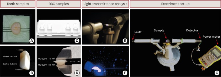
- Comparison of light-transmittance in dental tissues and dental composite restorations using incremental layering build-up with varying enamel resin layer thickness
- Rodrigo Rocha Maia, Dayane Oliveira, Tracy D'Antonio, Fang Qian, Frederick Skiff
- Restor Dent Endod 2018;43(2):e22. Published online April 16, 2018
- DOI: https://doi.org/10.5395/rde.2018.43.e22
-
 Abstract
 PDF
PDF PubReader
 ePub
ePub Objectives To evaluate and compare light-transmittance in dental tissues and dental composite restorations using the incremental double-layer technique with varying layer thickness.
Materials and Methods B1-colored natural teeth slabs were compared to dental restoration build-ups with A2D and B1E-colored nanofilled, supra-nanofilled, microfilled, and microhybrid composites. The enamel layer varied from 0.3, 0.5, or 1.2 mm thick, and the dentin layer was varied to provide a standardized 3.7 mm overall sample thickness (
n = 10). All increments were light-cured to 16 J/cm2 with a multi-wave LED (Valo, Ultradent). Using a spectrophotometer, the samples were irradiated by an RGB laser beam. A voltmeter recorded the light output signal to calculate the light-transmittance through the specimens. The data were analyzed using 1-way analysis of variance followed by thepost hoc Tukey's test (p = 0.05).Results Mean light-transmittance observed at thicker final layers of enamel were significantly lower than those observed at thinner final layers. Within 1.2 mm final enamel resin layer (FERL) thickness, all composites were similar to the dental tissues, with exception of the nanofilled composite. However, within 0.5 mm FERL thickness, only the supra-nanofilled composite showed no difference from the dental tissues. Within 0.3 mm FERL thickness, none of the composites were similar to the dental tissues.
Conclusions The supra-nanofilled composite had the most similar light-transmittance pattern when compared to the natural teeth. However, for other composites, thicker FERL have a greater chance to match the light-transmittance of natural dental tissues.
-
Citations
Citations to this article as recorded by
- Impact of water absorption on the translucency of single-shade and conventional resin composites: an in vitro comparative study
Ceyda Sari, Elifnur Aydemir Aydın
Odontology.2026;[Epub] CrossRef - 3-year randomized clinical trial to evaluate the performance of posterior composite restorations lined with ion-releasing materials
Basma Ahmed, Ramy Ahmed Wafaie, Hamdi H. Hamama, Salah Hasab Mahmoud
Scientific Reports.2024;[Epub] CrossRef - Investigation on the Biaxial Flexural Strength of Universal Shade Resin-Based Composites
Keiko Sakuma, Taku Horie, Takafumi Kishimoto, Mayumi Maesako, Shigetaka Tomoda, Morioki Fujitani, Akimasa Tsujimoto
Polymers.2024; 16(13): 1853. CrossRef - Fabrication of color-graded feldspathic dental prosthetics for aesthetic and restorative dentistry
Imam Akbar Sutejo, Jeehwan Kim, Sinuo Zhang, Chang Woo Gal, Yeong-Jin Choi, Honghyun Park, Hui-suk Yun
Dental Materials.2023; 39(6): 568. CrossRef - Spectrophotometric evaluation of restorative composite shades and their match with a classical shade guide
Rafael Melara, Luciana Mendonça, Fábio Herrmann Coelho-de-Souza, Juliana Nunes Rolla, Luciano de Souza Gonçalves
Restorative Dentistry & Endodontics.2021;[Epub] CrossRef - In vitro wear of dual‐cured bulkfill composites and flowable bulkfill composites
Jean‐François Roulet, Snigdha Gummadi, Hind S. Hussein, Nader Abdulhameed, Chiayi Shen
Journal of Esthetic and Restorative Dentistry.2020; 32(5): 512. CrossRef
- Impact of water absorption on the translucency of single-shade and conventional resin composites: an in vitro comparative study
- 2,441 View
- 19 Download
- 6 Crossref

Basic Researchs
- Effect of the difference in spectral outputs of the single and dual-peak LEDs on the microhardness and the color stability of resin composites
- Hye-Jung Park, Sung-Ae Son, Bock Hur, Hyeon-Cheol Kim, Yong-Hoon Kwon, Jeong-Kil Park
- J Korean Acad Conserv Dent 2011;36(2):108-113. Published online March 31, 2011
- DOI: https://doi.org/10.5395/JKACD.2011.36.2.108
-
Abstract
PDFPubReaderePub
Objectives To determine the effect of the spectral output of single and dual-peak light emitting diode (LED) curing lights on the microhardness and color stability of commercial resin composites formulated with camphorquinone and alternative photoinitiators in combination.
Materials and Methods Three light-polymerized resin composites (Z100 (3M ESPE), Tetric Ceram (Ivoclar Vivadent) and Aelite LS Posterior (Bisco)) with different photoinitiator systems were used. The resin composites were packed into a Teflon mold (8 mm diameter and 2 mm thickness) on a cover glass. After packing the composites, they were light cured with single-peak and dual-peak LEDs. The Knoop microhardness (KHN) and color difference (ΔE) for 30 days were measured. The data was analyzed statistically using a student's
t -test (p < 0.05).Results All resin composites showed improved microhardness when a third-generation dual-peak LED light was used. The color stability was also higher for all resin composites with dual-peak LEDs. However, there was a significant difference only for Aelite LS Posterior.
Conclusions The dual-peak LEDs have a beneficial effect on the microhardness and color stability of resin composites formulated with a combination of camphorquinone and alternative photoinitiators.
-
Citations
Citations to this article as recorded by- Effect of irradiance from curing units on the microhardness of composite - a systematic review
Neenu Francis, Rakesh R. Rajan, Vijay Kumar, Anju Varughese, Vineetha Karuveetil, C. M. Sapna
Evidence-Based Dentistry.2022;[Epub] CrossRef - Micro-computed tomography evaluation of volumetric polymerization shrinkage and degree of conversion of composites cured by various light power outputs
Pablo J. ATRIA, Camila S. SAMPAIO, Eduardo CÁCERES, Jessica FERNÁNDEZ, Andre F. REIS, Marcelo GIANNINI, Paulo G. COELHO, Ronaldo HIRATA
Dental Materials Journal.2018; 37(1): 33. CrossRef - Effect of the irradiance distribution from light curing units on the local micro-hardness of the surface of dental resins
Thomas Haenel, Berenika Hausnerová, Johannes Steinhaus, Richard B.T. Price, Braden Sullivan, Bernhard Moeginger
Dental Materials.2015; 31(2): 93. CrossRef - 1,3-Butadiene as an Adhesion Promoter Between Composite Resin and Dental Ceramic in a Dielectric Barrier Discharge Jet
Geum-Jun Han, Sung-No Chung, Bae-Hyeock Chun, Chang-Keun Kim, Kyu Hwan Oh, Byeong-Hoon Cho
Plasma Chemistry and Plasma Processing.2013; 33(2): 539. CrossRef - Optimal combination of 3-component photoinitiation system to increase the degree of conversion of resin monomers
Chang-Gyu Kim, Ho-Jin Moon, Dong-Hoon Shin
Journal of Korean Academy of Conservative Dentistry.2011; 36(4): 313. CrossRef
- Effect of irradiance from curing units on the microhardness of composite - a systematic review
- 1,995 View
- 3 Download
- 5 Crossref
- Power density of light curing units through resin inlays fabricated with direct and indirect composites
- Hoon-Sang Chang, Young-Jun Lim, Jeong-Mi Kim, Sung-Ok Hong
- J Korean Acad Conserv Dent 2010;35(5):353-358. Published online September 30, 2010
- DOI: https://doi.org/10.5395/JKACD.2010.35.5.353
-
Abstract
PDFPubReaderePub
Objectives The purpose of this study was to measure the power density of light curing units transmitted through resin inlays fabricated with direct composite (Filtek Z350, Filtek Supreme XT) and indirect composite (Sinfony).
Materials and Methods A3 shade of Z350, A3B and A3E shades of Supreme XT, and A3, E3, and T1 shades of Sinfony were used to fabricate the resin inlays in 1.5 mm thickness. The power density of a halogen light curing unit (Optilux 360) and an LED light curing unit (Elipar S10) through the fabricated resin inlays was measured with a hand held dental radiometer (Cure Rite). To investigate the effect of each composite layer consisting the resin inlays on light transmission, resin specimens of each shade were fabricated in 0.5 mm thickness and power density was measured through the resin specimens.
Results The power density through the resin inlays was lowest with the Z350 A3, followed by Supreme XT A3B and A3E. The power density was highest with Sinfony A3, E3, and T1 (
p < 0.05). The power density through 0.5 mm thick resin specimens was lowest with dentin shades, Sinfony A3, Z350 A3, Supreme XT A3B, followed by enamel shades, Supreme XT A3E and Sinfony E3. The power density was highest with translucent shade, Sinfony T1 (p < 0.05).Conclusions Using indirect lab composites with dentin, enamel, and translucent shades rather than direct composites with one or two shades could be advantageous in transmitting curing lights through resin inlays.
-
Citations
Citations to this article as recorded by- Comparison of polymerization shrinkage of dual-cure core build-up resin according to shade and curing mode
Yoorina Choi, Karl Lee, Hoon-Sang Chang
Oral Biology Research.2019; 43(4): 243. CrossRef - Early Hardness and Shear Bond Strength of Dual-cure Resin Cement Light Cured Through Resin Overlays With Different Dentin-layer Thicknesses
H-S Chang, J-W Kim
Operative Dentistry.2014; 39(4): 398. CrossRef - Effects of layering technique on the shade of resin overlays and the microhardness of dual cure resin cement
Hoon-Sang Chang, Sung-Ok Hong
Brazilian Oral Research.2014;[Epub] CrossRef - Light curing of dual cure resin cement
Hoon-Sang Chang
Restorative Dentistry & Endodontics.2013; 38(4): 266. CrossRef - Power density of various light curing units through resin inlays with modified layer thickness
Sung-Ok Hong, Yonghui Oh, Jeong-Bum Min, Jin-Woo Kim, Bin-Na Lee, Yun-Chan Hwang, In-Nam Hwang, Won-Mann Oh, Hoon-Sang Chang
Restorative Dentistry & Endodontics.2012; 37(3): 130. CrossRef
- Comparison of polymerization shrinkage of dual-cure core build-up resin according to shade and curing mode
- 1,836 View
- 2 Download
- 5 Crossref
Original Articles
- The effectiveness of sealing technique on in-office bleaching
- Yoon Lee, So-Ran Kwon, Jeong-Won Park
- J Korean Acad Conserv Dent 2008;33(5):463-471. Published online September 30, 2008
- DOI: https://doi.org/10.5395/JKACD.2008.33.5.463
-
Abstract
PDFPubReaderePub
This study investigated the clinical effectiveness and safety of sealed bleaching compared to conventional in-office bleaching using a randomized clinical trial of split arch design. Ten participants received a chairside bleaching treatment on the upper anterior teeth, and each side was randomly designated as sealed or control side. A mixture of Brite powder (PacDent, Walnut, USA), 3% hydrogen peroxide and carbamide peroxide (KoolWhite, PacDent, Walnut, USA) were used as bleaching agent. The control side was unwrapped and the experimental side was covered with a linear low density polyethylene (LLDPE) wrap for sealed bleaching. The bleaching gel was light activated for 1 hour. The tooth shades were evaluated before treatment, after treatment, and at one week check up by means of a visual shade (VS) assessment using a value oriented shade guide and a computer assisted shade assessment using a spectrophotometer (SP). The data were analyzed by paired t-test.
In the control and sealed groups, the visual shade scores after bleaching treatment and at check up showed statistically significant difference from the preoperative shade scores (p < .05). The shade scores of the sealed group were significantly lighter than the control immediately after bleaching and at the check-up appointment (p < 0.05). Compared to prebleaching status, the ΔE values at post-bleaching condition were 4.35 ± 1.38 and 5.08 ± 1.34 for the control and sealed groups, respectively. The ΔE values at check up were 3.73 ± 1.95 and 4.38 ± 2.08 for the control and sealed groups. ΔE values were greater for the sealed group both after bleaching (p < .05) and at check up (p < .05).
In conclusion, both ΔE and shade score changes were greater for the sealed bleaching group than the conventional bleaching group, effectively demonstrating the improvement of effectiveness through sealing.
- 2,915 View
- 4 Download
- Comparison of the residual stress of the nanofilled composites
- Jeong-won Park
- J Korean Acad Conserv Dent 2008;33(5):457-462. Published online September 30, 2008
- DOI: https://doi.org/10.5395/JKACD.2008.33.5.457
-
Abstract
PDFPubReaderePub
"Residual stress" can be developed during polymerization of the dental composite and it can be remained after this process was completed. The total amount of the force which applied to the composite restoration can be calculated by the sum of external and internal force. For the complete understanding of the restoration failure behavior, these two factors should be considered. In this experiment, I compared the residual stress of the recently developed nanofilled dental composite by ring slitting methods.
The composites used in this study can be categorized in two groups, one is microhybrid type-Z250, as control group, and nanofilled type-Grandio, Filtek Supreme, Ceram-X, as experimental ones. Composite ring was made and marked two reference points on the surface. Then measure the change of the distance between these two points before and after ring slitting. From the distance change, average circumferential residual stress (σθ) was calculated. In 10 minutes and 1 hour measurement groups, Filtek Supreme showed higher residual stress than Z250 and Ceram-X. In 24 hour group, Filtek showed higher stress than the other groups.
Following the result of this experiment, nanofilled composite showed similar or higher residual stress than Z250, and when comparing the Z250 and Filtek Supreme, which have quite similar matrix components, Filtek Supreme groups showed higher residual stress.
-
Citations
Citations to this article as recorded by- Microleakage of the experimental composite resin with three component photoinitiator systems
Ji-Hoon Kim, Dong-Hoon Shin
Journal of Korean Academy of Conservative Dentistry.2009; 34(4): 333. CrossRef
- Microleakage of the experimental composite resin with three component photoinitiator systems
- 1,790 View
- 4 Download
- 1 Crossref
- Ingredients and cytotoxicity of MTA and 3 kinds of Portland cements
- Seok-Woo Chang, Hyun-Mi Yoo, Dong Sung Park, Tae-Seok Oh, Kwang-Shik Bae
- J Korean Acad Conserv Dent 2008;33(4):369-376. Published online July 31, 2008
- DOI: https://doi.org/10.5395/JKACD.2008.33.4.369
-
Abstract
PDFPubReaderePub
The aim of this study was to compare the compositions and cytotoxicity of white ProRoot MTA (white mineral trioxide aggregate) and 3 kinds of Portland cements. The elements, simple oxides and phase compositions of white MTA (WMTA), gray Portland cement (GPC), white Portland cement (WPC) and fast setting cement (FSC) were measured by inductively coupled plasma atomic emission spectrometry (ICP-AES), X-ray fluorescence spectrometry (XRF) and X-ray diffractometry (XRD). Agar diffusion test was carried out to evaluate the cytotoxicity of WMTA and 3 kinds of Portland cements.
The results showed that WMTA and WPC contained far less magnesium (Mg), iron (Fe), manganese (Mn), and zinc (Zn) than GPC and FSC. FSC contained far more aluminum oxide (Al2O3) than WMTA, GPC, and WPC. WMTA, GPC, WPC and FSC were composed of main phases, such as tricalcicium silicate (3CaO·SiO2), dicalcium silicate (2CaO·SiO2), tricalcium aluminate (3CaO·Al2O3), and tetracalcium aluminoferrite (4CaO·Al2O3·Fe2O3). The significance of the differences in cellular response between WMTA, GPC, WPC and FSC was statistically analyzed by Kruskal-Wallis Exact test with Bonferroni's correction. The result showed no statistically significant difference (p > 0.05).
WMTA, GPC, WPC and FSC showed similar compositions. However there were notable differences in the content of minor elements, such as aluminum (Al), magnesium, iron, manganese, and zinc. These differences might influence the physical properties of cements.
-
Citations
Citations to this article as recorded by- Development of Multi-functional Composite Cement with Strength Improvement Using Disposable Waste Masks
Jong-Won Chung, Hyun-Kyoung Yang
Journal of Power System Engineering.2022; 26(3): 31. CrossRef - The effects of mineral trioxide aggregate on osteo/odontogenic potential of mesenchymal stem cells: a comprehensive and systematic literature review
Danial Babaki, Sanam Yaghoubi, Maryam M. Matin
Biomaterial Investigations in Dentistry.2020; 7(1): 175. CrossRef - Remineralization of demineralized dentin using a dual analog system
Neha Saxena, Stefan Habelitz, Grayson W. Marshall, Laurie B. Gower
Orthodontics & Craniofacial Research.2019; 22(S1): 76. CrossRef - Chemical analysis and biological properties of two different formulations of white portland cements
Hany Mohamed Aly Ahmed, Norhayati Luddin, Thirumulu Ponnuraj Kannan, Khairani Idah Mokhtar, Azlina Ahmad
Scanning.2016; 38(4): 303. CrossRef - In vitrocytotoxicity of four calcium silicate-based endodontic cements on human monocytes, a colorimetric MTT assay
Sedigheh Khedmat, Somayyeh Dehghan, Jamshid Hadjati, Farimah Masoumi, Mohammad Hossein Nekoofar, Paul Michael Howell Dummer
Restorative Dentistry & Endodontics.2014; 39(3): 149. CrossRef - Conservative approach of a symptomatic carious immature permanent tooth using a tricalcium silicate cement (Biodentine): a case report
Cyril Villat, Brigitte Grosgogeat, Dominique Seux, Pierre Farge
Restorative Dentistry & Endodontics.2013; 38(4): 258. CrossRef - Chemical characteristics of mineral trioxide aggregate and its hydration reaction
Seok-Woo Chang
Restorative Dentistry & Endodontics.2012; 37(4): 188. CrossRef - Physical and chemical properties of experimental mixture of mineral trioxide aggregate and glass ionomer cement
Yu-Na Jeong, So-Young Yang, Bum-Jun Park, Yeong-Joon Park, Yun-Chan Hwang, In-Nam Hwang, Won-Mann Oh
Journal of Korean Academy of Conservative Dentistry.2010; 35(5): 344. CrossRef - Biocompatibility of bioaggregate cement on human pulp and periodontal ligament (PDL) derived cells
Choo-Ryung Chung, Euiseong Kim, Su-Jung Shin
Journal of Korean Academy of Conservative Dentistry.2010; 35(6): 473. CrossRef - Physical properties of novel composite using Portland cement for retro-filling material
Sang-Jin Lee, Ok-In Cho, Jiwan Yum, Jeong-Kil Park, Bock Hur, Hyeon-Cheol Kim
Journal of Korean Academy of Conservative Dentistry.2010; 35(6): 445. CrossRef - A bioactivity study of Portland cement mixed with β-glycerophosphosphate on human pulp cell
Young-Hwan Oh, Young-Joo Jang, Yong-Bum Cho
Journal of Korean Academy of Conservative Dentistry.2009; 34(5): 415. CrossRef - Comparison of biocompatibility of four root perforation repair materials
Min-Kyung Kang, In-Ho Bae, Jeong-Tae Koh, Yun-Chan Hwang, In-Nam Hwang, Won-Mann Oh
Journal of Korean Academy of Conservative Dentistry.2009; 34(3): 192. CrossRef - Effects of condensation techniques and canal sizes on the microleakage of orthograde MTA apical plug in simulated canals
Deuk-Lim Nam, Jeong-Kil Park, Bock Hur, Hyeon-Cheol Kim
Journal of Korean Academy of Conservative Dentistry.2009; 34(3): 208. CrossRef
- Development of Multi-functional Composite Cement with Strength Improvement Using Disposable Waste Masks
- 2,078 View
- 3 Download
- 13 Crossref
- Shear bond strength of dentin bonding agents cured with a Plasma Arc curing light
- Youngchul Kwon, Sun-Young Kim, Sae-Joon Chung, Young-Chul Han, In-Bog Lee, Ho-Hyun Son, Chung-Moon Um, Byeong-Hoon Cho
- J Korean Acad Conserv Dent 2008;33(3):213-223. Published online May 31, 2008
- DOI: https://doi.org/10.5395/JKACD.2008.33.3.213
-
Abstract
PDFPubReaderePub
The objective of this study was to compare dentin shear bond strength (DSBS) of dentin bonding agents (DBAs) cured with a plasma arc (PAC) light curing unit (LCU) and those cured with a light emitting diode (LED) LCU. Optical properties were also analyzed for Elipar freelight 2 (3M ESPE); LED LCU, Apollo 95E (DMT Systems); PAC LCU and VIP Junior (Bisco); Halogen LCU. The DBAs used for DSBS test were Scotchbond Multipurpose (3M ESPE), Singlebond 2 (3M ESPE) and Clearfil SE Bond (Kuraray). After DSBS testing, fractured specimens were analyzed for failure modes with SEM.
The total irradiance and irradiance between 450 nm and 490 nm of the LCUs were different. LED LCU showed narrow spectral distribution around its peak at 462 nm whereas PAC and Halogen LCU showed a broad spectrum. There were no significant differences in mean shear bond strength among different LCUs (P > 0.05) but were significant differences among different DBAs (P < 0.001)
-
Citations
Citations to this article as recorded by- Temperature changes under demineralized dentin during polymerization of three resin-based restorative materials using QTH and LED units
Sayed-Mostafa Mousavinasab, Maryam Khoroushi, Mohammadreza Moharreri, Mohammad Atai
Restorative Dentistry & Endodontics.2014; 39(3): 155. CrossRef
- Temperature changes under demineralized dentin during polymerization of three resin-based restorative materials using QTH and LED units
- 2,087 View
- 2 Download
- 1 Crossref
- Surface roughness and color stability of various composite resins
- Sung-Yi Lee, Hyeon-Cheol Kim, Bock Hur, Jeong-Kil Park
- J Korean Acad Conserv Dent 2007;32(6):542-549. Published online November 30, 2007
- DOI: https://doi.org/10.5395/JKACD.2007.32.6.542
-
Abstract
PDFPubReaderePub
The purpose of this study was to evaluate the difference in the surface roughness after polishing and to evaluate the difference in color stability after immersion in a dye solution among four types of composite resin materials. Four light-polymerized composite resins (Shade A2) with different sized filler content (a nanofilled, a hybrid, a microfilled, a flowble) were used. Average surface roughness (Ra) was measured with a surface roughness tester (Surftest Formtracer) before and after polishing with aluminum oxide abrasive discs (Super-Snap). Color of specimens before and after staining with 2% methylene blue solution were measured using spectrophotometer (CM-3700d) with SCI geometries. The results of Ra and ΔE were analyzed by one-way analysis of variance (ANOVA), a Scheffe multiple comparison test and Student t-test (p = 0.05). After polishing, Ra values were decreased regardless of type of composite resins. In surface roughness after polishing and color stability after staining, nanofilled composite resin was not different with other composite resins except flowable resins.
-
Citations
Citations to this article as recorded by- Effect of contemporary polishing systems on hardness and roughness of one-shaded dental composites
Kivanc Dulger, Gencaga Purcek
Journal of the Australian Ceramic Society.2025; 61(3): 841. CrossRef - Physicomechanical properties and polymerization shrinkage of the newly developed radiopaque flowable composite derived from rice husk
Nor Ain Fatihah Azlisham, Yanti Johari, Dasmawati Mohamad, Mohd Firdaus Yhaya, Zuliani Mahmood
Polymer Composites.2025; 46(7): 5924. CrossRef - Highly Filled Flowable Composite Resins as Sole Restorative Materials: A Systematic Review
Konstantinos Tzimas, Eftychia Pappa, Maria Fostiropoulou, Efstratios Papazoglou, Christos Rahiotis
Materials.2025; 18(14): 3370. CrossRef - Effect of immersion and thermocycling in different beverages on the surface roughness of single- and multi-shade resin composites
Aiah A. El-Rashidy, Omar Shaalan, Rasha M. Abdelraouf, Nour A. Habib
BMC Oral Health.2023;[Epub] CrossRef - Degree of conversion and physicomechanical properties of newly developed flowable composite derived from rice husk using urethane dimethacrylate monomer
Nor Ain Fatihah Azlisham, Yanti Johari, Dasmawati Mohamad, Mohd Firdaus Yhaya, Zuliani Mahmood
Proceedings of the Institution of Mechanical Engineers, Part H: Journal of Engineering in Medicine.2023; 237(12): 1339. CrossRef - Translucency and Color Stability of a Simplified Shade Nanohybrid Composite after Ultrasonic Scaling and Air-Powder Polishing
Ksenia Babina, Maria Polyakova, Inna Sokhova, Vladlena Doroshina, Alexandr Zaytsev, Elena E. Nikonova, Gleb S. Budylin, Evgeny A. Shirshin, Christian Tantardini, Nina Novozhilova
Nanomaterials.2022; 12(24): 4465. CrossRef - Surface properties and color stability of dental flowable composites influenced by simulated toothbrushing
Guangyun LAI, Liya ZHAO, Jun WANG, Karl-Heinz KUNZELMANN
Dental Materials Journal.2018; 37(5): 717. CrossRef - Topography and surface roughness of fluid resins used as bioprotectors of mini-implants
Rogério Lacerda-Santos, Mirella de Fátima Liberato de Moura, Fabíola Galbiatti Carvalho, Hugo Lemes Carlo, Matheus Melo Pithon, Bruno Alessandro Silva Guedes de Lima, Tibério Andrade dos Passos
Applied Adhesion Science.2014;[Epub] CrossRef
- Effect of contemporary polishing systems on hardness and roughness of one-shaded dental composites
- 3,734 View
- 28 Download
- 8 Crossref
- Effect of intermediate resin hydrophilicity on bond strength of single step adhesive
- Yong-Sung Kim, Sang-Hyuk Park, Gi-Woon Choi, Kyoung-Kyu Choi
- J Korean Acad Conserv Dent 2007;32(5):445-458. Published online September 30, 2007
- DOI: https://doi.org/10.5395/JKACD.2007.32.5.445
-
Abstract
PDFPubReaderePub
The purpose of this study was to evaluate the bond strength of a new Single step system with different curing mode composites, and to evaluate the effect of the intermediate resins which have different hydrophilicity on bonding ability by means of the micro shear bond testing and TEM examination for the adhesive interface. The adhesive used in this study was an experimental single step system (Bisco Inc., Schaumburg, IL). Experimental groups were produced by using six kinds of intermediate resin having different hydrophilicity that was hydrophilic, hydrophobic and most hydrophobic resin and as filled or not after applying adhesive. Each experimental group was further divided into two subgroups whether the adhesive was light cured or not. Dual cured composite (Bis Core, Bisco Ltd., Schaumburg, IL) was placed on the adhesive layer as light cure or self cure mode. The results of bond strength were statistically analyzed using one way ANOVA and multiple comparisons are made using Tukey's test at α < 0.05 level.
The results of this study were as follows;
1. The application of intermediate resin did not increase the bond strength for light cured composite.
2. The bond strength of an experimental adhesive with self cured composite was significantly increased by the application of intermediate resin layer.
3. The bond strength of adhesive was irrespective of the cure or not of itself before intermediate resin layer applied.
4. As applied hydrophilic resin layer was, the initial bond strength was higher than both hydrophobic and most hydrophobic one used but there was no significance.
Using a single step adhesive with dual/self cured composite, the incompatibility between both of them should be solved by the application of intermediate hydrophobic resin to reduce the adhesive permeability. However, Single step adhesive can be used in the light cured composite restoration without any decrease of the initial bond strength.
-
Citations
Citations to this article as recorded by- The effect of priming etched dentin with solvent on the microtensile bond strength of hydrophobic dentin adhesive
Eun-Sook Park, Ji-Hyun Bae, Jong-Soon Kim, Jae-Hoon Kim, In-Bog Lee, Chang-Keun Kim, Ho-Hyun Son, Byeong-Hoon Cho
Journal of Korean Academy of Conservative Dentistry.2009; 34(1): 42. CrossRef
- The effect of priming etched dentin with solvent on the microtensile bond strength of hydrophobic dentin adhesive
- 1,778 View
- 1 Download
- 1 Crossref
- The effect of adhesive thickness on microtensile bond strength to the cavity wall
- Hwa-Eon Lee, Hyeon-Cheol Kim, Bock Hur, Jeong-Kil Park
- J Korean Acad Conserv Dent 2007;32(1):9-18. Published online January 31, 2007
- DOI: https://doi.org/10.5395/JKACD.2007.32.1.009
-
Abstract
PDFPubReaderePub
The purposes of this study were to examine the variability of adhesive thickness on the different site of the cavity wall when used total-etch system without filler and simplified self-etch system with filler and to evaluate the relationship between variable adhesive thickness and microtensile bond strength to the cavity wall.
A class I cavity in six human molars was prepared to expose all dentinal walls. Three teeth were bonded with a filled adhesive, Clearfil™ SE bond and the other three teeth were bonded with unfilled adhesives, Scotchbond™ Multi Purpose. Morphology and thickness of adhesive layer were examined using fluorescence microscope. Bonding agent thickness was measured at three points along the axial cavity wall, edge of cavity margin
(rim) (hlf) (ang) rim, hlf ang For both bonding agents, adhesive thickness of
ang rim hlf Adhesive thickness of internal angle of the cavity was significantly thicker than that of the cavity margin and the halfway cavity wall for both bonding agents. Microtensile bond strength of the thick adhesive layer at the internal angle of the cavity was higher than that of the thin adhesive layer at the cavity margin and the halfway cavity in the two bonding systems.
-
Citations
Citations to this article as recorded by- Bond strength of a 3-step total-etch bonding system to dentine – An improved approach
H. Hassan Elnadif, W. Palin, M.A. Hadis, B.W. Darvell
Dental Materials.2025; 41(5): 483. CrossRef - Evaluation of bonding effectiveness of a self-etch and an etch-and-rinse adhesive resin to un-treated and Er:Yag laser treated dentin using mini-interfacial fracture toughness test
Marjan Behroozibakhsh, Sotoudeh Davaie, Abbas Monzavi, Tayebeh Abazari, Sima Shahabi, Maryam Pirmoradian
Journal of Adhesion Science and Technology.2019; 33(11): 1201. CrossRef - The effect of Er,Cr:YSGG irradiation on microtensile bond strength of composite resin restoration
Jeong-Hye Son, Hyeon-Cheol Kim, Bock Hur, Jeong-Kil Park
Journal of Korean Academy of Conservative Dentistry.2010; 35(2): 134. CrossRef
- Bond strength of a 3-step total-etch bonding system to dentine – An improved approach
- 2,588 View
- 7 Download
- 3 Crossref
- The effect of different flute design and torque-controlled motor on the shaping ability of simulated resin root canals
- Hyoung-Mee Roh, Bock Huh, Hyeon-Cheol Kim, Jeong-Kil Park
- J Korean Acad Conserv Dent 2005;30(6):486-492. Published online November 30, 2005
- DOI: https://doi.org/10.5395/JKACD.2005.30.6.486
-
Abstract
PDFPubReaderePub
The purpose of this study was to compare the shaping ability of the two different Ni-Ti file systems and the two different engine systems in simulated canals.
A total of four groups of each 10 were tested. Each group was instrumented with HeroShaper®and Endo-Mate2® (Group HE), HeroShaper® and Tecnika® (Group HT), ProFile® and Endo-Mate2® (Group PE), and ProFile® and Tecnika® (Group PT).
Canal preparation time was recorded. The images of pre- and post-instrumented root canals were scanned and superimposed. The amounts of increased width and centering ratio were measured and calculated at apical 1, 3 and 5 mm levels.
These data were statistically analyzed with one-way ANOVA and Duncan's multiple range test
The results of this study were as follows;
1. Canal preparation time of HT group was the shortest (p < 0.05).
2. The amount of increased canal width in HE group was significantly larger than PT group at apical 1 mm level (p < 0.05). At apical 3 mm level, PT group was significantly smaller than other groups (p < 0.05). At apical 5 mm level, PE group was significantly larger than PT group (p < 0.05).
3. The amount of centering ratio in HE group was significantly larger than other groups (p < 0.05). At apical 5 mm level, HT group was significantly larger than PE group and PT group (p < 0.05).
Under the condition of this study, torque-controlled endodontic motor is safer than no torque controlled motor, especially when the active file is used.
- 1,304 View
- 0 Download
- The sustaining effect of three polymers on the release of chlorhexidine from a controlled release drug device for root canal disinfection
- Young-Bin Bok, Doug-Youn Lee, Chang-Young Lee, Kyung-Nam Kim, Kee-Yeon Kum
- J Korean Acad Conserv Dent 2004;29(6):548-554. Published online November 30, 2004
- DOI: https://doi.org/10.5395/JKACD.2004.29.6.548
-
Abstract
PDFPubReaderePub
The aim of this in vitro study was to evaluate the suitability of using chitosan, poly (lactide-co-glycolide) (PLGA), and polymethyl methacrylate (PMMA) to control the release of chlorhexidine digluconate (CHX) from a prototype of controlled release drug device (CRD) for root canal disinfection. Four different prototypes with different formulations were prepared. Group A (n = 12); The device (absorbent paper point) was loaded with CHX as control. Group B (n = 12); same as group A, but the device was coated with chitosan. In Groups C and D, the device was treated in the same way as group A and then coated three times with 5% PMMA (Group C, n = 12), or coated three times with 3% PLGA (Group D, n = 12). The devices were randomly allocated to experimental groups of 12 each.
All CRD prototypes were soaked in 3 mL distilled water. The concentrations of CHX were determined using a UV spectrophotometer. The surface characteristics of each prototype were observed using a scanning electron microscope.
The result showed that release rate of CHX was the greatest in the non-coated group, followed by the chitosan-coated group, the PLGA-coated group, and the PMMA-coated group (P < 0.05). Pores were observed on the surface of the prototypes that were coated with PLGA and PMMA. When the pore size was smaller, the release rate was lower. This data indicate that polymer coating can control the release rate of CHX from the CRD prototypes.
-
Citations
Citations to this article as recorded by- Effect of different chlorhexidine application times on microtensile bond strength to dentin in Class I cavities
Hyun-Jung Kang, Ho-Jin Moon, Dong-Hoon Shin
Restorative Dentistry & Endodontics.2012; 37(1): 9. CrossRef - Effect of chlorhexidine application on the bond strength of resin core to axial dentin in endodontic cavity
Yun-Hee Kim, Dong-Hoon Shin
Restorative Dentistry & Endodontics.2012; 37(4): 207. CrossRef
- Effect of different chlorhexidine application times on microtensile bond strength to dentin in Class I cavities
- 1,903 View
- 2 Download
- 2 Crossref
- The polymerization rate and the degree of conversion of composite resins by different light sources
- Joo-Hee Ryoo, In-Bog Lee, Hyun-Mee Yoo, Mi-Ja Kim, Chang-In Seok, Hyuk-Choon Kwon
- J Korean Acad Conserv Dent 2004;29(4):386-398. Published online July 31, 2004
- DOI: https://doi.org/10.5395/JKACD.2004.29.4.386
-
Abstract
PDFPubReaderePub
Objectives The purpose of this study was to observe the reaction kinetics and the degree of polymerization of composite resins when cured by different light sources and to evaluate the effectiveness of the blue Light Emitting Diode Light Curing Units (LED LCUs) compared with conventional halogen LCUs.
Materials and Methods First, thermal analysis was performed by a differential scanning calorimeter (DSC). The LED LCU (Elipar Freelight, 320 mW/cm2) and the conventional halogen LCU (XL3000, 400 mW/cm2) were used in this study for curing three composite resins (SureFil, Z-250 and AEliteFLO). Second, the degree of conversion was obtained in the composite resins cured according to the above curing mode with a FTIR. Third, the measurements of depth of cure were carried out in accordance with ISO 4049 standards. Statistical analysis was performed by two-way ANOVA test at 95% levels of confidence and Duncan's procedure for multiple comparisons.
Results The heat of cure was not statistically different among the LCUs (p > 0.05). The composites cured by the LED (Exp) LCUs were statistically more slowly polymerized than by the halogen LCU and the LED (Std) LCU (p < 0.05). The composite resin groups cured by the LED (Exp) LCUs had significantly greater degree of conversion value than by the halogen LCU and the LED (Std) LCU (p = 0.0002). The composite resin groups cured by the LED (Std) LCUs showed significantly greater depth of cure value than by the halogen LCU and the LED (Exp) LCU (p < 0.05).
-
Citations
Citations to this article as recorded by- Features of polymerization kinetics and heat realize of epoxy resin modified with silicone, silane and siloxane additives
Sergey Savotchenko, Ekaterina Kovaleva
Polymer Bulletin.2024; 81(15): 13419. CrossRef - Kinetic features of polymerization of epoxy resin modified by silicon‐containing additives and mineral fillers
Ekaterina G. Kovaleva, Sergey E. Savotchenko
Polymer Engineering & Science.2022; 62(1): 75. CrossRef - Characterization of curing behavior of UV-curable LSR for LED embedded injection mold
Joon-Sung Tae, Kyung-Gyu Yim, Byung-Ohk Rhee, Jae B. Kwak
Korea-Australia Rheology Journal.2016; 28(4): 247. CrossRef
- Features of polymerization kinetics and heat realize of epoxy resin modified with silicone, silane and siloxane additives
- 2,325 View
- 5 Download
- 3 Crossref
- Effect of rewetting agent on dentinal microtensile bond strength
- Hee-Young Kang, Young-Gon Cho, Jong-Uk Kim, Byung-Cheul Park, Sang-Hoon Yoo, Cheul-Hee Jin, Hee-Young Choi, Young-Jae Ki
- J Korean Acad Conserv Dent 2004;29(2):153-161. Published online March 31, 2004
- DOI: https://doi.org/10.5395/JKACD.2004.29.2.153
-
Abstract
PDFPubReaderePub
This study investigated that the effect of rewetting agent on dentinal microtensile bond strength (µTBS). Human molars were sectioned to expose the superficial dentin surfaces.
Samples were divided into two groups according to type of adhesives-Single Bond (S) and One-Step (O)], and again subdivided into five groups by different dentin surface treatment-dry for 15s (D), blot dry (BD) or dry for 15s, and rewet with different rewetting agents [distilled water (DW), Gluma Desensitizer (GD) and Aqua-Prep (AP)] for 30s. After application of adhesive, composite resin was built up on the bonding surface. Each tooth was sectioned to obtain stick with 1 mm2 cross sectional area and the µTBS was determined by EZ test.
In the S group, the mean µTBS of GD, AP and BD group was significantly higher than that of DW and D group (p < 0.05). In the O group, the mean µTBS of AP, GD, BD and DW group was significantly higher than that of D group (p < 0.05).
The data suggested that Gluma Desensitizer and Aqua-Prep could be successfully used as rewetting agents, and Distilled water could be acceptable in aceton based adhesive system only.
- 1,286 View
- 1 Download
- Dentin bond strength of bonding agents cured with Light Emitting Diode
- Sun-Young Kim, In-Bog Lee, Byeong-Hoon Cho, Ho-Hyun Son, Mi-Ja Kim, Chang-In Seok, Chung-Moon Um
- J Korean Acad Conserv Dent 2004;29(6):504-514. Published online January 14, 2004
- DOI: https://doi.org/10.5395/JKACD.2004.29.6.504
-
Abstract
PDFPubReaderePub
ABSTRACT This study compared the dentin shear bond strengths of currently used dentin bonding agents that were irradiated with an LED (Elipar FreeLight, 3M-ESPE) and a halogen light (VIP, BISCO). The optical characteristics of two light curing units were evaluated. Extracted human third molars were prepared to expose the occlusal dentin and the bonding procedures were performed under the irradiation with each light curing unit. The dentin bonding agents used in this study were Scotchbond Multipurpose (3M ESPE), Single Bond (3M ESPE), One-Step (Bisco), Clearfil SE bond (Kuraray), and Adper Prompt (3M ESPE). The shear test was performed by employing the design of a chisel-on-iris supported with a Teflon wall. The fractured dentin surface was observed with SEM to determine the failure mode.
The spectral appearance of the LED light curing unit was different from that of the halogen light curing unit in terms of maximum peak and distribution. The LED LCU (maximum peak in 465 ㎚) shows a narrower spectral distribution than the halogen LCU (maximum peak in 487 ㎚). With the exception of the Clearfil SE bond (
P < 0.05), each 4 dentin bonding agents showed no significant difference between the halogen light-cured group and the LED light-cured group in the mean shear bond strength (P > 0.05).The results can be explained by the strong correlation between the absorption spectrum of cam-phoroquinone and the narrow emission spectrum of LED.
- 1,608 View
- 0 Download
- The amounts and speed of polymerization shrinkage and microhardness in LED cured composites
- Sung-Ho Park, Su-Sun Kim, Yong-Sik Cho, Soon-Young Lee, Do-Hyun Kim, Yong-Joo Jang, Hyun-Sung Mun, Jung-Won Seo, Byung-Duk Noh
- J Korean Acad Conserv Dent 2003;28(4):354-359. Published online July 31, 2003
- DOI: https://doi.org/10.5395/JKACD.2003.28.4.354
-
Abstract
PDFPubReaderePub
This study evaluated the effectiveness of the light emitting diode(LED) units for composite curing. To compare its effectiveness with conventional quartz tungsten halogen (QTH) light curing unit, the microhardness of 2mm composite, Z250, which had been light cured by the LEDs (Ultralume LED2, FreeLight, Developing product D1) or QTH (XL 3000) were compared on the upper and lower surface. One way ANOVA with Tukey and Paired t-test was used at 95% levels of confidence. In addition, the amount of linear polymerization shrinkage was compared between composites which were light cured by QTH or LEDs using a custom-made linometer in 10s and 60s of light curing, and the amount of linear polymerization shrinkage was compared by one way ANOVA with Tukey.
The amount of polymerization shrinkage at 10s was
XL3000 > Ultralume 2, 40, 60> FreeLight, D1 (P<0.05)
The amount of polymerization shrinkage at 60s was
XL3000 > Ultralume 2, 60> Ultralume 2,40> FreeLight, D1 (P<0.05)
The microhardness on the upper and lower surface was as follows;
It was concluded that the LEDs produced lower polymerization shrinkage in 10s and 60s compared with QTH unit. In addition, the microhardness of samples which had been cured with LEDs was lower on the lower surfaces than the upper surfaces whereas there was no difference in QTH cured samples.
-
Citations
Citations to this article as recorded by- The polymerization rate and the degree of conversion of composite resins by different light sources
Joo-Hee Ryoo, In-Bog Lee, Hyun-Mee Yoo, Mi-Ja Kim, Chang-In Seok, Hyuk-Choon Kwon
Journal of Korean Academy of Conservative Dentistry.2004; 29(4): 386. CrossRef - Measurements of shrinkage stress and reduction of inter-cuspal distance in maxillary premolars resulting from polymerization of composites and compomers
Soon-Young Lee, Sung-Ho Park
Journal of Korean Academy of Conservative Dentistry.2004; 29(4): 346. CrossRef
- The polymerization rate and the degree of conversion of composite resins by different light sources
- 1,747 View
- 0 Download
- 2 Crossref
- Polymerization ability of several light curing sources on composite resin
- Hye-Jin Shin, Jin-Woo Kim, Kyung-Mo Cho
- J Korean Acad Conserv Dent 2003;28(2):156-161. Published online March 31, 2003
- DOI: https://doi.org/10.5395/JKACD.2003.28.2.156
-
Abstract
PDFPubReaderePub
The purpose of this study is to evaluate the polymerization ability of three different light sources by microhardness test. Stainless steel molds of 1, 2, 3, 4 and 5 mm in thickness of 7 mm in diameter were prepared. The hybrid composite Z100 was packed into the hole of the mold and curing light was activated for designated time. Three different light sources, conventional halogen, light emitting diode, and plasma arc, were used for curing of composite. Two different curing times applied; one is to follow the manufacturer's recommendation and the other is to extend the curing time of LED and plasma arc for balancing the light energy with halogen. Immediately after curing, the Vickers hardness was measured at the bottom of specimen.
The results were as follows.
The composite cured with LED showed equal to higher microhardnesss than halogen.
The composite was cured with plasma arc by manufacturer's recommendation showed lowest microhardness at all thickness. However, when curing time was extended, microhardness was higher than the others.
In conclusion, this study suggested that plasma arc needs properly extended curing time.
-
Citations
Citations to this article as recorded by- Power density of light curing units through resin inlays fabricated with direct and indirect composites
Hoon-Sang Chang, Young-Jun Lim, Jeong-Mi Kim, Sung-Ok Hong
Journal of Korean Academy of Conservative Dentistry.2010; 35(5): 353. CrossRef
- Power density of light curing units through resin inlays fabricated with direct and indirect composites
- 1,716 View
- 0 Download
- 1 Crossref
- EFFECT OF LIGHT SOURCE AND SHADE ON DEPTH OF CURE OF COMPOSITES
- Joon-Sok Na, Sun-Wa Jeong, Yun-Chan Hwang, Sun-Ho Kim, Chang Yun, Won-Mann Oh, In-Nam Hwang
- J Korean Acad Conserv Dent 2002;27(6):561-568. Published online January 14, 2002
- DOI: https://doi.org/10.5395/JKACD.2002.27.6.561
-
Abstract
PDFPubReaderePub
ABSTRACT Purpose of this research is estimating polymerization depth of different source of light. XL 3000 for halogen light, Apollo 95E for plasma arc light and Easy cure for LED light source were used in this study. Different shade (B1 & A3) resin composites (Esthet-X, Dentsply, U.S.A.) were used to measure depth of cure. 1, 2, and 3 mm thick samples were light cured for three seconds, six seconds or 10 seconds with Apollo 95E and they were light cured with XL-3000 and Easy cure for 10 seconds, 20 seconds, or 40 seconds. Vicker's hardness test carried out after store samples for 24 hours in distilled water.
Results were as following.
Curing time increases from all source of lights, curing depth increased(p<0.05).
Depth (that except 1mm group and 2mm group which lighten to halogen source of light) deepens in all groups, Vickers hardness decreased(p<0.05).
Vicker's hardness of A3 shade composite was lower in all depths more than B1 shade composites in group that do polymerization for 10 seconds and 20 seconds using halogen source of light(p<0.05), but group that do polymerization for 40 seconds did not show difference(p>0.05).
Groups that do polymerization using Plasma arc and LED source of light did not show Vicker's hardness difference according to color at surface and 1mm depth(p>0.05), but showed difference according to color at 2mm and 3mm depth(p<0.05). The results showed that Apollo 95E need more polymerization times than manufacturer's recommendation (3 seconds), and Easy cure need polymerization time of XL-3000 at least.
-
Citations
Citations to this article as recorded by- Power density of light curing units through resin inlays fabricated with direct and indirect composites
Hoon-Sang Chang, Young-Jun Lim, Jeong-Mi Kim, Sung-Ok Hong
Journal of Korean Academy of Conservative Dentistry.2010; 35(5): 353. CrossRef
- Power density of light curing units through resin inlays fabricated with direct and indirect composites
- 1,788 View
- 6 Download
- 1 Crossref
 First
First Prev
Prev

