Search
- Page Path
- HOME > Search
- Evaluation of the effects of different file systems and apical functions of integrated endodontic motors on debris extrusion: an ex vivo experimental study
- Sıla Nur Usta, Antonio Magan-Fernandez, Cumhur Aydın
- Restor Dent Endod 2025;50(2):e14. Published online April 14, 2025
- DOI: https://doi.org/10.5395/rde.2025.50.e14
-
 Abstract
 PDF
PDF PubReader
 ePub
ePub - Objectives
This study aimed to evaluate the effects of two different file systems operated with three apical functions of an endodontic motor integrated with an electronic apex locator on debris extrusion.
Methods
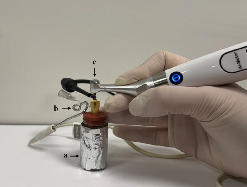
Sixty single-rooted teeth were prepared and divided into two main groups and three subgroups based on the file system (OneShape [Micro-Mega SA] and WaveOne [Dentsply Maillefer]) and apical function of the endodontic motor used (auto apical stop [AAS], auto apical reverse [AAR], and auto apical slowdown [ASD]). The teeth were mounted in pre-weighed glass tubes filled with 0.9% sodium chloride to complete the circuit with the apex locator. Files were advanced until the respective apical function (stop, reverse, or slowdown) was activated. The extruded debris was collected, dried, and weighed by subtracting pre-weighed values from post-weighed values. Preparation time was also recorded. Statistical analyses were performed to compare the groups.
Results
OneShape was associated with significantly less debris extrusion compared to WaveOne, regardless of the apical function (p < 0.05). The ASD function resulted in the least debris extrusion compared to AAS and AAR (p < 0.05). Preparation time was significantly longer in the ASD function (p < 0.05), while no differences were observed between the file systems (p > 0.05).
Conclusions
The OneShape file system and the ASD function produced the least amount of apical debris. While the ASD function requires more preparation time, its potential to minimize debris extrusion suggests it may reduce postoperative symptoms. -
Citations
Citations to this article as recorded by- Inflammatory Mediator Levels and Postoperative Pain Following Root Canal Shaping with Different Apical Actions: A Randomized Controlled Trial
Mustafa Mert Tulgar, Yağmur Kılıç, Oğuz Karalar, Huriye Erbak Yılmaz, Emrah Karataşlıoğlu
Journal of Endodontics.2025;[Epub] CrossRef
- Inflammatory Mediator Levels and Postoperative Pain Following Root Canal Shaping with Different Apical Actions: A Randomized Controlled Trial
- 4,065 View
- 214 Download
- 1 Crossref

- Effects of calcium silicate cements on neuronal conductivity
- Derya Deniz-Sungur, Mehmet Ali Onur, Esin Akbay, Gamze Tan, Fügen Daglı-Comert, Taner Cem Sayın
- Restor Dent Endod 2022;47(2):e18. Published online March 7, 2022
- DOI: https://doi.org/10.5395/rde.2022.47.e18
-
 Abstract
 PDF
PDF PubReader
 ePub
ePub Objectives This study evaluated alterations in neuronal conductivity related to calcium silicate cements (CSCs) by investigating compound action potentials (cAPs) in rat sciatic nerves.
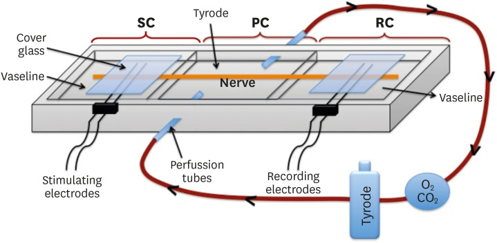
Materials and Methods Sciatic nerves were placed in a Tyrode bath and cAPs were recorded before, during, and after the application of test materials for 60-minute control, application, and recovery measurements, respectively. Freshly prepared ProRoot MTA, MTA Angelus, Biodentine, Endosequence RRM-Putty, BioAggregate, and RetroMTA were directly applied onto the nerves. Biopac LabPro version 3.7 was used to record and analyze cAPs. The data were statistically analyzed.
Results None of the CSCs totally blocked cAPs. RetroMTA, Biodentine, and MTA Angelus caused no significant alteration in cAPs (
p > 0.05). Significantly lower cAPs were observed in recovery measurements for BioAggregate than in the control condition (p < 0.05). ProRoot MTA significantly but transiently reduced cAPs in the application period compared to the control period (p < 0.05). Endosequence RRM-Putty significantly reduced cAPs.Conclusions Various CSCs may alter cAPs to some extent, but none of the CSCs irreversibly blocked them. The usage of fast-setting CSCs during apexification or regeneration of immature teeth seems safer than slow-setting CSCs due to their more favorable neuronal effects.
-
Citations
Citations to this article as recorded by- Endodontic Sealers and Innovations to Enhance Their Properties: A Current Review
Anna Błaszczyk-Pośpiech, Natalia Struzik, Maria Szymonowicz, Przemysław Sareło, Maria Wiśniewska-Wrona, Kamila Wiśniewska, Maciej Dobrzyński, Magdalena Wawrzyńska
Materials.2025; 18(18): 4259. CrossRef
- Endodontic Sealers and Innovations to Enhance Their Properties: A Current Review
- 1,377 View
- 19 Download
- 1 Web of Science
- 1 Crossref

- Shaping ability and apical debris extrusion after root canal preparation with rotary or reciprocating instruments: a micro-CT study
- Emmanuel João Nogueira Leal da Silva, Sara Gomes de Moura, Carolina Oliveira de Lima, Ana Flávia Almeida Barbosa, Waleska Florentino Misael, Mariane Floriano Lopes Santos Lacerda, Luciana Moura Sassone
- Restor Dent Endod 2021;46(2):e16. Published online February 25, 2021
- DOI: https://doi.org/10.5395/rde.2021.46.e16
-
 Abstract
 PDF
PDF PubReader
 ePub
ePub Objectives The aim of this study was to evaluate the shaping ability of the TruShape and Reciproc Blue systems and the apical extrusion of debris after root canal instrumentation. The ProTaper Universal system was used as a reference for comparison.
Materials and Methods Thirty-three mandibular premolars with a single canal were scanned using micro-computed tomography and were matched into 3 groups (
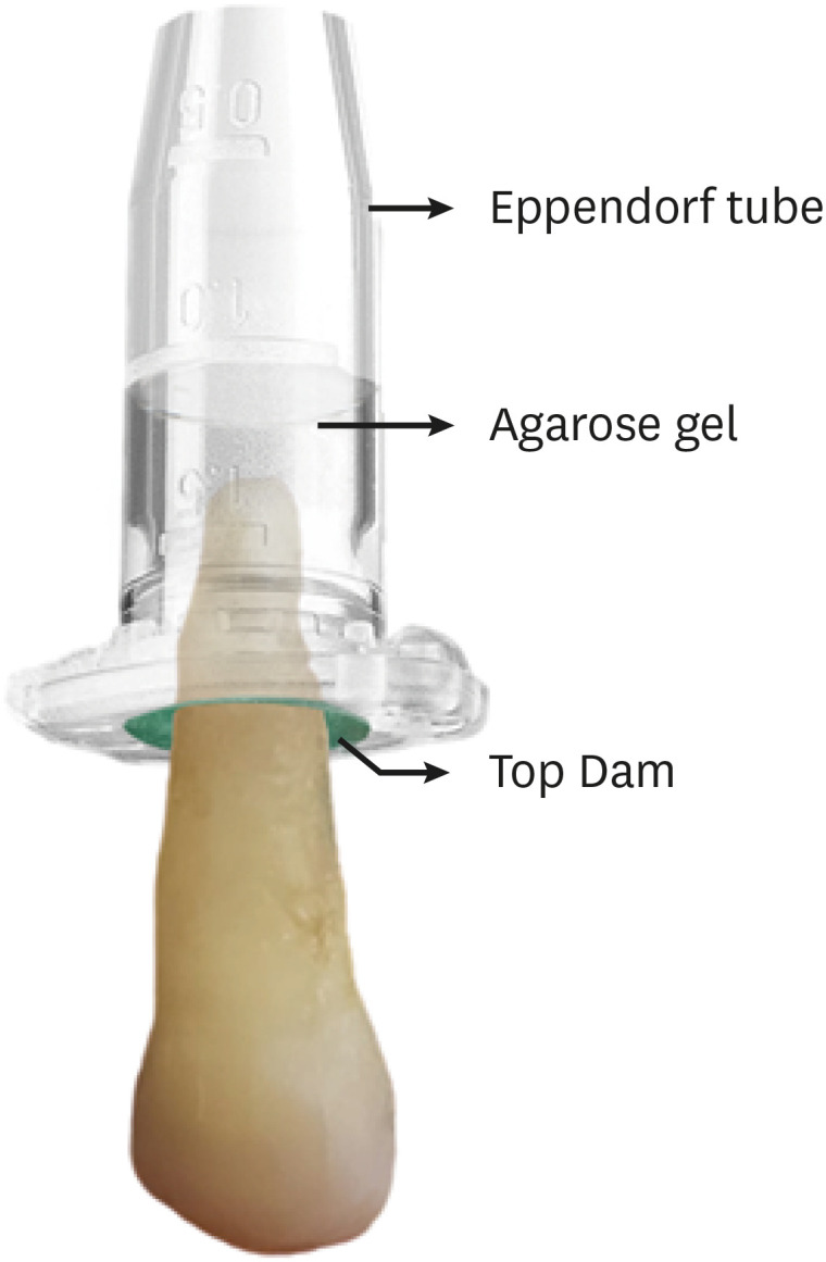
n = 11) according to the instrumentation system: TruShape, Reciproc Blue and ProTaper Universal. The teeth were accessed and mounted in an apparatus with agarose gel, which simulated apical resistance provided by the periapical tissue and enabled the collection of apically extruded debris. During root canal preparation, 2.5% sodium hypochlorite was used as an irrigant. The samples were scanned again after instrumentation. The percentage of unprepared area, removed dentin, and volume of apically extruded debris were analyzed. The data were analyzed using 1-way analysis of variance and the Tukey test for multiple comparisons at a 5% significance level.Results No significant differences in the percentage of unprepared area were observed among the systems (
p > 0.05). ProTaper Universal presented a higher percentage of dentin removal than the TruShape and Reciproc Blue systems (p < 0.05). The systems produced similar volumes of apically extruded debris (p > 0.05).Conclusions All systems caused apically extruded debris, without any significant differences among them. TruShape, Reciproc Blue, and ProTaper Universal presented similar percentages of unprepared area after root canal instrumentation; however, ProTaper Universal was associated with higher dentin removal than the other systems.
-
Citations
Citations to this article as recorded by- Evaluation of Silver-Ion-Coated Rotary Nickel Titanium Files - An In Vitro Study
Jhanvi H. Sadaria, Kondas V. Venkatesh, Dhanasekaran Sihivahanan
Indian Journal of Dental Research.2026;[Epub] CrossRef - Comparison of post-operative pain prevalence after single visit endodontic treatment with two NiTi rotary files - a randomized clinical trial
M. E. Khallaf, Yousra Aly, Amira Ibrahim Mohamed
Scientific Reports.2025;[Epub] CrossRef - A quantitative comparison of apically extruded debris during root canal preparation using NiTi full-sequence rotary and single-file rotary systems: An in vitro study
Pallavi Goel, R. Vikram, R. Anithakumari, M. S. Adarsha, M. E. Sudhanva
Endodontology.2024; 36(3): 235. CrossRef - Extrusion of Sodium Hypochlorite in Oval-Shaped Canals: A Comparative Study of the Potential of Four Final Agitation Approaches Employing Agarose-Embedded Mandibular First Premolars
Aalisha Parkar, Kulvinder Singh Banga, Ajinkya M. Pawar, Alexander Maniangat Luke
Journal of Clinical Medicine.2024; 13(10): 2748. CrossRef - Shaping Efficiency of Rotary and Reciprocating Kinematics of Engine-driven Nickel-Titanium Instruments in Moderate and Severely curved Root Canals Using Microcomputed Tomography: A Systematic Review of Ex Vivo Studies
Claudiu Călin, Ana-Maria Focșăneanu, Friedrich Paulsen, Andreea C. Didilescu, Tiberiu Niță
Journal of Endodontics.2024; 50(7): 907. CrossRef - Intracanal removal and apical extrusion of filling material after retreatment using rotary or reciprocating instruments: A new approach using human cadavers
Thamyres M. Monteiro, Victor O. Cortes‐Cid, Marilia F. V. Marceliano‐Alves, Andrea F. Campello, Luan F. Bastos, Ricardo T. Lopes, José F. Siqueira, Flávio R. F. Alves
International Endodontic Journal.2024; 57(1): 100. CrossRef - Assessment of debris extrusion on using automated irrigation device with conventional needle irrigation – An ex vivo study
Sahil Choudhari, Kavalipurapu Venkata Teja, Raja Kumar, Sindhu Ramesh
Saudi Endodontic Journal.2023; 13(3): 263. CrossRef - Postoperative pain perception and associated risk factors in children after continuous rotation versus reciprocating kinematics: A randomised prospective clinical trial
Ahmad Abdel Hamid Elheeny, Dania Ibrahem Sermani, Mahmoud Ahmed Abdelmotelb
Australian Endodontic Journal.2023; 49(S1): 345. CrossRef - A critical analysis of research methods and experimental models to study apical extrusion of debris and irrigants
Jale Tanalp
International Endodontic Journal.2022; 55(S1): 153. CrossRef - Quantitative evaluation of apically extruded debris using TRUShape, TruNatomy, and WaveOne Gold in curved canals
Nehal Nabil Roshdy, Reham Hassan
BDJ Open.2022;[Epub] CrossRef - Shaping ability of new reciprocating or rotary instruments with two cross‐sectional designs: An ex vivo study
Isabela G. Guedes, Renata C. V. Rodrigues, Marília F. Marceliano‐Alves, Flávio R. F. Alves, Isabela N. Rôças, José F. Siqueira
International Endodontic Journal.2022; 55(12): 1385. CrossRef
- Evaluation of Silver-Ion-Coated Rotary Nickel Titanium Files - An In Vitro Study
- 2,523 View
- 49 Download
- 8 Web of Science
- 11 Crossref

- Efficacy of reciprocating and rotary retreatment nickel-titanium file systems for removing filling materials with a complementary cleaning method in oval canals
- Said Dhaimy, Hyeon-Cheol Kim, Lamyae Bedida, Imane Benkiran
- Restor Dent Endod 2021;46(1):e13. Published online February 3, 2021
- DOI: https://doi.org/10.5395/rde.2021.46.e13
-
 Abstract
 PDF
PDF PubReader
 ePub
ePub Objectives This study aimed to evaluate and compare the efficacy of the S1 reciprocating system and the D-Race retreatment rotary system for filling material removal and the apical extrusion of debris.
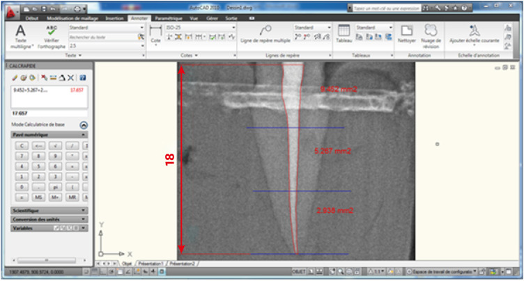
Materials and Methods Sixty-four freshly extracted maxillary canines were shaped with size 10 and size 15 K-files, instrumented using ProTaper Gold under irrigation with 2.5% sodium hypochlorite (NaOCl), obturated according to the principle of thermo-mechanical condensation with gutta-percha and zinc oxide eugenol sealer, and allowed to set for 3 weeks at 37°C. Subsequently, the teeth were divided into a control group (
n = 4), the D-Race rotary instrument group (n = 30), and the S1 reciprocating instrument group (n = 30). After classical retreatment, the canals were subjected to a complementary approach with the XP-Endo Shaper. Desocclusol was used as a solvent, and irrigation with 2.5% NaOCl was performed. Each group was divided into subgroups according to the timing of radiographic readings. The images were imported into a software program to measure the remaining filling material, the apical extrusion, and the root canal space. The data were statistically analyzed using the Z-test and JASP graphics software.Results No significant differences were found between the D-Race and S1 groups for primary retreatment; however, using a complementary cleaning method increased the removal of remnant filling (
p < 0.05).Conclusions Classical removal of canal filling material may not be sufficient for root canal disinfection, although a complementary finishing approach improved the results. Nevertheless, all systems left some debris and caused apical extrusion.
-
Citations
Citations to this article as recorded by- Effectiveness of different supplementary protocols for remaining filling material removal in endodontic reintervention: an integrative review
Amanda Freitas da Rosa, Bruna Venzke Fischer, Luiz Carlos de Lima Dias-Junior, Anna Victoria Costa Serique, Eduardo Antunes Bortoluzzi, Cleonice da Silveira Teixeira, Lucas da Fonseca Roberti Garcia
Odontology.2024; 112(1): 51. CrossRef - Critical analysis of research methods and experimental models to study removal of root filling materials
Mahdi A. Ajina, Pratik K. Shah, Bun San Chong
International Endodontic Journal.2022; 55(S1): 119. CrossRef - Economic analysis of the different endodontic instrumentation techniques used in the Unified Health System
Laura Paredes Merchan, Livia Fernandes Probst, Ana Clara Correa Duarte Simões, Augusto Cesar Santos Raimundo, Yuri Wanderley Cavalcanti, Denise de Fátima Barros Cavalcante, João Victor Frazão Câmara, Antonio Carlos Pereira
BMC Oral Health.2022;[Epub] CrossRef - Fabrication of a Potential Electrodeposited Nanocomposite for Dental Applications
Chun-Wei Chang, Chen-Han Tsou, Bai-Hung Huang, Kuo-Sheng Hung, Yung-Chieh Cho, Takashi Saito, Chi-Hsun Tsai, Chia-Chien Hsieh, Chung-Ming Liu, Wen-Chien Lan
Inorganics.2022; 10(10): 165. CrossRef - Influence of Filling Material Remnants on the Diffusion of Hydroxyl Ions in Endodontically Retreated Teeth: An Ex Vivo Study
Vania Portela Ditzel Westphalen, Marilisa Carneiro Leao Gabardo, Natanael Henrique Ribeiro Mattos, Camila Paiva Perin, Liliane Roskamp, Cristiano Miranda de Araújo, Luiz Fernando Fariniuk, Flares Baratto–Filho
The Journal of Contemporary Dental Practice.2022; 23(8): 768. CrossRef - Efficacy of Removing Thermafil and GuttaCore from Straight Root Canal Systems Using a Novel Non-Surgical Root Canal Re-Treatment System: A Micro-Computed Tomography Analysis
Vicente Faus-Llácer, Rubén Linero Pérez, Ignacio Faus-Matoses, Celia Ruiz-Sánchez, Álvaro Zubizarreta-Macho, Salvatore Sauro, Vicente Faus-Matoses
Journal of Clinical Medicine.2021; 10(6): 1266. CrossRef
- Effectiveness of different supplementary protocols for remaining filling material removal in endodontic reintervention: an integrative review
- 1,853 View
- 37 Download
- 7 Web of Science
- 6 Crossref

- Impact of root canal curvature and instrument type on the amount of extruded debris during retreatment
- Burcu Serefoglu, Gözde Kandemir Demirci, Seniha Miçooğulları Kurt, İlknur Kaşıkçı Bilgi, Mehmet Kemal Çalışkan
- Restor Dent Endod 2021;46(1):e5. Published online December 17, 2020
- DOI: https://doi.org/10.5395/rde.2021.46.e5
-
 Abstract
 PDF
PDF PubReader
 ePub
ePub Objectives The aim of the current study was to assess whether the amount of extruded debris differs for straight and severely curved root canals during retreatment using H-files, R-Endo, Reciproc and ProTaper Universal Retreatment (PTU-R) files. Additionally, the area of residual filling material was evaluated.
Materials and Methods Severely curved (
n = 104) and straight (n = 104) root canals of maxillary molar teeth were prepared with WaveOne Primary file and obturated with gutta-percha and AH Plus sealer. Root canal filling materials were removed with one of the preparation techniques: group 1: H-file; group 2: R-Endo; group 3: Reciproc; group 4: PTU-R (n = 26). The amount of extruded material and the area of the residual filling material was measured. The data were analyzed with 2-way analysis of variance (ANOVA) and 1-way ANOVA at the 0.05 significance level.Results Except for Reciproc group (
p > 0.05), PTU-R, R-Endo, and H-file systems extruded significantly more debris in severely curved canals (p < 0.05). Each file system caused more residual filling material in severely curved canals than in straight ones (p < 0.05).Conclusions All instruments used in this study caused apical debris extrusion. Root canal curvature had an effect on extruded debris, except for Reciproc system. Clinicians should be aware that the difficult morphology of the severely curved root canals is a factor increasing the amount of extruded debris during the retreatment procedure.
-
Citations
Citations to this article as recorded by- Comparative Analysis of Root Canal Curvature Measurement Methods for Permanent Mandibular Molars Distal Root: An Observational Study
Tanu Singh, Saurav Bathla, Anuraag Gurtu, Shubhi Gupta, Sana Saifi, Madhusudan Astekar
The Journal of Contemporary Dental Practice.2025; 26(10): 945. CrossRef - Do Continuous Rotating Endodontic Instruments Extrude Fewer Apical Debris Than Reciprocating Instruments in Non-Surgical Endodontic Retreatments? A Systematic Review
Francesco Puleio, Francesco Giordano, Ugo Bellezza, David Rizzo, Valentina Coppini, Roberto Lo Giudice
Applied Sciences.2024; 14(4): 1621. CrossRef - Intracanal removal and apical extrusion of filling material after retreatment using rotary or reciprocating instruments: A new approach using human cadavers
Thamyres M. Monteiro, Victor O. Cortes‐Cid, Marilia F. V. Marceliano‐Alves, Andrea F. Campello, Luan F. Bastos, Ricardo T. Lopes, José F. Siqueira, Flávio R. F. Alves
International Endodontic Journal.2024; 57(1): 100. CrossRef - Comparative analysis of methods for measuring root canal curvature based on periapical radiography: A laboratory study
Rafael Chies Hartmann, Eduardo Silva Ferraz, Theodoro Weissheimer, Jose Antônio Poli de Figueiredo, Giampiero Rossi‐Fedele, Maximiliano Schünke Gomes
International Endodontic Journal.2024; 57(12): 1848. CrossRef - Evaluation of apically extruded debris during root canal filling material removal in teeth with external apical root resorption: a comparison of different obturation techniques
Büşra Melike Çağlar, İsmail Uzun
BMC Oral Health.2024;[Epub] CrossRef - Evaluation of apically extruded debris using protaper universal, protaper next, one curve, Xp shaper, and edge file: An in vitro study
Murtada Qadir Muhaibes, Shatha Abdulkareem Alwakeel
Saudi Endodontic Journal.2024; 14(1): 31. CrossRef - A quantitative comparison of apically extruded debris during root canal preparation using NiTi full-sequence rotary and single-file rotary systems: An in vitro study
Pallavi Goel, R. Vikram, R. Anithakumari, M. S. Adarsha, M. E. Sudhanva
Endodontology.2024; 36(3): 235. CrossRef - In vitro evaluation of filling material removal and apical debris extrusion after retreatment using Reciproc blue, Hyflex EDM and ProTaper retreatment files
Passent Abdelnaby, Mohamed Ibrahim, Rania ElBackly
BMC Oral Health.2023;[Epub] CrossRef - A Comparative Study on the Shaping Ability and Cleaning Efficiency of Two Different Single-File Systems, Reciprocating Wave One Versus Continuous Rotation F360, Evaluated by Scanning Electron Microscope: An In Vitro Study
Arunkumar Samudrala, Chandrakanth Majeti, Kommineni Harika Chowdary, Lakshmi Bhavani Potru, Anusha Yaragani, Yata Prashanth Kumar, Gagandeep K Sidhu, Navneet S Kathuria
Cureus.2023;[Epub] CrossRef - COMPARATIVE EVALUATION OF THE EFFECT OF DIFFERENT ROTARY INSTRUMENT SYSTEMS ON THE AMOUNT OF APICALLY EXTRUDED DEBRIS
Recai ZAN, Bilge LENGER
Cumhuriyet Dental Journal.2022; 25(2): 172. CrossRef - A critical analysis of research methods and experimental models to study apical extrusion of debris and irrigants
Jale Tanalp
International Endodontic Journal.2022; 55(S1): 153. CrossRef - Critical analysis of research methods and experimental models to study removal of root filling materials
Mahdi A. Ajina, Pratik K. Shah, Bun San Chong
International Endodontic Journal.2022; 55(S1): 119. CrossRef
- Comparative Analysis of Root Canal Curvature Measurement Methods for Permanent Mandibular Molars Distal Root: An Observational Study
- 2,238 View
- 31 Download
- 8 Web of Science
- 12 Crossref

- Effects of the endodontic access cavity on apical debris extrusion during root canal preparation using different single-file systems
- Pelin Tüfenkçi, Koray Yılmaz, Mehmet Adigüzel
- Restor Dent Endod 2020;45(3):e33. Published online June 4, 2020
- DOI: https://doi.org/10.5395/rde.2020.45.e33
-
 Abstract
 PDF
PDF PubReader
 ePub
ePub Objectives This study was conducted to evaluate the effects of traditional and contracted endodontic cavity (TEC and CEC) preparation with the use of Reciproc Blue (RPC B) and One Curve (OC) single-file systems on the amount of apical debris extrusion in mandibular first molar root canals.
Materials and Methods Eighty extracted mandibular first molar teeth were randomly assigned to 4 groups (
n = 20) according to the endodontic access cavity shape and the single file system used for root canal preparation (reciprocating motion with the RCP B and rotary motion with the OC): TEC-RPC B, TEC-OC, CEC-RPC B, and CEC-OC. The apically extruded debris during preparation was collected in Eppendorf tubes. The amount of extruded debris was quantified by subtracting the weight of the empty tubes from the weight of the Eppendorf tubes containing the debris. Data were analyzed using 1-way analysis of variance with the Tukeypost hoc test. The level of significance was set atp < 0.05.Results The CEC-RPC B group showed more apical debris extrusion than the TEC-OC and CEC-OC groups (
p < 0.05). There were no statistically significant differences in the amount of apical debris extrusion among the TEC-OC, CEC-OC, and TEC-RPC B groups.Conclusions RPC B caused more apical debris extrusion in the CEC groups than did the OC single-file system. Therefore, it is suggested that the RPC B file should be used carefully in teeth with a CEC.
-
Citations
Citations to this article as recorded by- Comparative Evaluation of Periapical Expulsion Using Manual, Rotary, and Reciprocating Instrumentation With EndoVac Irrigation: An In Vitro Study
Sachin Metkari, Sanpreet S Sachdev, Pravin Patil, Manoj Ramugade, Kishor D Sapkale, Kulvinder S Banga, Dinesh Rao
Cureus.2025;[Epub] CrossRef - Comparison of Debris Extrusion and Preparation Time by Traverse, R‐Motion Glider C, and Other Glide Path Systems in Severely Curved Canals
Taher Al Omari, Layla Hassouneh, Khawlah Albashaireh, Alaa Dkmak, Rami Albanna, Ali Al-Mohammed, Ahmed Jamleh, Lucas da Fonseca Roberti Garcia
International Journal of Dentistry.2025;[Epub] CrossRef - Minimal İnvaziv Giriş Kavitelerinin Alt Kesici Dişlerdeki Apikal Ekstrüzyona Etkisi
İrem Haskarabağ, Cangül Keskin
Türk Diş Hekimliği Araştırma Dergisi.2025; 4(2): 75. CrossRef - Evaluation of apically extruded debris from root canal filling removal of the mesiobuccal canal of maxillary molars using XP shaper and protaper with two different irrigation
Sanaz Mirsattari, Maryam Zare Jahromi, Masoud Khabiri
Dental Research Journal.2024;[Epub] CrossRef - The Impact of Minimum Invasive Access Cavity Design on the Quality of Instrumentation of Root Canals of Maxillary Molars Using Cone-Beam Computed Tomography: An in Vitro Study
Fahad H Baabdullah, Samia M Elsherief , Rayan A Hawsawi, Hetaf S Redwan
Cureus.2024;[Epub] CrossRef - Assessment of Bacterial Load and Post-Endodontic Pain after One-Visit Root Canal Treatment Using Two Types of Endodontic Access Openings: A Randomized Controlled Clinical Trial
Ahmed M. Al-Ani, Ahmed H. Ali, Garrit Koller
Dentistry Journal.2024; 12(4): 88. CrossRef - The effect of different kinematics on apical debris extrusion with a single-file system
Taher M. N. Al Omari, Giusy Rita Maria La Rosa, Rami Haitham Issa Albanna, Abedelmalek Tabnjh, Flavia Papale, Eugenio Pedullà
Odontology.2023; 111(4): 910. CrossRef - The effects of laser and ultrasonic irrigation activation methods on smear and debris removal in traditional and conservative endodontic access cavities
Hüseyin Gündüz, Esin Özlek
Lasers in Medical Science.2023;[Epub] CrossRef - Influence of access cavity design, sodium hypochlorite formulation and XP‐endo Shaper usage on apical debris extrusion – A laboratory investigation
Jerry Jose, Aishuwariya Thamilselvan, Kavalipurapu Venkata Teja, Giampiero Rossi–Fedele
Australian Endodontic Journal.2023; 49(1): 6. CrossRef - Apically extruded debris, canal transportation, and shaping ability of nickel-titanium instruments on contracted endodontic cavities in molar teeth
Qinqin Zhang, Jingyi Gu, Jiadi Shen, Ming Ma, Ying Lv, Xin Wei
Journal of Oral Science.2023; 65(4): 203. CrossRef - Impact of contracted endodontic cavities on instrumentation efficacy—A systematic review
Manan Shroff, Karkala Venkappa Kishan, Nimisha Shah, Purnima Saklecha
Australian Endodontic Journal.2023; 49(1): 202. CrossRef - Present status and future directions – Minimal endodontic access cavities
Emmanuel João Nogueira Leal Silva, Gustavo De‐Deus, Erick Miranda Souza, Felipe Gonçalves Belladonna, Daniele Moreira Cavalcante, Marco Simões‐Carvalho, Marco Aurélio Versiani
International Endodontic Journal.2022; 55(S3): 531. CrossRef - Effect of guided conservative endodontic access and different file kinematics on debris extrusion in mesial root of the mandibular molars: An in vitro study
Sathish Sundar, Aswathi Varghese, KrithikaJ Datta, Velmurugan Natanasabapathy
Journal of Conservative Dentistry.2022; 25(5): 547. CrossRef - A critical analysis of research methods and experimental models to study apical extrusion of debris and irrigants
Jale Tanalp
International Endodontic Journal.2022; 55(S1): 153. CrossRef - Current strategies for conservative endodontic access cavity preparation techniques—systematic review, meta-analysis, and decision-making protocol
Benoit Ballester, Thomas Giraud, Hany Mohamed Aly Ahmed, Mohamed Shady Nabhan, Frédéric Bukiet, Maud Guivarc’h
Clinical Oral Investigations.2021; 25(11): 6027. CrossRef - Extrusion of debris with and without intentional foraminal enlargement – A systematic review and meta‐analysis
Ricardo Machado, Gislayne Vigarani, Tainara Macoppi, Ajinkya Pawar, Stella Maria Glaci Reinke, Ana Cristina Kovalik Gonçalves
Australian Endodontic Journal.2021; 47(3): 741. CrossRef - Apical debris extrusion of single-file systems in curved canals
Ecehan Hazar, Olcay Özdemir, Mustafa Murat Koçak, Baran Can Sağlam, Sibel Koçak
Endodontology.2021; 33(3): 128. CrossRef - Quantitative Evaluation of Apically Extruded Debris in Root Canals prepared by Single-file Reciprocating and Single File Rotary Instrumentation Systems: A Comparative In vitro Study
Sonal Sinha, Konark Singh, Anju Singh, Swati Priya, Avanindra Kumar, Sahil Kawle
Journal of Pharmacy and Bioallied Sciences.2021; 13(Suppl 2): S1398. CrossRef - THE INFLUENCE OF DIFFERENT PECKING DEPTH ON AMOUNT OF APICALLY EXTRUDED DEBRIS DURING ROOT CANAL PREPARATION
Fatih ÇAKICI, Busra UYSAL, Elif Bahar CAKİCİ, Adem GUNAYDIN
Atatürk Üniversitesi Diş Hekimliği Fakültesi Dergisi.2021; : 1. CrossRef
- Comparative Evaluation of Periapical Expulsion Using Manual, Rotary, and Reciprocating Instrumentation With EndoVac Irrigation: An In Vitro Study
- 2,409 View
- 23 Download
- 19 Crossref

-
Comparison of apical extrusion of intracanal bacteria by various glide-path establishing systems: an
in vitro study - Alberto Dagna, Rashid El Abed, Sameeha Hussain, Ibrahim H Abu-Tahun, Livia Visai, Federico Bertoglio, Floriana Bosco, Riccardo Beltrami, Claudio Poggio, Hyeon-Cheol Kim
- Restor Dent Endod 2017;42(4):316-323. Published online October 31, 2017
- DOI: https://doi.org/10.5395/rde.2017.42.4.316
-
 Abstract
 PDF
PDF PubReader
 ePub
ePub Objectives This study compared the amount of apically extruded bacteria during the glide-path preparation by using multi-file and single-file glide-path establishing nickel-titanium (NiTi) rotary systems.
Materials and Methods Sixty mandibular first molar teeth were used to prepare the test apparatus. They were decoronated, blocked into glass vials, sterilized in ethylene oxide gas, infected with a pure culture of
Enterococcus faecalis, randomly assigned to 5 experimental groups, and then prepared using manual stainless-steel files (group KF) and glide-path establishing NiTi rotary files (group PF with PathFiles, group GF with G-Files, group PG with ProGlider, and group OG with One G). At the end of canal preparation, 0.01 mL NaCl solution was taken from the experimental vials. The suspension was plated on brain heart infusion agar and colonies of bacteria were counted, and the results were given as number of colony-forming units (CFU).Results The manual instrumentation technique tested in group KF extruded the highest number of bacteria compared to the other 4 groups (
p < 0.05). The 4 groups using rotary glide-path establishing instruments extruded similar amounts of bacteria.Conclusions All glide-path establishment instrument systems tested caused a measurable apical extrusion of bacteria. The manual glide-path preparation showed the highest number of bacteria extruded compared to the other NiTi glide-path establishing instruments.
-
Citations
Citations to this article as recorded by- Apical Extrusion of Bacteria during Canal Preparation: A Systematic Review of Laboratory Studies
Thamyres M. Monteiro, Warley O. Silva, Jeferson O. Marques, Ana Carolina S.P. Guerra, Flávio R.F. Alves
Journal of Endodontics.2025; 51(7): 866. CrossRef - Glide Path in Endodontics: A Literature Review of Current Knowledge
Vlad Mircea Lup, Giulia Malvicini, Carlo Gaeta, Simone Grandini, Gabriela Ciavoi
Dentistry Journal.2024; 12(8): 257. CrossRef - Evaluation of apically extruded debris using protaper universal, protaper next, one curve, Xp shaper, and edge file: An in vitro study
Murtada Qadir Muhaibes, Shatha Abdulkareem Alwakeel
Saudi Endodontic Journal.2024; 14(1): 31. CrossRef - Effect of Multiple Glide Path Files on Apical Debris Extrusion in Severely Curved Mesial Roots of Mandibular Molars: An In Vitro Study
Niranjan Desai, Ashish S Bhadane, Nishant K Vyavahare, Dipali Y Shah, Akash S Kale, Simran K Chaudhari
Journal of Operative Dentistry & Endodontics.2024; 8(1): 1. CrossRef - Evaluation of Pain Following the Use of Different Single-file Glide Path Systems: A Randomized Clinical Trial
Zeliha Danaci, Kübra Yeşildal Yeter
Journal of Endodontics.2024; 50(2): 120. CrossRef - Impact of Different Glidepath Techniques on the Overall Performance of WaveOne Gold in an Artificial S-Shape Canal
Vlad Mircea Lup, Olivia Andreea Marcu, Carlo Gaeta, Gabriela Ciavoi
Dentistry Journal.2024; 12(6): 182. CrossRef - Influence of different irrigant activation methods on apical debris extrusion and bacterial elimination from infected root canals
KSadia Ada, Shibani Shetty, KB Jayalakshmi, PrasannaLatha Nadig, PG Manje Gowda, ArulK Selvan
Journal of Conservative Dentistry.2023; 26(1): 31. CrossRef - A Comparative Evaluation of the Apically Extruded Debris from Root Canals Prepared by R-Motion NiTi File System
Farah B. Al-Saffar, Hikmet A. Al-Gharrawi, Luca Testarelli
International Journal of Dentistry.2023; 2023: 1. CrossRef - Effect of glide path files with different metallurgy on intracanal bacterial extrusion by HyFlex electrical discharge machining file
Priyanka Soni, Pragya Kumar, Sonali Taneja, Anshi Jain
Endodontology.2022; 34(3): 168. CrossRef - Impact of kinematics on the efficiency and safety of an engine-driven file for glide path preparation in MB2 canals of maxillary molars
Larissa B. B. Araújo, Pedro H. S. Calefi, Murilo P. Alcalde, Giulio Gavini, Rodrigo R. Vivan, Marco Antonio Hungaro Duarte
Clinical Oral Investigations.2022; 27(3): 1153. CrossRef - Critical analysis of research methods and experimental models to study removal of root filling materials
Mahdi A. Ajina, Pratik K. Shah, Bun San Chong
International Endodontic Journal.2022; 55(S1): 119. CrossRef - A Comparative Evaluation of Apically Extruded Debris using Three Rotary and One Reciprocating Instrumentation Ni-Ti Systems
Maha Adnan Habeeb
Journal of Orofacial Sciences.2022; 14(2): 93. CrossRef - A critical analysis of research methods and experimental models to study apical extrusion of debris and irrigants
Jale Tanalp
International Endodontic Journal.2022; 55(S1): 153. CrossRef - Evaluation of type of kinematics on glide path procedures and torsional fatigue resistance after preparation of moderately curved canals
Murilo Priori Alcalde, Marco Antonio Hungaro Duarte, Pedro Henrique Souza Calefi, Victor de Moraes Cruz, Bruno Carvalho de Vasconcelos, Marcus Vinícius Reis Só, Rodrigo Ricci Vivan
Brazilian Oral Research.2021;[Epub] CrossRef - Analysis of a glide path creation necessity at the initial stages of endodontic treatment
Z. S. Khabadze, Yu. A. Generalova
Endodontics Today.2021; 19(1): 39. CrossRef - Influence of glide path kinematics during endodontic treatment on the occurrence and intensity of intraoperative and postoperative pain: a systematic review of randomized clinical trials
Thaís Christina Cunha, Felipe de Souza Matos, Luiz Renato Paranhos, Ítalo de Macedo Bernardino, Camilla Christian Gomes Moura
BMC Oral Health.2020;[Epub] CrossRef - Apically extruded debris produced during glide path preparation using R‐Pilot, WaveOne Gold Glider and ProGlider in curved root canals
Cangül Keskin, Özlem Sivas Yilmaz, Uğur Inan
Australian Endodontic Journal.2020; 46(3): 439. CrossRef - Influence of Negotiation, Glide Path, and Preflaring Procedures on Root Canal Shaping—Terminology, Basic Concepts, and a Systematic Review
Gianluca Plotino, Venkateshbabu Nagendrababu, Frederic Bukiet, Nicola M. Grande, Sajesh K. Veettil, Gustavo De-Deus, Hany Mohamed Aly Ahmed
Journal of Endodontics.2020; 46(6): 707. CrossRef - Mechanical Properties of Various Glide Path Preparation Nickel-titanium Rotary Instruments
Joo-Yeong Lee, Sang Won Kwak, Jung-Hong Ha, Ibrahim H. Abu-Tahun, Hyeon-Cheol Kim
Journal of Endodontics.2019; 45(2): 199. CrossRef - Effective Establishment of Glide-Path to Reduce Torsional Stress during Nickel-Titanium Rotary Instrumentation
Ibrahim H. Abu-Tahun, Sang Won Kwak, Jung-Hong Ha, Asgeir Sigurdsson, Mehmet Baybora Kayahan, Hyeon-Cheol Kim
Materials.2019; 12(3): 493. CrossRef - Postoperative pain after glide path preparation using manual, reciprocating and continuous rotary instruments: a randomized clinical trial
C. Keskin, Ö. Sivas Yilmaz, U. Inan, Ö. Özdemir
International Endodontic Journal.2019; 52(5): 579. CrossRef - Intraoperative Pain During Glide Path Creation with the Use of a Rotary or Reciprocating System
Pelin TUFENKCİ, Mehmet ADIGUZEL, Koray YILMAZ
Cumhuriyet Dental Journal.2019; 22(1): 66. CrossRef - Effects of Different Glide Path Files on Apical Debris Extrusion in Curved Root Canals
Betul Gunes, Kubra Yesildal Yeter
Journal of Endodontics.2018; 44(7): 1191. CrossRef
- Apical Extrusion of Bacteria during Canal Preparation: A Systematic Review of Laboratory Studies
- 1,459 View
- 11 Download
- 23 Crossref

- Accidental injury of the inferior alveolar nerve due to the extrusion of calcium hydroxide in endodontic treatment: a case report
- Yooseok Shin, Byoung-Duck Roh, Yemi Kim, Taehyeon Kim, Hyungjun Kim
- Restor Dent Endod 2016;41(1):63-67. Published online January 6, 2016
- DOI: https://doi.org/10.5395/rde.2016.41.1.63

-
 Abstract
 PDF
PDF PubReader
 ePub
ePub During clinical endodontic treatment, we often find radiopaque filling material beyond the root apex. Accidental extrusion of calcium hydroxide could cause the injury of inferior alveolar nerve, such as paresthesia or continuous inflammatory response. This case report presents the extrusion of calcium hydroxide and treatment procedures including surgical intervention. A 48 yr old female patient experienced Calcipex II extrusion in to the inferior alveolar canal on left mandibular area during endodontic treatment. After completion of endodontic treatment on left mandibular first molar, surgical intervention was planned under general anesthesia. After cortical bone osteotomy and debridement, neuroma resection and neurorrhaphy was performed, and prognosis was observed. But no improvement in sensory nerve was seen following surgical intervention after 20 mon. A clinician should be aware of extrusion of intracanal medicaments and the possibility of damage on inferior alveolar canal. Injectable type of calcium hydroxide should be applied with care for preventing nerve injury. The alternative delivery method such as lentulo spiral was suggested on the posterior mandibular molar.
-
Citations
Citations to this article as recorded by- Nicolau syndrome in endodontics: A narrative review on calcium hydroxide extrusion and its therapeutic risks
Manisha Chaudhary, Akash Kumar Giri, Ashok Ayer
Saudi Endodontic Journal.2026; 16(1): 12. CrossRef - Invasion of Calcium Hydroxide Preparations Leading to Severe Chemical Nerve Injury Treated Through Nerve Repair Using Artificial Nerve Conduit: A Case Report
Akihiro Nishiyama, Andreas Neff, Takahiro Nakada, Takaharu Ariizumi, Akira Iwasaki, Keisuke Sugahara, Akira Katakura, Hannah Wesley
Case Reports in Dentistry.2025;[Epub] CrossRef - Automatic localization of inferior alveolar nerve canal in panoramic dental images
Uma Maheswari Pandyan, Banumathi Arumugam, Ulaganathan Gurunathan, Shahul Hameed Kopuli Ashkar Ali
Signal, Image and Video Processing.2022; 16(5): 1389. CrossRef - Inferior alveolar nerve injury due to the extrusion of calcium hydroxide during endodontic treatment: A case report
Metin Berk Kasapoğlu, Gülce Ecem Doğancalı
Australian Endodontic Journal.2022; 48(2): 342. CrossRef - Inferior alveolar nerve canal segmentation by local features based neural network model
P. Uma Maheswari, A. Banumathi, G. Ulaganathan, R. Yoganandha
IET Image Processing.2022; 16(3): 703. CrossRef - Microsurgical Repair of Inferior Alveolar Nerve Injuries Associated With Endodontic Treatment: Results on Sensory Function and Relief of Pain
Keith A. Sonneveld, Kristopher L. Hasstedt, Roger A. Meyer, Shahrokh C. Bagheri
Journal of Oral and Maxillofacial Surgery.2021; 79(7): 1434. CrossRef - The significance of diagnosis and treatment planning in periapical lesion overfilled with calcium hydroxide paste
Kyoung-Hwa Jung, Eun-Young Kwon, Youn-Kyung Choi, So-Yeun Kim, Hye-Mi Jeon, Jeong-Kil Park
Journal of Dental Rehabilitation and Applied Science.2021; 37(2): 95. CrossRef - The anatomical relationship between the roots of erupted permanent teeth and the mandibular canal: a systematic review
Michał Puciło, Mariusz Lipski, Magdalena Sroczyk-Jaszczyńska, Aleksandra Puciło, Alicja Nowicka
Surgical and Radiologic Anatomy.2020; 42(5): 529. CrossRef - Massive extrusion of calcium hydroxide paste containing barium sulphate during endodontic treatment
Jéssica Montenegro Fonsêca, Natália Rangel Palmier, Gleyson Kleber Amaral‐Silva, Lady Paola Aristizabal Arboleda, José Flávio Affonso Almeida, Mario Fernando de Goes, Pablo Agustin Vargas, Marcio Ajudarte Lopes, Alan Roger Santos‐Silva
Australian Endodontic Journal.2020; 46(2): 257. CrossRef - The double-edged sword of calcium hydroxide in endodontics
Alan H. Gluskin, Gordon Lai, Christine I. Peters, Ove A. Peters
The Journal of the American Dental Association.2020; 151(5): 317. CrossRef - Endodontic-related inferior alveolar nerve injuries: A review and a therapeutic flow chart
R. Castro, M. Guivarc'h, J.M. Foletti, J.H. Catherine, C. Chossegros, L. Guyot
Journal of Stomatology, Oral and Maxillofacial Surgery.2018; 119(5): 412. CrossRef - Relationship between Root Apices and the Mandibular Canal: A Cone-beam Computed Tomographic Comparison of 3 Populations
Alex Lvovsky, Shir Bachrach, Hyeon-Cheol Kim, Ajinkya Pawar, Oleg Levinzon, Joe Ben Itzhak, Michael Solomonov
Journal of Endodontics.2018; 44(4): 555. CrossRef - A case of high density abnormality in x-ray findings of mandible caused by leakage of root canal filling paste
Haruko Kashiwamura, Kyoko Oka, Yoko Tuchihashi, Hanako Yoshioka, Mayumi Kato, Atsuko Baba, Toyohiro Kagawa, Kazuhiko Okamura, Masao Ozaki
Pediatric Dental Journal.2017; 27(3): 162. CrossRef - Oral dysesthesia
Christopher J. Spencer, Gary D. Klasser
The Journal of the American Dental Association.2017; 148(12): 941. CrossRef - Microsurgical Decompression of Inferior Alveolar Nerve After Endodontic Treatment Complications
Bernardo Bianchi, Andrea Ferri, Andrea Varazzani, Michela Bergonzani, Enrico Sesenna
Journal of Craniofacial Surgery.2017; 28(5): 1365. CrossRef
- Nicolau syndrome in endodontics: A narrative review on calcium hydroxide extrusion and its therapeutic risks
- 3,211 View
- 36 Download
- 15 Crossref

- An esthetic appliance for the management of crown-root fracture: a case report
- Sang-Min Jeon, Kang-Hee Lee, Bock-Young Jung
- Restor Dent Endod 2014;39(3):226-229. Published online May 22, 2014
- DOI: https://doi.org/10.5395/rde.2014.39.3.226
-
 Abstract
 PDF
PDF PubReader
 ePub
ePub Orthodontic extrusion is usually performed by means of a fixed orthodontic appliance that utilizes arch wire attached to adjacent teeth and transfers the desired force by elastic from the wire to the root. However, clinicians often encounter cases where the bonding required for tooth traction is not possible because the adjacent teeth have been restored with ceramic or veneer. The purpose of this case report is to describe a modified orthodontic extrusion appliance that is useful when conventional orthodontic treatment is not possible. The modified appliance was fabricated using an artificial tooth, clear plastic sheeting, and a braided fiber-reinforced composite strip that covered adjacent teeth without bonding. It satisfied the esthetic and functional needs of the patient and established the optimal biologic width.
-
Citations
Citations to this article as recorded by- Esthetic enhancement of a traumatized anterior tooth with a combination of forced eruption and tooth alignment: a case report
So-Hee Kang, Jung-Hong Ha, Myoung-Uk Jin, Sung-Kyo Kim, Young-Kyung Kim
Restorative Dentistry & Endodontics.2016; 41(3): 210. CrossRef
- Esthetic enhancement of a traumatized anterior tooth with a combination of forced eruption and tooth alignment: a case report
- 1,105 View
- 5 Download
- 1 Crossref

- Surgical management of a failed internal root resorption treatment: a histological and clinical report
- Saeed Asgary, Mohammad Jafar Eghbal, Leili Mehrdad, Sanam Kheirieh, Ali Nosrat
- Restor Dent Endod 2014;39(2):137-142. Published online March 21, 2014
- DOI: https://doi.org/10.5395/rde.2014.39.2.137
-
 Abstract
 PDF
PDF PubReader
 ePub
ePub This article presents the successful surgical management of a failed mineral trioxide aggregate (MTA) orthograde obturation of a tooth with a history of impact trauma and perforated internal root resorption. A symptomatic maxillary lateral incisor with a history of perforation due to internal root resorption and nonsurgical repair using MTA was referred. Unintentional overfill of the defect with MTA had occurred 4 yr before the initial visit. The excess MTA had since disappeared, and a radiolucent lesion adjacent to the perforation site was evident radiographically. Surgical endodontic retreatment was performed using calcium enriched mixture (CEM) cement as a repair material. Histological examination of the lesion revealed granulation tissue with chronic inflammation, and small fragments of MTA encapsulated within fibroconnective tissue. At the one and two year follow up exams, all signs and symptoms of disease had resolved and the tooth was functional. Complete radiographic healing of the lesion was observed two years after the initial visit. This case report illustrates how the selection of an appropriate approach to treatment of a perforation can affect the long term prognosis of a tooth. In addition, extrusion of MTA into a periradicular lesion should be avoided.
-
Citations
Citations to this article as recorded by- Managing Internal Inflammatory Root Resorption and Perforation of a Mandibular Primary Molar: A Case Report With 15 Months Follow‐Up
Mana Mowji, Motahareh Khosrojerdi
Clinical Case Reports.2025;[Epub] CrossRef - Endodontic management of internal replacement resorption of two maxillary central incisors with the aid of cone-beam computed tomography as the diagnostic tool: a case report and review of literature
Fatemeh Eskandari, Safoora Sahebi, Negar Ghorbani Jahandizi, Hossein Mofidi
Journal of Medical Case Reports.2025;[Epub] CrossRef - Removal of AH Plus Bioceramic Sealer from Artificial Internal Resorption Cavities Using Different Irrigation Activation Systems
Mine Büker, Meltem Sümbüllü, Emine Şimşek, Fadime Sena Sezer
Cumhuriyet Dental Journal.2025; 28(3): 383. CrossRef - Evaluation of the effectiveness of different supplemental cleaning techniques in the retreatment of roots with small simulated internal resorption cavities: an in vitro comparative study
Sine Güngör Us, Özgür Uzun, Nazlı Merve Güngör
BMC Oral Health.2025;[Epub] CrossRef - Comprehensive review of composition, properties, clinical applications, and future perspectives of calcium-enriched mixture (CEM) cement: a systematic analysis
Saeed Asgary, Mahtab Aram, Mahta Fazlyab
BioMedical Engineering OnLine.2024;[Epub] CrossRef - The various forms of tooth resorption
Jordan Samuel Blum
Australian Endodontic Journal.2024; 50(2): 191. CrossRef - Bioceramics in Endodontics: Updates and Future Perspectives
Xu Dong, Xin Xu
Bioengineering.2023; 10(3): 354. CrossRef - Imaging techniques and various treatment modalities used in the management of internal root resorption: A systematic review
R. S Digholkar, S D Aggarwal, P S Kurtarkar, P. B Dhatavkar, V L Neil, D N Agarwal
Endodontology.2023; 35(2): 85. CrossRef - Treatment of Teeth with Root Resorptions: A Case Report and Systematic Review
Damla Erkal, Abdullah Başoğlu, Damla Kırıcı, Nezahat Arzu Kayar, Simay Koç, Kürşat Er
Galician Medical Journal.2023;[Epub] CrossRef - Effects of calcium silicate cements on neuronal conductivity
Derya Deniz-Sungur, Mehmet Ali Onur, Esin Akbay, Gamze Tan, Fügen Daglı-Comert, Taner Cem Sayın
Restorative Dentistry & Endodontics.2022;[Epub] CrossRef - Mineral trioxide aggregate and other bioactive endodontic cements: an updated overview – part II: other clinical applications and complications
M. Torabinejad, M. Parirokh, P. M. H. Dummer
International Endodontic Journal.2018; 51(3): 284. CrossRef - Periodontal healing following non-surgical repair of an old perforation with pocket formation and oral communication
Saeed Asgary, Prashant Verma, Ali Nosrat
Restorative Dentistry & Endodontics.2018;[Epub] CrossRef - Conservative Management of Class 4 Invasive Cervical Root Resorption Using Calcium-enriched Mixture Cement
Saeed Asgary, Ali Nosrat
Journal of Endodontics.2016; 42(8): 1291. CrossRef - Importance of CBCT in the management plan of upper canine with internal resorption
Roberto Fornara, Dario Re Cecconi
Giornale Italiano di Endodonzia.2015; 29(2): 70. CrossRef
- Managing Internal Inflammatory Root Resorption and Perforation of a Mandibular Primary Molar: A Case Report With 15 Months Follow‐Up
- 1,935 View
- 11 Download
- 14 Crossref

- Clinical evaluation of a new extraction method for intentional replantation
- Yong-Hoon Choi, Ji-Hyun Bae
- J Korean Acad Conserv Dent 2011;36(3):211-218. Published online May 31, 2011
- DOI: https://doi.org/10.5395/JKACD.2011.36.3.211
-
 Abstract
 PDF
PDF PubReader
 ePub
ePub Purpose Intentional replantation (IR) is a suitable treatment option when nonsurgical retreatment and periradicular surgery are unfeasible. For successful IR, fracture-free safe extraction is crucial step. Recently, a new extraction method of atraumatic safe extraction (ASE) for IR has been introduced.
Patients and Methods Ninety-six patients with the following conditions who underwent IR at the Department of Conservative Dentistry, Seoul National University Bundang Hospital, in 2010 were enrolled in this study: failed nonsurgical retreatment and periradicular surgery not recommended because of anatomical limitations or when rejected by the patient. Preoperative orthodontic extrusive force was applied for 2-3 weeks to increase mobility and periodontal ligament volume. A Physics Forceps was used for extraction and the success rate of ASE was assessed.
Results Ninety-six premolars and molars were treated by IR. The complete success rate (no crown and root fracture) was 93% (
n = 89); the limited success rates because of partial root tip fracture and partial osteotomy were 2% (n = 2) and 5% (n = 5), respectively. The clinical and overall success rates of ASE were 95% and 100%, respectively; no failure was observed.Conclusions ASE can be regarded as a reproducible, predictable method of extraction for IR.
-
Citations
Citations to this article as recorded by- Bone Loss and Soft Tissue Loss Following Orthodontic Extraction Using Conventional Forceps versus Physics Forceps: A Prospective Split Mouth Study
D. Alden Schnyder Jason, S. Gidean Arularasan, Murugesan Krishnan, M. P. Santhosh Kumar, Saravanan Lakshmanan
Journal of Maxillofacial and Oral Surgery.2025; 24(1): 301. CrossRef - Survival outcomes of third molar autotransplantation according to impaction severity: a retrospective cohort study
Kang-Hee Lee, Yong-Suk Choi, Pil-Young Yun, Ji-Young Yoon, Jeong-Kui Ku
Journal of the Korean Association of Oral and Maxillofacial Surgeons.2025; 51(4): 198. CrossRef - Minimally Invasive Extraction System Benex—Clinical Evaluation and Comparison
Lyubomir Chenchev, Vasilena Ivanova, Krikor Giragosyan, Tasho Gavrailov, Ivan Chenchev
Dentistry Journal.2024; 12(8): 234. CrossRef - Minimally invasive extractions with physics forceps – clinical evaluation and comparison
Lyubomir I. Chenchev, Vasilena V. Ivanova, Ivan L. Chenchev, Hristo I. Daskalov
Folia Medica.2024; 66(2): 235. CrossRef - Orthodontic Extrusion vs. Surgical Extrusion to Rehabilitate Severely Damaged Teeth: A Literature Review
Martina Cordaro, Edoardo Staderini, Ferruccio Torsello, Nicola Maria Grande, Matteo Turchi, Massimo Cordaro
International Journal of Environmental Research and Public Health.2021; 18(18): 9530. CrossRef - Comparison of the efficiency of arm force versus arm force plus wrist movement in closed method extractions an observational study
Prashanth Sundaram, Saravanan Kandasamy, Reena Rachel John, K. C. Keerthana Sri
National Journal of Maxillofacial Surgery.2021; 12(2): 250. CrossRef - Surgical extrusion of a maxillary premolar after orthodontic extrusion: a retrospective study
Yong-Hoon Choi, Hyo-Jung Lee
Journal of the Korean Association of Oral and Maxillofacial Surgeons.2019; 45(5): 254. CrossRef - A Cone-beam Computed Tomographic Study of Apical Surgery–related Morphological Characteristics of the Distolingual Root in 3-rooted Mandibular First Molars in a Chinese Population
Xiao Zhang, Ning Xu, Hanguo Wang, Qing Yu
Journal of Endodontics.2017; 43(12): 2020. CrossRef - Influence of Apical Root Resection on the Biomechanical Response of a Single-rooted Tooth—Part 2: Apical Root Resection Combined with Periodontal Bone Loss
Youngjune Jang, Hyoung-Taek Hong, Heoung-Jae Chun, Byoung-Duck Roh
Journal of Endodontics.2015; 41(3): 412. CrossRef - Comparison Between Physics and Conventional Forceps in Simple Dental Extraction
Mohamed H. El-Kenawy, Wael Mohamed Said Ahmed
Journal of Maxillofacial and Oral Surgery.2015; 14(4): 949. CrossRef - Clinical outcome of intentional replantation with preoperative orthodontic extrusion: a retrospective study
Y. H. Choi, J. H. Bae, Y. K. Kim, H. Y. Kim, S. K. Kim, B. H. Cho
International Endodontic Journal.2014; 47(12): 1168. CrossRef - Sealing Ability of Three Different Materials Used as Retrograde Filling
Ji-Hoon Park, Seung-Bok Kang, Yong-Hoon Choi, Ji-Hyun Bae
Journal of Korean Dental Science.2012; 5(2): 60. CrossRef - Cone-Beam Computed Tomography Study of Incidence of Distolingual Root and Distance from Distolingual Canal to Buccal Cortical Bone of Mandibular First Molars in a Korean Population
Sin-Young Kim, Sung-Eun Yang
Journal of Endodontics.2012; 38(3): 301. CrossRef
- Bone Loss and Soft Tissue Loss Following Orthodontic Extraction Using Conventional Forceps versus Physics Forceps: A Prospective Split Mouth Study
- 1,652 View
- 9 Download
- 13 Crossref

- Influence of plugger penetration depth on the apical extrusion of root canal sealer in Continuous Wave of Condensation Technique
- Ho-Young So, Young-Mi Lee, Kwang-Keun Kim, Ki-Ok Kim, Young-Kyung Kim, Sung-Kyo Kim
- J Korean Acad Conserv Dent 2004;29(5):439-445. Published online January 14, 2004
- DOI: https://doi.org/10.5395/JKACD.2004.29.5.439
-
 Abstract
 PDF
PDF PubReader
 ePub
ePub ABSTRACT The purpose of this study was to evaluate the influence of plugger penetration depth on the apical extrusion of root canal sealer during root canal obturation with Continuous Wave of Condensation Technique.
Root canals of forty extracted human teeth were divided into four groups and were prepared up to size 40 of 0.06 taper with ProFile. After drying, canals of three groups were filled with Continuous Wave of Condensation Technique with System B™ and different plugger penetration depths of 3, 5, and 7 mm from the apex. Canals of one group were filled with cold lateral compaction technique as a control. Canals were filled with non-standardized master gutta-percha cones and 0.02 mL of Sealapex. Apical extruded sealer was collected in a container and weighed. Data was analyzed with one-way ANOVA and Duncan’s Multiple Range Test. 3 and 5 mm penetration depth groups in Continuous Wave of Condensation Technique showed significantly more extrusion of root canal sealer than 7 mm penetration depth group (
p < 0.05). However, there was no significant difference between 7 mm depth group in Continuous Wave of Condensation Technique and cold lateral compaction group (p < 0.05).The result of this study demonstrates that deeper plugger penetration depth causes more extrusion of root canal sealer in root canal obturation by Continuous Wave of Condensation Technique. Therefore, special caution is needed when plugger penetration is deeper in the canal in Continuous Wave of Condensation Technique to minimize the amount of sealer extrusion beyond apex.
-
Citations
Citations to this article as recorded by- Influence of plugger penetration depth on the area of the canal space occupied by gutta-percha
Young Mi Lee, Ho-young So, Young Kyung Kim, Sung Kyo Kim
Journal of Korean Academy of Conservative Dentistry.2006; 31(1): 66. CrossRef
- Influence of plugger penetration depth on the area of the canal space occupied by gutta-percha
- 1,247 View
- 7 Download
- 1 Crossref


 KACD
KACD

 First
First Prev
Prev


