Previous issues
- Page Path
- HOME > Browse articles > Previous issues
- Dental care for patients taking antiresorptive drugs: a literature review
- Minju Song
- Restor Dent Endod 2019;44(4):e42. Published online November 1, 2019
- DOI: https://doi.org/10.5395/rde.2019.44.e42
-
 Abstract
 PDF
PDF PubReader
 ePub
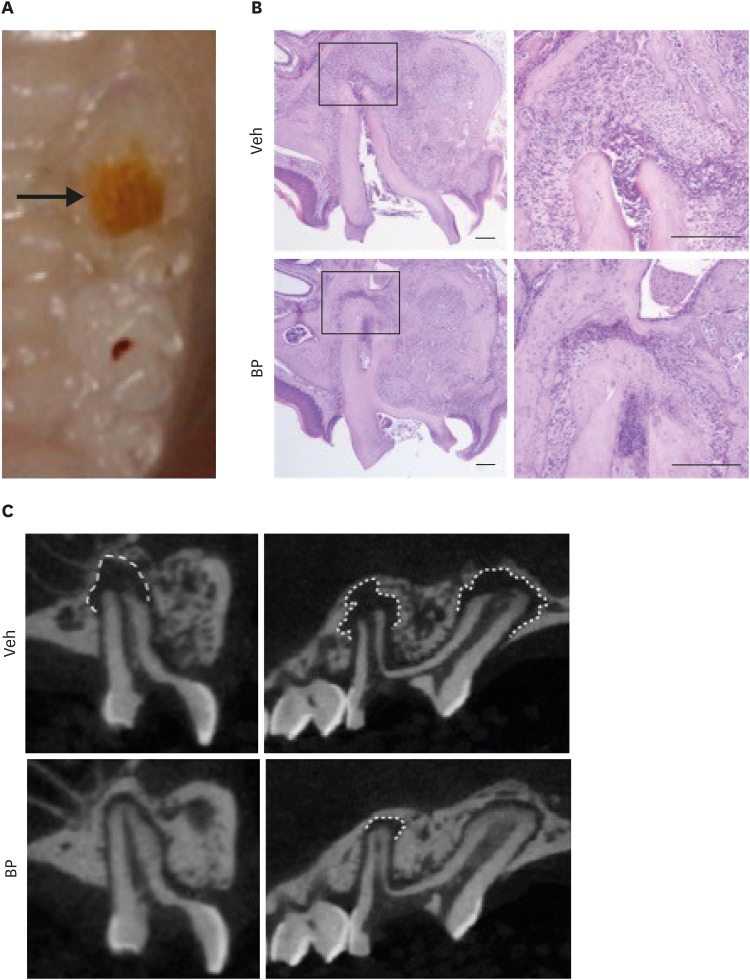
ePub Antiresorptive drugs (ARDs), such as bisphosphonates or denosumab, that prevent bone resorption are widely used in patients with osteoporosis or with cancer that has metastasized to the bones. Although osteonecrosis of the jaw (ONJ) is a well-documented complication of ARD use, the benefits ARDs outweigh the complication. Thus, research has focused on finding ways to prevent or reduce the risk of developing ONJ. Dentists, as part of a multi-professional team, have a critical role in preventing ONJ. However, many dentists tend to hesitate to provide dental care to patients with ONJ, or tend to think that it is a problem to be dealt with by oral surgeons. This review gives an overview of ARD-related ONJ and provides the guidelines for dental care in patients taking ARDs to lower the risk of developing ONJ.
-
Citations
Citations to this article as recorded by- Successful prevention of medication-related osteonecrosis of the jaw after dental extractions by socket preservation with alloplast plus tetracycline in patients taking antiresorptive drugs
Liang-Ho Lin, Chun-Hsiang Wang, Shin-Yu Lu
Journal of Dental Sciences.2026; 21(1): 468. CrossRef - Dynamic Navigation in Endodontics: Scope, Benefits, and Challenges - A Systematic Review
Ankita Kapoor, Ragavi Alagarsamy, Babu Lal, Amal Singh Rana, Amandeep Kaur, Sidhartha Sharma, Ajay Logani
Journal of Endodontics.2025; 51(7): 879. CrossRef - Biosimilars in osteoporosis treatment: focus on denosumab
Matti Aapro, Peyman Hadji, Daniele Santini, Ralf Schmidmaier, Richard Eastell
Expert Opinion on Biological Therapy.2025; 25(8): 887. CrossRef - Assessment of the occurrence of apical periodontitis and endodontically treated/non-treated teeth in a Lower Austrian patient population treated for osteoporosis: a cohort study
Pascal Grün, Marius Meier, Johannes Dittrich, Arb Gjergjindreaj, Dragan Ströbele, Florian Pfaffeneder-Mantai, Sepideh Hatamikia, Margrit-Ann Geibel, Dritan Turhani
Annals of Medicine & Surgery.2024; 86(9): 5049. CrossRef - Endodontic and periapical status of patients with osteoporosis
Selin Goker Kamalı, Dilek Turkaydın
The Journal of the American Dental Association.2024; 155(12): 1022. CrossRef - Postoperative pain in oncological patients subjected to nonsurgical root canal treatment: a prospective case-control study
Kaline Romeiro, Luciana F. Gominho, Isabela N. Rôças, José F. Siqueira Jr
Clinical Oral Investigations.2024;[Epub] CrossRef - Periodontal ligament‐associated protein‐1 promotes osteoclastogenesis in mice by modulating TGF‐β1/Smad1 pathway
Shuang Liu, Xijiao Yu, Qiushuang Guo, Shuaiqi Zhao, Kaixian Yan, Meng Hou, Fuxiang Bai, Shu Li
Journal of Periodontology.2024; 95(2): 146. CrossRef - What Is the Appropriate Antibiotic Administration During Tooth Extractions in Patients Receiving High-Dose Denosumab?
Eiji Iwata, Takumi Hasegawa, Hiroaki Ohori, Toshiya Oko, Tsutomu Minamikawa, Daisuke Miyai, Masaki Kobayashi, Naoki Takata, Shungo Furudoi, Junichiro Takeuchi, Kosuke Matsumoto, Akira Tachibana, Masaya Akashi
Cureus.2024;[Epub] CrossRef - Perfil Odontológico dos Pacientes em Uso de Bisfosfonatos em um Hospital Oncológico
Jade Fontenele Tagliabue, Lísia Daltro Borges Alves, Héliton Spíndola Antunes
Revista Brasileira de Cancerologia.2024;[Epub] CrossRef - Analysis of the Degree of Information of Dental Surgeons about Antiresorptive Drugs According to the Time Since Graduation in Dentistry
Flávia Godinho Costa Wanderley Rocha, Roberto Paulo Correia de Araújo
Pesquisa Brasileira em Odontopediatria e Clínica Integrada.2023;[Epub] CrossRef - Osteonecrosis of the Jaw and Antiresorptive Agents in Benign and Malignant Diseases: A Critical Review Organized by the ECTS
Athanasios D Anastasilakis, Jessica Pepe, Nicola Napoli, Andrea Palermo, Christos Magopoulos, Aliya A Khan, M Carola Zillikens, Jean-Jacques Body
The Journal of Clinical Endocrinology & Metabolism.2022; 107(5): 1441. CrossRef - Accuracy of Dynamic Navigation for Non-Surgical Endodontic Treatment: A Systematic Review
Egle Marija Jonaityte, Goda Bilvinaite, Saulius Drukteinis, Andres Torres
Journal of Clinical Medicine.2022; 11(12): 3441. CrossRef - Periapical status in patients affected by osteoporosis: A retrospective clinical study
Erika Cadoni, Francesca Ideo, Giuseppe Marongiu, Silvia Mezzena, Luca Frigau, Quirico Mela, Antonio Capone, Henry F. Duncan, Elisabetta Cotti
Clinical and Experimental Dental Research.2022; 8(5): 1068. CrossRef - LONG-TERM USE OF DENOSUMAB IN GIANT CELL TUMORS AND VERTEBRAL ANEURYSMAL BONE CYSTS
Pedro Luis Bazán, Micaela Cinalli, Felipe Lanari Zabiaur, Roberto Castelli, Claudio Silveri, José Luis Monayer, Enrique Gustavo Gobbi, Alejandro Maria Steverlynck
Coluna/Columna.2022;[Epub] CrossRef - A 5-year retrospective cohort study of denosumab induced medication related osteonecrosis of the jaw in osteoporosis patients
Seoyeon Jung, Jaeyeon Kim, Jin Hoo Park, Ki-Yeol Kim, Hyung Jun Kim, Wonse Park
Scientific Reports.2022;[Epub] CrossRef - Management of Patients under Treatment with Monoclonal Antibodies and New Biological Therapies
Marta Amigo-Basilio, Covadonga Álvarez-González, Carlos Cobo-Vázquez, Isabel Leco-Berrocal, Luis Miguel Sáez-Alcaide, Cristina Méniz-García
Applied Sciences.2021; 11(11): 4865. CrossRef - Medication-Related Osteonecrosis of the Jaw, a Hidden Enemy. An Integrative Review
Odel Chediak-Barbur
Universitas Odontologica.2021;[Epub] CrossRef - A Retrospective Observational Study of Risk Factors for Denosumab-Related Osteonecrosis of the Jaw in Patients with Bone Metastases from Solid Cancers
Satoe Okuma, Yuhei Matsuda, Yoshiki Nariai, Masaaki Karino, Ritsuro Suzuki, Takahiro Kanno
Cancers.2020; 12(5): 1209. CrossRef
- Successful prevention of medication-related osteonecrosis of the jaw after dental extractions by socket preservation with alloplast plus tetracycline in patients taking antiresorptive drugs
- 3,918 View
- 52 Download
- 18 Crossref

- Cyclic fatigue resistance of the WaveOne Gold Glider, ProGlider, and the One G glide path instruments in double-curvature canals
- Damla Kırıcı, Alper Kuştarcı
- Restor Dent Endod 2019;44(4):e36. Published online September 9, 2019
- DOI: https://doi.org/10.5395/rde.2019.44.e36
-
 Abstract
 PDF
PDF PubReader
 ePub
ePub Objectives The aim of this study was to compare the cyclic fatigue resistance of the WaveOne Gold Glider, ProGlider and One G glide path instruments in artificial double-curvature canals.
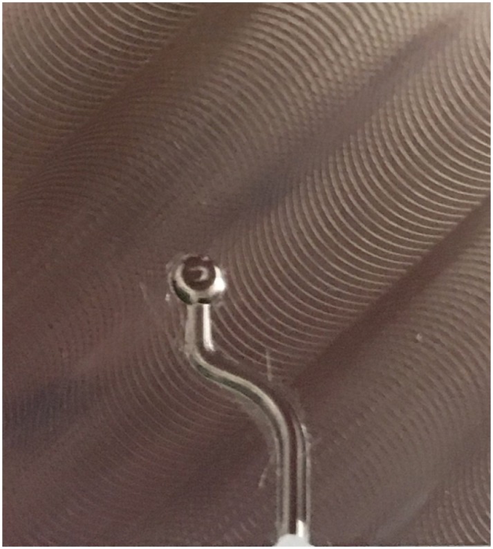
Materials and Methods This study included 15 WaveOne Gold Glider (size 15/0.08), 15 ProGlider (size 16/0.08), and 15 One G (size 16/0.06) nickel titanium files. The files were used in accordance with the manufacturer's instructions until they were broken in artificial double-curvature canals made of stainless steel. The time to fracture was recorded via a digital stopwatch and the number of rotations until fracture was also calculated. The data were statistically analyzed via the Kruskal-Wallis test.
Results The highest average number of rotations until fracture of the files was found for the WaveOne Gold Glider, followed by ProGlider and One G in order. Statistically significant differences were present between all groups of files (
p < 0.05).Conclusions In our study, the resistance of the WaveOne Gold Glider nickel-titanium (Ni-Ti) file to cyclic fatigue in S-shaped curved canals was found to be higher than that of the ProGlider and One G Ni-Ti files.
-
Citations
Citations to this article as recorded by- Glide Path in Endodontics: A Literature Review of Current Knowledge
Vlad Mircea Lup, Giulia Malvicini, Carlo Gaeta, Simone Grandini, Gabriela Ciavoi
Dentistry Journal.2024; 12(8): 257. CrossRef - In Vitro Evaluation of the Dynamic Cyclic Fatigue Resistance of a New TruNatomy Glider File after Different Cycles of Use
Lorena Ferreira Rego, Juliana Delatorre Bronzato, Alana Pinto Carôso Souza, Adriana de-Jesus-Soares, Marcos Frozoni
Journal of Endodontics.2024; 50(5): 619. CrossRef - Comparative Evaluation of the Cyclic Fatigue Resistance of WaveOne Gold in Reciprocation, ProGlider in Rotary Motion, and Manual Files in a Reciprocating Handpiece Within Simulated Curved Canals: An In Vitro Study
Shivangi M Pujara, Hardik B Shah, Leena H Jobanputra
Cureus.2024;[Epub] CrossRef - Comparative analysis of torsional and cyclic fatigue resistance of ProGlider, WaveOne Gold Glider, and TruNatomy Glider in simulated curved canal
Pedro de Souza Dias, Augusto Shoji Kato, Carlos Eduardo da Silveira Bueno, Rodrigo Ricci Vivan, Marco Antonio Hungaro Duarte, Pedro Henrique Souza Calefi, Rina Andréa Pelegrine
Restorative Dentistry & Endodontics.2023;[Epub] CrossRef - Negotiability of mesiobuccal canals in maxillary first molars using different path file systems
Maryam Gharechahi, Mandana Khajehpour, Ali Hamedi, Maryam Peighoun
Dental Research Journal.2023;[Epub] CrossRef - Cyclic Fatigue Resistance of Glide Path Rotary Files: A Systematic Review of in Vitro Studies
Israa Ashkar, José Luis Sanz, Leopoldo Forner
Materials.2022; 15(19): 6662. CrossRef - Evaluation of design, mechanical properties, and torque/force generation of heat-treated NiTi glide path instruments
Soram Oh, Ji-Yeon Seo, Ji-Eun Lee, Hyun-Jung Kim, Ji-Hyun Jang, Seok Woo Chang
BMC Oral Health.2022;[Epub] CrossRef - Body temperature fatigue behaviour of reciprocating and rotary glide path instruments in sodium hypochlorite solutions alone or combined with etidronate
Dario Perez‐Villalba, José C. Macorra, Juan J. Perez‐Higueras, Ove A. Peters, Ana Arias
Australian Endodontic Journal.2021; 47(3): 450. CrossRef
- Glide Path in Endodontics: A Literature Review of Current Knowledge
- 1,509 View
- 11 Download
- 8 Crossref

-
The effect of individualization of fiberglass posts using bulk-fill resin-based composites on cementation: an
in vitro study - Rodrigo Barros Esteves Lins, Jairo Matozinho Cordeiro, Carolina Perez Rangel, Thiago Bessa Marconato Antunes, Luís Roberto Marcondes Martins
- Restor Dent Endod 2019;44(4):e37. Published online October 18, 2019
- DOI: https://doi.org/10.5395/rde.2019.44.e37
-
 Abstract
 PDF
PDF PubReader
 ePub
ePub Objectives This study evaluated the bond strength of various fiberglass post cementation techniques using different resin-based composites.
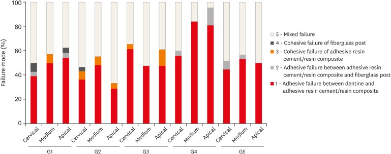
Materials and Methods The roots from a total of 100 bovine incisors were randomly assigned to 5 treatment groups: G1, post + Scotchbond Multi-Purpose (SBMP) + RelyX ARC luting agent; G2, relined post (Filtek Z250) + SBMP + RelyX ARC; G3, individualized post (Filtek Z250) + SBMP; G4, individualized post (Filtek Bulk-Fill) + SBMP; G5, individualized post (Filtek Bulk-Fill Flow) + SBMP. The samples were subjected to the push-out (
n = 10) and pull-out (n = 10) bond strength tests. Data from the push-out bond strength test were analyzed using 2-way analysis of variance (ANOVA) with the Bonferronipost hoc test, and data from the pull-out bond strength test were analyzed using 1-way ANOVA.Results The data for push-out bond strength presented higher values for G2 and G5, mainly in the cervical and middle thirds, and the data from the apical third showed a lower mean push-out bond strength in all groups. No significant difference was noted for pull-out bond strength among all groups. The most frequent failure modes observed were adhesive failure between dentine and resin and mixed failure.
Conclusions Fiberglass post cementation using restorative and flowable bulk-fill composites with the individualization technique may be a promising alternative to existing methods of post cementation.
-
Citations
Citations to this article as recorded by- EVALUATION OF PUSH-OUT BOND STRENGTH OF GLASS FIBER POSTS USING DIFFERENT LUTING CEMENTS
Jannah Mohammed, Maha Agha
BULLETIN OF STOMATOLOGY AND MAXILLOFACIAL SURGERY.2025; : 274. CrossRef - Effects of a relined fiberglass post with conventional and self-adhesive resin cement
Wilton Lima dos Santos Junior, Marina Rodrigues Santi, Rodrigo Barros Esteves Lins, Luís Roberto Marcondes Martins
Restorative Dentistry & Endodontics.2024;[Epub] CrossRef - Fracture resistance of weakened roots restored with relined or milled CAD-CAM glass fiber posts
Belizane das Graças Oliveira MAIA, Thais da Silva Alves SANTOS, Cláudio Antonio Talge CARVALHO, Francielle Silvestre VERNER, Rafael Binato JUNQUEIRA
Dental Materials Journal.2023; 42(1): 92. CrossRef - Evaluation of pretreatments on intra‐radicular dentin bond strength of self‐adhesive resin cements
Marina Rodrigues Santi, Rodrigo Barros Esteves Lins, Beatriz Ometto Sahadi, Luís Roberto Marcondes Martins, Jorge Rodrigo Soto‐Montero
Journal of Esthetic and Restorative Dentistry.2022; 34(7): 1051. CrossRef - Comparison of the Mechanical Properties and Push-out Bond Strength of Self-adhesive and Conventional Resin Cements on Fiber Post Cementation
MR Santi, RBE Lins, BO Sahadi, JR Soto-Montero, LRM Martins
Operative Dentistry.2022; 47(3): 346. CrossRef - Glass fiber posts
Renata Pereira, Rodrigo Barros Esteves Lins, Victória Castelan Rodrigues, Débora Alves Nunes Leite Lima, Luís Roberto Marcondes Martins, Flávio Henrique Baggio Aguiar
Brazilian Journal of Oral Sciences.2020; 19: e207508. CrossRef
- EVALUATION OF PUSH-OUT BOND STRENGTH OF GLASS FIBER POSTS USING DIFFERENT LUTING CEMENTS
- 1,310 View
- 10 Download
- 6 Crossref

- Effect of vibratory stimulation on pain during local anesthesia injections: a clinical trial
- Sajedeh Ghorbanzadeh, Hoda Alimadadi, Nazanin Zargar, Omid Dianat
- Restor Dent Endod 2019;44(4):e40. Published online October 24, 2019
- DOI: https://doi.org/10.5395/rde.2019.44.e40
-
 Abstract
 PDF
PDF PubReader
 ePub
ePub Objectives This study aimed to assess the effect of DentalVibe on the level of pain experienced during anesthetic injections using 2 different techniques.
Materials and Methods This randomized crossover clinical trial evaluated 60 patients who required 2-session endodontic treatment. Labial infiltration (LI) anesthesia was administered in the anterior maxilla of 30 patients, while inferior alveolar nerve block (IANB) was performed in the remaining 30 patients. 1.8 mL of 2% lidocaine was injected at a rate of 1 mL/min using a 27-gauge needle. DentalVibe was randomly assigned to either the first or second injection session. A visual analog scale was used to determine participants' pain level during needle insertion and the anesthetic injection. The paired
t -test was applied to assess the efficacy of DentalVibe for pain reduction.Results In LI anesthesia, the pain level was 12.0 ± 15.5 and 38.1 ± 21.0 during needle insertion and 19.1 ± 16.1 and 48.9 ± 24.6 during the anesthetic injection using DentalVibe and the conventional method, respectively. In IANB, the pain level was 14.1 ± 15.9 and 35.1 ± 20.8 during needle insertion and 17.3 ± 14.2 and 39.5 ± 20.8 during the anesthetic injection using DentalVibe and the conventional method, respectively. DentalVibe significantly decreased the level of pain experienced during needle insertion and the anesthetic injection in anterior LI and mandibular IANB anesthesia.
Conclusions The results suggest that DentalVibe can be used to reduce the level of pain experienced by adult patients during needle insertion and anesthetic injection.
-
Citations
Citations to this article as recorded by- Effectiveness of cold vibratory stimuli on pain perception governing infiltration anesthesia in the maxillary arch in children: a randomized controlled clinical trial
Hoda Mohamed Hambouta, Dina Aly Sharaf, Nadia Aziz Wahba
BMC Oral Health.2025;[Epub] CrossRef - Comparative Evaluation of Pain Reduction with Vibration Using Speech Stimulator vs 2% Lignocaine Topical Gel Application Prior to Greater Palatine Nerve Block in 7–12-year-old Children: A Randomized Controlled Trial
Santhosh Prasanth, Padmakumari B, Bhageeradhi D, Seethal B
International Journal of Clinical Pediatric Dentistry.2025; 18(10): 1278. CrossRef - Effect of Vibration on Acute and Chronic Back Pain After Spinal Anesthesia: A Randomized Clinical Trial
Shervin Shahinpour, Fatemeh Refahi, Nader Ali Nazemian
Anesthesiology and Pain Medicine.2024;[Epub] CrossRef - Vibrating toothbrush, ice, or topical anesthetic agent to reduce pain of local anesthetic injection in 5- to 12-year-old children undergoing dental procedures — a randomized controlled trial
Meenu Mittal, Ashok Kumar, Radhika Chopra, Gurvinder Kaur, Sarang Sharma
Ain-Shams Journal of Anesthesiology.2023;[Epub] CrossRef - A Clinical Comparison of Pain Perception and Behavior in Children Using Conventional and Vibraject Injection Techniques
KS Mukunda, Calvin Hilary, C Preeja, G Midhun Mohan
World Journal of Dentistry.2022; 13(6): 623. CrossRef - Topical Anesthesia in Pediatric Dentistry: An Update
International Journal of Clinical Pediatric Dentistry.2022; 15(2): 240. CrossRef - Effect of Photobiomodulation Therapy with 915 nm Diode Laser on Pain Perception during Local Anesthesia of Maxillary Incisors: A Randomized Controlled Trial
Hamid Kermanshah, Nasim Chiniforush, Mahsa Kolahdouz Mohammadi, Fariba Motevasselian
Photochemistry and Photobiology.2022; 98(6): 1471. CrossRef - Comparative evaluation of pain perception during conventional greater palatine injections versus the use of a novel barovibrotactile device - In vivo study
Aishwarya Avinash Gangawane, Sonal Bhavesh Shah, Tanvi Eknath Malankar, Anmol Mathur, Shriya Shrirang Ginde, Manne Lakshmi Priyanka
Journal of Oral Biology and Craniofacial Research.2022; 12(5): 542. CrossRef - Efficacy of Higher Gauged Needles or Topical Pre-Cooling for Pain Reduction during Local Anesthesia Injection: A Split-Mouth Randomized Trial
Srikanth Gadicherla, Mihika Mahandwan, Shane Quek Yi Xuan, Kalyana-Chakravarthy Pentapati
Pesquisa Brasileira em Odontopediatria e Clínica Integrada.2021;[Epub] CrossRef - Auto-controlled Syringe vs Insulin Syringe for Palatal Injections in Children: A Randomized Crossover Trial
Sunny P Tirupathi, Srinitya Rajasekhar, Pushpalatha Tummakomma, Aishwarya Arya Gangili, Abdul Rehman Ahmed Khan, Mohammed Khurramuddin, Usha Purumandla
The Journal of Contemporary Dental Practice.2020; 21(6): 604. CrossRef
- Effectiveness of cold vibratory stimuli on pain perception governing infiltration anesthesia in the maxillary arch in children: a randomized controlled clinical trial
- 1,428 View
- 10 Download
- 10 Crossref

- The effect of preheating resin composites on surface hardness: a systematic review and meta-analysis
- Ali A. Elkaffas, Radwa I. Eltoukhy, Salwa A. Elnegoly, Salah H. Mahmoud
- Restor Dent Endod 2019;44(4):e41. Published online October 29, 2019
- DOI: https://doi.org/10.5395/rde.2019.44.e41
-
 Abstract
 PDF
PDF PubReader
 ePub
ePub Objectives This paper presents a systematic review and meta-analysis of the effect of preheating on the hardness of nanofilled, nanoceramic, nanohybrid, and microhybrid resin composites.
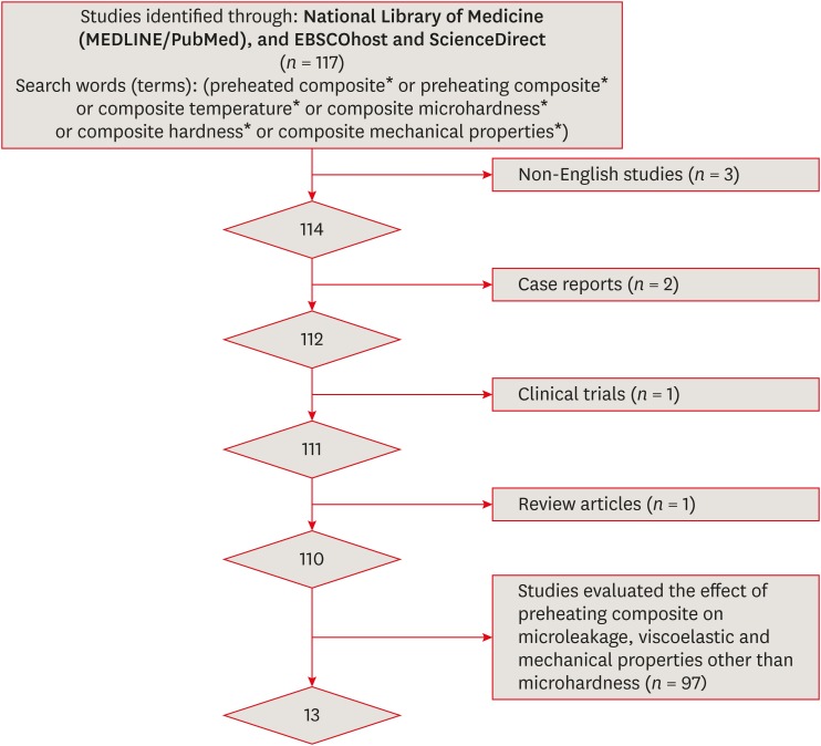
Materials and Methods An electronic search of papers on MEDLINE/PubMed, ScienceDirect, and EBSCOhost was performed. Only
in vitro studies were included. Non-English studies, case reports, clinical trials, and review articles were excluded. A meta-analysis of the reviewed studies was conducted to quantify differences in the microhardness of the Z250 microhybrid resin composite using the Comprehensive Meta-Analysis software.Results Only 13 studies met the inclusion criteria for this systematic review. The meta-analysis showed that there were significant differences between the non-preheated and preheated modes for both the top and bottom surfaces of the specimens (
p < 0.05). The microhardness of the Z250 resin composite on the top surface in the preheated mode (78.1 ± 2.9) was higher than in the non-preheated mode (67.4 ± 4.0;p < 0.001). Moreover, the microhardness of the Z250 resin composite on the bottom surface in the preheated mode (71.8 ± 3.8) was higher than in the non-preheated mode (57.5 ± 5.7,p < 0.001).Conclusions Although the results reported in the reviewed studies showed great variability, sufficient scientific evidence was found to support the hypothesis that preheating can improve the hardness of resin composites.
-
Citations
Citations to this article as recorded by- Influence of preheating and water storage on the color, whiteness, and translucency of modern resin‐based composites
Corina Mirela Prodan, Cristina Gasparik, Javier Ruiz‐López, Diana Dudea
Journal of Esthetic and Restorative Dentistry.2025; 37(2): 533. CrossRef - Effects of pre-heating on physical–mechanical–chemical properties of contemporary resin composites
Thamires Bueno, Nivien Masoud, Anna Akkus, Italo Silva, Karen McPherson, Adilson Yoshio Furuse, Fabio Rizzante
Odontology.2025; 113(1): 135. CrossRef - The effects of a carbonated beverage on the optical properties and microhardness of preheated bulk-fill composite resin restorations
Nancy Soliman Farghal, Ayya Abu Shamleh, Osamah Al Hurmuzi, Okba Mahmoud
Frontiers in Oral Health.2025;[Epub] CrossRef - Evaluation of Degree of Conversion, Flexural Strength, and Microhardness of a Novel Flowable Resin Composite
Bengü Doğu Kaya, Selinsu Öztürk, Nazlı Zeynep Kuzu, Ayşe Aslı Şenol, Erkut Kahramanoğlu, Pınar Yılmaz Atalı, Bilge Tarçın
Selcuk Dental Journal.2025; 12(2): 202. CrossRef - Clinical performance of different bulk‐fill composite resin systems in classIIcavities: A 2‐year randomized clinical trial
Badria Goda, Kareem Hamdi, Radwa I. Eltoukhy, Ashraf I. Ali, Salah Hasab Mahmoud
Journal of Esthetic and Restorative Dentistry.2024; 36(8): 1122. CrossRef - Comparative evaluation of microleakage in Class II cavities restored with snowplow technique using flowable or preheated packable bulk-fill composite resin as gingival increment by dye extraction method: An in vitro study
M. A. Ranjini, V. Geetha, B. Vedavathi, H. K. Ashok, Akshata J. Airsang, S. Swathi
Journal of Conservative Dentistry and Endodontics.2024; 27(11): 1158. CrossRef - Influence of Light‐Curing Time and Increment Thickness on the Properties of Bulk Fill Composite Resins With Distinct Application Systems
Carlos Rocha Gomes Torres, Taiana Paola Prado, Daniele Mara da Silva Ávila, Cesar Rogério Pucci, Alessandra Bühler Borges, Heng Bo Jiang
International Journal of Dentistry.2024;[Epub] CrossRef - Last Generation Bis-GMA Free Composite For Indirect Posterior Restorations: A Case Report
M. Delgado
Endodontics Today.2024; 21(4): 305. CrossRef - The clinical performance of dental resin composite repeatedly preheated: A randomized controlled clinical trial
Mahmoud Elkady, Safaa Abdelhakim, Mona Riad
Journal of Dentistry.2024; 144: 104940. CrossRef - Preheating effect on microhardness and depth of cure of three bulk-fill composite resins: An in vitro study
Aashna Sunil Sahetia, Divya Rupesh Jain, Padmaja Panditrao Sirsat, Meenal N. Gulve, Swapnil J. Kolhe, Surbhi P. Patel
Endodontology.2024;[Epub] CrossRef - Evaluation of Shear Bond Strength of Lithium Disilicate Veneers Using Pre-heated Resin Composite With Two Conventional Resin Cements: An In Vitro Study
Ghalia Akyle, Hassan Achour
Cureus.2024;[Epub] CrossRef - Effect of Glass Fiber Reinforcement on Marginal Microleakage in Class II Composite Restorations: An In Vitro Pilot Study
Csaba Dudás, Emánuel Kardos, Melinda Székely, Lea Ádám, Zsuzsanna Bardocz-Veres, Evelyn Szőllősi, Kinga Mária Jánosi, Bernadette Kerekes-Máthé
Dentistry Journal.2024; 12(12): 410. CrossRef - Effect of preheating on the physicochemical properties and bond strength of composite resins utilized as dental cements: An in vitro study
Carolina Carramilo Raposo, Luanna Marinho Sereno Nery, Edilausson Moreno Carvalho, Paulo Vitor Campos Ferreira, Diego Machado Ardenghi, José Bauer, Darlon Martins Lima
The Journal of Prosthetic Dentistry.2023; 129(1): 229.e1. CrossRef - Examining the Impact of Preheating on the Fracture Toughness and Microhardness of Composite Resin: A Systematic Review
Jay Bhopatkar, Anuja Ikhar, Manoj Chandak, Aditya Patel, Paridhi Agrawal
Cureus.2023;[Epub] CrossRef - In Vitro Effect of Mouthrinses on the Microhardness of Three Different Nanohybrid Composite Resins
Jhonn Luis Bernaldo-Faustino, Julissa Amparo Dulanto-Vargas, Kilder Maynor Carranza-Samanez, Carlos A. Munoz-Viveros
International Journal of Dentistry.2023; 2023: 1. CrossRef - Efecto del precalentamiento en la microdureza superficial de seis resinas compuestas
Gloria Cristina Moreno Abello, Kavhas Castro, Paula Alejandra Ovalle Barrera, Paula Bernal, Laura Catalina Lara Hernández
Universitas Odontologica.2023;[Epub] CrossRef - Wear and Color Stability of Preheated Bulk-fill and Conventional Resin Composites
AA Abdulmajeed, AA Suliman, BJ Selivany, A Altitinchi, TA Sulaiman
Operative Dentistry.2022; 47(5): 585. CrossRef - Comparison of Mechanical Properties of a Self-Adhesive Composite Cement and a Heated Composite Material
Anastazja Skapska, Zenon Komorek, Mariusz Cierech, Elzbieta Mierzwinska-Nastalska
Polymers.2022; 14(13): 2686. CrossRef - Effects of ionizing radiation on surface properties of current restorative dental materials
Débora Michelle Gonçalves de Amorim, Aretha Heitor Veríssimo, Anne Kaline Claudino Ribeiro, Rodrigo Othávio de Assunção e Souza, Isauremi Vieira de Assunção, Marilia Regalado Galvão Rabelo Caldas, Boniek Castillo Dutra Borges
Journal of Materials Science: Materials in Medicine.2021;[Epub] CrossRef - Quality assessment tools used in systematic reviews of in vitro studies: A systematic review
Linh Tran, Dao Ngoc Hien Tam, Abdelrahman Elshafay, Thao Dang, Kenji Hirayama, Nguyen Tien Huy
BMC Medical Research Methodology.2021;[Epub] CrossRef - Preheated composite: Innovative approach for aesthetic restoration
Reema N Asani, Vandana J Gade, Kalyani G Umale, Rachana Gawande, Rohit R Amburle, Raksha R Kusumbe, Purva P Kale, Priya R Kosare
Archives of Dental Research.2021; 11(2): 103. CrossRef
- Influence of preheating and water storage on the color, whiteness, and translucency of modern resin‐based composites
- 2,590 View
- 25 Download
- 21 Crossref

-
Cyclic fatigue resistance of M-Pro and RaCe Ni-Ti rotary endodontic instruments in artificial curved canals: a comparative
in vitro study - Hadeer Mostafa El Feky, Khalid Mohammed Ezzat, Marwa Mahmoud Ali Bedier
- Restor Dent Endod 2019;44(4):e44. Published online November 7, 2019
- DOI: https://doi.org/10.5395/rde.2019.44.e44
-
 Abstract
 PDF
PDF PubReader
 ePub
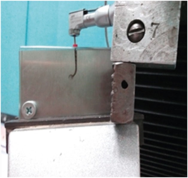
ePub Objectives To compare the flexural cyclic fatigue resistance and the length of the fractured segments (FLs) of recently introduced M-Pro rotary files with that of RaCe rotary files in curved canals and to evaluate the fracture surface by scanning electron microscopy (SEM).
Materials and Methods Thirty-six endodontic files with the same tip size and taper (size 25, 0.06 taper) were used. The samples were classified into 2 groups (n = 18): the M-Pro group (M-Pro IMD) and the RaCe group (FKG). A custom-made simulated canal model was fabricated to evaluate the total number of cycles to failure and the FL. SEM was used to examine the fracture surfaces of the fragmented segments. The data were statistically analyzed and comparisons between the 2 groups for normally distributed numerical variables were carried out using the independent Student's
t -test. Ap value less than 0.05 was considered to indicate statistical significance.Results The M-Pro group showed significantly higher resistance to flexural cyclic fatigue than the RaCe group (
p < 0.05), but there was no significant difference in the FLs between the 2 groups (p ≥ 0.05).Conclusions Thermal treatment of nickel-titanium instruments can improve the flexural cyclic fatigue resistance of rotary endodontic files, and the M-Pro rotary system seems to be a promising rotary endodontic file.
-
Citations
Citations to this article as recorded by- The Effect of Canal Curvature and Different Manufacturing Processes of Five Different NiTi Rotary Files on Cyclic Fatigue Resistance
Panupat Phumpatrakom, Awiruth Klaisiri, Sukitti Techapatiphandee, Thippawan Saekow, Panuroot Aguilar
European Journal of General Dentistry.2025; 14(03): 264. CrossRef - EndoMagic Gold M06 Eğelerinde Boyut ve Konikliğin Döngüsel Yorgunluğa Etkisi: Bir İn Vitro Çalışma
Bircan Kuloğlu, Ayşe Çoban, Hatice Büyüközer Özkan
Akdeniz Diş Hekimliği Dergisi.2025; 4(3): 212. CrossRef - Evaluatation of two nickle-titanium systems’ (Neolix and X Pro Gold) resistance to fracture after immersion in sodium hypochlorite.
Solmaz Araghi, Abbas Delvarani, Faeze dehghan, Parisa Kaghazloo
journal of research in dental sciences.2024; 21(1): 17. CrossRef - Endodontic Ni–Ti Rotary Instruments for Glide-path, Are They Still Necessary and How to Think about the Ideal Instrument?
Shilpa Bhandi, Rodolfo Reda, Luca Testarelli, Elisa Maccari
The Journal of Contemporary Dental Practice.2024; 25(6): 505. CrossRef - Comparative evaluation of cyclic fatigue resistance of thermomechanically treated NiTi rotary instruments in simulated curved canals with two different radii of curvature: An in vitro Study
Tahira Hamid, Azhar Malik, Ajay Kumar, Shamim Anjum
Journal of Conservative Dentistry and Endodontics.2024; 27(4): 393. CrossRef - New heat-treated vs electropolished nickel-titanium instruments used in root canal treatment: Influence of autoclave sterilization on surface roughness
Rahaf A. Almohareb, Reem Barakat, Fatimah Albohairy, Hannes C. Schniepp
PLOS ONE.2022; 17(3): e0265226. CrossRef - The Effect of Taper and Apical Diameter on the Cyclic Fatigue Resistance of Rotary Endodontic Files Using an Experimental Electronic Device
Vicente Faus-Llácer, Nirmine Hamoud Kharrat, Celia Ruiz-Sánchez, Ignacio Faus-Matoses, Álvaro Zubizarreta-Macho, Vicente Faus-Matoses
Applied Sciences.2021; 11(2): 863. CrossRef
- The Effect of Canal Curvature and Different Manufacturing Processes of Five Different NiTi Rotary Files on Cyclic Fatigue Resistance
- 2,102 View
- 10 Download
- 7 Crossref

- Influence of 10-MDP concentration on the adhesion and physical properties of self-adhesive resin cements
- Kazuhiko Shibuya, Naoko Ohara, Serina Ono, Kumiko Matsuzaki, Masahiro Yoshiyama
- Restor Dent Endod 2019;44(4):e45. Published online November 12, 2019
- DOI: https://doi.org/10.5395/rde.2019.44.e45

-
 Abstract
 PDF
PDF PubReader
 ePub
ePub Objectives Self-adhesive resin cements contain functional monomers that enable them to adhere to the tooth structure without a separate adhesive or etchant. One of the most stable functional monomers used for chemical bonding to calcium in hydroxyapatite is 10-methacryloyloxydecyl dihydrogen phosphate (10-MDP). The aim of this study was to evaluate the influence of the10-MDP concentration on the bond strength and physical properties of self-adhesive resin cements.
Materials and Methods We used experimental resin cements containing 3 different concentrations of 10-MDP: 3.3 wt% (RC1), 6.6 wt% (RC2), or 9.9 wt% (RC3). The micro-tensile bond strength of each resin cement to dentin and a hybrid resin block (Estenia C&B, Kuraray Noritake Dental) was measured, and the fractured surface morphology was analyzed. Further, the flexural strength of the resin cements was measured using the three-point bending test. The water sorption and solubility of the cements following 30 days of immersion in water were measured.
Results The bond strength of RC2 was significantly higher than that of RC1. There was no significant difference between the bond strength of RC2 and that of RC3. The water sorption of RC3 was higher than that of any other cement. There were no significant differences in the three-point bending strength or water solubility among all three types of cements.
Conclusions Within the limitations of this study, it is suggested that 6.6 wt% 10-MDP showed superior properties than 3.3 wt% or 9.9 wt% 10-MDP in self-adhesive resin cement.
-
Citations
Citations to this article as recorded by- Bonding effectiveness of 10-MDP containing resin composite cements: a systematic review with meta-analysis
Sofia Bignotto de Carvalho, Lívia Maiumi Uehara, João Marcos Carvalho-Silva, Andréa Cândido dos Reis
International Journal of Adhesion and Adhesives.2026; 146: 104260. CrossRef - Comparative Evaluation of Color Stability in Bioactive and Conventional Resin Cements Under Thermal Stress Conditions
Alaa Turkistani, Hanin E. Yeslam
Biomimetics.2025; 10(7): 432. CrossRef - Influence of temperature and curing modes on polymerization of self-adhesive resin cements
Hae-In Kim, Jin-Woo Kim, Se-Hee Park, Kyung-Mo Cho
Korean Journal of Dental Materials.2025; 52(3): 143. CrossRef - Clinical Performance and Retention of Partial Implant Restorations Cemented with Fuji Plus® and DentoTemp™: A Retrospective Clinical Study with Mechanical Validation
Sergiu-Manuel Antonie, Laura-Cristina Rusu, Ioan-Achim Borsanu, Remus Christian Bratu, Emanuel-Adrian Bratu
Medicina.2025; 61(12): 2183. CrossRef - A thorough assessment of 10-MDP primers in modern dental adhesive systems
Ahmed A Abduljawad, Harraa SM Salih, Omar F Tawfiq
Journal of Baghdad College of Dentistry.2024; 36(3): 79. CrossRef - Material properties and finite element analysis of adhesive cements used for zirconia crowns on dental implants
Megha Satpathy, Hai Pham, Shreya Shah
Computer Methods in Biomechanics and Biomedical Engineering.2024; : 1. CrossRef - Clinical reliability of self-adhesive luting resins compared to other adhesive procedures: A systematic review and meta-analysis
Mohammed Ahmed Alghauli, Ahmed Yaseen Alqutaibi, Sebastian Wille, Matthias Kern
Journal of Dentistry.2023; 129: 104394. CrossRef - Influence of autoclave sterilization on bond strength between zirconia frameworks and Ti-base abutments using different resin cements
Reinhold Lang, Karl-Anton Hiller, Lena Kienböck, Katrin Friedl, Karl-Heinz Friedl
The Journal of Prosthetic Dentistry.2022; 127(4): 617.e1. CrossRef - Varying 10-methacryloyloxydecyl dihydrogen phosphate (10-MDP) level improves polymerisation kinetics and flexural strength in self-adhesive, remineralising composites
António H.S. Delgado, Nazanin Owji, Paul Ashley, Anne M. Young
Dental Materials.2021; 37(9): 1366. CrossRef - Investigating a Commercial Functional Adhesive with 12-MDPB and Reactive Filler to Strengthen the Adhesive Interface in Eroded Dentin
Madalena Belmar da Costa, António HS Delgado, Tomás Amorim Afonso, Luís Proença, Ana Sofia Ramos, Ana Mano Azul
Polymers.2021; 13(20): 3562. CrossRef
- Bonding effectiveness of 10-MDP containing resin composite cements: a systematic review with meta-analysis
- 2,559 View
- 15 Download
- 10 Crossref

- Pulp revascularization with and without platelet-rich plasma in two anterior teeth with horizontal radicular fractures: a case report
- Edison Arango-Gómez, Javier Laureano Nino-Barrera, Gustavo Nino, Freddy Jordan, Henry Sossa-Rojas
- Restor Dent Endod 2019;44(4):e35. Published online August 20, 2019
- DOI: https://doi.org/10.5395/rde.2019.44.e35
-
 Abstract
 PDF
PDF PubReader
 ePub
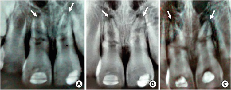
ePub Pulp revascularization is an alternative treatment in immature traumatized teeth with necrotic pulp. However, this procedure has not been reported in horizontal root fractures. This is a case report of a 9-year-old patient with multiple horizontal root fractures in 2 upper central incisors that were successfully treated with pulp revascularization. The patient presented for treatment 2 years after the initial trauma, and revascularization was attempted after the initial treatment with calcium hydroxide had failed. Prior to pulp revascularization, cone-beam computed tomography and autoradiograms demonstrated multiple horizontal fractures in the middle and apical thirds of the roots of the 2 affected teeth. Revascularization was performed in both teeth; platelet-rich plasma (PRP) was used in one tooth (#11) and the conventional method (blood clot) was used in the other tooth (#21). Clinical and radiographic follow-up over 4 years demonstrated pulp calcification in the PRP-treated tooth. Neither of the 2 teeth were lost, and the root canal calcification of tooth #11 was greater than that of tooth #21. This case suggests that PRP-based pulp revascularization may be an alternative for horizontal root fractures.
-
Citations
Citations to this article as recorded by- Platelet-Rich Plasma and Platelet-Rich Fibrin in Endodontics: A Scoping Review
Simão Rebimbas Guerreiro, Carlos Miguel Marto, Anabela Paula, Joana Rita de Azevedo Pereira, Eunice Carrilho, Manuel Marques-Ferreira, Siri Vicente Paulo
International Journal of Molecular Sciences.2025; 26(12): 5479. CrossRef - Dental pulp mesenchymal stem cells-response to fibrin hydrogel reveals ITGA2 and MMPs expression
David Tong, Stéphanie Gobert, Alicia Reuzeau, Jean-Christophe Farges, Marianne Leveque, Marie Bolon, Arthur Costantini, Marielle Pasdeloup, Jérôme Lafont, Maxime Ducret, Mourad Bekhouche
Heliyon.2024; 10(13): e32891. CrossRef - Pulp regeneration treatment using different bioactive materials in permanent teeth of pediatric subjects
Dina Abdellatif, Alfredo Iandolo, Giuseppina De Benedetto, Francesco Giordano, Davide Mancino, Edouard Euvrard, Massimo Pisano
Journal of Conservative Dentistry and Endodontics.2024; 27(5): 458. CrossRef - Retreatment of a Failed Regenerative Endodontic Treatment in an Immature Tooth with a Horizontal Root Fracture: A Case Report
Zaher Marjy, Iris Slutzky-Goldberg
International Journal of Clinical Pediatric Dentistry.2024; 17(10): 1168. CrossRef - The Impact of the Preferred Reporting Items for Case Reports in Endodontics (PRICE) 2020 Guidelines on the Reporting of Endodontic Case Reports
Sofian Youssef, Phillip Tomson, Amir Reza Akbari, Natalie Archer, Fayjel Shah, Jasmeet Heran, Sunmeet Kandhari, Sandeep Pai, Shivakar Mehrotra, Joanna M Batt
Cureus.2023;[Epub] CrossRef - Evaluation of postoperative pain and healing following regenerative endodontics using platelet‐rich plasma versus conventional endodontic treatment in necrotic mature mandibular molars with chronic periapical periodontitis. A randomized clinical trial
Yassmin Elsayed Ahmed, Geraldine Mohamed Ahmed, Angie Galal Ghoneim
International Endodontic Journal.2023; 56(4): 404. CrossRef - Regenerative endodontic procedures for two traumatized mature anterior teeth with transverse root fractures
Jing Lu, Bill Kahler
BMC Oral Health.2022;[Epub] CrossRef - Are platelet concentrate scaffolds superior to traditional blood clot scaffolds in regeneration therapy of necrotic immature permanent teeth? A systematic review and meta-analysis
Qianwei Tang, Hua Jin, Song Lin, Long Ma, Tingyu Tian, Xiurong Qin
BMC Oral Health.2022;[Epub] CrossRef - Platelet-Rich Fibrin Used as a Scaffold in Pulp Regeneration: Case Series
Ceren ÇİMEN, Selin ŞEN, Elif ŞENAY, Tuğba BEZGİN
Cumhuriyet Dental Journal.2021; 24(1): 113. CrossRef - Plasma rico en plaquetas en Odontología: Revisión de la literatura
Hugo Anthony Rosas Rozas, Hugo Leoncio Rosas Cisneros
Yachay - Revista Científico Cultural.2021; 10(1): 536. CrossRef
- Platelet-Rich Plasma and Platelet-Rich Fibrin in Endodontics: A Scoping Review
- 2,428 View
- 41 Download
- 10 Crossref

- Guided endodontics: a case report of maxillary lateral incisors with multiple dens invaginatus
- Afzal Ali, Hakan Arslan
- Restor Dent Endod 2019;44(4):e38. Published online October 21, 2019
- DOI: https://doi.org/10.5395/rde.2019.44.e38
-
 Abstract
 PDF
PDF PubReader
 ePub
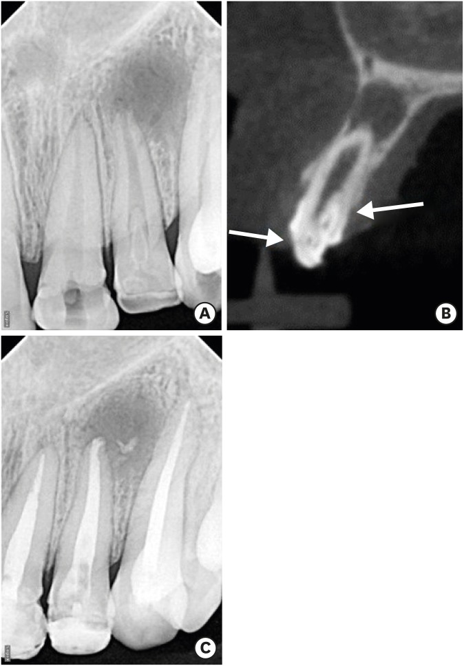
ePub Navigation of the main root canal and dealing with a dens invaginatus (DI) is a challenging task in clinical practice. Recently, the guided endodontics technique has become an alternative method for accessing root canals, surgical cavities, and calcified root canals without causing iatrogenic damage to tissue. In this case report, the use of the guided endodontics technique for two maxillary lateral incisors with multiple DIs is described. A 16-year-old female patient was referred with the chief complaint of pain and discoloured upper front teeth. Based on clinical and radiographic findings, a diagnosis of pulp necrosis and chronic periapical abscess associated with double DI (Oehler's type II) was established for the upper left lateral maxillary incisor (tooth #22). Root canal treatment and the sealing of double DI with mineral trioxide aggregate was planned for tooth #22. For tooth #12 (Oehler's type II), preventive sealing of the DI was planned. Minimally invasive access to the double DI and the main root canal of tooth #22, and to the DI of tooth #12, was achieved using the guided endodontics technique. This technique can be a valuable tool because it reduces chair-time and, more importantly, the risk of iatrogenic damage to the tooth structure.
-
Citations
Citations to this article as recorded by- Guided endodontics in the application of personalized mini-invasive treatment in clinical cases: a literature review
Shuangshuang Ren, Wanping Wang, Mingyue Cheng, Wenyue Tang, Yue Zhao, Leiying Miao
The Saudi Dental Journal.2025;[Epub] CrossRef - Navigating Calcified Challenges: Guided Endodontic Treatment of a Maxillary Central Incisor
Saide Nabavi, Sara Navabi, Iman Shiezadeh, SeyedehZahra JamaliMotlagh
Clinical Case Reports.2025;[Epub] CrossRef - Successful nonsurgical management of Oehler’s type III dens invaginatus in maxillary lateral incisor: A case report as per CARE guidelines
Anshul Sachdeva, Gurdeep Singh Gill, Adel Al Obied, Suraj Arora, Ali Y. Alsaeed, Waled Abdulmalek Alanesi, Gotam Das
Medicine.2025; 104(31): e42725. CrossRef - Guided Endodontics in Managing Root Canal Treatment for Anomalous Teeth—A Narrative Review
Pouya Sabanik, Mohammad Samiei, Shiva Tavakkoli Avval, Bruno Cavalcanti
Australian Endodontic Journal.2025;[Epub] CrossRef - Efficacy of Computer-aided Static Navigation on Accuracy of Guided Endodontic Root Canal Treatment: A Systematic Review and Meta-analysis
Ashish Jain, Rahul D Rao, Meenakshi R Verma, Rishabhkumar N Jain, Shreya Sivasailam, Anandita Sinha
World Journal of Dentistry.2024; 14(11): 1004. CrossRef - Application of personalized templates in minimally invasive management of coronal dens invaginatus: a report of two cases
Mingming Li, Guosong Wang, Fangzhi Zhu, Han Jiang, Yingming Yang, Ran Cheng, Tao Hu, Ru Zhang
BMC Oral Health.2024;[Epub] CrossRef - Endodontic management of severely calcified mandibular anterior teeth using guided endodontics: A report of a case and a review of the literature
Mina Davaji, Sahar Karimpour
Saudi Endodontic Journal.2024; 14(2): 245. CrossRef - Application of an Endodontic Static Guide in Fiber Post Removal from a Compromised Tooth
Mehran Farajollahi, Omid Dianat, Samaneh Gholami, Shima Saber Tahan, Sivakumar Nuvvula
Case Reports in Dentistry.2023;[Epub] CrossRef - The Impact of the Preferred Reporting Items for Case Reports in Endodontics (PRICE) 2020 Guidelines on the Reporting of Endodontic Case Reports
Sofian Youssef, Phillip Tomson, Amir Reza Akbari, Natalie Archer, Fayjel Shah, Jasmeet Heran, Sunmeet Kandhari, Sandeep Pai, Shivakar Mehrotra, Joanna M Batt
Cureus.2023;[Epub] CrossRef - Effectiveness of guided endodontics in locating calcified root canals: a systematic review
F. Peña-Bengoa, M. Valenzuela, M. J. Flores, N. Dufey, K. P. Pinto, E. J. N. L. Silva
Clinical Oral Investigations.2023; 27(5): 2359. CrossRef - Expert consensus on digital guided therapy for endodontic diseases
Xi Wei, Yu Du, Xuedong Zhou, Lin Yue, Qing Yu, Benxiang Hou, Zhi Chen, Jingping Liang, Wenxia Chen, Lihong Qiu, Xiangya Huang, Liuyan Meng, Dingming Huang, Xiaoyan Wang, Yu Tian, Zisheng Tang, Qi Zhang, Leiying Miao, Jin Zhao, Deqin Yang, Jian Yang, Junqi
International Journal of Oral Science.2023;[Epub] CrossRef - Prevalence and morphological analysis of dens invaginatus in anterior teeth using cone beam computed tomography: A systematic review and meta-analysis
Guilherme Nilson Alves dos Santos, Manoel Damião Sousa-Neto, Helena Cristina Assis, Fabiane Carneiro Lopes-Olhê, André L. Faria-e-Silva, Matheus L. Oliveira, Jardel Francisco Mazzi-Chaves, Amanda Pelegrin Candemil
Archives of Oral Biology.2023; 151: 105715. CrossRef - Root Maturation of an Immature Dens Invaginatus Despite Unsuccessful Revitalization Procedure: A Case Report and Recommendations for Educational Purposes
Julia Ludwig, Marcel Reymus, Alexander Winkler, Sebastian Soliman, Ralf Krug, Gabriel Krastl
Dentistry Journal.2023; 11(2): 47. CrossRef - Guided Endodontics as a Personalized Tool for Complicated Clinical Cases
Wojciech Dąbrowski, Wiesława Puchalska, Adam Ziemlewski, Iwona Ordyniec-Kwaśnica
International Journal of Environmental Research and Public Health.2022; 19(16): 9958. CrossRef - Present status and future directions – Guided endodontics
Thomas Connert, Roland Weiger, Gabriel Krastl
International Endodontic Journal.2022; 55(S4): 995. CrossRef - Treatment options for dens in dente: state-of-art literature review
Volodymyr Fedak
Ukrainian Dental Journal.2022; 1(1): 37. CrossRef - Dens Invaginatus: Clinical Implications and Antimicrobial Endodontic Treatment Considerations
José F. Siqueira, Isabela N. Rôças, Sandra R. Hernández, Karen Brisson-Suárez, Alessandra C. Baasch, Alejandro R. Pérez, Flávio R.F. Alves
Journal of Endodontics.2022; 48(2): 161. CrossRef - Guided Endodontics: Static vs. Dynamic Computer-Aided Techniques—A Literature Review
Diana Ribeiro, Eva Reis, Joana A. Marques, Rui I. Falacho, Paulo J. Palma
Journal of Personalized Medicine.2022; 12(9): 1516. CrossRef - Application of CBCT Data and Three-Dimensional Printing for Endodontic Diagnosis and Treatment
Srinidhi Vishnu Ballulaya, Neha Taufin, Nenavath Deepthi, Venu Babu Devella
Nigerian Journal of Experimental and Clinical Biosciences.2021; 9(3): 206. CrossRef - When to consider the use of CBCT in endodontic treatment planning in adults
Nisha Patel, Andrew Gemmell, David Edwards
Dental Update.2021; 48(11): 932. CrossRef
- Guided endodontics in the application of personalized mini-invasive treatment in clinical cases: a literature review
- 2,815 View
- 93 Download
- 20 Crossref

- A case report of multiple bilateral dens invaginatus in maxillary anteriors
- Shin Hye Chung, You-Jeong Hwang, Sung-Yeop You, Young-Hye Hwang, Soram Oh
- Restor Dent Endod 2019;44(4):e39. Published online October 21, 2019
- DOI: https://doi.org/10.5395/rde.2019.44.e39
-
 Abstract
 PDF
PDF PubReader
 ePub
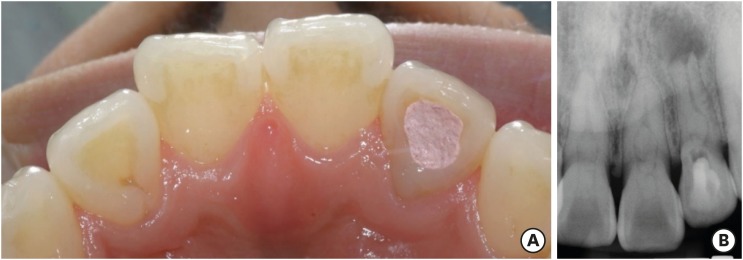
ePub The present report presents a case of dens invaginatus (DI) in a patient with 4 maxillary incisors. A 24-year-old female complained of swelling of the maxillary left anterior region and discoloration of the maxillary left anterior tooth. The maxillary left lateral incisor (tooth #22) showed pulp necrosis and a chronic apical abscess, and a periapical X-ray demonstrated DI on bilateral maxillary central and lateral incisors. All teeth responded to a vitality test, except tooth #22. The anatomic form of tooth #22 was similar to that of tooth #12, and both teeth had lingual pits. In addition, panoramic and periapical X-rays demonstrated root canal calcification, such as pulp stones, in the maxillary canines, first and second premolars, and the mandibular incisors, canines, and first premolars bilaterally. The patient underwent root canal treatment of tooth #22 and non-vital tooth bleaching. After a temporary filling material was removed, the invaginated mass was removed using ultrasonic tips under an operating microscope. The working length was established, and the root canal was enlarged up to #50 apical size and obturated with gutta-percha and AH 26 sealer using the continuous wave of condensation technique. Finally, non-vital bleaching was performed, and the access cavity was filled with composite resin.
-
Citations
Citations to this article as recorded by- The use of three-dimensional-printed guides, static navigation, and bioactive materials to treat bilateral and double dens invaginatus
Parth Patel, Nidhi Bharti, Ankit Arora, C. Nimisha Shah
Saudi Endodontic Journal.2025; 15(2): 207. CrossRef - Endodontic Management of Dens in Dente – A Systematic Review of Case Reports and Case Series
Sanket Dilip Aras, Anamika Chetan Borkar, Sonal Kale, Sayali Maral, Prakriti Jaggi, Shailendra Sonawane
Journal of the International Clinical Dental Research Organization.2024; 16(1): 17. CrossRef - Dens invaginatus of fourteen teeth in a pediatric patient
Momoko Usuda, Tatsuya Akitomo, Mariko Kametani, Satoru Kusaka, Chieko Mitsuhata, Ryota Nomura
Pediatric Dental Journal.2023; 33(3): 240. CrossRef - The Impact of the Preferred Reporting Items for Case Reports in Endodontics (PRICE) 2020 Guidelines on the Reporting of Endodontic Case Reports
Sofian Youssef, Phillip Tomson, Amir Reza Akbari, Natalie Archer, Fayjel Shah, Jasmeet Heran, Sunmeet Kandhari, Sandeep Pai, Shivakar Mehrotra, Joanna M Batt
Cureus.2023;[Epub] CrossRef - Root Maturation of an Immature Dens Invaginatus Despite Unsuccessful Revitalization Procedure: A Case Report and Recommendations for Educational Purposes
Julia Ludwig, Marcel Reymus, Alexander Winkler, Sebastian Soliman, Ralf Krug, Gabriel Krastl
Dentistry Journal.2023; 11(2): 47. CrossRef - Conservative Management of Infraorbital Space Infection Secondary to Type III B Dens Invaginatus: A Case Report
Ashima Goyal, Aditi Kapur, Manoj A Jaiswal, Gauba Krishan, Raja Raghu, Sanjeev K Singh
Journal of Postgraduate Medicine, Education and Research.2022; 56(4): 192. CrossRef
- The use of three-dimensional-printed guides, static navigation, and bioactive materials to treat bilateral and double dens invaginatus
- 2,416 View
- 28 Download
- 6 Crossref


 KACD
KACD


 First
First Prev
Prev


