
Previous issues
- Page Path
- HOME > Browse articles > Previous issues
- Volume 49 (2); May 2024
-


Review Articles
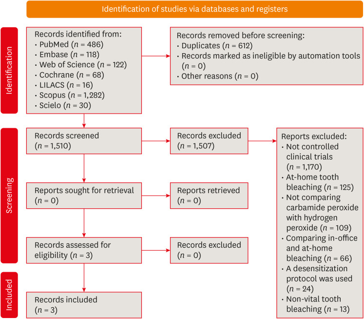
- Can carbamide peroxide be as effective as hydrogen peroxide for in-office tooth bleaching and cause less sensitivity? A systematic review
- Patrick Wesley Marques de Boa, Kaiza de Sousa Santos, Francisca Jennifer Duarte de Oliveira, Boniek Castillo Dutra Borges
- Restor Dent Endod 2024;49(2):e14. Published online March 20, 2024
- DOI: https://doi.org/10.5395/rde.2024.49.e14
-
 Abstract
 PDF
PDF PubReader
 ePub
ePub This study aimed to answer the question through a systematic review: Can carbamide peroxide be as effective as hydrogen peroxide and cause less in-office bleaching sensitivity? A literature survey was performed in PubMed/MEDLINE, Embase, Scopus, ISI Web of Science, and gray literature. Primary clinical trials that compared the efficacy or the in-office bleaching sensitivity between carbamide and hydrogen peroxides were included. The risk of bias was evaluated using the RoB2. The certainty of the evidence was assessed using the GRADE approach. DPI training significantly improved the mean scores of the dental undergraduates from 7.53 in the pre-DPI-training test to 9.01 in the post-DPI-training test (
p < 0.001). After 6 weeks, the mean scores decreased marginally to 8.87 in the retention test (p = 0.563). DPI training increased their confidence level from 5.68 pre-DPI training to 7.09 post-DPI training. The limited evidence suggests that the 37% carbamide peroxide may be similarly effective to the 35% hydrogen peroxide for bleaching teeth in-office and causes less bleaching sensitivity. However, more well-designed split-mouth clinical trials are necessary to strengthen the evidence.-
Citations
Citations to this article as recorded by
- Impact of nanostructured additives in tooth bleaching agents on enhancing color change and reducing side effects: a scoping review
Patrick Wesley Marques de Boa, Kaiza de Sousa Santos, Aleph Matthews da Silva Souza, Arnóbio Antônio da Silva-Júnior, Boniek Castillo Dutra Borges
Clinical Oral Investigations.2025;[Epub] CrossRef - Quantitative and Qualitative Assessment of Enamel Surface Roughness Following High-Concentration Peroxide Bleaching: A Comparative In Vitro Study
Mamnoon Ghafir, Nida Mehmood, Leeza Bharati, Shreya Bhukal, Ritika Sethi, Aanchal Chaudhary, Seema Gupta
Cureus.2025;[Epub] CrossRef - Using violet light during in-office tooth bleaching to enhance the efficacy of carbamide peroxide without increasing bleaching sensitivity: a systematic review and meta-analysis
Mariana Silva de Bessa, Kaiza de Sousa Santos, Patrick Wesley Marques de Boa, Francisca Jennifer Duarte de Oliveira, Bárbara Faria de Sá Barbosa, Boniek Castillo Dutra Borges
Lasers in Medical Science.2025;[Epub] CrossRef - Influence of Different Light-Activated Bleaching Gels on Pulp Chamber Temperature: An In Vitro Study
Mandana Karimi, Elmira Ataee, Ladan Ranjbar Omrani, Mahdi Abbasi, Elham Ahmadi
Avicenna Journal of Dental Research.2024; 16(4): 225. CrossRef
- Impact of nanostructured additives in tooth bleaching agents on enhancing color change and reducing side effects: a scoping review
- 13,610 View
- 211 Download
- 1 Web of Science
- 4 Crossref

- The prevalence of apical periodontitis in patients prior to hematopoietic cell transplantation: a systematic review
- Letícia Tainá de Oliveira Lemes, Carolina Horn Troian-Michel, Theodoro Weissheimer, Marcus Vinicius Reis Só
- Restor Dent Endod 2024;49(2):e22. Published online May 9, 2024
- DOI: https://doi.org/10.5395/rde.2024.49.e22
-
Abstract
PDF
 Supplementary MaterialPubReaderePub
 Objectives This systematic review addressed the question: “What is the prevalence of apical periodontitis in patients prior to hematopoietic cell transplantation?”
Materials and Methods A systematic search was conducted in MEDLINE/PubMed, Cochrane Library, Scopus, Web of Science, Embase, and Grey Literature Report. Eligibility criteria were based on the condition, content, and population strategy: the condition was the radiographic prevalence of apical periodontitis, the content comprised patients scheduled for hematopoietic stem cell transplantation, and the population consisted of adult and pediatric patients. The revised Risk of Bias in Nonrandomized Studies of Exposure tool was used to assess the quality of studies. The Grading Recommendations Assessments, Development, and Evaluation (GRADE) tool was used to assess the quality of evidence.
Results Eight studies were included in this review. The average number of patients with apical periodontitis was 15.65% (range, 2.1%–43.34%). One study was classified as having a very high risk of bias, 1 with a high risk of bias, and 6 with some concern for bias. GRADE analysis showed a very low certainty of evidence. Significant limitations concerning the absence of control over confounding variables were identified.
Conclusions With the caveat of the very low quality of evidence in the studies reviewed, there was a low to moderate prevalence of apical periodontitis in patients prior to undergoing hematopoietic cell transplantation.
- 3,003 View
- 63 Download
Research Articles
- Impact of different agitation methods on smear layer cleaning of mesial canals with accentuated curvature
- Abel Teves Cordova, Murilo Priori Alcalde, Michel Espinosa Klymus, Leonardo Rigoldi Bonjardim, Rodrigo Ricci Vivan, Marco Antonio Hungaro Duarte
- Restor Dent Endod 2024;49(2):e12. Published online March 4, 2024
- DOI: https://doi.org/10.5395/rde.2024.49.e12
-
Abstract
PDFPubReaderePub
Objectives This study evaluated the impact of different methods of irrigant agitation on smear layer removal in the apical third of curved mesial canals of 3 dimensionally (D) printed mandibular molars.
Materials and Methods Sixty 3D-printed mandibular second molars were used, presenting a 70° curvature and a Vertucci type II configuration in the mesial root. A round cavity was cut 2 mm from the apex using a trephine of 2 mm in diameter, 60 bovine dentin disks were made, and a smear layer was formed. The dentin disks had the adaptation checked in the apical third of the teeth with wax. The dentin disks were evaluated in environmental scanning electron microscope before and after the following irrigant agitation methods: G1(PIK Ultrasonic Tip), G2 (Passive Ultrasonic Irrigation with Irrisonic– PUI), G3 (Easy Clean), G4 (HBW Ultrasonic Tip), G5 (Ultramint X Ultrasonic tip), and G6 (conventional irrigation-CI) (
n = 10). All groups were irrigated with 2.5% sodium hypochlorite and 17% ethylenediaminetetraacetic acid.Results All dentin disks were 100% covered by the smear layer before treatment, and all groups significantly reduced the percentage of the smear layer after treatment. After the irrigation protocols, the Ultra-X group showed the lowest coverage percentage, statistically differing from the conventional, PIK, and HBW groups (
p < 0.05). There was no significant difference among Ultramint X, PUI-Irrisonic, and Easy Clean (p > 0.05). None of the agitation methods could remove the smear layer altogether.Conclusions Ultramint X resulted in the most significant number of completely clean specimens.
-
Citations
Citations to this article as recorded by- A new cleaning protocol in minimally invasive endodontic surgery: RUA (“retro irrigant activation”)
Dina Abdellatif, Davide Mancino, Massimo Pisano, Sara De Fontaine, Alfredo Iandolo
Journal of Conservative Dentistry and Endodontics.2025; 28(3): 297. CrossRef - Impact of the use of high-power 810-nm diode laser as monotherapy on the clinical and tomographic success of the treatment of teeth with periapical lesions: an observational clinical study
Fabricio Hinojosa Pedraza, Abel Victor Isidro Teves-Cordova, Murilo Priori Alcalde, Marco Antonio Hungaro Duarte
Restorative Dentistry & Endodontics.2025; 50(2): e15. CrossRef - Smear layer removal comparing conventional irrigation, passive ultrasonic irrigation, EndoActivator System, and a new sonic device (Perfect Clean System) by scanning electron microscopy: An ex vivo study
Bruna Fernanda Alionço Gonçalves, Divya Reddy, Ricardo Machado, Paulo César Soares Júunior, Sérgio Aparecido Ignácio, Douglas Augusto Fernandes Couto, Karine Santos Frasquetti, Vânia Portela Ditzel Westphalen, Everdan Carneiro, Ulisses Xavier da Silva Net
PLOS ONE.2024; 19(12): e0314940. CrossRef
- A new cleaning protocol in minimally invasive endodontic surgery: RUA (“retro irrigant activation”)
- 3,592 View
- 157 Download
- 2 Web of Science
- 3 Crossref
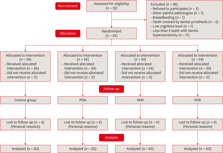
- Single-session associative protocol for dentin hypersensitivity management: a 1-year randomized, blinded clinical study
- Thayna Carolina Zeni, Poliana Maria de Faveri Cardoso, Rafael da Silva Vanolli, Márcio José Mendonça, Julio Katuhide Ueda, Veridiana Camilotti
- Restor Dent Endod 2024;49(2):e15. Published online March 20, 2024
- DOI: https://doi.org/10.5395/rde.2024.49.e15
-
Abstract
PDFPubReaderePub
Objectives This study aimed to establish a single-session associative protocol for non-restorative management of dentin hypersensitivity (DH).
Materials and Methods Twenty-four individuals with DH and a minimum sensitivity level of 4 on the visual analog scale (VAS) were selected. The study was conducted in a split-mouth design, with each participant (
n = 20) having at least 1 affected tooth in all quadrants. The management protocols consisted of control group: universal adhesive, Neural Desensitizing Protocol group: 5% potassium nitrate, Mixed Desensitizing Protocol (PAM) group: 5% sodium fluoride and 5% potassium nitrate, Remineralizing Desensitizing Protocol (PDR) group: surface-partially reacted glass technology photopolymerizable varnish. Evaluations were performed immediately after application, at 1 week, 1 month, 2 months, and 12 months using the VAS sensitivity test.Results The scores were subjected to statistical analysis using the Friedman test (
p < 0.05), Durbin-Conover test (p < 0.05), and Wilcoxon test (p < 0.05). At the 12-month evaluation, all groups showed statistically significant differences compared to the initial assessment. For the evaluation after 12 months, there was a statistically significant difference between the PAM group, the control group, and the PDR group.Conclusions It can be concluded that all groups were effective in controlling DH, but there were significant results in the control group and PDR group. The clinical relevance of this study is to demonstrate that the application of single-session desensitizing protocols can be effective in controlling DH for up to 12 months.
Trial Registration Brazilian Clinical Trials Registry Identifier:
RBR-4r63d7s -
Citations
Citations to this article as recorded by- Mesoporous Bioactive Glass: Preparation, Characterisation, and Emerging Applications in Regenerative Medicine and Dentistry
Bakhtawar Mobeen, Nawshad Muhammad, Minati Choudhury, Ayesha Feroz, Sandleen Feroz
International Dental Journal.2026; 76(2): 109454. CrossRef - Effect of different material protocols on the control of dentin hypersensitivity: a split-mouth randomized controlled clinical trial
Júlia Marques Martins, Maria Fernanda Ferreira Nogueira, Guilherme José Pimentel Lopes de Oliveira, Alexandre Coelho Machado, Paulo César de Freitas Santos Filho, Hugo Lemes Carlo, Carlos José Soares, Gisele Rodrigues da Silva
Clinical Oral Investigations.2026;[Epub] CrossRef - In vivo and in situ evaluation of innovative approaches in dentin hypersensitivity treatment
Heba Abd El-Fattah Mohamed, Dina Ezzeldin Mohamed, Elhassan Hassanein, Heba El-din Salah El-din Hamza
BMC Oral Health.2025;[Epub] CrossRef - Publication trends and scientific profile of clinical trials on universal adhesives in dentistry: A metrics-based review
Aurélio de Oliveira Rocha, Lucas Menezes dos Anjos, Michael Willian Favoreto, Michely Cristina Goebel, Bruno Henriques, Alessandra Reis, Alessandro D. Loguercio, Mariane Cardoso
Journal of Dentistry.2025; 161: 105965. CrossRef - EVALUATION OF PUSH-OUT BOND STRENGTH OF GLASS FIBER POSTS USING DIFFERENT LUTING CEMENTS
Jannah Mohammed, Maha Agha
BULLETIN OF STOMATOLOGY AND MAXILLOFACIAL SURGERY.2025; : 274. CrossRef - EVALUATION OF PUSH-OUT BOND STRENGTH OF GLASS FIBER POSTS USING DIFFERENT LUTING CEMENTS
Jannah Mohammed, Jannah Mohammed
BULLETIN OF STOMATOLOGY AND MAXILLOFACIAL SURGERY.2025; : 274. CrossRef - CLINICAL AND BEHAVIORAL DETERMINANTS OF DENTIN SENSITIVITY AMONG DENTAL STUDENTS: AN INSTITUTIONAL CROSS-SECTIONAL STUDY
Giuseppe Eliseo ALLOCCA, Alexandrina MUNTEAN , Cristian Doru OLTEANU , Sorana Maria BUCUR
Medicine and Materials.2025; 5(2): 73. CrossRef - Desensitizing efficacy of a universal dentin adhesive containing mesoporous bioactive glass on dentin hypersensitivity: a randomized clinical trial with a split-mouth model
Hyun-Jung Kim, Soram Oh, Jiyoung Kwon, Kyoung-Kyu Choi, Ji-Hyun Jang, Duck-Su Kim
Scientific Reports.2024;[Epub] CrossRef
- Mesoporous Bioactive Glass: Preparation, Characterisation, and Emerging Applications in Regenerative Medicine and Dentistry
- 8,147 View
- 169 Download
- 5 Web of Science
- 8 Crossref
- Prevalence of apical periodontitis and quality of root canal treatment in an adult Kuwaiti sub-population: a cross-sectional study
- Abdulrahman A. Alhailaa, Saad A Al-Nazhan, Mazen A Aldosimani
- Restor Dent Endod 2024;49(2):e16. Published online March 22, 2024
- DOI: https://doi.org/10.5395/rde.2024.49.e16
-
Abstract
PDFPubReaderePub
Objectives This cross-sectional study evaluated the prevalence of apical periodontitis (AP) and the technical quality of root canal fillings in an adult Kuwaiti subpopulation using cone-beam computed tomography (CBCT) images.
Materials and Methods Two experienced examiners analyzed 250 CBCT images obtained from Kuwaiti patients aged 15–65 years who attended government dental specialist clinics between January 2019 and September 2020. The assessment followed the radiographic scoring criteria proposed by De Moor for periapical status and the technical quality of root canal filling. Chi-square and Fisher’s exact tests were used for statistical analysis, with significance level set at
p < 0.05.Results Among the 2,762 examined teeth, 191 (6.91%) exhibited radiographic signs of AP, and 176 (6.37%) had undergone root canal filling. AP prevalence in root canal-treated teeth was 32.38%, with a significant difference between males and females. Most of the endodontically treated teeth exhibited adequate root canal filling (71.5%).
Conclusions The study demonstrated a comparable prevalence of AP and satisfactory execution of root canal treatment compared to similar studies in different countries.
-
Citations
Citations to this article as recorded by- RISK FACTORS FOR CHRONIC APICAL PERIODONTITIS ACCORDING TO THE CASE-CONTROL STUDY
N. Bagryantseva
Vrach.2026; : 43. CrossRef - A Retrospective Study of CBCT-Based Detection of Endodontic Failures and Periapical Lesions in a Romanian Cohort
Oana Andreea Diaconu, Lelia Mihaela Gheorghiță, Anca Gabriela Gheorghe, Mihaela Jana Țuculină, Maria Cristina Munteanu, Cătălina Alexandra Iacov, Virginia Maria Rădulescu, Mihaela Ionescu, Adina Andreea Mirea, Carina Alexandra Bănică
Journal of Clinical Medicine.2025; 14(18): 6364. CrossRef
- RISK FACTORS FOR CHRONIC APICAL PERIODONTITIS ACCORDING TO THE CASE-CONTROL STUDY
- 6,980 View
- 109 Download
- 1 Web of Science
- 2 Crossref
- Pulp stones: any relevance with the levels of serum calcium, parathyroid hormone, vitamin D and uric acid
- Ceyda Gürhan, Ercan Saruhan
- Restor Dent Endod 2024;49(2):e17. Published online March 26, 2024
- DOI: https://doi.org/10.5395/rde.2024.49.e17
-
Abstract
PDFPubReaderePub
Objectives This study evaluated the effect of serum calcium, parathyroid hormone (PTH), vitamin D, and uric acid levels on pulp stone formation.
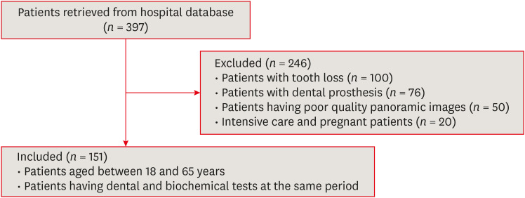
Materials and Methods Patients who were admitted to the Muğla Sıtkı Koçman University, Faculty of Dentistry, Department of Oral and Maxillofacial Radiology for dental complaints were registered. Among these patients, individuals who had routine biochemical tests at the same period in the Outpatient Clinics of Muğla Sıtkı Koçman University Training and Research Hospital were included in the study. The patients with at least 1 pulp stone on panoramic radiographs recorded as the “pulp stone group” while patients without any pulp stones were the “control group”. Demographic data and serum levels of calcium, PTH, vitamin D, and uric acid were retrospectively evaluated in both groups. Student
t -test or Mann-WhitneyU test was used to evaluate the differences between the groups.Results Among 151 patients, dental pulp stone was detected in 53.6% of patients, and 82.7% of these patients were female. Female sex and pulp stone formation were significantly associated (
p = 0.001). The mean age of the pulp stone group was 43.9, while it was 39.9 in the control group, without any significant correlation between age and pulp stone (p > 0.05). Similarly, there were no significant differences in serum levels of PTH, vitamin D, uric acid and calcium between groups (p > 0.05).Conclusions According to the present study, the effect of dental factors rather than systemic factors should be considered primarily in pulp stone formation.
-
Citations
Citations to this article as recorded by- A novel deep learning-based pipeline architecture for pulp stone detection on panoramic radiographs
Ceyda Gürhan, Hasan Yiğit, Selim Yılmaz, Cihat Çetinkaya
Oral Radiology.2025; 41(2): 285. CrossRef - Vitamin D deficiency and oral health: a systematic review of literature
Saida Ziada, Aws Wishahe, Najet Mabrouk, Souad Sahtout
BMC Oral Health.2025;[Epub] CrossRef - Association between pulp stones and systemic diseases: a retrospective study using digital panoramic radiographs in a Turkish population
Buket Beytaş Alğan, Mustafa Murat Koçak, Sibel Koçak, Baran Can Sağlam
BMC Oral Health.2025;[Epub] CrossRef
- A novel deep learning-based pipeline architecture for pulp stone detection on panoramic radiographs
- 5,076 View
- 121 Download
- 4 Web of Science
- 3 Crossref
- Effects of a relined fiberglass post with conventional and self-adhesive resin cement
- Wilton Lima dos Santos Junior, Marina Rodrigues Santi, Rodrigo Barros Esteves Lins, Luís Roberto Marcondes Martins
- Restor Dent Endod 2024;49(2):e18. Published online March 27, 2024
- DOI: https://doi.org/10.5395/rde.2024.49.e18
-
Abstract
PDFPubReaderePub
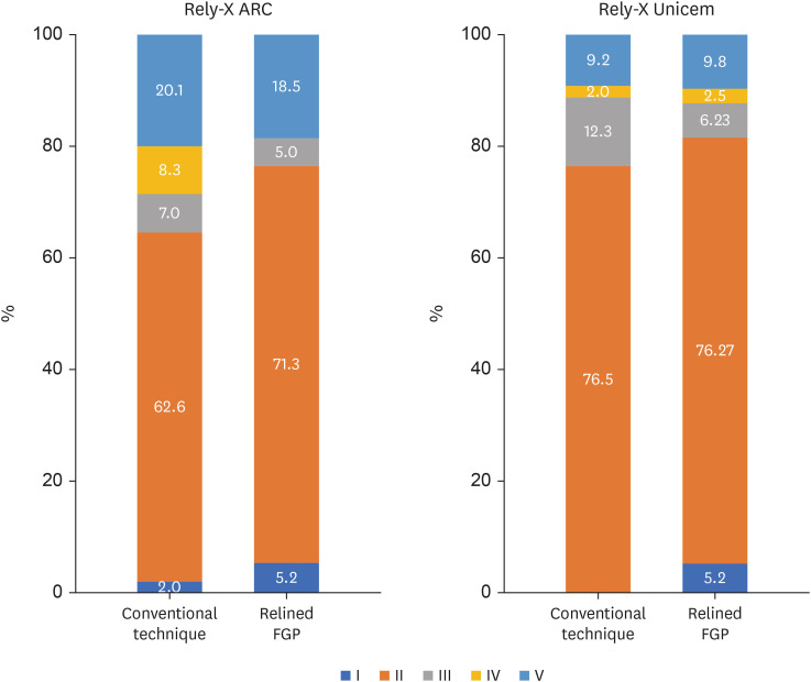
Objectives This study was conducted to evaluate the mechanical properties of relined and non-relined fiberglass posts when cemented to root canal dentin using a conventional dual-cure resin cement or a self-adhesive resin cement.
Materials and Methods Two types of resin cements were utilized: conventional and self-adhesive. Additionally, 2 cementation protocols were employed, involving relined and non-relined fiberglass posts. In total, 72 bovine incisors were cemented and subjected to push-out bond strength testing (
n = 10) followed by failure mode analysis. The cross-sectional microhardness (n = 5) was assessed along the root canal, and interface analyses (n = 3) were conducted using scanning electron microscopy (SEM). Data from the push-out bond strength and cross-sectional microhardness tests were analyzed via 3-way analysis of variance and the Bonferronipost-hoc test (α = 0.05).Results For non-relined fiberglass posts, conventional resin cement exhibited higher push-out bond strength than self-adhesive cement. Relined fiberglass posts yielded comparable results between the resin cements. Type II failure was the most common failure mode for both resin cements, regardless of cementation protocol. The use of relined fiberglass posts improved the cross-sectional microhardness values for both cements. SEM images revealed voids and bubbles in the incisors with non-relined fiberglass posts.
Conclusions Mechanical properties were impacted by the cementation protocol. Relined fiberglass posts presented the highest push-out bond strength and cross-sectional microhardness values, regardless of the resin cement used (conventional dual-cure or self-adhesive). Conversely, for non-relined fiberglass posts, the conventional dual-cure resin cement yielded superior results to the self-adhesive resin cement.
-
Citations
Citations to this article as recorded by- Push-Out Bond Strength of Different Luting Cements Following Post Space Irrigation with 2% Chitosan: An In Vitro Study
Shimaa Rifaat, Ahmed Rahoma, Hind Muneer Alharbi, Sawsan Jamal Kazim, Shrouq Ali Aljuaid, Basmah Omar Alakloby, Faraz A. Farooqi, Noha Taymour
Prosthesis.2025; 7(1): 18. CrossRef
- Push-Out Bond Strength of Different Luting Cements Following Post Space Irrigation with 2% Chitosan: An In Vitro Study
- 6,414 View
- 158 Download
- 1 Web of Science
- 1 Crossref
- Nanoleakage of apical sealing using a calcium silicate-based sealer according to canal drying methods
- Yoon-Joo Lee, Kyung-Mo Cho, Se-Hee Park, Yoon Lee, Jin-Woo Kim
- Restor Dent Endod 2024;49(2):e20. Published online April 19, 2024
- DOI: https://doi.org/10.5395/rde.2024.49.e20

-
Abstract
PDFPubReaderePub
Objectives This study investigated the nanoleakage of root canal obturations using calcium silicate-based sealer according to different drying methods.
Materials and Methods Fifty-two extracted mandibular premolars with a single root canal and straight root were selected for this study. After canal preparation with a nickel-titanium rotary file system, the specimens were randomly divided into 4 groups according to canal drying methods (1: complete drying, 2: blot drying/distilled water, 3: blot drying/NaOCl, 4: aspiration only). The root canals were obturated using a single-cone filling technique with a calcium silicate–based sealer. Nanoleakage was evaluated using a nanoflow device after 24 hours, 1 week, and 1 month. Data were collected twice per second at the nanoscale and measured in nanoliters per second. Data were statistically analyzed using the Kruskal-Wallis and Mann–Whitney
U -tests (p < 0.05).Results The mean flow rate measured after 24 hours showed the highest value among the time periods in all groups. However, the difference in the flow rate between 1 week and 1 month was not significant. The mean flow rate of the complete drying group was the highest at all time points. After 1 month, the mean flow rate in the blot drying group and the aspiration group was not significantly different.
Conclusions Within the limitations of this study, the canal drying method had a significant effect on leakage and sealing ability in root canal obturations using a calcium silicate-based sealer. Thus, a proper drying procedure is critical in endodontic treatment.
-
Citations
Citations to this article as recorded by- Effects of interactions between sealers and irrigants on the physicochemical and surface characteristics of endodontic sealers
Hye-In Kim, Yeon-Ju You, Yemi Kim, Jin-Woo Kim, Young-Eun Jang
Clinical Oral Investigations.2026;[Epub] CrossRef - Assessment of Shear Bond Strength of MTA and ApaCal ART Liners to a Universal Bonding Agent: An In Vitro Study
Angel Kurian, Banibrata Lahiri, Archana A Thomas, Gautham Anilkumar, Fawaz Pullishery, Reshma Raju
The Journal of Contemporary Dental Practice.2026; 27(3): 254. CrossRef
- Effects of interactions between sealers and irrigants on the physicochemical and surface characteristics of endodontic sealers
- 3,610 View
- 134 Download
- 1 Web of Science
- 2 Crossref
Statistical Research Article
- An elaboration on sample size determination for correlations based on effect sizes and confidence interval width: a guide for researchers
- Mohamad Adam Bujang
- Restor Dent Endod 2024;49(2):e21. Published online May 2, 2024
- DOI: https://doi.org/10.5395/rde.2024.49.e21
-
Abstract
PDFPubReaderePub
Objectives This paper aims to serve as a useful guide for sample size determination for various correlation analyses that are based on effect sizes and confidence interval width.
Materials and Methods Sample size determinations are calculated for Pearson’s correlation, Spearman’s rank correlation, and Kendall’s Tau-b correlation. Examples of sample size statements and their justification are also included.
Results Using the same effect sizes, there are differences between the sample size determination of the 3 statistical tests. Based on an empirical calculation, a minimum sample size of 149 is usually adequate for performing both parametric and non-parametric correlation analysis to determine at least a moderate to an excellent degree of correlation with acceptable confidence interval width.
Conclusions Determining data assumption(s) is one of the challenges to offering a valid technique to estimate the required sample size for correlation analyses. Sample size tables are provided and these will help researchers to estimate a minimum sample size requirement based on correlation analyses.
-
Citations
Citations to this article as recorded by- Model Selection Challenges in Non-Stationary Precipitation Estimation: The Role of AIC, BIC, and Covariate Choice
Murat Yegin, Gulsah Karakaya, Elcin Kentel
Water Resources Management.2026;[Epub] CrossRef - Sampling Methods and Sample Size Determination in Clinical Research: An Educational Review
Azzam Zrineh, Maysa Al‐Usta, Abdallah Alwawi
Journal of General and Family Medicine.2026;[Epub] CrossRef -
Phytochemical investigation of
Calophyllum sundaicum
P.F.Stevens: isolation, structure elucidation, and biological potential
Mas Atikah Lizazman, Vivien Yi Mian Jong, Nor Hisam Zamakshshari, Yiizamy Suffian, Mohamad Izwan Bin Ismail, Enis Nadia Md Yusof, Nurr Maria Ulfa Binti Seruji
Natural Product Research.2026; : 1. CrossRef - How Participants Are Selected in Intelligent Vehicle Human-Machine Interaction Usability Evaluation: A Systematic Review of Current Practices
Xiaofang Yuan, Datao Zhou, Yidi Sun, Yu Wu
International Journal of Human–Computer Interaction.2026; : 1. CrossRef - A long-term exploratory study of source code quality issues in open-source Python projects
Liviu-Marian Berciu, Simona Claudia Motogna, Arthur-Jozsef Molnar
Software Quality Journal.2026;[Epub] CrossRef - Apego entre iguales y su relación con la percepción del sexismo en adolescentes
Azucena Prado-Espinoza, Jesus Chavez-Parillo, Rosario Castelo-Collado, Ruth Soto-Yana, Juan Coaquira-Mamani, Magnolia Sierra-Delgado
Revista Estudios Psicológicos.2026; 6(1): 7. CrossRef - Introducing Palliative Care: Family Caregivers’ Knowledge, Exposure, and Preferred Messaging
Elaine Wittenberg, Joy V. Goldsmith, Sierra Forrest, Hanna G. Lee, Eva YN Yuen
American Journal of Hospice and Palliative Medicine®.2026;[Epub] CrossRef - The indirect effect of perfectionism between anxiety, self-esteem, aesthetic pressure, and eating disorder risk in competitive figure skaters
Leiva Fernández Cordero, Carlos Marchena-Giráldez, Alejandra Cruz Corbella, Dra. Elena Ruiz-Sancho
Eating Behaviors.2026; : 102085. CrossRef - Evaluating the reclamation performance of earth block-mortar combinations
Erik Pelicaen, Rafael Novais Passarelli, Veerle Vandersmissen, Elke Knapen
Construction and Building Materials.2026; 529: 146479. CrossRef - Relationship between physical activity and ankle osteoarthritis: Implications for metabolic diseases
Woo Sub Kim, Hee-jin Yang, Ji Hye Choi, Hee Soo Han, Dong Yeon Lee, Kyoung Min Lee, Zulkarnain Jaafar
PLOS One.2026; 21(5): e0348766. CrossRef - Unveiling the Clinical and Biochemical Impact of Vitamin D Deficiency in Indian Rheumatoid Arthritis Patients: A Cross-Sectional Analysis
Shalini AS, Jayashankar CA, Venkataramana Kandi, Venkata Bharat Kumar Pinnelli, Surendra Babu T, Koshy T Sam, Manish GR, Snigdha Reddy, Akshay AS
Cureus.2026;[Epub] CrossRef - Classifying family resilience trajectory of patients undergoing lung cancer surgery: a longitudinal study
Shu-Rui Gao, Jie Zhu, Hui Yan, Hao Xu, Xin-Yue Wu, Gui-Hui Luo, Xu-Ting Li, Man Ye
Supportive Care in Cancer.2026;[Epub] CrossRef - ICT engagement and technostress among Iranian EFL teachers: a person–environment fit perspective
Reza Nejati
Cogent Education.2026;[Epub] CrossRef - Association of Perceived Recovery and Sleep Quality with Shoulder Pain and Functional Limitation in Overhead Athletes: A Cross-Sectional Study
Hafiza Aroofa, Anam Fatima, Anosha Shabbir, Laiba Ehsan, Fatima, Ashley Nudrat, Sarwat Mehmood
The Healer Journal of Physiotherapy and Rehabilitation Sciences.2026; 6(4): 89. CrossRef - Informal learning spaces and student experience: a mixed-methods case study at an Austrian university
Filiz Keser Aschenberger, Christina Ipser, Gregor Radinger, Sonja Brachtl
Frontiers in Education.2026;[Epub] CrossRef - Scale of Alopecia Areata Distress (SAAD‐41): Initial Validation Part 2—Scoring, Preliminary Reference Values and Validity
Kristina Gorbatenko‐Roth, Mattea Trinitapoli, Sarah Wood, Irmina Wallander, Jaime Nugent, Maria Hordinsky
JEADV Clinical Practice.2026;[Epub] CrossRef - AI-assisted Assessment of Software Engineering Coursework: Tool-support and an Empirical Study
Vəhid Gəruslu, Zafar Jafarov, Aytan Mövsümova, Atif Namazov, Niall Hurson, Hüseyn Mirzayev
Journal of Systems and Software.2026; : 113056. CrossRef - Return to work in young and middle-aged colorectal cancer survivors: Factors influencing self-efficacy, fear, resilience, and financial toxicity
Dan Hu, Yue Li, Hua Zhang, Lian-Lian Wang, Wen-Wen Liu, Xin Yang, Ming-Zhao Xiao, Hao-Ling Zhang, Juan Li
World Journal of Gastroenterology.2025;[Epub] CrossRef - Return to work in young and middle-aged colorectal cancer survivors: Factors influencing self-efficacy, fear, resilience, and financial toxicity
Dan Hu, Yue Li, Hua Zhang, Lian-Lian Wang, Wen-Wen Liu, Xin Yang, Ming-Zhao Xiao, Hao-Ling Zhang, Juan Li
World Journal of Gastroenterology.2025;[Epub] CrossRef - Predictive validity of obstacle-crossing test variations in identifying fallers after inpatient rehabilitation for stroke
Prudence Plummer, Megan E. Schliep, Lina Jallad, Ehsan Sinaei, Jody A. Feld, Vicki S. Mercer
Topics in Stroke Rehabilitation.2025; 32(6): 631. CrossRef - Global NDVI-LST Correlation: Temporal and Spatial Patterns from 2000 to 2024
Ehsan Rahimi, Pinliang Dong, Chuleui Jung
Environments.2025; 12(2): 67. CrossRef - Increased functional connectivity of motor regions and dorsolateral prefrontal cortex in musicians with focal hand dystonia
Stine Alpheis, Christopher Sinke, Julian Burek, Tillmann H. C. Krüger, Eckart Altenmüller, Daniel S. Scholz
Journal of Neurology.2025;[Epub] CrossRef - Expanded span of control, leadership and management performance, work-related stress, and job satisfaction among first-line managers: A repeated cross-sectional study
Jonas Svanström, Bernice Skytt, Maria Lindberg, Magnus Lindberg
WORK: A Journal of Prevention, Assessment & Rehabilitation.2025; 81(3): 2952. CrossRef - The Dilemma and Wisdom in Translating p Values: A Collaborative Approach to Strengthening Scientific Validity
Mohamad Adam Bujang, Suyan Tian
BioMed Research International.2025;[Epub] CrossRef - Comparison of Condylar Position Discrepancies Assessed Using an Optical Jaw Tracking System and a Conventional Condylar Position Indicator
Joana Silva, Eugénio Martins, Alberto Canabez, Domingo Martin, Conchita Martin
Prosthesis.2025; 7(2): 40. CrossRef - Examining the link between intensive care unit nurses’ burnout and perceived quality of life: a multicenter cross-sectional study
Hazel Novela Villagracia, Tajah Ali Akhdair, Salwa Abd El Gawad Sallam, Rico William A. Villagracia, Bushra Alshammari, Awatif M. Alrasheeday, Shaimaa Mohamed Nageeb, Lea L. Dando, Odeta A. Nacubuan, Turki Ahmed Alsaif, Sage Mesias Raguindin, Ingrid Jacin
BMC Nursing.2025;[Epub] CrossRef - Microstructural Engineering of Porous Polymethylsilsesquioxane via Solvothermal Synthesis in Diverse Solvents
Stefanie Beatrice Hauser, Gabriella Saraiva, Chiara Hasenfratz, Mengmeng Li, Zahra Mazrouei-Sebdani, Wim J. Malfait, Shanyu Zhao
ACS Applied Materials & Interfaces.2025; 17(17): 25634. CrossRef - Playfulness of Preschool-Aged Children With Autism in a Sensory Integration Room
Sinem Kars, Esra Aki
Clinical Pediatrics.2025; 64(11): 1538. CrossRef - Fish community responses to habitat alteration: Interactions, biomass shifts, and the value of imperfect data
Eric A. Bonk, Robert H. Hanner, Adrienne J. Bartlett, Gerald R. Tetreault
Environmental Biology of Fishes.2025; 108(7): 1047. CrossRef - Osteoprotegerin and its ligands RANKL and TRAIL in falciparum, vivax, and knowlesi malaria
Arya Sheela Nair, John Woodford, Jessica Loughland, Dean Andrew, Kim Piera, Fiona Amante, Timothy William, Matthew J. Grigg, James S. McCarthy, Nicholas M. Anstey, Michelle J. Boyle, Bridget E. Barber
iScience.2025; 28(6): 112768. CrossRef - Text Analysis of Corporate Cryptocurrency Disclosures in Varying Market Conditions
Ramy Elitzur, Wendy Rotenberg
Journal of Alternative Finance.2025; 2(3): 302. CrossRef - Evaluation of the Influence of Intervention Tools Used in Nutrition Education Programs: A Mixed Approach
Luca Muzzioli, Costanza Gimbo, Maria Pintavalle, Silvia Migliaccio, Lorenzo M. Donini
Nutrients.2025; 17(15): 2460. CrossRef - Path Analysis Reveals Plant Pod Number as the Key Trait for Indirect Selection in Segregating Generations For Pigeonpea Grain Yield
Carlos Antonio Fernandes Santos, Antonio Elton da Silva Costa
Revista de Gestão Social e Ambiental.2025; 19(8): e013079. CrossRef - Analysis of the current status of attitudes toward aging and its influencing factors in elderly maintenance hemodialysis patients in remote areas: a cross-sectional study
Hao-jie Zeng, Zheng-juan Shi, Mei-ying Shen, Sheng-jing Li, Xiang Peng
Geriatric Nursing.2025; 65: 103553. CrossRef - Exploratory Data Analysis of a North American Whole Building Life Cycle Assessment datasets
Yang Shen, Brad Benke, Milad Ashtiani, Monica Huang, Kathrina Simonen
Building and Environment.2025; 286: 113655. CrossRef - Dengue disease severity in humans is augmented by waning Japanese encephalitis virus immunity
Sidharth Malhotra, Birendra P. Gupta, Surendra Uranw, Chinmay Kumar Mantri, Abhay P.S. Rathore, Ashley L. St. John
Science Translational Medicine.2025;[Epub] CrossRef - Investigation of Anthropogenic and Emerging Contaminants in Sinkholes (Cenotes) of the Great Mayan Aquifer, Yucatán Peninsula
Sarah Kopczynski, Rayna Nolen, David Hala, Fernanda Lases-Hernández, Wendy Escobedo-Hinojosa, Flor Arcega-Cabrera, Ismael Oceguera-Vargas, Antonietta Quigg
Archives of Environmental Contamination and Toxicology.2025; 89(3): 279. CrossRef - Bahasa Indonesia version of Weight Stigma Exposure Inventory (WeSEI): Translation and validation among young adults
Kamolthip Ruckwongpatr, Jian-An Su, I-Hua Chen, Nadia Bevan, Ira Nurmala, Muthmainnah Muthmainnah, Lutfi Agus Salim, Asma Nadia, Musheer A. Aljaberi, Mark D. Griffiths, Chung-Ying Lin
Acta Psychologica.2025; 261: 105748. CrossRef - Clinician-Caregiver Engagement in Older Adult Care. Development of a Validated Caregiver Experience Survey to Inform the Optimization of the Caregiver Role
Ronaye T Gilsenan, Rhonda E Schwartz, Iris A Gutmanis
Journal of Patient Experience.2025;[Epub] CrossRef - Evaluating the quantity and spatial density of macrophage-like cells in patients with retinal vascular disease and healthy subjects via non-invasive retinal imaging
Farhad Ghaseminejad, Thomas J. van Rijssen, Parsa Khatami, Pedro L. Rissoli, Ricky Chen, Yudan Chen, Brendan Tao, Myeong Jin Ju, Faisal Beg, Eduardo V. Navajas
International Journal of Retina and Vitreous.2025;[Epub] CrossRef - Sperm DNA Fragmentation in Normozoospermic Men Is Associated with Blastocyst Formation and Quality in Conventional In Vitro Fertilization
Yusaku Mori, Linji Chen, Shogo Nishii, Miwa Sakamoto, Makoto Ohara, Akihiko Sekizawa, Sho-Ichi Yamagishi
Journal of Clinical Medicine.2025; 14(24): 8892. CrossRef - The Impact of Depression on Defense Mechanisms in Adults: The Moderating Role of Attachment Style
Andra-Iuliana Tanase, Amelia-Damiana Trifu, Simona Trifu
Behavioral Sciences.2025; 16(1): 57. CrossRef - The Relationship Between Emotional Intelligence and Self-Compassion Among Medical Students
Evelina Linkevičiūtė, Dalia Antinienė
Baltic Journal of Sport and Health Sciences.2025; 4(137): 4. CrossRef - The Role of the Basophil Activation Test in the Diagnosis of Drug-Induced Anaphylaxis
Maria Czarnobilska, Małgorzata Bulanda, Ewa Czarnobilska, Wojciech Dyga, Marcel Mazur
Diagnostics.2024; 14(18): 2036. CrossRef - Food insecurity impacts diet quality and adherence to the gluten‐free diet in youth with celiac disease
Xinyi Wang, Sven Anders, Zhiqian Jiang, Marcia Bruce, Dominica Gidrewicz, Margaret Marcon, Justine M. Turner, Diana R. Mager
Journal of Pediatric Gastroenterology and Nutrition.2024; 79(6): 1180. CrossRef - Fuel Load Models for Different Tree Vegetation Types in Sichuan Province Based on Machine Learning
Hongrong Wang, Haoquan Chen, Hanmin Sheng, Kai Chen, Chen Dong, Zhiqiang Min
Forests.2024; 16(1): 42. CrossRef
- Model Selection Challenges in Non-Stationary Precipitation Estimation: The Role of AIC, BIC, and Covariate Choice
- 20,574 View
- 581 Download
- 38 Web of Science
- 46 Crossref
Case Report
- Garre’s osteomyelitis of the mandible managed by nonsurgical re-endodontic treatment
- Heegyun Kim, Jiyoung Kwon, Hyun-Jung Kim, Soram Oh, Duck-Su Kim, Ji-Hyun Jang
- Restor Dent Endod 2024;49(2):e13. Published online March 18, 2024
- DOI: https://doi.org/10.5395/rde.2024.49.e13
-
Abstract
PDFPubReaderePub
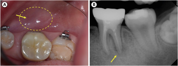
Chronic osteomyelitis with proliferative periostitis, known as Garre’s osteomyelitis, is a type of osteomyelitis characterized by a distinctive gross thickening of the periosteum of bones. Peripheral reactive bone formation can be caused by mild irritation or infection. Garre’s osteomyelitis is usually diagnosed in children and young adults, and the mandible is more affected than the maxilla. The following is a case report of a 12-year-old female patient with Garre’s osteomyelitis of the mandible due to an infection of a root canal-treated tooth. Without surgical intervention, the patient’s symptoms were relieved through nonsurgical root canal re-treatment with long-term calcium hydroxide placement. A cone-beam computed tomography image obtained 6 months after treatment completion displayed complete healing of the periapical lesion and resolution of the peripheral reactive buccal bone. Due to the clinical features of Garre's osteomyelitis, which is characterized by thickening of the periosteum, it can be mistaken for other diseases such as fibrous dysplasia. It is important to correctly diagnose Garre's osteomyelitis based on its distinctive clinical features to avoid unnecessary surgical intervention, and it can lead to minimally invasive treatment options.
-
Citations
Citations to this article as recorded by- Endodontic Intervention in Chronic Osteomyelitis With Proliferative Periostitis: A Rare Case Report and Scoping Review
Gabriel Lima Braz, Ana Paula Neutzling Gomes, Lisandrea Rocha Schardosim, Nadia de Souza Ferreira, Jose Francisco Gomez-Sosa
Case Reports in Dentistry.2026;[Epub] CrossRef - How Clinical and Radiological Findings in Chronic Mandibular Osteomyelitis Do Not Always Correlate: Diagnostic Dilemmas in Dental-Related Bone Inflammations
Kamil Nelke, Ömer Uranbey, Ece Gülbağ, Büşra Ekinci, Burcu Gürsoytrak, Angela Rosa Caso, Michał Gontarz, Maciej Janeczek, Piotr Kuropka, Maciej Dobrzyński
Diagnostics.2026; 16(10): 1427. CrossRef - Focal osteomyelitis with proliferative periostitis
Zarah Yakoob
South African Dental Journal.2025; 79(09): 508. CrossRef - Garré’s osteomyelitis of the mandible in an adolescent: a case report
Wiem Feki, Imen Haddar, Marwa Bahloul, Zeineb Mnif, Thouraya Kammoun, Ines Maaloul
Journal of Medical Case Reports.2025;[Epub] CrossRef - Garré’s Chronic Sclerosing Osteomyelitis: An Overview of Clinical and Radiologic Features
Mohamed Fadil, Ayman Farouki, Rachida Saouab, Hassan En-nouali, Jamal El Fenni, Zakariya Toufga
Oxford Medical Case Reports.2025;[Epub] CrossRef
- Endodontic Intervention in Chronic Osteomyelitis With Proliferative Periostitis: A Rare Case Report and Scoping Review
- 11,168 View
- 239 Download
- 4 Web of Science
- 5 Crossref
Letter to the Editor
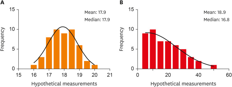
- It is normal to be “not-normal”: reporting of correct descriptive statistics in dental research
- Snigdho Das
- Restor Dent Endod 2024;49(2):e19. Published online April 12, 2024
- DOI: https://doi.org/10.5395/rde.2024.49.e19
- 2,110 View
- 48 Download
 First
First Prev
Prev

