-
Color discrepancy of single-shade composites at different distances from the interface measured using cell phone images
-
Márcia Luciana Carregosa Santana, Gabriella de Jesus Santos Livi, André Luis Faria-e-Silva
-
Restor Dent Endod 2024;49(1):e7. Published online January 24, 2024
-
DOI: https://doi.org/10.5395/rde.2024.49.e7
-
-
 Abstract Abstract
 PDF PDF PubReader PubReader ePub ePub
- Objectives
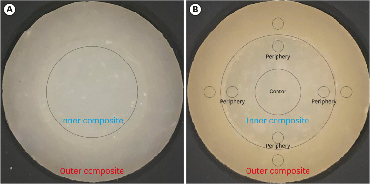
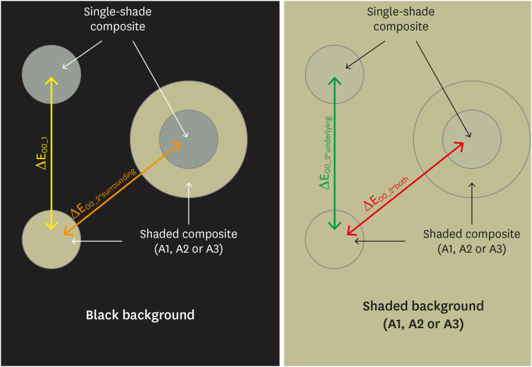
This study aimed to evaluate the impact of substrate color and interface distance on the color adjustment of 2 single-shade composites, Vittra APS Unique and Charisma Diamond One. Materials and MethodsDual disc-shaped specimens were created using Vittra APS Unique or Charisma Diamond One as the center composite, surrounded by shaded composites (A1 or A3). Color measurements were taken with a spectrophotometer against a gray background, recording the color coordinates in the CIELAB color space. Illumination with a light-correcting device and image acquisition using a polarizing filter-equipped cell phone were performed on specimens over the same background. Image processing software was used to measure the color coordinates in the center and periphery of the inner composite and in the outer composite. The color data were then converted to CIELAB coordinates and adjusted using data from the spectrophotometer. Color differences (ΔE00) between the center/periphery of single-shade and outer composites were calculated, along with color changes in single-shade composites caused by different outer composites. Color differences for the inner composites surrounded by A1 and A3 were also calculated. Data were analyzed using repeated-measures analysis of variance (α = 0.05). ResultsThe results showed that color discrepancies were lowest near the interface and when the outer composite was whiter (A1). Additionally, Charisma Diamond One exhibited better color adjustment ability than Vittra APS Unique. ConclusionsColor discrepancies between the investigated single-shade composites diminished towards the interface with the surrounding composite, particularly when the latter exhibited a lighter shade.
-
Citations
Citations to this article as recorded by  - Evaluation of color stability in single-shade composite resins using spectrophotometer and cross-polarized mobile photography
Hatice Tepe, Ozge Celiksoz, Batu Can Yaman
BMC Oral Health.2025;[Epub] CrossRef
-
288
View
-
20
Download
-
1
Web of Science
-
1
Crossref
-
Effects of surrounding and underlying shades on the color adjustment potential of a single-shade composite used in a thin layer
-
Mariana Silva Barros, Paula Fernanda Damasceno Silva, Márcia Luciana Carregosa Santana, Rafaella Mariana Fontes Bragança, André Luis Faria-e-Silva
-
Restor Dent Endod 2023;48(1):e7. Published online December 29, 2022
-
DOI: https://doi.org/10.5395/rde.2023.48.e7
-
-
 Abstract Abstract
 PDF PDF PubReader PubReader ePub ePub
- Objectives
This study aimed to evaluate the surrounding and underlying shades’ effect on the color adjustment potential (CAP) of a single-shade composite used in a thin layer. Materials and MethodsCylinder specimens (1.0 mm thick) were built with the Vittra APS Unique composite, surrounded (dual specimens) or not (simple specimens) by a control composite (shade A1, A2, or A3). Simple specimens were also built only with the control composites. Each specimen’s color was measured against white and black backgrounds or the simple control specimens with a spectrophotometer (CIELAB system). The whiteness index for dentistry (WID) and translucency parameters (TP00) were calculated for simple specimens. Differences (ΔE00) in color between the simple/dual specimens and the controls were calculated. The CAP was calculated based on the ratios between data from simple and dual specimens. ResultsThe Vittra APS Unique composite showed higher WID and TP00 values than the controls. The highest values of ΔE00 were observed among simple specimens. The color measurements of Vittra APS Unique (simple or dual) against the control specimens presented the lowest color differences. Only surrounding the single-shade composite with a shaded composite barely impacted the ΔE00. The highest CAP values were obtained using a shaded composite under simple or dual specimens. ConclusionsThe CAP of Vittra APS Unique was strongly affected by the underlying shade, while surrounding this composite with a shaded one barely affected its color adjustment.
-
Citations
Citations to this article as recorded by  - At‐Home and In‐Office Bleaching Protocols on the Color Match of Restorations Made With Single‐Shade Composites
Luciana Vasconcelos Ramos, Dayana Fernandes Rocha Aparicio, André Luis Faria‐e‐Silva, Maíra do Prado, Andréa Vaz Braga Pintor, Marcela Baraúna Magno
Journal of Esthetic and Restorative Dentistry.2025;[Epub] CrossRef - Evaluation of color matching of three single-shade composites employing simulated 3D printed cavities with different thicknesses using CIELAB and CIEDE2000 color difference formulae
Engin Kariper, Aylin Cilingir
REVIEWS ON ADVANCED MATERIALS SCIENCE.2025;[Epub] CrossRef - Influence of cavity wall thickness on the color adjustment potential of single-shade resin composites
Fabrício Luscino Alves de Castro, Letícia Brandão Durand
The Journal of the American Dental Association.2024; 155(7): 605. CrossRef - Assessing color mismatch in single-shade composite resins for enamel replacement
Rafaella Mariana Fontes de Bragança, Diana Leyva Del Rio, Luiz Alves Oliveira-Neto, William Michael Johnston
The Journal of Prosthetic Dentistry.2024; 132(3): 613.e1. CrossRef - Color discrepancy of single-shade composites at different distances from the interface measured using cell phone images
Márcia Luciana Carregosa Santana, Gabriella de Jesus Santos Livi, André Luis Faria-e-Silva
Restorative Dentistry & Endodontics.2024;[Epub] CrossRef - Is It Possible for Single-shade Composites to Mimic the Color, Lightness, Chroma, and Hue of Other Single-shade Composites? An In Vitro Study
M Buldur, G Ayan
Operative Dentistry.2024; 49(6): 691. CrossRef - Color evaluation of a one-shade used for restoration of non-carious cervical lesions: an equivalence randomized clinical trial
Michael Willian Favoreto, Amanda de Oliveira de Miranda, Thalita P. Matos, Andrea dos Santos de Castro, Mylena de Abreu Cardoso, Julia Beatriz, Jenny Collantes-Acuña, Alessandra Reis, Alessandro Dourado Loguercio
BMC Oral Health.2024;[Epub] CrossRef - Influence of Thickness on the Translucency Parameter and Whiteness Index of Single-Shade Resin Composites
Ö Yağcı, M Fidan
Operative Dentistry.2024; 49(2): 189. CrossRef - A Comparative Study of the Sensitivity and Specificity of the Ishihara Test With Various Displays
Thomas Klinke, Wolfgang Hannak, Klaus Böning, Holger Jakstat
International Dental Journal.2024; 74(4): 892. CrossRef - Color match evaluation using instrumental method for three single-shade resin composites before and after in-office bleaching
Aylin Cilingir, Engin Kariper
REVIEWS ON ADVANCED MATERIALS SCIENCE.2023;[Epub] CrossRef - The role of interface distance and underlying substrate on the color adjustment potential of single‐shade composites
Gabriella Jesus Santos de Livi, Tauan Rosa Santana, Rafaella Mariana Fontes Bragança, Rosa Maria Viana de Bragança Garcez, André Luis Faria‐e‐Silva
Journal of Esthetic and Restorative Dentistry.2023; 35(8): 1279. CrossRef
-
473
View
-
23
Download
-
10
Web of Science
-
11
Crossref
-
The effectiveness of the supplementary use of the XP-endo Finisher on bacteria content reduction: a systematic review and meta-analysis
-
Ludmila Smith de Jesus Oliveira, Rafaella Mariana Fontes de Bragança, Rafael Sarkis-Onofre, André Luis Faria-e-Silva
-
Restor Dent Endod 2021;46(3):e37. Published online June 18, 2021
-
DOI: https://doi.org/10.5395/rde.2021.46.e37
-
-
 Abstract Abstract
 PDF PDF Supplementary Material Supplementary Material PubReader PubReader ePub ePub
- Objectives
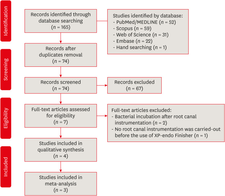
This systematic review evaluated the efficacy of the supplementary use of the XP-endo Finisher on bacteria content reduction in the root canal system. Materials and MethodsIn-vitro studies evaluating the use of the XP-endo Finisher on bacteria content were searched in four databases in July 2020. Two authors independently screened the studies for eligibility. Data were extracted, and risk of bias was assessed. Data were meta-analyzed by using random-effects model to compare the effect of the supplementary use (experimental) or not (control) of the XP-endo Finisher on bacteria counting reduction, and results from different endodontic protocols were combined. Four studies met the inclusion criteria while 1 study was excluded from the meta-analysis due to its high risk of bias and outlier data. The 3 studies that made it to the meta-analysis had an unclear risk of bias for at least one criterion. ResultsNo heterogeneity was observed among the results of the studies included in the meta-analysis. The study excluded from the meta-analysis assessing the bacteria counting deep in the dentin demonstrated further bacteria reduction upon the use of the XP-endo Finisher. ConclusionsThis systematic review found no evidence supporting the supplementary use of the XP-endo Finisher on further bacteria counting the reduction in the root canal.
-
Citations
Citations to this article as recorded by  - Characteristics and Effectiveness of XP‐Endo Files and Systems: A Narrative Review
Sarah M. Alkahtany, Rana Alfadhel, Aseel AlOmair, Sarah Bin Durayhim, Kee Y. Kum
International Journal of Dentistry.2024;[Epub] CrossRef - Impact XP-endo finisher on the 1-year follow-up success of posterior root canal treatments: a randomized clinical trial
Ludmila Smith de Jesus Oliveira, Fabricio Eneas Diniz de Figueiredo, Janaina Araújo Dantas, Maria Amália Gonzaga Ribeiro, Carlos Estrela, Manoel Damião Sousa-Neto, André Luis Faria-e-Silva
Clinical Oral Investigations.2023; 27(12): 7595. CrossRef - Comparative analysis of the effectiveness of modern irrigants activation techniques in the process of mechanical root canal system treatment (Literature review)
Anatoliy Potapchuk, Vasyl Almashi, Arsenii Horzov, Victor Buleza
InterConf.2023; (34(159)): 200. CrossRef - Methodological quality assessment criteria for the evaluation of laboratory‐based studies included in systematic reviews within the specialty of Endodontology: A development protocol
Venkateshbabu Nagendrababu, Paul V. Abbott, Christos Boutsioukis, Henry F. Duncan, Clovis M. Faggion, Anil Kishen, Peter E. Murray, Shaju Jacob Pulikkotil, Paul M. H. Dummer
International Endodontic Journal.2022; 55(4): 326. CrossRef
-
287
View
-
10
Download
-
3
Web of Science
-
4
Crossref
-
Color assessment of resin composite by using cellphone images compared with a spectrophotometer
-
Rafaella Mariana Fontes de Bragança, Rafael Ratto Moraes, André Luis Faria-e-Silva
-
Restor Dent Endod 2021;46(2):e23. Published online April 5, 2021
-
DOI: https://doi.org/10.5395/rde.2021.46.e23
-
-
 Abstract Abstract
 PDF PDF PubReader PubReader ePub ePub
- Objectives
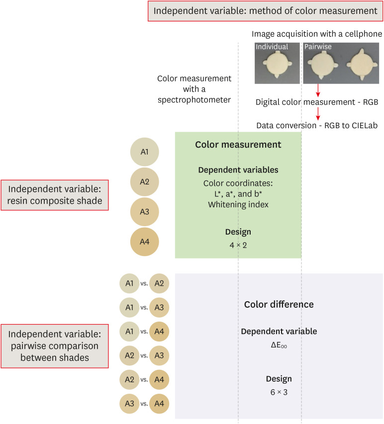
This study assessed the reliability of digital color measurements using images of resin composite specimens captured with a cellphone. Materials and MethodsThe reference color of cylindrical specimens built-up with the use of resin composite (shades A1, A2, A3, and A4) was measured with a portable spectrophotometer (CIELab). Images of the specimens were obtained individually or pairwise (compared shades in the same photograph) under standardized parameters. The color of the specimens was measured in the images using RGB system and converted to CIELab system using image processing software. Whiteness index (WID) and color differences (ΔE00) were calculated for each color measurement method. For the cellphone, the ΔE00 was calculated between the pairs of shades in separate images and in the same image. Data were analyzed using 2-way repeated-measures analysis of variance (α = 0.05). Linear regression models were used to predict the reference ΔE00 values of those calculated using color measured in the images. ResultsImages captured with the cellphone resulted in different WID values from the spectrophotometer only for shades A3 and A4. No difference to the reference ΔE00 was observed when individual images were used. In general, a similar ranking of ΔE00 among resin composite shades was observed for all methods. Stronger correlation coefficients with the reference ΔE00 were observed using individual than pairwise images. ConclusionsThis study showed that the use of cellphone images to measure the color difference seems to be a feasible alternative providing outcomes similar to those obtained with the spectrophotometer.
-
Citations
Citations to this article as recorded by  - Evaluation of color stability in single-shade composite resins using spectrophotometer and cross-polarized mobile photography
Hatice Tepe, Ozge Celiksoz, Batu Can Yaman
BMC Oral Health.2025;[Epub] CrossRef - Color discrepancy of single-shade composites at different distances from the interface measured using cell phone images
Márcia Luciana Carregosa Santana, Gabriella de Jesus Santos Livi, André Luis Faria-e-Silva
Restorative Dentistry & Endodontics.2024;[Epub] CrossRef - How the Translucency and Color Stability of Single-Shade Universal Resin Composites Are Affected by Coffee?
Büşra Özdemir, Betül Kübra Kurucu Karadeniz, Seyit Bilal Özdemir, Ömer Akbulut
Current Research in Dental Sciences.2024; 34(4): 270. CrossRef - Color Image Expression through CIE L*a*b* System in Foods
Hyun-Woong Choi, Seong-Eun Park, Hong-Seok Son
Journal of the Korean Society of Food Science and Nutrition.2023; 52(2): 223. CrossRef - Comparative Evaluation of VITA Shade Guide and Various Composite Shades Using Spectrophotometer, Digital Single-lens Reflex, and Cellphone: An In Vitro Study
Aman Verma, Sonali Taneja, Surabhi Ghosh
World Journal of Dentistry.2023; 14(9): 803. CrossRef - Comparison of instrumental methods for color change assessment of Giomer resins
Luiza de Almeida Queiroz Ferreira, Rogéli Tibúrcio Ribeiro da Cunha Peixoto, Cláudia Silami de Magalhães, Tassiana Melo Sá, Monica Yamauti, Francisca Daniele Moreira Jardilino
Restorative Dentistry & Endodontics.2022;[Epub] CrossRef
-
383
View
-
7
Download
-
2
Web of Science
-
6
Crossref
-
Effects of solvent volatilization time on the bond strength of etch-and-rinse adhesive to dentin using conventional or deproteinization bonding techniques
-
José Aginaldo de Sousa Júnior, Márcia Luciana Carregosa Santana, Fabricio Eneas Diniz de Figueiredo, André Luis Faria-e-Silva
-
Restor Dent Endod 2015;40(3):202-208. Published online March 17, 2015
-
DOI: https://doi.org/10.5395/rde.2015.40.3.202
-
-
 Abstract Abstract
 PDF PDF PubReader PubReader ePub ePub
- Objectives
This study determined the effect of the air-stream application time and the bonding technique on the dentin bond strength of adhesives with different solvents. Furthermore, the content and volatilization rate of the solvents contained in the adhesives were also evaluated. Materials and MethodsThree adhesive systems with different solvents (Stae, SDI, acetone; XP Bond, Dentsply De Trey, butanol; Ambar, FGM, ethanol) were evaluated. The concentrations and evaporation rates of each adhesive were measured using an analytical balance. After acid-etching and rinsing, medium occlusal dentin surfaces of human molars were kept moist (conventional) or were treated with 10% sodium hypochlorite for deproteinization. After applying adhesives over the dentin, slight air-stream was applied for 10, 30 or 60 sec. Composite cylinders were built up and submitted to shear testing. The data were submitted to ANOVA and Tukey's test (α = 0.05). ResultsStae showed the highest solvent content and Ambar the lowest. Acetone presented the highest evaporation rate, followed by butanol. Shear bond strengths were significantly affected only by the factors of 'adhesive' and 'bonding technique' (p < 0.05), while the factor 'duration of air-stream' was not significant. Deproteinization of dentin increased the bond strength (p < 0.05). Stae showed the lowest bond strength values (p < 0.05), while no significant difference was observed between XP Bond and Ambar. ConclusionsDespite the differences in content and evaporation rate of the solvents, the duration of air-stream application did not affect the bond strength to dentin irrespective of the bonding technique.
-
Citations
Citations to this article as recorded by  - Effect of adhesive air-drying time on bond strength to dentin: A systematic review and meta-analysis
Mohamed M. Awad, Ali Alrahlah, Jukka P. Matinlinna, Hamdi Hosni Hamama
International Journal of Adhesion and Adhesives.2019; 90: 154. CrossRef
-
195
View
-
1
Download
-
1
Crossref
-
Bond strength of self-adhesive resin cements to composite submitted to different surface pretreatments
-
Victor Hugo dos Santos, Sandro Griza, Rafael Ratto de Moraes, André Luis Faria-e-Silva
-
Restor Dent Endod 2014;39(1):12-16. Published online January 20, 2014
-
DOI: https://doi.org/10.5395/rde.2014.39.1.12
-
-
 Abstract Abstract
 PDF PDF PubReader PubReader ePub ePub
- Objectives
Extensively destroyed teeth are commonly restored with composite resin before cavity preparation for indirect restorations. The longevity of the restoration can be related to the proper bonding of the resin cement to the composite. This study aimed to evaluate the microshear bond strength of two self-adhesive resin cements to composite resin. Materials and MethodsComposite discs were subject to one of six different surface pretreatments: none (control), 35% phosphoric acid etching for 30 seconds (PA), application of silane (silane), PA + silane, PA + adhesive, or PA + silane + adhesive (n = 6). A silicone mold containing a cylindrical orifice (1 mm2 diameter) was placed over the composite resin. RelyX Unicem (3M ESPE) or BisCem (Bisco Inc.) self-adhesive resin cement was inserted into the orifices and light-cured. Self-adhesive cement cylinders were submitted to shear loading. Data were analyzed by two-way ANOVA and Tukey's test (p < 0.05). ResultsIndependent of the cement used, the PA + Silane + Adhesive group showed higher microshear bond strength than those of the PA and PA + Silane groups. There was no difference among the other treatments. Unicem presented higher bond strength than BisCem for all experimental conditions. ConclusionsPretreatments of the composite resin surface might have an effect on the bond strength of self-adhesive resin cements to this substrate.
-
Citations
Citations to this article as recorded by  - An Innovative Method of Permanent Retention on Veneered Crowns
Yugandhar Garlapati, Sampath Krishna Veni, Jashva Vamsi Kogila, Polisetty Siva Krishna, K. N. Anand Kumar
Journal of Indian Orthodontic Society.2025;[Epub] CrossRef - Influence of mechanochemical treatment and oxygen inhibited layer on the adhesion of self-adhesive resin cement to bulk-fill composite resin
Sreya Dutta, Samikhya Priyadarsani Sahu, Anushka Arora, Srikant Natarajan, Abhishek Parolia, Manuel Thomas
Cumhuriyet Dental Journal.2024; 27(2): 79. CrossRef - Substrate Rigidity Effect on CAD/CAM Restorations at Different Thicknesses
César Rogério Pucci, Ana Paula Valente Pinho Mafetano, Alexandre Luiz Souto Borges, Guilherme Schmitt de Andrade, Amanda Maria de Oliveira Dal Piva, Cornelis J. Kleverlaan, João Paulo Mendes Tribst
European Journal of Dentistry.2023; 17(04): 1020. CrossRef - Microgap Formation between a Dental Resin-Matrix Computer-Aided Design/Computer-Aided Manufacturing Ceramic Restorative and Dentin after Various Surface Treatments and Artificial Aging
Alexandros Galanopoulos, Dimitrios Dionysopoulos, Constantinos Papadopoulos, Petros Mourouzis, Kosmas Tolidis
Applied Sciences.2023; 13(4): 2335. CrossRef - Dental Luting Cements: An Updated Comprehensive Review
Artak Heboyan, Anna Vardanyan, Mohmed Isaqali Karobari, Anand Marya, Tatevik Avagyan, Hamid Tebyaniyan, Mohammed Mustafa, Dinesh Rokaya, Anna Avetisyan
Molecules.2023; 28(4): 1619. CrossRef - Effect of full-step versus simplified resin cement luting strategies on the push-out bond strength of indirect resin composite restorations bonded to dentin
Bianca Cristina Dantas da Silva, Isabelle Helena Gurgel de Carvalho, Taciana Emília Leite Vila-Nova, Gabriela Monteiro de Araújo, Boniek Castillo Dutra Borges, Marília Regalado Galvão Rabelo Caldas, Isauremi Vieira de Assunção, Mutlu Özcan, Rodrigo Othávi
Journal of Adhesion Science and Technology.2023; 37(24): 3552. CrossRef - Effect of various polymerization protocols on the cytotoxicity of conventional and self-adhesive resin-based luting cements
Ece Irem Oguz, Ufuk Hasanreisoglu, Sadullah Uctasli, Mutlu Özcan, Mehmet Kiyan
Clinical Oral Investigations.2020; 24(3): 1161. CrossRef - Repair bond strength of resin composite to three aged CAD/CAM blocks using different repair systems
Pinar Gul, Latife Altınok-Uygun
The Journal of Advanced Prosthodontics.2020; 12(3): 131. CrossRef - Evaluation of the Surface Characteristics of Dental CAD/CAM Materials after Different Surface Treatments
Konstantinos Papadopoulos, Kimon Pahinis, Kyriaki Saltidou, Dimitrios Dionysopoulos, Effrosyni Tsitrou
Materials.2020; 13(4): 981. CrossRef - Adhesive Systems Used in Indirect Restorations Cementation: Review of the Literature
Cristian Abad-Coronel, Belén Naranjo, Pamela Valdiviezo
Dentistry Journal.2019; 7(3): 71. CrossRef - Effects of different etching methods and bonding procedures on shear bond strength of orthodontic metal brackets applied to different CAD/CAM ceramic materials
S. Kutalmış Buyuk, Ahmet Serkan Kucukekenci
The Angle Orthodontist.2018; 88(2): 221. CrossRef - Ceramic repairs with resins: silanization protocols
Teresa Cristina Vasconcelos dos Santos
Journal of Dental Health, Oral Disorders & Therapy.2018;[Epub] CrossRef - Influence of different surface treatments on bond strength of novel CAD/CAM restorative materials to resin cement
Meltem Bektaş Kömürcüoğlu, Elçin Sağırkaya, Ayça Tulga
The Journal of Advanced Prosthodontics.2017; 9(6): 439. CrossRef - Adhesive bonding to polymer infiltrated ceramic
Judith SCHWENTER, Fredy SCHMIDLI, Roland WEIGER, Jens FISCHER
Dental Materials Journal.2016; 35(5): 796. CrossRef - Orthodontic bracket bonding to glazed full-contour zirconia
Ji-Young Kwak, Hyo-Kyung Jung, Il-Kyung Choi, Tae-Yub Kwon
Restorative Dentistry & Endodontics.2016; 41(2): 106. CrossRef - Effect of Silanization on Microtensile Bond Strength of Different Resin Cements to a Lithium Disilicate Glass Ceramic
Cristina Parise Gré, Renan C de Ré Silveira, Shizuma Shibata, Carlo TR Lago, Luiz CC Vieira
The Journal of Contemporary Dental Practice.2016; 17(2): 149. CrossRef - Effects of air abrasion with alumina or glass beads on surface characteristics of CAD/CAM composite materials and the bond strength of resin cements
ARAO Nobuaki, YOSHIDA Keiichi, SAWASE Takashi
Journal of Applied Oral Science.2015; 23(6): 629. CrossRef - Resin cement to indirect composite resin bonding: Effect of various surface treatments
Omer Kirmali, Cagatay Barutcugil, Osman Harorli, Alper Kapdan, Kursat Er
Scanning.2015; 37(2): 89. CrossRef - Impact of different adhesives on work of adhesion between CAD/CAM polymers and resin composite cements
Christine Keul, Manuel Müller-Hahl, Marlis Eichberger, Anja Liebermann, Malgorzata Roos, Daniel Edelhoff, Bogna Stawarczyk
Journal of Dentistry.2014; 42(9): 1105. CrossRef - Effect of Plasma Deposition Using Low-Power/Non-thermal Atmospheric Pressure Plasma on Promoting Adhesion of Composite Resin to Enamel
Geum-Jun Han, Jae-Hoon Kim, Sung-No Chung, Bae-Hyeock Chun, Chang-Keun Kim, Byeong-Hoon Cho
Plasma Chemistry and Plasma Processing.2014; 34(4): 933. CrossRef - Bonding efficacy of a self-adhesive resin cement to enamel and dentin
Linhu Wang, Haixing Xu, Songyang Li, Bin Shi, Rong Li, Mingfu Ye, Jing Yang
Journal of Wuhan University of Technology-Mater. Sci. Ed..2014; 29(6): 1307. CrossRef
-
224
View
-
2
Download
-
21
Crossref
|


