Abstract
-
Objectives
This study aimed to evaluate the impact of substrate color and interface distance on the color adjustment of 2 single-shade composites, Vittra APS Unique and Charisma Diamond One.
-
Materials and Methods
Dual disc-shaped specimens were created using Vittra APS Unique or Charisma Diamond One as the center composite, surrounded by shaded composites (A1 or A3). Color measurements were taken with a spectrophotometer against a gray background, recording the color coordinates in the CIELAB color space. Illumination with a light-correcting device and image acquisition using a polarizing filter-equipped cell phone were performed on specimens over the same background. Image processing software was used to measure the color coordinates in the center and periphery of the inner composite and in the outer composite. The color data were then converted to CIELAB coordinates and adjusted using data from the spectrophotometer. Color differences (ΔE00) between the center/periphery of single-shade and outer composites were calculated, along with color changes in single-shade composites caused by different outer composites. Color differences for the inner composites surrounded by A1 and A3 were also calculated. Data were analyzed using repeated-measures analysis of variance (α = 0.05).
-
Results
The results showed that color discrepancies were lowest near the interface and when the outer composite was whiter (A1). Additionally, Charisma Diamond One exhibited better color adjustment ability than Vittra APS Unique.
-
Conclusions
Color discrepancies between the investigated single-shade composites diminished towards the interface with the surrounding composite, particularly when the latter exhibited a lighter shade.
-
Keywords: Color; Composite dental resin; Dental restauration; Permanent; Photography dental; Spectrophotometry
INTRODUCTION
Single-shade composites have been designed to enhance the predictability of achieving color-matched restorations that blend seamlessly with the existing tooth structure. These materials exhibit improved translucency, allowing them to mirror the surrounding tooth color within the composite [
1,
2,
3]. As a result, this optical phenomenon enables better color adaptation, ensuring that the single-shade composite effectively matches the natural tooth structure in various clinical situations [
4,
5,
6]. Based on this innovative concept, manufacturers claim that using single-shade composites could eliminate the need for shade selection in direct restorative procedures.
The color adjustment ability of single-shade composites has been extensively studied. Many of these investigations involve dual specimens, where the evaluated material is encircled by a chromatic substrate, such as composites in different shades [
1,
2,
3,
7,
8]. However, due to limitations in spectrophotometer reading apertures, only the color of the inner composite is typically measured. This indirect method involves comparing the color of the inner composite in dual specimens with a specimen made solely of the other material. Additionally, the color of this last specimen is compared with one made exclusively of the single-shade composite without any surrounding color effect. Then, the color adjustment potential is calculated by assessing the reduction in color differences achieved when surrounding the single-shade composite with the control material [
1].
Furthermore, this methodology doesn't permit the evaluation of how the distance from the interface with the outer composite impacts the color adjustment ability of single-shade composites. To address this limitation, proper color measurement in specimen images can be utilized [
9,
10]. Equipped with high-resolution sensors and advanced image processing capabilities, several smartphones enable the capture of high-quality images even in automatic mode. This empowers non-experts to acquire adequate images for color measurement using readily available and cost-effective devices. However, it is crucial to ensure that the colors in the images correctly represent the specimens for reliable results. To improve the reliability of the findings, using neutral gray references during image acquisition and ensuring proper illumination conditions is recommended [
9,
10,
11,
12,
13,
14,
15,
16]. Nevertheless, confirming the method’s accuracy is vital by comparing it to a validated technique, such as using a spectrophotometer [
14,
17].
Therefore, this study aimed to assess the impact of the color of the surrounding substrate and the distance from the interface on the color difference between 2 single-shade composites and the surrounding substrate. It is hypothesized that the color discrepancy between the single-shade composites and the surrounding substrate remains unaffected by the distance from the interface or the substrate color. Furthermore, it is hypothesized that the assessed single-shade composites demonstrate similar patterns in color adaptation to the surrounding composite’s shade.
MATERIALS AND METHODS
Experimental design
This study investigated 3 independent variables: “location of measurement,” “single-shade composite,” and “surrounding shade,” each with 2 levels. The measurement locations were the center and periphery of the specimens, which were fabricated using 2 single-shade composites: Charisma Diamond One (Kulzer Dental, Wehrheim, Germany) and Vittra APS Unique (FGM, Joinvile, SC, Brazil). These single-shade composites were encircled by another composite in either shade A1 or A3. The study focused on 2 dependent variables: the color differences between the inner single-shaded composite and the outer composite and the color alteration of single-shade composites resulting from modifications in the outer composite shade.
Specimen preparation
Dual disc-shaped specimens were produced using a matrix having a 16-mm internal diameter and a 2-mm depth. A 10-mm diameter metal cylinder was placed at the center of the mold, and the mold was subsequently filled with Forma composite (Ultradent, Indaiatuba, SP, Brazil). To ensure adequate light coverage of the entire composite surface, the light-curing unit tip (Radii-Cal, SDI, Victoria, Australia; internal diameter approximately 6.0 mm) was positioned 2 mm away from the mold. Due to this increased distance, the light-curing time was extended to 40 seconds, exceeding the recommended time for dental composites, to compensate for the reduced irradiance. To ensure complete and uniform polymerization, the light-curing unit tip was gradually moved between each photoactivation, overlapping different areas of the specimen until the entire surface was cured [
13]. Subsequently, the central metal cylinder was lowered, creating a 10-mm diameter space. This space was then filled with one of the 6 single-shade composites, and each composite was light-cured for 40 seconds. After polishing with aluminum oxide discs (Sof-lex; 3M ESPE, St. Paul, MN, USA) under water coolant, all specimens were immersed in distilled water for 24 hours prior to color measurement.
The sample size was predetermined for the repeated measures (RMs) analysis of variance (ANOVA) (within factors) with 2 groups (composites) and 4 measurements (2 distances vs. 2 surrounding shades). We specified a Cohen's effect size of 0.6, a type error of 5%, a power test of 80%, and a correlation among RMs of 0.5. Based on these parameters, a minimum sample size of 6 was determined.
Color measurement of specimens with a spectrophotometer
To assess the color of the inner composite, we used a spectrophotometer (SP60; X-Rite, Grand Rapids, MI, USA) in reflectance mode. The measurements were taken over the gray part of the ColorChecker grayscale (X-Rite) using a 2° observer angle and illuminant D65 [
18,
19]. The color coordinates of the gray background were L* = 73.1, a* = 0.5, and b* = 0.2. The spectrophotometer has an 8 mm aperture diameter. These instrumental color readings were done to adjust the color coordinates obtained with the specimens’ images later. We used no coupling agent between the specimen and the background [
17,
20]. The recorded color coordinates were L*, a*, and b*.
For image-based color assessment, the specimens were positioned on the gray portion of the ColorChecker grayscale, and the light-correcting device Smile Lite MDP (Smile Line St-Imier, Bern, Switzerland) was placed 5 cm away from the specimens. The device was equipped with a cross-polarizing filter provided by the manufacturer. Images of the specimens were captured using an iPhone 8 Plus (Apple, Cupertino, CA, USA). To ensure consistency, the white balance was adjusted using Adobe Photoshop Lightroom Classic software (Adobe Systems, San José, CA, USA) based on the neutral gray background of the images.
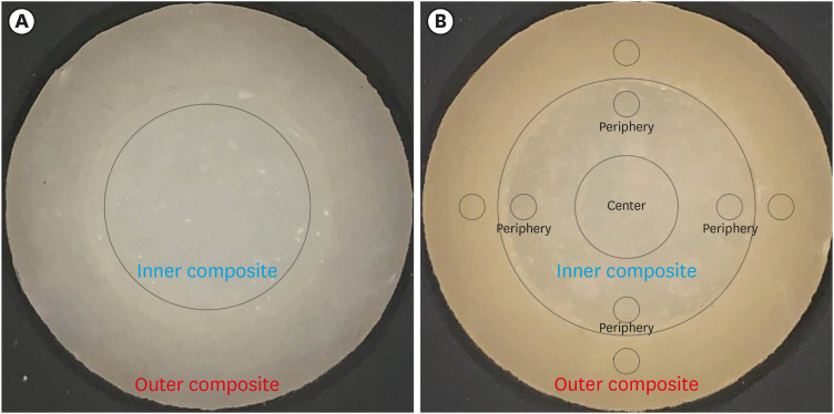
Specific measurement areas were defined using CorelDraw Graphics Suite X8 software (Corel Corporation, Ottawa, ON, Canada). An 8-mm diameter circle was drawn at the center of the inner composite (
Figure 1A), corresponding to the region measured with the spectrophotometer (aperture of 8 mm). The color measurement from this area was used to adjust the color coordinates. The images with the defined measurement areas were saved at 600 dpi in the RGB color system, using the .jpg format. Additionally, 4 1-mm diameter circles were drawn in the periphery of the inner composite and another 4 in a similar position on the outer composite. Furthermore, a 4-mm diameter circle was drawn at the center of the inner composite (
Figure 1B). This second image was also saved in the .jpg format.
Figure 1Illustrative specimens’ images, highlighting the delimited areas used for color measurements. (A) An 8-mm diameter area in the center of the specimen was utilized to adjust the values obtained from image measurements with those obtained from the spectrophotometer. (B) Four 1-mm diameter areas were delimited in the outer composite, along with 4 similar areas in the periphery of the inner single-shade composite. Additionally, a centered 4-mm diameter area was also delimited for measurement purposes.

The open-source image processing software ImageJ (NIH, Bethesda, MD, USA) was employed to measure the color of the defined areas in the images. The RGB values were then converted into CIELAB coordinates using an MS Excel spreadsheet based on the EasyRGB software (Logicol S.l.r., Trieste, Italy). The RGB data was initially converted to the CIE 1931 XYZ color space before obtaining the CIELAB values. The conversion utilized reference values of X=95.047, Y=100.000, and Z=108.883, considering a 1931 2° supplementary standard observer and the CIE D65 standard illuminant [
18,
19,
21].
For color difference calculation, linear regressions were employed to predict the values of each CIELAB color coordinate measured with a spectrophotometer based on the values obtained from the images. This process involved the insertion of the raw image data as “x” values into the regression equations, and the resulting “y” values were defined as the adjusted values.
Color differences were calculated within the same specimen by comparing the adjusted color coordinates of the inner composite to those of the outer composite, both at the periphery and the center. For the periphery, the difference between the 1-mm circle in the inner composite and its corresponding circle in the outer composite was determined, resulting in 4 values. The center color difference was calculated by comparing the average color coordinates of 4 circles in the outer composite to those measured in the center 4-mm circle of the inner composite.
The CIEDE2000 formula, expressed as follows, was used for all color difference calculations [
22,
23]:
In this equation, ΔL', ΔC', and ΔH' represent the changes in luminosity, chroma, and hue, respectively. SL, SC, and SH are the weighted functions for each component, and KL, KC, and KH are the weighted factors for Lightness, Chroma, and Hue, where KL = KC = KH = 1. RT is the interactive term between chroma and hue differences.
In addition, color differences between the inner composite surrounded by composite A1 and the inner composite surrounded by composite A3 were calculated using the same formula. For comparisons involving periphery data, the average color coordinates from the 4 circles were utilized. The ΔE
00 values of 0.8 and 1.8 were employed as 50:50% thresholds for perceptibility and acceptability, respectively, to qualitatively assess the data [
24].
For data analysis, normality was assessed using the Shapiro-Wilk test, and variance homogeneity was evaluated using Levene’s test. RMs ANOVA was employed as the statistical tool. For calculated ΔE
00 values between the inner and outer composites, 3 independent variables were considered: “single-shade composite,” “location of measurement,” and “surrounding shade.” The last 2 variables were defined as repetition measures factors. However, the factor “surrounding shade” was not included in the analysis of ΔE
00 calculated by differences due to changing the outer composite shade. All analyses were conducted at a 95% confidence level and performed using the open statistical platform Jamovi 1.6.15 (
www.jamovi.org). The statistician remained blinded to the experimental conditions.
RESULTS
Regressions for color coordinates adjustments
Figure 2A and 2B present color data obtained using a spectrophotometer or calculated from RGB values captured in images with a cellphone. The figure also showcases the linear regressions applied to each color coordinate and the resulting equations used for adjusting the color coordinates. The strongest correlation between spectrophotometer data and image measurements was found for the coordinate a* (R = 0.956), while the weakest correlation was observed for L* (R = 0.645).
Figure 2Scatter plots illustrating the color coordinates data of the specimens’ center, which were measured using both the spectrophotometer and the images. Each plot represents a specific color coordinate: (A) L* coordinate, (B) a* coordinate, and (C) b* coordinate. The linear regression equations for each plot are also displayed, depicting the relationships between the measured values obtained from both methods.

Color differences between inner and outer composites
Table 1 displays the results of the repeated-measures ANOVA, while
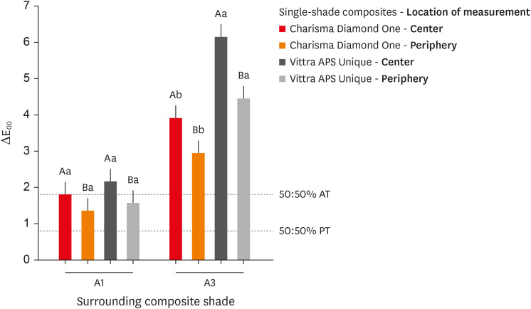
Figure 3 represents the pairwise comparisons. Color differences in the center of specimens were consistently lower than those in the periphery, regardless of the surrounding composite shade or the single-shade composite being evaluated. There was no significant difference between the single-shade composites when the outer composite was A1. However, when the outer composite was A3, Charisma Diamond One showed lower ΔE
00 values than Vittra APS Unique. All ΔE
00 values were above the 50:50% perceptibility threshold, indicating noticeable color variations [
24]. Regardless of the single-shade composite evaluated, the ΔE
00 values in the periphery of the specimen were the only ones below the 50:50% acceptability threshold [
24].
Table 1Results of the repeated measures analysis of variance for color differences between inner and outer composites
|
Variables |
Sum of squares |
df |
Mean square |
F |
p value |
|
Within subjects effects |
|
|
|
|
|
|
Location of evaluation |
8.677 |
1 |
8.6769 |
148.36 |
< 0.001 |
|
Location of evaluation * surrounding shade |
1.677 |
1 |
1.6769 |
28.67 |
< 0.001 |
|
Location of evaluation * single-shade composite |
0.504 |
1 |
0.5040 |
8.62 |
0.010 |
|
Location of evaluation * surrounding shade * single-shade composite |
0.221 |
1 |
0.2205 |
3.77 |
0.070 |
|
Residual |
0.936 |
16 |
0.0585 |
|
|
|
Between subjects effects |
|
|
|
|
|
|
Surrounding shade |
69.72 |
1 |
69.722 |
208.0 |
< 0.001 |
|
Single-shade composite |
11.65 |
1 |
11.653 |
34.8 |
< 0.001 |
|
Surrounding shade * single-shade composite |
6.23 |
1 |
6.233 |
18.6 |
< 0.001 |
|
Residual |
5.36 |
16 |
0.335 |
|
|
Figure 3
Color differences measured between the inner single-shade composites and the surrounding shaded composite. Letters indicating the statistical differences in Tukey’s test should be analyzed separately for each surrounding composite shade. Uppercase letters compare the locations of evaluation within the same composite, while lowercase letters compare the single-shade composites within the same location. Statistical differences are denoted by distinct letters (p < 0.05).
AT, acceptability threshold; PT, perceptibility threshold.

Color differences observed by changing the outer composites
Table 2 and
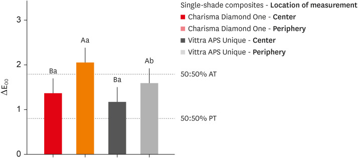
Figure 4 show the results of the repeated-measures ANOVA and pairwise comparisons, respectively. Regardless of the composite used, higher ΔE
00 values were consistently observed in the periphery than in the center, regardless of the composite used. There was no significant difference in ΔE
00 values between the single-shade composites in the center. However, in the periphery, Charisma Diamond One showed significantly higher ΔE00 values than Vittra APS Unique. All color differences exceeded the 50:50% perceptibility threshold [
24]. Except for Charisma Diamond One in the periphery, all other ΔE
00 values remained below the 50:50% acceptability threshold [
24].
Table 2Results of the repeated measures analysis of variance for color differences observed by changing the outer composites
|
Variables |
Sum of squares |
df |
Mean square |
F |
p value |
|
Within subjects effects |
|
|
|
|
|
|
Location of evaluation |
7.890 |
1 |
7.8905 |
82.03 |
< 0.001 |
|
Location of evaluation * single-shade composite |
0.426 |
1 |
0.4264 |
4.43 |
0.041 |
|
Residual |
4.617 |
48 |
0.0962 |
|
|
|
Between subjects effects |
|
|
|
|
|
|
Single-shade composite |
2.47 |
1 |
2.474 |
3.87 |
0.055 |
|
Residual |
30.68 |
48 |
0.639 |
|
|
Figure 4
Color differences measured between specimens with outer composite A1 and those with A3 within the same single-shade composite. Uppercase letters compare the locations of evaluation within the same composite, while lowercase letters compare the single-shade composites within the same location. Statistical differences are denoted by distinct letters (p < 0.05).
AT, acceptability threshold; PT, perceptibility threshold.

DISCUSSION
The findings from this study revealed that color discrepancies between the evaluated single-shade composites and the surrounding composites were lower near the interface, particularly when a lighter outer composite was used. Additionally, Charisma Diamond One displayed fewer color discrepancies than Vittra APS Unique. Both evaluated single-shade composites demonstrated better color adjustment near the interface, with Charisma Diamond One exhibiting superior adjustment compared to Vittra APS Unique. As a result, none of the tested hypotheses in the study can be accepted.
Using specimen images allows for color assessment in specific areas and the determination of color discrepancies within the same specimen [
9,
10]. However, ensuring the accuracy of color measurements is crucial for obtaining reliable results. While using images is not a universally validated method for color measurements, previous research has demonstrated that by standardizing image acquisition and correcting white balance using a neutral gray card, it is possible to obtain reliable results comparable to those obtained with spectrophotometers [
10,
14]. Many studies that assess color using specimen images utilize DSLR cameras. Despite the enduring advantages of DSLRs in terms of sensor size, lens quality, and manual control, the burgeoning convenience, portability, and affordability of smartphones have propelled them to the forefront of various imaging applications. In this study, we employed a smartphone camera to showcase its suitability for capturing precise images, circumventing the need for specialized expertise [
13,
25]. Also, proper specimen illumination conditions are vital to obtaining accurate color images [
15].
Illumination with the D65 standard illuminant, recommended by the CIE (Commission Internationale de l'Éclairage) for color evaluation, was simulated using a light-correcting device with 6 LEDs emitting light at a temperature of 5500°K, representing daylight illumination [
19]. Additionally, a cross-polarizing filter was employed to minimize shiny reflections that could potentially affect color measurements in the specimen images [
12,
16]. Despite these meticulously controlled illumination conditions, the images exhibited a tendency to appear darker (lower L* values), yellower (higher a* values), and redder (higher b* values) compared to the "true" color of the specimens as measured by the spectrophotometer. Linear regressions were employed to harmonize the color coordinates and improve the consistency of the measured color. The chromatic coordinates a* and b* exhibited strong correlations with the spectrophotometer data (nearly perfect for b*), while the correlation for lightness (L*) was moderate. The positive correlation coefficients indicated that the data from the images and the spectrophotometer followed similar trends. However, increasing one value measured in images yielded a smaller increase in the corresponding spectrophotometer value, with coefficients ranging between 0.24 (for L*) and 0.44 (for b*). For the chromatic coordinates (a* and b*), regression coefficients less than 1 and predictors with negative values were necessary to rectify the overestimated values in the image-based measurements. Conversely, only minor discrepancies were observed in L* values between the 2 color evaluation methods, supporting the reliability of the methodology used for color assessment based on cellphone images.
The study’s findings revealed that the color discrepancy between the single-shade composites and the outer composites diminished as the proximity to the interface between the materials increased, irrespective of the specific single-shade composite under evaluation. This improvement in color blending of the single-shade composites near the interface can be attributed to their inherent translucency, allowing the color of the surrounding substrate to influence their overall appearance [
2,
3,
8]. As expected, the mirroring effect of the surrounding color is anticipated to diminish as the distance from the interface increases. It is crucial to emphasize that a gradual decrease in color blending between the composite and the surrounding substrate does not necessarily imply a color mismatch at the restoration's center. In other words, while the color blending may become less apparent towards the center, it does not necessarily indicate that the color will appear off or mismatched in that area.
Achieving an imperceptible restoration heavily relies on precise color adjustment near the interface. The color differences between the single-shade and outer composites consistently exceeded the 50:50 perceptibility threshold [
24]. This indicates that most people would likely notice the difference in color between the 2 composites within the specimens. However, the color discrepancies observed in the study were acceptable near the interface when the surrounding composite was A1, regardless of the single-shade composite being evaluated. On the other hand, when the single-shaded composites were surrounded by a composite A3, the color discrepancies were deemed unacceptable. This discrepancy was particularly significant for the composite Vittra APS Unique, where the ΔE
00 values calculated in the periphery were approximately 3 times above the 50:50 acceptability threshold [
24].
It is important to highlight that both single-shade composites demonstrated significant color adjustment to the surrounding shade, even in the center of the specimens where the color discrepancies were more noticeable. In previous studies, color adjustment potential was calculated based on the relative reduction of color differences between a single-shade composite surrounded by another one, compared to the color difference of these 2 materials placed side-by-side without any interface [
1,
2,
3,
7,
8]. In contrast, our analysis focused solely on the color changes in the single-shade composites caused by modifying the shade of the outer composite. The results revealed that these color changes (adjustments) were clinically perceptible even in the center of the specimens, with no notable difference between the evaluated single-shade composites. As expected, the color changes caused by modifying the outer composite shade were more pronounced near the interface. In this area, the composite Charisma Diamond One exhibited the highest values of color changes, surpassing the 50:50 acceptability threshold [
24]. This better color adjustment observed for Charisma Diamond One can be attributed to its higher translucency compared to Vittra APS Unique [
10]. However, this difference in light transmission between the materials, which allows for mirroring the surrounding color, tends to reduce as the distance from the interface increases.
The current study demonstrated that the color adjustment capability of the assessed single-shade composites to the surrounding color might not be adequate to achieve restorations that are imperceptible, particularly when dealing with darker substrates at the restoration margins. It is important to acknowledge that our study employed a gray-neutral background, and different outcomes might be observed with more chromatic backgrounds. In fact, earlier studies have indicated that the color adjustment of single-shade composites is strongly dependent on the background having a similar color to the surrounding substrate [
3,
10]. Consequently, further studies that modify both the underlying and surrounding substrates could provide additional insights and elucidate the color adjustment potential of various single-shade composites. By examining a wider range of background colors, we can gain a better understanding of how different materials respond to various clinical scenarios, ultimately contributing to achieving optimal color matching for dental restorations.
While standardizing specimen illumination with a light-correcting device ensures consistent lighting conditions, replicating the exact results obtained in this study using other cellphone devices remains challenging. The quality of images captured by cellphone cameras depends on various factors, including sensor size and quality, lens quality, and image processing algorithms. These factors can vary significantly across different cellphone models, potentially leading to color discrepancies. However, despite these inherent variations, the reliability of color data can be enhanced by employing a calibrated neutral gray card and spectrophotometer measurements to calibrate image colors. These procedures were implemented in the present study to ensure data consistency.
CONCLUSIONS
The findings of the study revealed that the color discrepancy between the evaluated single-shade composites and the surrounding composite was significantly lower when the shade of the latter was A1 compared to A3. Additionally, an increased color blending effect was observed for the single-shade composites as they approached the interface. Charisma Diamond One exhibited superior color adjustment capabilities when compared to Vittra APS Unique.
ACKNOWLEDGEMENTS
G.J.S.L. is grateful to The National Council of Scientific and Technological Development (CNPq) for the research fellowship.
-
Funding: This study received partial financial support from the Coordination for the Improvement of Higher Education Personnel (CAPES), Brazil, under Finance Code 001.
-
Conflict of Interest: No potential conflict of interest relevant to this article was reported.
-
Author Contributions:
Conceptualization: Santana MLC, Livi GJS, Faria-e-Silva AL.
Data curation: Faria-e-Silva AL.
Formal analysis: Faria-e-Silva AL.
Funding acquisition: Faria-e-Silva AL.
Investigation: Santana MLC, Livi GJS.
Methodology: Santana MLC, Livi GJS.
Project administration: Faria-e-Silva AL.
Supervision: Faria-e-Silva AL.
Validation: Faria-e-Silva AL.
Writing - original draft: Santana MLC, Livi GJS, Faria-e-Silva AL.
Writing - review & editing: Santana MLC, Livi GJS, Faria-e-Silva AL.
REFERENCES
- 1. Trifkovic B, Powers JM, Paravina RD. Color adjustment potential of resin composites. Clin Oral Investig 2018;22:1601-1607.ArticlePubMedPDF
- 2. Barros MS, Silva PF, Santana ML, Bragança RM, Faria-E-Silva AL. Effect of surrounded shade and specimen’s thickness on color adjustment potential of a single-shade composite. Braz Dent J 2022;33:126-132.ArticlePubMedPMC
- 3. Barros MS, Silva PF, Santana ML, Bragança RM, Faria-E-Silva AL. Effects of surrounding and underlying shades on the color adjustment potential of a single-shade composite used in a thin layer. Restor Dent Endod 2023;48:e7.ArticlePubMedPMCPDF
- 4. de Abreu JL, Sampaio CS, Benalcázar Jalkh EB, Hirata R. Analysis of the color matching of universal resin composites in anterior restorations. J Esthet Restor Dent 2021;33:269-276.ArticlePubMedPDF
- 5. Korkut B, Ünal T, Can E. Two-year retrospective evaluation of monoshade universal composites in direct veneer and diastema closure restorations. J Esthet Restor Dent 2023;35:525-537.ArticlePubMedPDF
- 6. Forabosco E, Consolo U, Mazzitelli C, Kaleci S, Generali L, Checchi V. Effect of bleaching on the color match of single-shade resin composites. J Oral Sci 2023;65:232-236.ArticlePubMed
- 7. Durand LB, Ruiz-López J, Perez BG, Ionescu AM, Carrillo-Pérez F, Ghinea R, et al. Color, lightness, chroma, hue, and translucency adjustment potential of resin composites using CIEDE2000 color difference formula. J Esthet Restor Dent 2021;33:836-843.ArticlePubMedPDF
- 8. Barros MS, Silva PF, Santana ML, Bragança RM, Faria-E-Silva AL. Background and surrounding colors affect the color blending of a single-shade composite. Braz Oral Res 2023;37:e035.ArticlePubMed
- 9. de Melo Oliveira I, Santana TR, Correia AC, Fontes LS, Griza S, Faria-E-Silva AL. Color heterogeneity and individual color changes in dentin and enamel bleached in the presence of a metallic orthodontic bracket. J Esthet Restor Dent 2021;33:262-268.ArticlePubMedPDF
- 10. de Livi GJ, Santana TR, Bragança RM, de Bragança Garcez RM, Faria-E-Silva AL. The role of interface distance and underlying substrate on the color adjustment potential of single-shade composites. J Esthet Restor Dent 2023;35:1279-1285.ArticlePubMed
- 11. Tam WK, Lee HJ. Dental shade matching using a digital camera. J Dent 2012;40(Supplement 2):e3-ee10.Article
- 12. Lazar R, Culic B, Gasparik C, Lazar C, Dudea D. The accuracy of dental shade matching using cross-polarization photography. Int J Comput Dent 2019;22:343-351.PubMed
- 13. de Bragança RM, Moraes RR, Faria-E-Silva AL. Color assessment of resin composite by using cellphone images compared with a spectrophotometer. Restor Dent Endod 2021;46:e23.PubMedPMC
- 14. Rondón LF, Ramírez R, Pecho OE. Comparison of visual shade matching and photographic shade analysis. J Esthet Restor Dent 2022;34:374-382.ArticlePubMedPDF
- 15. Brokos I, Polychronakis N, Polyzois G, Lagouvardos P, Krejci I. Illuminant metameric effects on interbrand and intrabrand color differences of direct composite resins. J Prosthet Dent 2022;128:1342-1349.ArticlePubMed
- 16. Yilmaz B, Dede DÖ, Diker E, Fonseca M, Johnston WM, Küçükekenci AS. Effect of cross-polarization filters on the trueness of colors obtained with a single-lens reflex camera, macro lens, and a ring flash. J Esthet Restor Dent 2023;35:878-885.ArticlePubMed
- 17. Soares KD, Bragança RM, Leal PC, Schneider LF, Faria-e-Silva AL. Is it possible to determine the optical properties of resin composites with clinical spectrophotometers? Color Res Appl 2022;47:706-716.ArticlePDF
- 18. ISO 11664-2:2007. Colorimetry — Part 2: CIE standard illuminants. Geneva: International Organization for Standardization; 2007.
- 19. ISO/CIE 11664-1:2019. Colorimetry — Part 1: CIE standard colorimetric observers. Geneva: International Organization for Standardization; 2019.
- 20. Araujo FS, Barros MC, Santana ML, de Jesus Oliveira LS, Silva PF, Lima GD, et al. Effects of adhesive used as modeling liquid on the stability of the color and opacity of composites. J Esthet Restor Dent 2018;30:427-433.ArticlePubMedPDF
- 21. Carney MN, Johnston WM. A novel regression model from RGB image data to spectroradiometric correlates optimized for tooth colored shades. J Dent 2016;51:45-48.ArticlePubMedPMC
- 22. Luo MR, Cui BR, Rigg B. The development of the CIE 2000 colour-difference formula: CIEDE2000. Color Res Appl 2001;26:340-350.Article
- 23. Sharma G, Wu W, Dalal EM. The CIEDE2000 color-difference formula: Implementation notes, supplementary test data, and mathematical observations. Color Res Appl 2005;30:21-30.Article
- 24. Paravina RD, Ghinea R, Herrera LJ, Bona AD, Igiel C, Linninger M, et al. Color difference thresholds in dentistry. J Esthet Restor Dent 2015;27(Supplement 1):S1-S9.ArticlePubMedPDF
- 25. Nixon M, Outlaw F, Leung TS. Accurate device-independent colorimetric measurements using smartphones. PLoS One 2020;15:e0230561.ArticlePubMedPMC
 , Gabriella de Jesus Santos Livi2
 , André Luis Faria-e-Silva1,2


 KACD
KACD
 ePub Link
 Cite
Cite

