
Articles
- Page Path
- HOME > Restor Dent Endod > Volume 49(4); 2024 > Article
- Review Article Comparative evaluation of the biological response of conventional and resin modified glass ionomer cement on human cells: a systematic review
-
Shishir Singh1
 , Gaurav Kulkarni1, R S Mohan Kumar2, Romi Jain3, Ameya M Lokhande1, Teena K Sitlaney1, Musharraf H F Ansari1, Navin S Agarwal1
 -
Restor Dent Endod 2024;49(4):e41.
DOI: https://doi.org/10.5395/rde.2024.49.e41
Published online: November 1, 2024
1Department of Conservative Dentistry and Endodontics, TPCT’s Terna Dental College, Maharashtra, India.
2Department of Conservative Dentistry and Endodontics, Priyadarshini Dental College, Tamilnadu, India.
3Department of Public Health Dentistry, TPCT’s Terna Dental College, Maharashtra, India.
- Correspondence to Shishir Singh, BDS, MDS, PhD. Department of Conservative Dentistry and Endodontics, TPCT’s Terna Dental College, Nerul, Navi Mumbai, Maharashtra 400706. India. drshishirs@gmail.com
• Received: April 29, 2024 • Revised: May 31, 2024 • Accepted: July 1, 2024
Copyright © 2024. The Korean Academy of Conservative Dentistry
This is an Open Access article distributed under the terms of the Creative Commons Attribution Non-Commercial License (https://creativecommons.org/licenses/by-nc/4.0/) which permits unrestricted non-commercial use, distribution, and reproduction in any medium, provided the original work is properly cited.
Abstract
-
Trial Registration PROSPERO Identifier: CRD42023426021
INTRODUCTION
Dental cements are crucial in dentistry, serving as foundations, protective layers, fillings, or adhesives for dental devices, with diverse options chosen based on specific needs. Their careful selection significantly impacts dental restoration success. Typically, these cements solidify by mixing powder and liquid to meet various criteria, including safeguarding tooth tissues, resisting tension and pressure, forming durable bonds, and biocompatibility and impermeability. Along with ease of use, low solubility, radiopacity, optimal working times, high resistance, suitable viscosity, and aesthetics, it is imperative for the cements to have the ability to ensure that a dental treatment accomplishes its intended purpose effectively, it should operate without causing any unwanted local or systemic effects for the patient. Instead, it should stimulate the most suitable beneficial response from cells or tissues in the particular scenario, ultimately maximizing the therapy’s practical effectiveness in clinical settings [1].
Dental cements are categorized by purpose, composition, and properties, including glass ionomer cements (GICs), introduced in 1969 by Wilson and Kent in London, which release fluoride to inhibit caries through a consistent process, acting as a fluoride reservoir for long-term effectiveness.
Extensive research has probed the biocompatibility of GICs, revealing that these cements generally fall within biocompatible parameters, although initial pulp reactions may occur, with uncertain long-term effects on pulp tissue [2,3]. GICs come in various forms and are set within 2–3 minutes, fully hardening in up to 48 hours [4]. Adhering to the recommended powder-liquid ratio is crucial. GICs’ unique attributes, including fluoride release, biocompatibility, direct bonding to teeth, good marginal adaptation, and dentin-like elasticity, make them versatile for dental procedures such as restorations, liners, luting, crowns, and bridges. However, they have limitations due to suboptimal mechanical properties, slow setting, high solubility, and sensitivity to moisture, restricting their use in stress-prone areas like posterior teeth [2,4]. Efforts to improve GICs have involved modifying glass powder formulations with elements like resin, hydroxyapatite, fibers, and nano-sized particles, with mixed effects on their properties [5].
Resin modified glass ionomer cements (RMGICs), introduced in 1991, aimed to overcome the limitations of traditional GICs by incorporating elements like hydroxyethyl methacrylate (HEMA), triethylene glycol dimethacrylate (TEGDMA) and initiators for polymerization into the basic GIC composition. This hybrid approach combines acid-base and polymerization reactions, reducing microleakage while retaining the benefits of fluoride release and tooth bonding. RMGICs are easier to handle and bond well with composite materials and can be cured on command thereby improving its handling properties and increasing acceptance among the clinicians. However, assessing the biocompatibility of these materials is crucial, especially when compared to conventional GICs, which share similar compositions and reactions [6]. The biological response of RMGIC is believed to be compromised due to the release of components like HEMA, which can have cytotoxic effects on pulp cells, posing potential risks to dental professionals and patients.
Evaluating dental material biocompatibility is crucial for patient and dental professional well-being, aiding in the selection of suitable dental cements. Some commonly used dental cements have exhibited cytotoxic effects on both soft and hard tissues, potentially affecting the long-term success of restorations [7,8]. The degree of cytotoxicity varies among cements, depending on their specific composition and the leachable components within them. For instance, GICs were initially thought to be toxic due to fluoride, but subsequent studies revealed other major components as the culprits [5,9]. Conversely, resin-based cements, including RMGICs, tend to exhibit higher cytotoxicity due to the presence of monomers like HEMA, TEGDMA, and bisphenol A-glycidyl methacrylate, which can adversely affect various oral tissues Strict adherence to recommended polymerization times is essential to mitigate these effects [3,10].
The diffusion of these monomers into dentin allows them to interact with pulp cells, potentially disrupting cell function, inhibiting differentiation, and reducing the formation of mineral nodules within teeth [3,11,12]. Monomers may also induce immune responses, leading to allergies and hypersensitivity [13,14,15]. Additionally, certain monomers like HEMA and TEGDMA have been linked to genotoxic effects, including increased micronucleus formation and chromosomal aberrations [16,17]. Clinical, radiographic, and histological evaluations are critical in assessing the biocompatibility of dental materials, covering aspects like cell viability, metabolism, morphology, inflammatory responses, tissue organization, and more.
RMGIC has superior mechanical and physical properties, superior handling, and better adhesiveness as compared to conventional GIC. However, literature shows conflicting views of pulpal response towards RMGIC in deep cavities [3,6].
The current scientific literature lacks a systematic review directly comparing the biological response in terms of biocompatibility and cytotoxicity of conventional GIC and RMGIC when used in direct or indirect pulp capping or pulpotomy. Thus, the objective of this systematic review is to evaluate and compare the biological response (biocompatibility and cytotoxicity) of resin modified glass ionomer cement in contrast to conventional glass ionomer cement on human cells.
MATERIALS AND METHODS
This systematic review was reported according to the Preferred Reporting Items for Systematic Reviews and Meta-Analyses (PRISMA) and the protocol was registered in the International Prospective Register of Systematic Reviews database (PROSPERO) with registration number CRD42023426021. Also, a well-defined review question was developed by using the patient Population, Intervention, Comparison, and Outcome (PICO) framework.
The aim of the present review is to investigate the biological response of GIC and RMGIC which are commonly used restorative materials.
The following PICO framework was developed for a systematic review.
Population: Human carious tooth/iatrogenic exposed pulp undergone Indirect or direct pulp capping or pulpotomy procedure with GIC or RMGIC or cell culture (odontoblasts, fibroblast, dental pulp stem cells or any other cells) exposed to material extracts of conventional GIC or RMGIC.
Intervention/Exposure: Treatment of human carious tooth/Iatrogenic exposure of pulp using Indirect pulp capping, direct pulp capping and pulpotomy procedure OR exposure of cell culture (odontoblasts, fibroblast, dental pulp stem cells or any other cell) to the material extract of conventional GIC (Inocid-L 30, Ketac-Fil, Ketac- Molar or any other commercially available conventional GIC).
Comparison: Treatment of human carious tooth/Iatrogenic exposure of pulp using Indirect pulp capping, Direct pulp capping and pulpotomy procedures using RMGIC or exposure of cell culture (odontoblasts, fibroblast, dental pulp stem cells or any other cell) to the material extract of RMGIC (Vivaglass, Vitremer 3M, Vitrebond or any other commercially available RMGIC).
Outcome: The main outcome is in terms of biological response to the included material. It was based on histological, clinical, and radiographic evaluation. The histological evaluation included cell viability, cellular metabolism, cell morphology, odontoblastic changes, inflammatory cell infiltration, reactionary dentin formation, presence of microorganisms, tissue disorganization, etc. Clinical parameters included the presence or absence of pain, swelling, sinus or periodontal pockets, tenderness on percussion, or any other symptoms. Radiographic parameters like changes in the periapical region, root resorption, or any other method of evaluation were included.
Among conventional GIC and RMGIC, which material is better in terms of biological response?
1. Inclusion criteria
The inclusion criteria were 1) Articles reporting parallel and split-mouth clinical trials, randomized controlled trials, non-randomized controlled trials, prospective studies, and in vitro studies on human permanent teeth; 2) Studies in which conventional GIC and RMGIC are used for direct or indirect pulp capping; 3) Studies in which cell culture (odontoblasts, fibroblast, dental pulp stem cells or any other cells) is exposed to material extracts of conventional GIC or RMGIC is used; 4) Studies in which biological response was assessed by using histological, clinical and radiographic criteria; and 5) Articles in English language or other languages where English translation is possible.
2. Exclusion criteria
The inclusion criteria were 1) Case reports, reviews and expert opinion; 2) Studies with only abstracts without the availability of full text; 3) Studies on primary teeth; and 4) Animal studies.
The following electronic bibliographic databases were searched: MEDLINE/PubMed, PubMed Central, EBSCO, Cochrane Central Register of Controlled Trials as well as Google Scholar. Other searches included hand searches of articles, grey literature, citations as well as cross-references.
The following terminologies were searched: ((((((GIC) OR (Glass ionomer cement)) OR (Ketac-Molar)) OR (Ketac-Fil)) OR (Glass-Ionomer Cements)) AND ((((((Resin modified glass ionomer cement) OR (RMGIC)) OR (Light curing GIC)) OR (Vivaglass,)) OR (Vitremer 3M)) OR (Vitrebond))) AND ((((((((Biocompatibility) OR (Pulpal response)) OR (Human Odontoblast)) OR (Gingival fibroblast)) OR (Dental pulp stem cells)) OR (Cytotoxicity)) OR (Primary culture)) OR (Histopathology)) Filters: Free full text, Full text.
The study selection was done by 2 reviewers (SS) and (RJ). During the first step, the articles from the different databases were imported with software (Mendeley), and duplicate articles were eliminated. Then the titles and abstracts of the articles were reviewed to eliminate the irrelevant articles. In the final step, the articles were filtered through the full reading of each one of them. During each step, a third reviewer (GK) could be consulted if the 2 reviewers were not able to resolve disagreement through discussion.
Data extraction was done in 4 domains: 1) Identification of the study (type of the study, article title; journal title; authors; country of the study; language; publication year; host institution of the study); 2) Methodological characteristics (study objective or research question; sample characteristics, e.g., sample size, age, type of procedure performed, type of material used, type of cells used, different concentration of material extract, methods of evaluation, statistical analyses, etc.; 3) Main findings (Histological, clinical, and radiographic outcome); and 4) Conclusions or remarks.
The assessment of the risk of bias was done by 2 reviewers (RJ and SS), to analyze the methodological quality of the articles included. For the assessment of the non-randomized clinical trials, the Methodological Index for Non-Randomized Studies (MINORS) tool was used [18]. This tool has 8 items for non-comparative studies and 12 items for comparative studies. On this scale, items are scored 0 (not reported), 1 (reported but inadequate), or 2 (reported and adequate). The global ideal score is 16 for non-comparative studies and 24 for comparative studies.
To assess the risk of bias in the in vitro studies, the modified scale of Animal Research: Reporting of In Vivo Experiments (ARRIVE) and Consolidated Standards of Reporting Trials (CONSORT) was used (the highest score is 25, acceptability range is 18–25) [19].
RESULTS
Initial screening found 537 studies, yielding 408 screened abstracts after removing duplicates. Of these, 46 full texts were considered eligible. However, 16 articles could not be retrieved in full. After a thorough assessment, 9 papers were included for review, while 21 were excluded for various reasons (refer to the PRISMA flow diagram and Figure 1 for details on excluded articles).
Figure 1
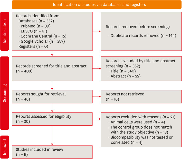
Summary of the inclusion and screening of articles following the PRISMA approach.
PRISMA, Preferred Reporting Items for Systematic Reviews and Meta-Analyses.
Out of 10 included studies 3 studies were nonrandomised clinical studies whereas 6 studies were in vitro studies [20,21,22,23,24,25,26,27,28]. Three studies assessed histological response by using premolars [20,21,22]. Six studies used various cell lines namely human gingival fibroblast (HGF), human dental pulp fibroblast (HDPF), stem cell human exfoliated deciduous teeth (SHED), and human odontoblast cell line (MDPC-23) [23,24,25,26,27,28]. Odontoblastic changes inflammatory response, tertiary dentin formation, presence of microorganisms, assessment of morphological changes, lactate dehydrogenase release (LDH), cell viability percentage, RNA expression for RPL13, stromal cell-derived factor-α value, interleukin (IL)-8, IL-6, Nitrito, etc. were the different parameters which were considered to assess the biological response of conventional and RMGICs. Table 1 shows the general characteristics of the studies included in the review, and Tables 2 and 3 show details of various parameters used to assess the biocompatibility and cytotoxicity of conventional and RMGIC.
Table 1
General characteristics of the included studies
| Author and year | Study design | Materials tested | Experimental model | Exposure duration | Cell line | Variables studies |
|---|---|---|---|---|---|---|
| Eskandarizadeh et al. (2015) [21] | Non-randomized controlled trial | RMGIC (Vivaglass) GIC (Ionocid) Calcium hydroxide (Dycal) | 30 human premolars | 5 and 30 days | None | Odontoblastic changes, inflammatory response, tertiary dentin formation, and presence of microorganisms |
| Ribeiro et al. (2020) [20] | Non-randomized controlled trial | RMGIC (Riva LC) GIC (Riva SC) Calcium hydroxide (Dycal) | 26 human premolars + 4 controls | 7 and 30 days | None | Inflammatory reaction, tissue disorganization, reactionary dentin formation, and bacteria |
| Mousavinasab et al. (2008) [22] | Non-randomized controlled trial | RMGIC (Vivaglass) GIC (Chem bond Superior) Calcium hydroxide (Dycal) | 55 human premolars | 7, 30 and 60 days | None | Odontoblastic changes, inflammatory cell infiltration, and reactionary dentin formation |
| Leyhausen et al. (1998) [23] | In vitro study (histological studies) | RMGICs (Ionoseal, Vitrebond, Compoglass) and GIC (Ketac Fil) | HGF | 48 hours | Cytotoxicity | Morphology and growth characteristics by PCM |
| Rodriguez et al. (2013) [24] | In vitro study (Histological studies) | RMGIC (Vitrebond) GIC (Ketac Molar) | HGF | 72 hours | Cytotoxicity | Morphological changes by PCM and LDH |
| Mohd Zainal Abidin et al. (2015) [25] | In vitro study (histological studies) | Fuji IX GPExtra | SHED | 72 hours | Cytotoxicity | Cell viability percentage and IC50 |
| Fuji II LC | ||||||
| Koohpeima et al. (2017) [26] | In vitro study (histological studies) | Fuji II | HGF | 24 hours | Cytotoxicity | percentage of cell viability of HGF at 25, 50, 75 and 100% |
| Fuji II LC | ||||||
| de Souza Costa et al. (2003) [27] | In vitro study (histological studies) | Fuji IX, Ketac Molar | Human odontoblast cell line (MDPC-23) | 72 hours | Cytotoxicity | Cell number, cell morphology, cell metabolism |
| Vitrebond, Vitremer, Fuji II LC | ||||||
| Sun et al. (2011) [28] | In vitro study (histological studies) | Fuji II, Fuji II LC, Vitrmer | Human pulp cells, 3T3 mouse fibroblast | 1 and 3 days | Cytotoxicity | Cell number, cell morphology and cell metabolism |
Table 2
Biocompatibility and cytotoxicity of conventional GIC and RMGIC
| Author and year | Cytotoxicity test used | Results for conventional GICs | Results for resin modified GICs | Author’s conclusion | ||
|---|---|---|---|---|---|---|
| Leyhausen et al. (1998) [23] | DNA Intercalating fluorochrome assay | Ketac Fil Applicap | Vitrebond | Light cure GICs revealed cytotoxic effects when compared to conventional GIC which had no or slight alterations in cell lines. | ||
| - Growth of the primary HGF: Day 1: 107 ± 19.5, Day 9: 103 ± 4 | - Growth of the primary HGF: Day 1: 15.7 ± 13.9, Day 9: 68.8 ± 6.8 | |||||
| Rodriguez et al. (2013) [24] | PCM, LDH assay, EPXMA analysis | Ketac Molar | Vitrebond | Morphological, biochemical, and micro-analytical indicators suggested that RMGIC causes greater alteration that points towards necrosis as compared to conventional GIC. | ||
| - Number of fibroblasts: 52.2% ± 20.4% | - Number of fibroblasts: 3.9% ± 5% | |||||
| - LDH release: 11.04% ± 21.69% | - LDH release: 38.46% ± 7.29% | |||||
| - Intracellular levels (Na): 85.79 ± 58.03 mmol/kg | - Intracellular elements levels (Na): 187.03 ± 113.11 mmol/kg | |||||
| - Intracellular levels (K): 272.72 ± 75.69 mmol/kg | - Intracellular elements levels (K): 186.74 ± 132.06 mmol/kg | |||||
| - Intracellular elements levels (Cl): 115.89 ± 75.69 mmol/kg | - Intracellular elements levels (Cl): 153.12 ± 57.28 mmol/kg | |||||
| Mohd Zainal Abidin et al. (2015) [25] | MTT assay | Fuji IX GPExtra | Fuji II LC | RMGIC exhibited cytotoxic effect on SHED as well as the least favorable cell viability among all the groups. | ||
| IC50 = 45 mg/mL | IC50 = 31.2 5 mg/mL | |||||
| Koohpeima et al. (2017) [26] | MTT assay | Fuji II | Fuji II LC | Study showed that cytotoxic effect of conventional GIC was significantly lesser as compared to other modified GICs. | ||
| - Percentage cell viability: at 25% = 100.80% ± 8.17%, at 50% = 122.64% ± 3.76%, at 75% = 125.15% ± 3.92%, and at 100% = 134.86% ± 0.65% | - Percentage cell viability: at 25% = 98.45% ± 7.24%, at 50% = 102.50% ± 6.16%, at 75% = 5.41% ± 9.16%, at 100% = 112.29% ± 3.85% | |||||
| de Souza Costa et al. (2003) [27] | MTT assay, SEM | Fuji IX, Ketac Molar | Vitrebond, Vitremer, Fuji II LC | Study concluded that Vitremer and Vitrebond (RMGIC) were more cytopathic than Fuji IX GP and Ketac Molar. | ||
| - Reduction in cell metabolism: Fuji IX = 40.3%, Ketac Molar = 42.5% | - Reduction in cell metabolism: Vitrebond = 79.1%, Vitremer = 83.9%, Fuji II LC = 53.75% | |||||
| - Reduction in cell number: Fuji IX = 29.5%, Ketac Molar = 32.5% | - Reduction in cell number: Vitrebond = 74.5%, Vitremer = 75.5%, Fuji II LC = 45.5% | |||||
| Sun et al. (2011) [28] | MTT assay, WST-1 assay | Fuji II | Fuji II LC, Vitremer | Study revealed that Fuji II and Fuji II LC are not cytotoxic to human pulp cells but Vitremer is very cytotoxic. Cytotoxicity was dose-dependent. | ||
| - Cell viability: Day 1: 100.3% ± 6.3%, Day 3: 98.8% ± 7.8% | - Cell viability: Day 1: 88.0% ± 11%, Day 3: 105.9% ± 10.3% | |||||
Values are in mean ± standard deviation.
GIC, glass ionomer cement; RMGIC, resin modified glass ionomer cement; HGF, human gingival fibroblast; PCM, phase contrast microscopy; LDH, lactate dehydrogenase release; EPXMA, Electron Probe Microanalyzer; MTT, 3-(4,5-dimethylthiazol-2-yl)-2,5-diphenyltetrazolium bromide, IC50, half maximal inhibitory concentration; SHED, stem cell human exfoliated deciduous teeth; SEM, scanning electron microscopy; WST-1, Water-Soluble Tetrazolium 1.
Table 3
Histological response of pulp to RMGIC and conventional GIC
| Histological events | Material | 5–7 days | 30 days | Total | |||||||
|---|---|---|---|---|---|---|---|---|---|---|---|
| No | Mild | Moderate | Severe | No | Mild | Moderate | Severe | ||||
| Ribeiro et al. (2020) [20] | |||||||||||
| Inflammatory response | RMGIC | 1 | 3 | 1 | 0 | 3 | 2 | 0 | 0 | 10 | |
| GIC | 2 | 3 | 0 | 0 | 5 | 0 | 0 | 0 | 10 | ||
| Tissue disorganization | RMGIC | 1 | 4 | 0 | 0 | 3 | 2 | 0 | 0 | 10 | |
| GIC | 2 | 3 | 0 | 0 | 4 | 1 | 0 | 0 | 10 | ||
| Reactionary dentin formation | RMGIC | 4 | 1 | 0 | 0 | 5 | 0 | 0 | 0 | 10 | |
| GIC | 5 | 0 | 0 | 0 | 4 | 1 | 0 | 0 | 10 | ||
| Bacteria | RMGIC | 5 | 0 | 0 | 0 | 3 | 2 | 0 | 0 | 10 | |
| GIC | 5 | 0 | 0 | 0 | 4 | 1 | 0 | 0 | 10 | ||
| Eskandarizadeh et al. (2015) [21] | |||||||||||
| Inflammatory response | RMGIC | 2 | 2 | 1 | 0 | 3 | 2 | 0 | 0 | 10 | |
| GIC | 2 | 2 | 0 | 1 | 3 | 1 | 0 | 1 | 10 | ||
| Odontoblastic changes | RMGIC | 0 | 3 | 2 | - | 2 | 1 | 2 | - | 10 | |
| GIC | 1 | 3 | 1 | - | 3 | 1 | 0 | - | 10 | ||
| Mousavinasab et al. (2008) [22] | |||||||||||
| Inflammatory cell infiltration | RMGIC | 0 | 2 | 6 | 0 | 3 | 2 | 0 | 0 | 13 | |
| GIC | 2 | 1 | 4 | 0 | 2 | 2 | 2 | 0 | 13 | ||
| Odontoblastic changes | RMGIC | 3 | 1 | 4 | 0 | 2 | 1 | 2 | 0 | 13 | |
| GIC | 2 | 2 | 3 | 0 | 2 | 2 | 2 | 0 | 13 | ||
| Reactionary dentin formation | RMGIC | 8 | 0 | 0 | 0 | 2 | 3 | 0 | 0 | 13 | |
| GIC | 7 | 0 | 0 | 0 | 5 | 1 | 0 | 0 | 13 | ||
Six studies have used various cell cultures (odontoblasts, fibroblast, dental pulp stem cells, or any other cell) and exposed them to the material extract of conventional GIC and resin modified GIC to assess the cytotoxicity [23,24,25,26,27,28]. They have used various cytotoxicity tests like DNA Intercalating fluorochrome assay, Phase contrast microscopy, LDH assay, EPXMA analysis, TRIPAN blue, MTT assay, cytokine detection by enzyme-linked immunosorbent assay and reverse transcription-quantitative polymerase chain reaction, Griess methods for nitric oxide release mRNA expression and Water-Soluble Tetrazolium 1 assay. All 6 studies showed that the cytotoxic effect of conventional GIC was significantly lesser as compared to RMGICs (Table 2). Three studies have compared the histological response of pulp to RMGIC and conventional GIC in terms of inflammatory response, tissue disorganization, reactionary dentin formation, number of bacteria, odontoblastic changes at 5–7 days and 30 days follow-up [20,21,22]. All these are recorded as no changes, mild, moderate, and severe changes. These studies have shown favorable results with conventional GIC as compared to RMGIC (Table 3).
The 3 non-randomized controlled clinical trials presented a score on the MINORS tool as 13 or 14 out of 16 points (Table 4), therefore they can be considered as “low- risk” of bias studies. Concerning the in vitro studies, 2 studies showed a moderate risk of bias whereas the 5 other studies presented a low risk of bias according to the modified ARRIVE and CONSORT scale (Table 5).
Table 4
Risk of bias/quality assessment for non-randomized clinical trials using MINORS tool
| Study | A clearly stated aim | Inclusion of consecutive patients | Prospective collection of data | Endpoints appropriate to the aim of the study | Unbiased assessment of the study endpoint | Follow-up period appropriate to the aim of the study | Loss to follow up less than 5% | Prospective calculation of the study size | Total |
|---|---|---|---|---|---|---|---|---|---|
| Eskandarizadeh et al. (2015) [21] | 2 | 2 | 2 | 2 | 0 | 2 | 2 | 1 | 13 |
| Ribeiro et al. (2020) [20] | 2 | 2 | 2 | 2 | 1 | 2 | 2 | 1 | 14 |
| Mousavinasab et al. (2023) [22] | 2 | 2 | 2 | 2 | 1 | 2 | 2 | 1 | 14 |
Table 5
Measurement of the risk of bias/quality assessment of in vitro studies (histopathological studies) with the modified ARRIVE and CONSORT scale (acceptability range 21–28).
| Studies | Title | Abstract | Introduction | Introduction | Methods: Study design | Methods: experimental procedures | Method: Sample size | Method: Statistical procedure | Result | Discussion | Potential conflicts | Publication | Total score |
|---|---|---|---|---|---|---|---|---|---|---|---|---|---|
| Leyhausen et al. (1998) [23] | 1 | 2 | 3 | 2 | 2 | 2 | 2 | 3 | 2 | 1 | 1 | 0 | 22 |
| Mohd Zainal Abidin et al. (2015) [25] | 0 | 3 | 2 | 2 | 2 | 3 | 1 | 2 | 2 | 1 | 0 | 1 | 19 |
| Rodriguez et al. (2013) [24] | 1 | 2 | 3 | 1 | 2 | 3 | 2 | 3 | 3 | 1 | 1 | 1 | 23 |
| Koohpeima et al. (2017) [26] | 0 | 3 | 3 | 1 | 1 | 2 | 1 | 3 | 3 | 2 | 1 | 1 | 21 |
| de Souza Costa et al. (2003) [27] | 0 | 2 | 1 | 2 | 1 | 1 | 1 | 2 | 2 | 1 | 1 | 1 | 15 |
| Sun et al. (2011) [28] | 1 | 1 | 2 | 2 | 3 | 1 | 2 | 3 | 2 | 1 | 1 | 21 |
DISCUSSION
The present systematic review exhibits information, based on scientific evidence, related to the biological response of conventional GIC compared to RMGIC. The aim of this study was to analyze the toxicity of RMGIC in contrast to conventional GIC on the human carious tooth or exposed pulp undergoing direct or indirect pulp capping or human cell culture exposed to extracts of the test materials.
Numerous factors, including the heat damage caused during cavity preparation, the existence of bacteria, their metabolites as well as the toxicity of substances released from dental materials can induce pulp damage [20]. Applying dental materials in cavities with thin remaining dentin thickness (RDT) tests the biologic behavior and response of both the dental material and the pulp dentin complex. Research has shown that maintaining a 0.5 mm layer of dentin between the pulp and the restorative material effectively shields the pulpal tissue from the harmful effects associated with the materials used. Factors such as wide dentinal tubules and increased moisture levels can facilitate the penetration of potentially harmful chemical residues from resin-based dental products including bonding agents, which could pose risks to the health of the nearby pulp tissue. Therefore, the use of a biocompatible liner in cases of deep dentinal caries is advisable [24,29,30].
Resin-based dental materials contain components having cytopathic properties [31]. The primary resin monomer used in bonding agents RMGIC is HEMA, whose potential for toxicity is well known. HEMA induces DNA strand breakage, leading to apoptosis and genotoxic effects in culture media [32]. When ethanol elution was employed to extract HEMA from RMGIC, it notably led to a reduction in the material’s cytotoxicity to pulp cells. Moreover, HEMA hinders the secretion of inflammatory cytokines by immune cells, thereby compromising the host’s defense response [33].
de Souza Costa et al. [34] demonstrated that there was no alteration in the pulp tissue when a light-cured RMGIC was used as a pulp capping agent in very deep cavities in non-conditioned dentin (i.e., RDT up to 0.3 mm). On the other hand, the application of a restorative RMGIC to dentin that had been conditioned with polyacrylic acid led to significant and permanent alterations in the human dentin-pulp complex [34].
The dental materials and their constituent parts undergo initial in vitro testing for mutagenesis and cytotoxicity. Understanding the protective properties of the dentin-pulp complex is crucial for optimizing material usage and formulating indications to prevent serious pulp damage. In order to maintain vitality, the dentin-pulp complex can adapt to a variety of stimuli and trigger a defense response. The substance will cause a biological reaction when it comes in contact with living tissues and its cytotoxicity may cause changes in metabolism up to cellular death. The first step in determining a material’s biocompatibility is to conduct cytotoxicity tests [35].
In vitro studies conducted by Leyhausen et al. [23], Rodriguez et al. [24], Mohd Zainal Abidin et al. [25], Koohpeima et al. [26] and de Souza Costa et al. [27] evaluated the cytotoxicity of conventional GIC and RMGIC using experimental cells derived from human i.e., HGF, SHED, HDPF, and MDPC-23. The results of these studies commonly stated that the cytotoxic effects of conventional GIC were significantly lesser than RMGIC. In another study included in this systematic review conducted by Sun et al. [28], the researchers examined the effects of 6 different contemporary dental restorative materials on human primary cells in vitro. The findings showed that Fuji II and Fuji II LC were not harmful to human pulp cells, while Vitremer exhibited significant cytotoxicity, likely attributed to its high content of TEGDMA. It was also concluded that cytotoxicity was dose-dependent [28].
Eskandarizadeh et al. [21], Ribeiro et al. [20] and Mousavinasab et al. [22] conducted controlled clinical trials by creating deep cavities prepared in human premolars. They lined the cavities using conventional GIC and RMGIC and then restored them with composite resin. After time intervals of 5, 7, or 30 days, the teeth were extracted, and processed for histological evaluation of the pulp, presence of microorganisms and the RDT between the cavity floor and the pulp was measured. They provided insight into important elements related to the use of RMGIC as a liner in deep cavities, such as the inflammatory response, the presence of bacteria, the formation of tertiary dentin, odontoblastic alterations, and tissue disorganization. Notably, the results of these clinical trials show that RMGICs typically cause a higher early-stage inflammation than conventional GICs, even though both cement types cause some inflammation after 5 and 7 days. Nonetheless, the overall rate of severe inflammation for both groups remains low, suggesting an acceptable level of biocompatibility. The authors attributed the low-level presence of microorganisms to insufficient isolation or contamination during the procedure.
Tertiary dentin formation after the pulp capping procedure is often thought of as a crucial long-term parameter determining clinical success. In the above-mentioned studies, there were no significant differences between the amount of tertiary dentin formed by conventional GIC and RMGIC. However, one must keep in mind that the amount of tertiary dentin formed is not always indicative of a successful clinical pulpal response. Additional factors, such as tissue disorganization and odontoblastic alterations, also contribute to an overall satisfactory level of biocompatibility of both test materials.
CONCLUSIONS
In the current systematic review, 7 out of 9 included studies presented low risk of bias whereas 2 studies showed moderate risk of bias. The evidence in this review suggests favorable results with conventional GIC as compared to RMGIC in terms of biological response of pulp when used in vital pulp therapies. Further long-term clinical trials are needed to effectively establish the conclusion.
-
Conflict of Interest: No potential conflict of interest relevant to this article was reported.
-
Author Contributions:
Conceptualization: Singh S, Kulkarni G, Mohan Kumar RS, Jain R.
Data curation: Singh S, Kulkarni G, Jain R.
Formal analysis: Jain R, Lokhande AM, Sitlaney TK, Ansari MHF.
Investigation: Singh S, Jain R, Lokhande AM, Sitlaney TK, Ansari MHF.
Methodology: Singh S, Kulkarni G, Mohan Kumar RS, Jain R, Lokhande AM, Sitlaney TK, Ansari MHF.
Project administration: Singh S, Jain R, Lokhande AM, Sitlaney TK, Ansari MHF, Agarwal NS.
Software: Jain R, Lokhande AM, Sitlaney TK, Ansari MHF, Agarwal NS.
Supervision: Singh S, Kulkarni G, Mohan Kumar RS, Jain R.
Validation: Singh S, Kulkarni G, Mohan Kumar RS, Agarwal NS.
Visualization: Singh S, Kulkarni G, Mohan Kumar RS, Jain R, Lokhande AM, Sitlaney TK, Ansari MHF, Agarwal NS.
Writing - original draft: Singh S, Kulkarni G, Mohan Kumar RS, Jain R, Lokhande AM, Sitlaney TK, Ansari MHF, Agarwal NS.
Writing - review & editing: Singh S, Kulkarni G, Mohan Kumar RS, Lokhande AM, Sitlaney TK.
- 1. Anusavice KJ. Phillips’ science of dental materials. 11th ed. Philadelphia, PA: WB Saunders; 2003.
- 2. Sidhu SK. Glass-ionomer cement restorative materials: a sticky subject? Aust Dent J 2011;56(Supplement 1):23-30.Article
- 3. Nicholson JW, Czarnecka B. The biocompatibility of resin-modified glass-ionomer cements for dentistry. Dent Mater 2008;24:1702-1708.ArticlePubMed
- 4. Mount GJ. Some physical and biological properties of glass ionomer cement. Int Dent J 1995;45:135-140.PubMed
- 5. Ching HS, Luddin N, Kannan TP, Ab Rahman I, Abdul Ghani NR. Modification of glass ionomer cements on their physical-mechanical and antimicrobial properties. J Esthet Restor Dent 2018;30:557-571.ArticlePubMedPDF
- 6. Genaro LE, Anovazzi G, Hebling J, Zuanon AC. Glass Ionomer cement modified by resin with incorporation of nanohydroxyapatite: in vitro evaluation of physical-biological properties. Nanomaterials (Basel) 2020;10:1412.PubMedPMC
- 7. Rodriguez LC, Saba JN, Chung KH, Wadhwani C, Rodrigues DC. In vitro effects of dental cements on hard and soft tissues associated with dental implants. J Prosthet Dent 2017;118:31-35.PubMed
- 8. Bajantri P, Rodrigues SJ, Shama Prasada K, Pai UY, Shetty T, Saldanha S, et al. Cytotoxicity of dental cements on soft tissue associated with dental implants. Int J Dent 2022;2022:4916464.ArticlePubMedPMCPDF
- 9. Plant CG, Tobias RS, Rippin JW, Brooks JW, Browne RM. A study of the relationship among pulpal response, microbial microleakage, and particle heterogeneity in a glass-ionomer-base material. Dent Mater 1991;7:217-224.ArticlePubMed
- 10. Diemer F, Stark H, Helfgen EH, Enkling N, Probstmeier R, Winter J, et al. In vitro cytotoxicity of different dental resin-cements on human cell lines. J Mater Sci Mater Med 2021;32:4.PubMedPMC
- 11. Geurtsen W, Spahl W, Leyhausen G. Residual monomer/additive release and variability in cytotoxicity of light-curing glass-ionomer cements and compomers. J Dent Res 1998;77:2012-2019.ArticlePubMedPDF
- 12. About I, Camps J, Mitsiadis TA, Bottero MJ, Butler W, Franquin JC. Influence of resinous monomers on the differentiation in vitro of human pulp cells into odontoblasts. J Biomed Mater Res 2002;63:418-423.ArticlePubMed
- 13. Sandberg E, Bergenholtz G, Eklund C, Dahlgren UI. HEMA bound to self-protein promotes auto-antibody production in mice. J Dent Res 2002;81:633-636.ArticlePubMedPDF
- 14. Becher R, Kopperud HM, Al RH, Samuelsen JT, Morisbak E, Dahlman HJ, et al. Pattern of cell death after in vitro exposure to GDMA, TEGDMA, HEMA and two compomer extracts. Dent Mater 2006;22:630-640.PubMed
- 15. Paranjpe A, Bordador LC, Wang MY, Hume WR, Jewett A. Resin monomer 2-hydroxyethyl methacrylate (HEMA) is a potent inducer of apoptotic cell death in human and mouse cells. J Dent Res 2005;84:172-177.ArticlePubMedPDF
- 16. Schweikl H, Schmalz G, Spruss T. The induction of micronuclei in vitro by unpolymerized resin monomers. J Dent Res 2001;80:1615-1620.ArticlePubMedPDF
- 17. Bakopoulou A, Mourelatos D, Tsiftsoglou AS, Giassin NP, Mioglou E, Garefis P. Genotoxic and cytotoxic effects of different types of dental cement on normal cultured human lymphocytes. Mutat Res 2009;672:103-112.ArticlePubMed
- 18. Slim K, Nini E, Forestier D, Kwiatkowski F, Panis Y, Chipponi J. Methodological index for non-randomized studies (minors): development and validation of a new instrument. ANZ J Surg 2003;73:712-716.ArticlePubMedPDF
- 19. Ramamoorthi M, Bakkar M, Jordan J, Tran SD. Osteogenic potential of dental mesenchymal stem cells in preclinical studies: a systematic review using modified arrive and consort guidelines. Stem Cells Int 2015;2015:378368.ArticlePubMedPMCPDF
- 20. Ribeiro AP, Sacono NT, Soares DG, Bordini EA, de Souza Costa CA, Hebling J. Human pulp response to conventional and resin-modified glass ionomer cements applied in very deep cavities. Clin Oral Investig 2020;24:1739-1748.ArticlePubMedPDF
- 21. Eskandarizadeh A, Parizi MT, Goroohi H, Badrian H, Asadi A, Khalighinejad N. Histological assessment of pulpal responses to resin modified glass ionomer cements in human teeth. Dent Res J 2015;12:144-149.
- 22. Mousavinasab M, Namazikhah MS, Sarabi N, Jajarm HH, Bidar M, Ghavamnasiri M. Histopathology study on pulp response to glass ionomers in human teeth. J Calif Dent Assoc 2008;36:51-55.ArticlePubMed
- 23. Leyhausen G, Abtahi M, Karbakhsch M, Sapotnick A, Geurtsen W. Biocompatibility of various light-curing and one conventional glass-ionomer cement. Biomaterials 1998;19:559-564.PubMed
- 24. Rodriguez IA, Ferrara CA, Campos-Sanchez F, Alaminos M, Echevarría JU, Campos A. An in vitro biocompatibility study of conventional and resin-modified glass ionomer cements. J Adhes Dent 2013;15:541-546.PubMed
- 25. Mohd Zainal Abidin R, Luddin N, Shamsuria Omar N, Mohamed Aly Ahmed H. Cytotoxicity of fast-set conventional and resin-modified glass ionomer cement polymerized at different times on shed. J Clin Pediatr Dent 2015;39:235-240.ArticlePubMedPDF
- 26. Koohpeima F, Mokhtari MJ, Doozandeh M, Jowkar Z, Yazdanshenas F. Comparison of cytotoxicity of new nanohybrid composite, giomer, glass ionomer and silver reinforced glass ionomer using human gingival fibroblast cell line. J Clin Pediatr Dent 2017;41:368-373.ArticlePubMedPDF
- 27. de Souza Costa CA, Hebling J, Garcia-Godoy F, Hanks CT. In vitro cytotoxicity of five glass-ionomer cements. Biomaterials 2003;24:3853-3858.ArticlePubMed
- 28. Sun J, Weng Y, Song F, Xie D. In vitro responses of human pulp cells and 3T3 mouse fibroblasts to six contemporary dental restoratives. J Biomed Sci Eng 2011;4:18-28.
- 29. Hebling J, Giro EM, Costa CA. Human pulp response after an adhesive system application in deep cavities. J Dent 1999;27:557-564.ArticlePubMed
- 30. de Souza Costa CA, Hebling J, Scheffel DL, Soares DG, Basso FG, Ribeiro AP. Methods to evaluate and strategies to improve the biocompatibility of dental materials and operative techniques. Dent Mater 2014;30:769-784.ArticlePubMed
- 31. Souza PP, Aranha AM, Hebling J, Giro EM, de Souza Costa CA. In vitro cytotoxicity and in vivo biocompatibility of contemporary resin-modified glass-ionomer cements. Dent Mater 2006;22:838-844.PubMed
- 32. Gallorini M, Cataldi A, di Giacomo V. HEMA-induced cytotoxicity: oxidative stress, genotoxicity and apoptosis. Int Endod J 2014;47:813-818.PubMed
- 33. Stanislawski L, Daniau X, Lauti A, Goldberg M. Factors responsible for pulp cell cytotoxicity induced by resin-modified glass ionomer cements. J Biomed Mater Res 1999;48:277-288.ArticlePubMed
- 34. de Souza Costa CA, Ribeiro AP, Giro EM, Randall RC, Hebling J. Pulp response after application of two resin modified glass ionomer cements (RMGICs) in deep cavities of prepared human teeth. Dent Mater 2011;27:e158-e170.ArticlePubMed
- 35. Modena KC, Calvo AM, Sipert CR, Colombini-Ishikiriama BL, Dionísio TJ, Navarro MF, et al. Molecular response of pulp fibroblasts after stimulation with pulp capping materials. Braz Dent J 2020;31:244-251.ArticlePubMed
REFERENCES
Tables & Figures
REFERENCES
Citations
Citations to this article as recorded by 
- Thermal Aging-Induced Alterations in Surface and Interface Topography of Bio-Interactive Dental Restorative Materials Assessed by 3D Non-Contact Profilometry
Zehra Güner, Gökçe Keçeci, Sadık Olguner, Hakan Çandar, Ayşenur Güngör Borsöken, Lezize Sebnem Turkun
Coatings.2026; 16(1): 53. CrossRef - Gümüş Nanopartikülle Modifiye Edilmiş Geleneksel Cam İyonomer Simanların Antimikrobiyal Etkinliği: Sistematik Derleme
Feyza Nur Altan, Zeynep Aslı Güçlü
Sağlık Bilimleri Dergisi.2026; 35(1): 190. CrossRef - Surface Roughness and Color Stability of Conventional Glass Ionomer Cement Reinforced With a Nanofiller Synthesized by the Coprecipitation Method
Neven S. Aref, Kaliannan Durairaj
International Journal of Biomaterials.2026;[Epub] CrossRef - Cytotoxicity of hexagonal boron nitride incorporated conventional and resin modified glass ionomer cements: in vitro study
Songul Kilic, Sema Yazici Akbiyik, Demet Kacaroglu
BMC Oral Health.2026;[Epub] CrossRef - Potential of Nano-Gum Arabic on the Physical, Mechanical, Adhesive, Optical, and Biological Performance of Glass Ionomer Cement: A Comprehensive In Vitro Study
Marwa Beleidy, Soha A. Hassan, Rania Rashad Omar Taha, Yousra Nashaat, Yasmine Alaa El-din
BMC Oral Health.2026;[Epub] CrossRef - Advanced Platelet-Rich Fibrin Plus Sealed Exclusively with Glass Ionomer Cement: Setting a New Standard for Healing, Aesthetics and Predictive Modelling in Regenerative Endodontics
Dubravka Turjanski, Dragutin Lisjak, Petra Bučević Sojčić, Jelena Valpotić, Tea Borojević Renić, Kristina Goršeta, Domagoj Glavina
Materials.2025; 18(18): 4421. CrossRef - The conventional glass ionomers – A forgotten paradigm
Shishir Singh
Journal of Conservative Dentistry and Endodontics.2024; 27(12): 1201. CrossRef
 ePub Link
 Cite
CiteComparative evaluation of the biological response of conventional and resin modified glass ionomer cement on human cells: a systematic review

Figure 1 Summary of the inclusion and screening of articles following the PRISMA approach.PRISMA, Preferred Reporting Items for Systematic Reviews and Meta-Analyses.
Figure 1
Comparative evaluation of the biological response of conventional and resin modified glass ionomer cement on human cells: a systematic review
General characteristics of the included studies
| Author and year | Study design | Materials tested | Experimental model | Exposure duration | Cell line | Variables studies |
|---|---|---|---|---|---|---|
| Eskandarizadeh | Non-randomized controlled trial | RMGIC (Vivaglass) GIC (Ionocid) Calcium hydroxide (Dycal) | 30 human premolars | 5 and 30 days | None | Odontoblastic changes, inflammatory response, tertiary dentin formation, and presence of microorganisms |
| Ribeiro | Non-randomized controlled trial | RMGIC (Riva LC) GIC (Riva SC) Calcium hydroxide (Dycal) | 26 human premolars + 4 controls | 7 and 30 days | None | Inflammatory reaction, tissue disorganization, reactionary dentin formation, and bacteria |
| Mousavinasab | Non-randomized controlled trial | RMGIC (Vivaglass) GIC (Chem bond Superior) Calcium hydroxide (Dycal) | 55 human premolars | 7, 30 and 60 days | None | Odontoblastic changes, inflammatory cell infiltration, and reactionary dentin formation |
| Leyhausen | RMGICs (Ionoseal, Vitrebond, Compoglass) and GIC (Ketac Fil) | HGF | 48 hours | Cytotoxicity | Morphology and growth characteristics by PCM | |
| Rodriguez | RMGIC (Vitrebond) GIC (Ketac Molar) | HGF | 72 hours | Cytotoxicity | Morphological changes by PCM and LDH | |
| Mohd Zainal Abidin | Fuji IX GPExtra | SHED | 72 hours | Cytotoxicity | Cell viability percentage and IC50 | |
| Fuji II LC | ||||||
| Koohpeima | Fuji II | HGF | 24 hours | Cytotoxicity | percentage of cell viability of HGF at 25, 50, 75 and 100% | |
| Fuji II LC | ||||||
| de Souza Costa | Fuji IX, Ketac Molar | Human odontoblast cell line (MDPC-23) | 72 hours | Cytotoxicity | Cell number, cell morphology, cell metabolism | |
| Vitrebond, Vitremer, Fuji II LC | ||||||
| Sun | Fuji II, Fuji II LC, Vitrmer | Human pulp cells, 3T3 mouse fibroblast | 1 and 3 days | Cytotoxicity | Cell number, cell morphology and cell metabolism |
RMGIC, resin modified glass ionomer cement; GIC, glass ionomer cement; IC50, half maximal inhibitory concentration; HGF, human gingival fibroblast; PCM, phase contrast microscopy; LDH, lactate dehydrogenase release; SHED, stem cell of human exfoliated deciduous teeth.
Biocompatibility and cytotoxicity of conventional GIC and RMGIC
| Author and year | Cytotoxicity test used | Results for conventional GICs | Results for resin modified GICs | Author’s conclusion | ||
|---|---|---|---|---|---|---|
| Leyhausen | DNA Intercalating fluorochrome assay | Ketac Fil Applicap | Vitrebond | Light cure GICs revealed cytotoxic effects when compared to conventional GIC which had no or slight alterations in cell lines. | ||
| - Growth of the primary HGF: Day 1: 107 ± 19.5, Day 9: 103 ± 4 | - Growth of the primary HGF: Day 1: 15.7 ± 13.9, Day 9: 68.8 ± 6.8 | |||||
| Rodriguez | PCM, LDH assay, EPXMA analysis | Ketac Molar | Vitrebond | Morphological, biochemical, and micro-analytical indicators suggested that RMGIC causes greater alteration that points towards necrosis as compared to conventional GIC. | ||
| - Number of fibroblasts: 52.2% ± 20.4% | - Number of fibroblasts: 3.9% ± 5% | |||||
| - LDH release: 11.04% ± 21.69% | - LDH release: 38.46% ± 7.29% | |||||
| - Intracellular levels (Na): 85.79 ± 58.03 mmol/kg | - Intracellular elements levels (Na): 187.03 ± 113.11 mmol/kg | |||||
| - Intracellular levels (K): 272.72 ± 75.69 mmol/kg | - Intracellular elements levels (K): 186.74 ± 132.06 mmol/kg | |||||
| - Intracellular elements levels (Cl): 115.89 ± 75.69 mmol/kg | - Intracellular elements levels (Cl): 153.12 ± 57.28 mmol/kg | |||||
| Mohd Zainal Abidin | MTT assay | Fuji IX GPExtra | Fuji II LC | RMGIC exhibited cytotoxic effect on SHED as well as the least favorable cell viability among all the groups. | ||
| IC50 = 45 mg/mL | IC50 = 31.2 5 mg/mL | |||||
| Koohpeima | MTT assay | Fuji II | Fuji II LC | Study showed that cytotoxic effect of conventional GIC was significantly lesser as compared to other modified GICs. | ||
| - Percentage cell viability: at 25% = 100.80% ± 8.17%, at 50% = 122.64% ± 3.76%, at 75% = 125.15% ± 3.92%, and at 100% = 134.86% ± 0.65% | - Percentage cell viability: at 25% = 98.45% ± 7.24%, at 50% = 102.50% ± 6.16%, at 75% = 5.41% ± 9.16%, at 100% = 112.29% ± 3.85% | |||||
| de Souza Costa | MTT assay, SEM | Fuji IX, Ketac Molar | Vitrebond, Vitremer, Fuji II LC | Study concluded that Vitremer and Vitrebond (RMGIC) were more cytopathic than Fuji IX GP and Ketac Molar. | ||
| - Reduction in cell metabolism: Fuji IX = 40.3%, Ketac Molar = 42.5% | - Reduction in cell metabolism: Vitrebond = 79.1%, Vitremer = 83.9%, Fuji II LC = 53.75% | |||||
| - Reduction in cell number: Fuji IX = 29.5%, Ketac Molar = 32.5% | - Reduction in cell number: Vitrebond = 74.5%, Vitremer = 75.5%, Fuji II LC = 45.5% | |||||
| Sun | MTT assay, WST-1 assay | Fuji II | Fuji II LC, Vitremer | Study revealed that Fuji II and Fuji II LC are not cytotoxic to human pulp cells but Vitremer is very cytotoxic. Cytotoxicity was dose-dependent. | ||
| - Cell viability: Day 1: 100.3% ± 6.3%, Day 3: 98.8% ± 7.8% | - Cell viability: Day 1: 88.0% ± 11%, Day 3: 105.9% ± 10.3% | |||||
Values are in mean ± standard deviation.
GIC, glass ionomer cement; RMGIC, resin modified glass ionomer cement; HGF, human gingival fibroblast; PCM, phase contrast microscopy; LDH, lactate dehydrogenase release; EPXMA, Electron Probe Microanalyzer; MTT, 3-(4,5-dimethylthiazol-2-yl)-2,5-diphenyltetrazolium bromide, IC50, half maximal inhibitory concentration; SHED, stem cell human exfoliated deciduous teeth; SEM, scanning electron microscopy; WST-1, Water-Soluble Tetrazolium 1.
Histological response of pulp to RMGIC and conventional GIC
| Histological events | Material | 5–7 days | 30 days | Total | |||||||
|---|---|---|---|---|---|---|---|---|---|---|---|
| No | Mild | Moderate | Severe | No | Mild | Moderate | Severe | ||||
| Ribeiro | |||||||||||
| Inflammatory response | RMGIC | 1 | 3 | 1 | 0 | 3 | 2 | 0 | 0 | 10 | |
| GIC | 2 | 3 | 0 | 0 | 5 | 0 | 0 | 0 | 10 | ||
| Tissue disorganization | RMGIC | 1 | 4 | 0 | 0 | 3 | 2 | 0 | 0 | 10 | |
| GIC | 2 | 3 | 0 | 0 | 4 | 1 | 0 | 0 | 10 | ||
| Reactionary dentin formation | RMGIC | 4 | 1 | 0 | 0 | 5 | 0 | 0 | 0 | 10 | |
| GIC | 5 | 0 | 0 | 0 | 4 | 1 | 0 | 0 | 10 | ||
| Bacteria | RMGIC | 5 | 0 | 0 | 0 | 3 | 2 | 0 | 0 | 10 | |
| GIC | 5 | 0 | 0 | 0 | 4 | 1 | 0 | 0 | 10 | ||
| Eskandarizadeh | |||||||||||
| Inflammatory response | RMGIC | 2 | 2 | 1 | 0 | 3 | 2 | 0 | 0 | 10 | |
| GIC | 2 | 2 | 0 | 1 | 3 | 1 | 0 | 1 | 10 | ||
| Odontoblastic changes | RMGIC | 0 | 3 | 2 | - | 2 | 1 | 2 | - | 10 | |
| GIC | 1 | 3 | 1 | - | 3 | 1 | 0 | - | 10 | ||
| Mousavinasab | |||||||||||
| Inflammatory cell infiltration | RMGIC | 0 | 2 | 6 | 0 | 3 | 2 | 0 | 0 | 13 | |
| GIC | 2 | 1 | 4 | 0 | 2 | 2 | 2 | 0 | 13 | ||
| Odontoblastic changes | RMGIC | 3 | 1 | 4 | 0 | 2 | 1 | 2 | 0 | 13 | |
| GIC | 2 | 2 | 3 | 0 | 2 | 2 | 2 | 0 | 13 | ||
| Reactionary dentin formation | RMGIC | 8 | 0 | 0 | 0 | 2 | 3 | 0 | 0 | 13 | |
| GIC | 7 | 0 | 0 | 0 | 5 | 1 | 0 | 0 | 13 | ||
RMGIC, resin modified glass ionomer cement; GIC, glass ionomer cement.
Risk of bias/quality assessment for non-randomized clinical trials using MINORS tool
| Study | A clearly stated aim | Inclusion of consecutive patients | Prospective collection of data | Endpoints appropriate to the aim of the study | Unbiased assessment of the study endpoint | Follow-up period appropriate to the aim of the study | Loss to follow up less than 5% | Prospective calculation of the study size | Total |
|---|---|---|---|---|---|---|---|---|---|
| Eskandarizadeh | 2 | 2 | 2 | 2 | 0 | 2 | 2 | 1 | 13 |
| Ribeiro | 2 | 2 | 2 | 2 | 1 | 2 | 2 | 1 | 14 |
| Mousavinasab | 2 | 2 | 2 | 2 | 1 | 2 | 2 | 1 | 14 |
MINOTRS, Methodological Index For Non-Randomized Studies.
Measurement of the risk of bias/quality assessment of in vitro studies (histopathological studies) with the modified ARRIVE and CONSORT scale (acceptability range 21–28).
| Studies | Title | Abstract | Introduction | Introduction | Methods: Study design | Methods: experimental procedures | Method: Sample size | Method: Statistical procedure | Result | Discussion | Potential conflicts | Publication | Total score |
|---|---|---|---|---|---|---|---|---|---|---|---|---|---|
| Leyhausen | 1 | 2 | 3 | 2 | 2 | 2 | 2 | 3 | 2 | 1 | 1 | 0 | 22 |
| Mohd Zainal Abidin | 0 | 3 | 2 | 2 | 2 | 3 | 1 | 2 | 2 | 1 | 0 | 1 | 19 |
| Rodriguez | 1 | 2 | 3 | 1 | 2 | 3 | 2 | 3 | 3 | 1 | 1 | 1 | 23 |
| Koohpeima | 0 | 3 | 3 | 1 | 1 | 2 | 1 | 3 | 3 | 2 | 1 | 1 | 21 |
| de Souza Costa | 0 | 2 | 1 | 2 | 1 | 1 | 1 | 2 | 2 | 1 | 1 | 1 | 15 |
| Sun | 1 | 1 | 2 | 2 | 3 | 1 | 2 | 3 | 2 | 1 | 1 | 21 |
ARRIVE, Animal Research: Reporting of In Vivo Experiments; CONSORT, Consolidated Standards of Reporting Trials.
Table 1 General characteristics of the included studies
RMGIC, resin modified glass ionomer cement; GIC, glass ionomer cement; IC50, half maximal inhibitory concentration; HGF, human gingival fibroblast; PCM, phase contrast microscopy; LDH, lactate dehydrogenase release; SHED, stem cell of human exfoliated deciduous teeth.
Table 2 Biocompatibility and cytotoxicity of conventional GIC and RMGIC
Values are in mean ± standard deviation.
GIC, glass ionomer cement; RMGIC, resin modified glass ionomer cement; HGF, human gingival fibroblast; PCM, phase contrast microscopy; LDH, lactate dehydrogenase release; EPXMA, Electron Probe Microanalyzer; MTT, 3-(4,5-dimethylthiazol-2-yl)-2,5-diphenyltetrazolium bromide, IC50, half maximal inhibitory concentration; SHED, stem cell human exfoliated deciduous teeth; SEM, scanning electron microscopy; WST-1, Water-Soluble Tetrazolium 1.
Table 3 Histological response of pulp to RMGIC and conventional GIC
RMGIC, resin modified glass ionomer cement; GIC, glass ionomer cement.
Table 4 Risk of bias/quality assessment for non-randomized clinical trials using MINORS tool
MINOTRS, Methodological Index For Non-Randomized Studies.
Table 5 Measurement of the risk of bias/quality assessment of in vitro studies (histopathological studies) with the modified ARRIVE and CONSORT scale (acceptability range 21–28).
ARRIVE, Animal Research: Reporting of In Vivo Experiments; CONSORT, Consolidated Standards of Reporting Trials.

