
Search
- Page Path
- HOME > Search
Research Articles
- How protocol, posts, and experience affect fracture detection in multi-rooted teeth using cone-beam computed tomography: an ex vivo experimental study
- Gleica Dal’ Ongaro Savegnago, Gabriela Marzullo de Abreu, Carolina Baumgratz Spiger, Lucas Machado Maracci, Wislem Miranda de Mello, Gabriela Salatino Liedke
- Restor Dent Endod 2025;50(3):e23. Published online July 24, 2025
- DOI: https://doi.org/10.5395/rde.2025.50.e23
-
 Abstract
 PDF
PDF PubReader
 ePub
ePub - Objectives
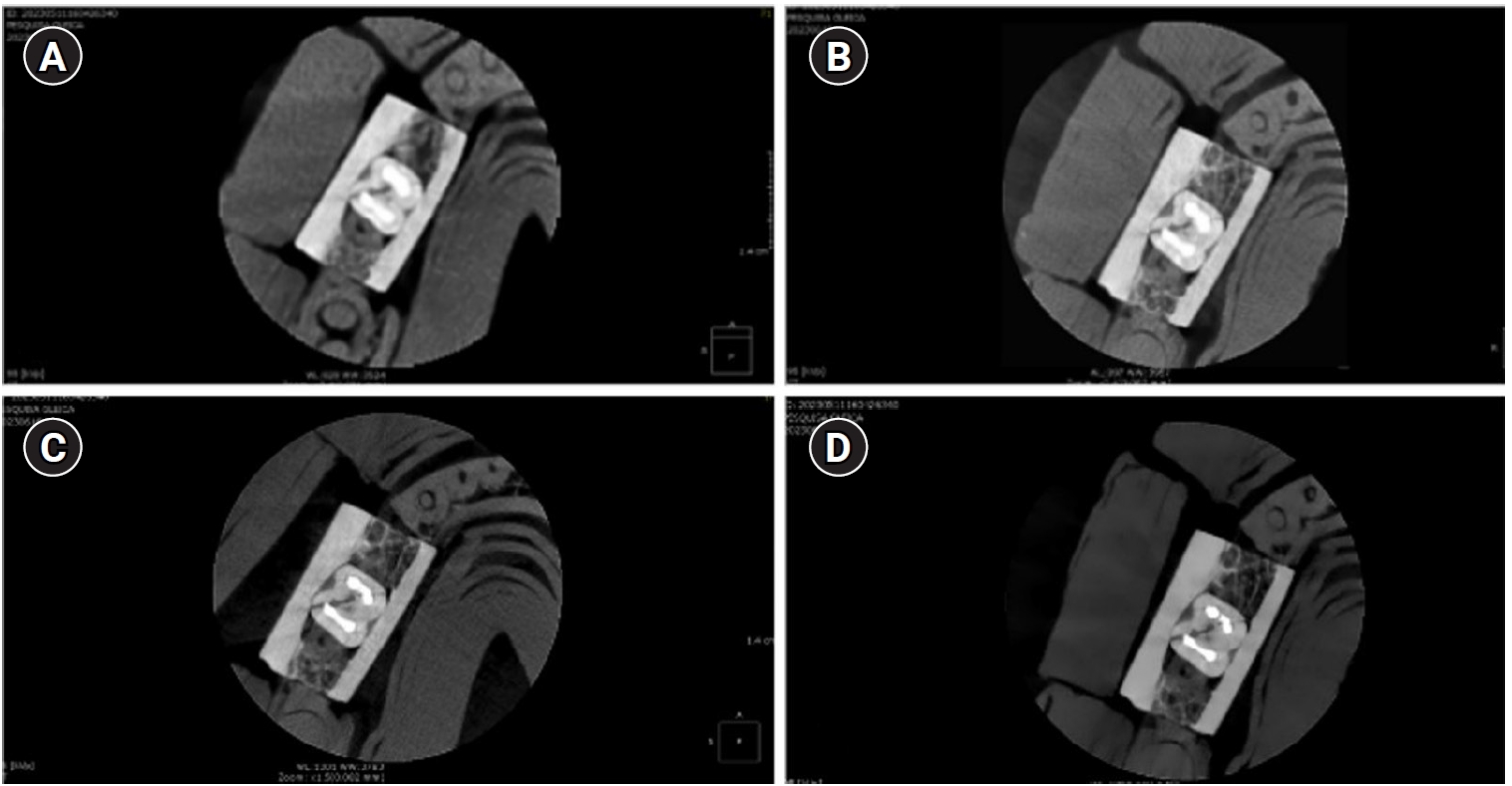
This study aimed to evaluate the influence of cone-beam computed tomography (CBCT) acquisition protocol, the presence of intraradicular metal post, and examiner experience on the detection of complete root fractures in multi-rooted teeth.
Methods
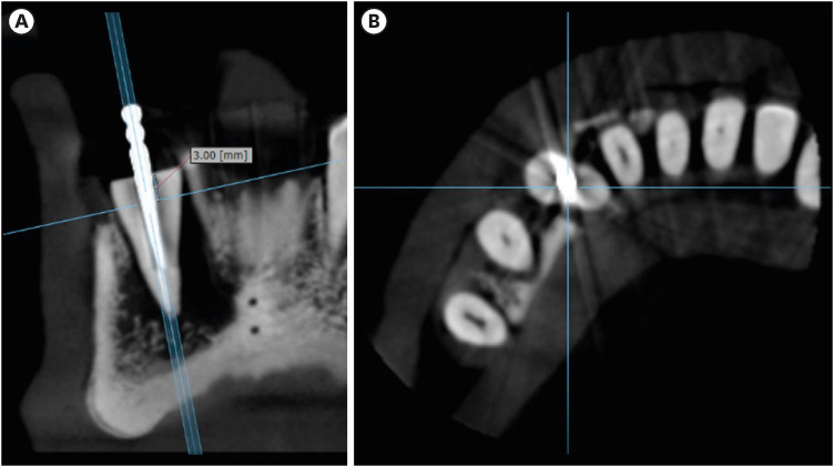
Twenty human molar teeth filled with gutta-percha were placed into artificial alveoli created in bovine ribs. The sample was divided into two groups based on the presence or absence of intraradicular posts in the distal roots. CBCT scans were obtained using four acquisition protocols with varying voxel sizes (0.28, 0.2, 0.125, and 0.80 mm). Following the creation of controlled fractures using a chisel and hammer, CBCT imaging was repeated, resulting in 160 images. Five examiners assessed the images using OnDemand software (KaVo Dental GmbH). Sensitivity, specificity, and accuracy were calculated for each examiner, CBCT protocol, and post-condition. Statistical comparisons were performed using Cochran’s Q test and McNemar test, and a significance level of 5%.
Results
In teeth without metallic posts, sensitivity, specificity, and accuracy values exceeded 0.70, 0.70, and 0.80, respectively. However, the presence of metallic posts significantly reduced diagnostic performance, particularly in low-resolution protocols evaluated by less-experienced examiners.
Conclusions
CBCT acquisition protocols should be selected based on the presence of metallic posts to optimize root fracture detection in multi-rooted teeth. Examiner experience also plays a critical role in diagnostic accuracy.
- 2,953 View
- 107 Download

-
Influence of CBCT parameters on image quality and the diagnosis of vertical root fractures in teeth with metallic posts: an
ex vivo study - Larissa Pereira Lagos de Melo, Polyane Mazucatto Queiroz, Larissa Moreira-Souza, Mariana Rocha Nadaes, Gustavo Machado Santaella, Matheus Lima Oliveira, Deborah Queiroz Freitas
- Restor Dent Endod 2023;48(2):e16. Published online April 27, 2023
- DOI: https://doi.org/10.5395/rde.2023.48.e16
-
Abstract
PDFPubReaderePub
Objectives The aim of this study was to evaluate the influence of peak kilovoltage (kVp) and a metal artifact reduction (MAR) tool on image quality and the diagnosis of vertical root fracture (VRF) in cone-beam computed tomography (CBCT).
Materials and Methods Twenty single-rooted human teeth filled with an intracanal metal post were divided into 2 groups: control (
n = 10) and VRF (n = 10). Each tooth was placed into the socket of a dry mandible, and CBCT scans were acquired using a Picasso Trio varying the kVp (70, 80, 90, or 99), and the use of MAR (with or without). The examinations were assessed by 5 examiners for the diagnosis of VRF using a 5-point scale. A subjective evaluation of the expression of artifacts was done by comparing random axial images of the studied protocols. The results of the diagnoses were analyzed using 2-way analysis of variance and the Tukeypost hoc test, the subjective evaluations were compared using the Friedman test, and intra-examiner reproducibility was evaluated using the weighted kappa test (α = 5%).Results The kVp and MAR did not influence the diagnosis of VRF (
p > 0.05). According to the subjective classification, the 99 kVp protocol with MAR demonstrated the least expression of artifacts, while the 70 kVp protocol without MAR led to the most artifacts.Conclusions Protocols with higher kVp combined with MAR improved the image quality of CBCT examinations. However, those factors did not lead to an improvement in the diagnosis of VRF.
-
Citations
Citations to this article as recorded by
- Digital Dentistry Society Quality Forum: Clinical recommendations on cone-beam computed tomography for the digital dentistry workflow
Hugo Gaêta-Araujo, Rocharles Cavalcante Fontenele, Reinhilde Jacobs
Digital Dentistry Journal.2026; 3(1): 100065. CrossRef - Photon‐Counting CT for Diagnosing Vertical Root Fractures in Teeth With Metal Posts: An Ex Vivo Comparative Analysis With Four CBCT Devices
Renata M. S. Leal, Fernanda B. Fagundes, Maria F. S. A. Bortoletto, Samuel C. Kluthcovsky, Walter Coudyzer, Bruno C. Cavenago, Reinhilde Jacobs, Rocharles Cavalcante Fontenele
International Endodontic Journal.2026; 59(4): 718. CrossRef - Cone‑beam computed tomography in endodontics: Historical development, technical principles, clinical applications and operative outcomes (Review)
Anshuman Shetty, Shivprasad Rai
World Academy of Sciences Journal.2026; 8(4): 1. CrossRef - Diagnostic Performance of Iterative Reconstruction of Cone-beam Computed Tomography for Detecting Vertical Root Fractures in the Presence of Metal Artifacts
Matheus Barros-Costa, Gustavo Santaella, Christiano Oliveira-Santos, Deborah Queiroz Freitas, William C. Scarfe, Francisco Carlos Groppo
Journal of Endodontics.2025; 51(6): 715. CrossRef - Radiographic and Clinical Outcomes of Laser-Enhanced Disinfection in Endodontic Therapy
Janos Kantor, Sorana Maria Bucur, Eugen Silviu Bud, Victor Nimigean, Ioana Maria Crișan, Mariana Păcurar
Journal of Clinical Medicine.2025; 14(12): 4055. CrossRef - Exploring Diagnostic Reliability of CBCT for Vertical Root Fractures: A Systematic Review and Meta‐Analytical Approach
Luiz Carlos de Lima Dias-Junior, Diego Leonardo de Souza, Adriana Pinto Bezerra, Marcio Correa, Cleonice da Silveira Teixeira, Eduardo Antunes Bortoluzzi, Lucas da Fonseca Roberti Garcia, Stefano Corbella
International Journal of Dentistry.2025;[Epub] CrossRef - Deep learning for dentomaxillofacial cone-beam computed tomography image quality enhancement: A pilot study
Ali Nazari, Seyed Mohammad Yousef Najafi, Reza Abbasi, Hossein Mohammad-Rahimi, Parisa Motie, Mina Iranparvar Alamdari, Mehdi Hosseinzadeh, Ruben Pauwels, Falk Schwendicke
Imaging Science in Dentistry.2025; 55(3): 271. CrossRef - Diagnostic Accuracy of Intraoral, Extraoral and Cone Beam Computed Tomography (CBCT)-Generated Bitewings for Detecting Approximal Caries and Periodontal Bone Loss
Jyoti Mago, Alan G Lurie, Aadarsh Gopalakrishna, Aditya Tadinada
Cureus.2025;[Epub] CrossRef - Vertical root fracture diagnosis in teeth with metallic posts: Impact of metal artifact reduction and sharpening filters
Débora Costa Ruiz, Lucas P. Lopes Rosado, Rocharles Cavalcante Fontenele, Amanda Farias-Gomes, Deborah Queiroz Freitas
Imaging Science in Dentistry.2024; 54(2): 139. CrossRef - Comparing standard- and low-dose CBCT in diagnosis and treatment decisions for impacted mandibular third molars: a non-inferiority randomised clinical study
Kuo Feng Hung, Andy Wai Kan Yeung, May Chun Mei Wong, Michael M. Bornstein, Yiu Yan Leung
Clinical Oral Investigations.2024;[Epub] CrossRef
- Digital Dentistry Society Quality Forum: Clinical recommendations on cone-beam computed tomography for the digital dentistry workflow
- 3,963 View
- 64 Download
- 7 Web of Science
- 10 Crossref
- Effect of post space preparation drills on the incidence of root dentin defects
- Thaíse Ayres Bezerra Zuli, Orlando Aguirre Guedes, Gislaine Figueiredo Zarza Arguello Gonçalves, Aurélio Rosa da Silva Júnior, Álvaro Henrique Borges, Andreza Maria Fábio Aranha
- Restor Dent Endod 2020;45(4):e53. Published online October 16, 2020
- DOI: https://doi.org/10.5395/rde.2020.45.e53
-
Abstract
PDFPubReaderePub
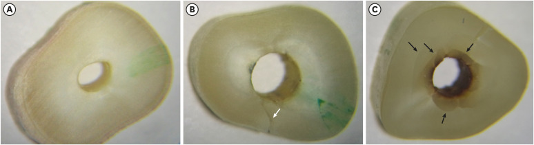
Objectives This study investigated the incidence of root dentin defects after the use of different post space preparation (PSP) drills.
Materials and Methods Seventy-two bovine incisors were selected and obtained 14-mm-long root sections. Twelve roots served as controls with no intervention (G1). The 60 root canals remaining were instrumented using the crown-down technique with the ProTaper Next system and obturated using the lateral condensation technique. Specimens were randomly distributed into 5 groups (
n = 12) according to the operative steps performed: G2, root canal instrumentation and filling (I+F); G3, I+F and PSP with Gates-Glidden drills; G4, I+F and PSP with Largo-Peeso reamers; G5, I+F and PSP with Exacto drill; and G6, I+F and PSP with WhitePost drill. Roots were sectioned at 3, 6, 9, and 12 mm from the apex, and digital images were captured. The presence of root dentin defects was recorded. Data were analyzed by the χ2 test, withp < 0.05 considered to indicate statistical significance.Results Root dentin defects were observed in 39.6% of the root sections. No defects were observed in G1. G5 had significantly more cracks and craze lines than G1, G2, and G3 (
p < 0.05), and more fractures than G1, G2, G3, and G4 (p < 0.05). When all root sections were analyzed together, significantly more defects were observed at the 12-mm level than at the 3-mm level (p < 0.05).Conclusions PSP drills caused defects in the root dentin. Gates-Glidden drills caused fewer root defects than Largo-Peeso reamers and Exacto drills.
-
Citations
Citations to this article as recorded by- Fracture Strength of CAD/CAM Endocrown and Post-Core Restorations with Fiber Strip Reinforcement in Mandibular Premolars
Kerem Yılmaz, Hakan Aydın, Zeynep Soylu, Özge Çiloğlu, Esma Fatıma Delican, Mehmet Mustafa Özarslan, Fehmi Gönüldaş
Journal of Functional Biomaterials.2026; 17(5): 248. CrossRef - Evaluation of dentinal crack formation during post space preparation using different fiber post systems with micro-computed tomography
Ayşe Nur Kuşuçar, Damla Kırıcı
BMC Oral Health.2025;[Epub] CrossRef - Fracture and Crack Behavior of Weakened Incisors Restored With Fiber Posts, Polyethylene Reinforcement, or 3D-Printed Endocrowns
Diana Codas-Duarte, Laís L Pelozo, Jardel F Mazzi-Chaves, Fabiane C Lopes-Olhê, Manoel D Sousa-Neto, Aline E Souza-Gabriel
Cureus.2025;[Epub] CrossRef - Selecting drill size for post space preparation based on final endodontic radiographs: An in vitro study
Farzaneh Farid, Julfikar Haider, Marjan Sadeghpour Shahab, Nika Rezaeikalantari
Technology and Health Care.2024; 32(4): 2575. CrossRef - Cone Beam Computed Tomography Analysis of Post Space in Bifurcated Premolars Using ParaPost and Peeso Reamer Drills
Abdulaziz Saleh Alqahtani, Omar Nasser Almonabhi, Abdulmajeed Moh. Almutairi, Reem R. Alnatsha
The Open Dentistry Journal.2024;[Epub] CrossRef - A Comparative Evaluation of Real-Time Guided Dynamic Navigation and Conventional Techniques for Post Space Preparation During Post Endodontic Management: An In Vitro Study
Sherifa Shervani, Sihivahanan Dhanasekaran, Vijay Venkatesh
Cureus.2024;[Epub] CrossRef - The effect of ultrasonic vibration protocols for cast post removal on the incidence of root dentin defects
Giulliano C. Serpa, Orlando A. Guedes, Neurinelma S. S. Freitas, Julio A. Silva, Carlos Estrela, Daniel A. Decurcio
Journal of Oral Science.2023; 65(3): 190. CrossRef
- Fracture Strength of CAD/CAM Endocrown and Post-Core Restorations with Fiber Strip Reinforcement in Mandibular Premolars
- 3,460 View
- 39 Download
- 7 Crossref
Case Reports
- Fiber-reinforced composite resin bridges: an alternative method to treat root-fractured teeth
- Gun Heo, Eun-Hye Lee, Jin-Woo Kim, Kyung-Mo Cho, Se-Hee Park
- Restor Dent Endod 2020;45(1):e8. Published online December 27, 2019
- DOI: https://doi.org/10.5395/rde.2020.45.e8
-
Abstract
PDFPubReaderePub
The replacement of missing teeth, especially in the anterior region, is an essential part of dental practice. Fiber-reinforced composite resin bridges are a conservative alternative to conventional fixed dental prostheses or implants. It is a minimally invasive, reversible technique that can be completed in a single visit. The two cases presented herein exemplify the treatment of root-fractured anterior teeth with a natural pontic immediately after extraction.
-
Citations
Citations to this article as recorded by- Prosthodontic Aspects of Splinting the Mandibular Anterior Teeth by Fiber Reinforced Composites
Hrelja Miroslav, Laškarin Mirko, Čimić Samir, Kraljević Sonja, Dulčić Nikša, Badel Tomislav
Journal of Dental Problems and Solutions.2025; 12(1): 004. CrossRef - Current Evidence on the Fiber-reinforced Composite Bridges
Ramesh Chowdhary, Sunil Kumar Mishra
International Journal of Prosthodontics and Restorative Dentistry.2023; 12(4): 159. CrossRef - Bridging the Gap: A Case Report of Tooth Replacement using Resin-Bonded Fiber- Reinforced Composite Resin
Vineet Sharma, Sumit Bhansali, Sonal Priya Bhansali
Journal of Pierre Fauchard Academy (India Section).2023; : 66. CrossRef - Reconstruction of Natural Smile and Splinting with Natural Tooth Pontic Fiber‐Reinforced Composite Bridge
Maryam S. Tavangar, Fatemeh Aghaei, Massoumeh Nowrouzi, Andrea Scribante
Case Reports in Dentistry.2022;[Epub] CrossRef
- Prosthodontic Aspects of Splinting the Mandibular Anterior Teeth by Fiber Reinforced Composites
- 2,766 View
- 23 Download
- 4 Crossref
- Microsurgical re-treatment of an endodontically treated tooth with an apically located incomplete vertical root fracture: a clinical case report
- Silvio Taschieri, Massimo Del Fabbro, Ahmed El Kabbaney, Igor Tsesis, Eyal Rosen, Stefano Corbella
- Restor Dent Endod 2016;41(4):316-321. Published online June 21, 2016
- DOI: https://doi.org/10.5395/rde.2016.41.4.316
-
Abstract
PDFPubReaderePub
Although it is challenging, the early diagnosis of a vertical root fracture (VRF) is crucial in order to ensure tooth preservation. The purpose of this clinical case report was to describe reparative surgery performed to treat a tooth affected by an incomplete VRF. A 26 year old male patient was suspected to have a VRF in a maxillary left central incisor, and an exploratory flap was performed in order to confirm the diagnosis. After detecting the fracture, the lesion was surgically treated, the fracture and the infected root-end were removed, and a platelet-rich plasma membrane was used to cover the defect in order to prevent bacterial migration. A 24 month clinical and radiological follow-up examination showed that the tooth was asymptomatic and that the healing process was in progress. The surgical approach described here may be considered an effective treatment for a combined endodontic-periodontal lesion originating from an incomplete VRF and a recurrent periapical lesion.
-
Citations
Citations to this article as recorded by- Biomechanical perspectives on dentine cracks and fractures: Implications in their clinical management
Sishi Chen, Dwayne Arola, Domenico Ricucci, Brian E. Bergeron, John A. Branton, Li-sha Gu, Franklin R. Tay
Journal of Dentistry.2023; 130: 104424. CrossRef - Efficacy of Autologous Platelet Concentrates in Regenerative Endodontic Treatment: A Systematic Review of Human Studies
Joanna Metlerska, Irini Fagogeni, Alicja Nowicka
Journal of Endodontics.2019; 45(1): 20. CrossRef - The preservation of teeth with root-originated fractures
Eyal Rosen, Ilan Beitlitum, Igor Tsesis
Evidence-Based Endodontics.2018;[Epub] CrossRef
- Biomechanical perspectives on dentine cracks and fractures: Implications in their clinical management
- 3,150 View
- 28 Download
- 3 Crossref
- Esthetic enhancement of a traumatized anterior tooth with a combination of forced eruption and tooth alignment: a case report
- So-Hee Kang, Jung-Hong Ha, Myoung-Uk Jin, Sung-Kyo Kim, Young-Kyung Kim
- Restor Dent Endod 2016;41(3):210-217. Published online June 1, 2016
- DOI: https://doi.org/10.5395/rde.2016.41.3.210
-
Abstract
PDFPubReaderePub
Exposing sound structure of a subgingivally fractured tooth using orthodontic extrusion is considered to be a conservative way to re-establish biologic width without sacrificing esthetics or jeopardizing periodontal support of neighboring teeth. When a misaligned tooth is traumatically involved, a more comprehensive approach combining tooth extrusion and re-alignment may be necessary for a successful restorative outcome. This case report describes a successful esthetic management of a patient with complicated crown-root fracture on the maxillary right central incisor and pre-existing malocclusion in the maxillary anterior region. Forced eruption along with re-alignment of teeth by orthodontic movement seems to allow re-positioning of the fracture line to a favorable position and correction of crowding, providing a better esthetic result.
-
Citations
Citations to this article as recorded by- Effects of systematic bisphosphonate use in patients under orthodontic treatment: a systematic review
Vasileios F Zymperdikas, Maria P Yavropoulou, Eleftherios G Kaklamanos, Moschos A Papadopoulos
European Journal of Orthodontics.2020; 42(1): 60. CrossRef - In vitro retention efficiency of temporary type zinc oxide cement for orthodontic forced eruption
Renato Nieto-Aguilar, Deyanira Serrato-Ochoa, Rafael Medina-Navarro, Asdrúbal Aguilera-Méndez, Karina Denisse Morales-Soto, Juan Pablo Loyola-Rodriguez, Antonio Campos, Miguel Alaminos
International Orthodontics.2019; 17(1): 96. CrossRef
- Effects of systematic bisphosphonate use in patients under orthodontic treatment: a systematic review
- 2,740 View
- 22 Download
- 2 Crossref
- Management of horizontal root fractures by fabrication of canine protected occlusion using composite resin
- Joo-Hee Shin, Ryan Jin-Young Kim
- Restor Dent Endod 2012;37(3):180-184. Published online August 29, 2012
- DOI: https://doi.org/10.5395/rde.2012.37.3.180
-
Abstract
PDFPubReaderePub
Traumatic injuries of the face often involve root fractures especially in anterior teeth. The prognosis and the treatment of the root fracture depend on the extent of the fracture line, general health and patient compliance. This case report outlines a new conservative trial treatment modality to stabilize the maxillary central incisors with horizontal root fracture on the cervical to middle third by fabricating canine guidance to remove loading on the traumatized maxillary central incisors during eccentric movements and thus inducing spontaneous healing of the fractured line between the fragments. Radiographs after thirty months showed adequate healing with no signs of pathological changes including root resorption, ankylosis or displacement. Long term follow-up revealed that vitality, stability and aesthetics were maintained and the patient was satisfied with the outcome.
-
Citations
Citations to this article as recorded by- Healing after horizontal root fractures: 3 cases with 2-year follow-up
Yoorina Choi, Sung-Ok Hong, Seok-Ryun Lee, Kyung-San Min, Su-Jung Park
Restorative Dentistry & Endodontics.2014; 39(2): 126. CrossRef
- Healing after horizontal root fractures: 3 cases with 2-year follow-up
- 1,926 View
- 4 Download
- 1 Crossref
- Treatment of crown-root fracture with a modified crown fragment reattachment technique
- Chang-Won Song, Min-Ju Song, Su-Jung Shin, Jeong-Won Park
- J Korean Acad Conserv Dent 2010;35(5):395-401. Published online September 30, 2010
- DOI: https://doi.org/10.5395/JKACD.2010.35.5.395
-
Abstract
PDFPubReaderePub
The development of adhesive dentistry has allowed that the crown fragment reattachment can be another option in the treatment of crown fracture. However, additional crown lengthening procedure or extrusion of the tooth may be necessary in the treatment of crown root fracture because subgingival fracture line in close proximity to the alveolar bone leads to challenges for restorative procedure and the violation of the biologic width. This case report presents a modified crown fragment reattachment technique of crown root fracture with pulp exposure, which was done without additional crown lengthening procedures. After the endodontic treatment, the patient was treated using a post insertion and the fragment reattachment technique, which made it possible to preserve the space for the biologic width and maintain a dry surgical field for adequate adhesion through the modification of the fractured coronal fragment. Since a coronal fracture was occurred and reattached afterward, it was observed that the coronal fragment was well maintained without the additional loss of periodontal attachment through 2-year follow up.
- 1,468 View
- 23 Download
Original Article
- Fracture resistance of crown-root fractured teeth repaired with dual-cured composite resin and horizontal posts
- Seok-Woo Chang, Yong-Keun Lee, Seung-Hyun Kyung, Hyun-Mi Yoo, Tae-Seok Oh, Dong-Sung Park
- J Korean Acad Conserv Dent 2009;34(5):383-389. Published online September 30, 2009
- DOI: https://doi.org/10.5395/JKACD.2009.34.5.383
-
Abstract
PDFPubReaderePub
The purpose of this study was to investigate the fracture resistance of crown-root fractured teeth repaired with dual-cured composite resin and horizontal posts. 48 extracted human premolars were assigned to control group and three experimental groups. Complete crown-root fractures were experimentally induced in all control and experimental teeth. In the control group, the teeth (n=12) were bonded with resin cement and endodontically treated. Thereafter, the access cavities were sealed with dual-cured composite resin. In composite resin core - post group (n=12), the teeth were endodontically treated and access cavities were sealed with dual-cured composite resin. In addition, the fractured segments in this group were fixed using horizontal posts. In composite resin core group (n=12), the teeth were endodontically treated and the access cavities were filled with dual-cured composite resin without horizontal posts. In bonded amalgam group (n=12), the teeth were endodontically treated and the access cavities were sealed with bonded amalgam. Experimental complete crown-root fractures were induced again on repaired control and experimental teeth. The ratio of fracture resistance to original fracture resistance was analyzed with Kruskal-Wallis test. The results showed that teeth in control and composite resin core - post group showed significantly higher resistance to re-fracture than those in amalgam core group (
p < 0.05). The resistance to refracture was high in the order of composite resin - post group, control group, composite resin group and bonded amalgam group. Within the scope of this study, the use of horizontal post could be beneficial in increasing the fracture resistance of previously fractured teeth.
- 1,657 View
- 4 Download
Case Report
- Anterior esthetic improvement through orthodontic extrusive remodeling and single-unit implantation in a fractured upper lateral incisor with alveolar bone loss: A case report
- Soo-Youn Hwang, Won-Jun Shon, Young-Chul Han, Kwang-Shik Bae, Seung-Ho Back, WooCheol Lee, Kee-Yeon Kum
- J Korean Acad Conserv Dent 2008;33(1):39-44. Published online January 31, 2008
- DOI: https://doi.org/10.5395/JKACD.2008.33.1.039
-
Abstract
PDFPubReaderePub
The treatment of esthetic areas with single-tooth implants represents a new challenge for the clinician. In 1993, a modification of the forced eruption technique, called "orthodontic extrusive remodelling," was proposed as a way to augment both soft- and hard-tissue profiles at potential implant sites. This case report describes augmentation of the coronal soft and hard tissues around a fractured maxillary lateral incisor associated with alveolar bone loss, which was achieved by forced orthodontic extrusion before implant placement. Through these procedures we could reconstruct esthetics and function in a hopeless tooth diagnosed with subgingival root fracture by trauma.
- 1,102 View
- 2 Download
 First
First Prev
Prev

