
Search
- Page Path
- HOME > Search
Research Articles
- The effect of limonene extract on the adhesion of different endodontic cements to root dentin: an in vitro experimental study
- Nayara Lima Ferraz Aguiar, Eduardo José Soares, Guilherme Nilson Alves dos Santos, Anna Luísa Araújo Pimenta, Laryssa Karla Romano, Ricardo Gariba Silva, Fernanda de Carvalho Panzeri
- Restor Dent Endod 2025;50(2):e16. Published online May 12, 2025
- DOI: https://doi.org/10.5395/rde.2025.50.e16
-
 Abstract
 PDF
PDF PubReader
 ePub
ePub - Objectives
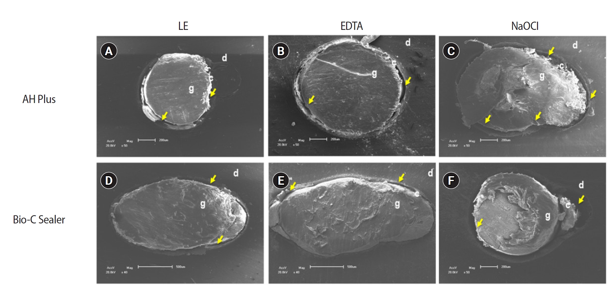
The study aimed to evaluate the effect of limonene extract (LE) on push-out bond strength (BS) to root dentin in endodontically treated teeth.
Methods
Single-rooted teeth were selected and instrumented using the reciprocating technique, then divided into three groups based on the final irrigating solution: 2.5% sodium hypochlorite (NaOCl), 17% ethylenediaminetetraacetic acid (EDTA), and 5% LE. The roots were further divided (n = 12) and obturated using the single-cone technique with epoxy resin-based (ERB) or bioceramic sealer (Bio-C). After 3 days, the roots were sectioned into 2-mm slices, obtaining two slices from each root third. Push-out BS testing was conducted at 0.5 mm/min, followed by failure pattern and adhesive interface analysis using scanning electron microscopy. Push-out BS data were analyzed by three-way analysis of variance and Tukey post-hoc test (p < 0.05).
Results
ERB showed higher BS when irrigated with EDTA (5.0 ± 2.3 MPa) compared to NaOCl (1.8 ± 1.1 MPa) (p = 0.0005), particularly in the cervical third. LE yielded intermediate values without significant differences from the other irrigants (3.5 ± 1.9 MPa) (p > 0.05). For Bio-C, the highest BS was observed in the apical third, especially with LE (9.4 ± 5.0 MPa), differing from other thirds and final irrigating solutions (p < 0.05). Mixed failure patterns were most prevalent, regardless of the irrigant solutions.
Conclusions
The combination of LE with Bio-C demonstrated superior BS in the apical third, suggesting its potential as a final irrigating solution in endodontic treatments.
- 3,383 View
- 242 Download

- Bone repair in defects filled with AH Plus sealer and different concentrations of MTA: a study in rat tibiae
- Jessica Emanuella Rocha Paz, Priscila Oliveira Costa, Albert Alexandre Costa Souza, Ingrid Macedo de Oliveira, Lucas Fernandes Falcão, Carlos Alberto Monteiro Falcão, Maria Ângela Area Leão Ferraz, Lucielma Salmito Soares Pinto
- Restor Dent Endod 2021;46(4):e48. Published online September 2, 2021
- DOI: https://doi.org/10.5395/rde.2021.46.e48
-
Abstract
PDFPubReaderePub
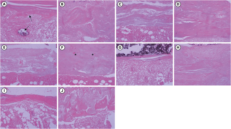
Objectives This study aimed to evaluate the effects on bone repair of different concentrations of mineral trioxide aggregate (MTA) added to AH Plus.
Materials and Methods Bone tissue reactions were evaluated in 30 rats (
Rattus norvegicus ) after 7 and 30 days. In the AH + MTA10, AH + MTA20, and AH + MTA30 groups, defects in the tibiae were filled with AH Plus with MTA in proportions of 10%, 20% and 30%, respectively; in the MTA-FILL group, MTA Fillapex was used; and in the control group, no sealer was used. The samples were histologically analyzed to assess bone union and maturation. The Kruskal-Wallis and Mann-Whitney tests were performed for multiple pairwise comparisons (p ≤ 0.05).Results At the 7-day time point, AH + MTA10 was superior to MTA-FILL with respect to bone union, and AH + MTA20 was superior to MTA-FILL with respect to bone maturity (
p < 0.05). At the 30-day time point, both the AH + MTA10 and AH + MTA20 experimental sealers were superior not only to MTA-FILL, but also to AH + MTA30 with respect to both parameters (p < 0.05). The results of the AH + MTA10 and AH + MTA20 groups were superior to those of the control group for both parameters and experimental time points (p < 0.05).Conclusions The results suggest the potential benefit of using a combination of these materials in situations requiring bone repair.
-
Citations
Citations to this article as recorded by
- Analysis of the cytotoxicity and bioactivity of CeraSeal, BioRoot™ and AH Plus® sealers in pre-osteoblast lineage cells
Luciano Aparecido de Almeida-Junior, Giuliana de Campos Chaves Lamarque, Henry Herrera, Maya Fernanda Manfrin Arnez, Francine Lorencetti-Silva, Raquel Assed Bezerra Silva, Léa Assed Bezerra Silva, Francisco Wanderley Garcia Paula-Silva
BMC Oral Health.2024;[Epub] CrossRef - A Review of the research methods and progress of biocompatibility evaluation of root canal sealers
Xiliang Yang, Tianxia Zheng, Nuoya Yang, Zihan Yin, Wuliang Wang, Yuhong Bai
Australian Endodontic Journal.2023; 49(S1): 508. CrossRef - Effect of Vitapex Combined with AH-Plus Paste on Inflammation in Middle-Aged and Elderly Patients with Periodontal-Endodontic Disease
Rong Hu, Fulan Zhang, Xiangyu Guo, Youren Jing, Xiaowan Lin, Liping Tian, Min Tang
Computational and Mathematical Methods in Medicine.2022; 2022: 1. CrossRef
- Analysis of the cytotoxicity and bioactivity of CeraSeal, BioRoot™ and AH Plus® sealers in pre-osteoblast lineage cells
- 2,803 View
- 17 Download
- 4 Web of Science
- 3 Crossref
- Effects of radiation therapy on the dislocation resistance of root canal sealers applied to dentin and the sealer-dentin interface: a pilot study
- Pallavi Yaduka, Rubi Kataki, Debosmita Roy, Lima Das, Shachindra Goswami
- Restor Dent Endod 2021;46(2):e22. Published online March 29, 2021
- DOI: https://doi.org/10.5395/rde.2021.46.e22
-
Abstract
PDFPubReaderePub
Objectives This study evaluated and compared the effects of radiation therapy on the dislocation resistance of AH Plus and BioRoot RCS applied to dentin and the sealer-dentin interface.
Materials and Methods Thirty single-rooted teeth were randomly assigned to 2 groups (
n = 15 each): AH Plus (Dentsply DeTrey) and BioRoot RCS (Septodont). Each group was subdivided into control and experimental groups. The experimental group was subjected to a total radiation dose of 60 Gy. The root canals of all samples were cleaned, shaped, and obturated using the single-cone technique. Dentin slices (1 mm) were sectioned from each root third for the push-out test and scanning electron microscopy (SEM) was done to examine the sealer-dentin interface. The failure mode was determined using stereomicroscopy. Bond strength data were analyzed by the independentt -test, 1-way analysis of variance, and the Tukeypost hoc test (α = 0.05).Results Significantly lower bond strength was observed in irradiated teeth than non-irradiated teeth in the AH Plus group (
p < 0.05). The BioRoot RCS group showed no significant reduction in bond strength after irradiation (p > 0.05) and showed a higher post-irradiation bond strength (209.92 ± 172.26 MPa) than the AH Plus group. SEM revealed slightly larger gap-containing regions in irradiated specimens from both groups.Conclusions The dislocation resistance of BioRoot RCS was not significantly changed by irradiation and was higher than that of AH Plus. BioRoot RCS may be the sealer of choice for root canal treatment in patients undergoing radiation therapy.
-
Citations
Citations to this article as recorded by- The impact of radiotherapy on endodontic treatment: a scoping review
Guilherme Pauletto, Giovanna Isabel Mittmann Voigt, Sidnei Flores de Pellegrin, Yasmin Padoin, Carlos Alexandre Souza Bier
Odontology.2026; 114(1): 24. CrossRef - Effects of radiotherapy dose and endodontic irrigants on universal resin cement bonding to root dentin: mechanical and interfacial analyses
Lívia Ribeiro, Luíz Carlos de Lima Dias-Júnior, Paulo Henrique dos Santos, Mariana Comparotto Minamisako, Paulo Marcelo Rodrigues, Vicente Ribeiro Netto, Bruno Alexandre Pacheco de Castro Henriques, Renata Gondo Machado, Cleonice da Silveira Teixeira, Luc
International Journal of Adhesion and Adhesives.2026; 146: 104252. CrossRef - Push-out bond strength of bioceramic-based sealers following different irrigation activation techniques
Fatma Begüm Peker, Hümeyra Çapkın, Ahsen Narbay
Journal of Applied Biomaterials & Functional Materials.2026;[Epub] CrossRef - Endodontic Management Before, During and After Head and Neck Radiotherapy: Biological, Diagnostic and Clinical Considerations in Head and Neck Cancer Patients
Jing‐zhi Ma, Franklin Tay
International Endodontic Journal.2026;[Epub] CrossRef - Effect of radiation therapy on the micro push-out bond strength of tricalcium silicate-based repair cements used for furcation perforation sealing
Emanuelle Luize Meurer, Bruna Venzke Fischer, Luíz Carlos de Lima Dias-Júnior, Augusto Vanni Bodanezi, Mariana Comparotto Minamisako, Paulo Marcelo Rodrigues, Vicente Ribeiro Netto, Cleonice da Silveira Teixeira, Lucas da Fonseca Roberti Garcia
International Journal of Adhesion and Adhesives.2026; 150: 104390. CrossRef - Impact of radiation therapy regimen on the dislodgement resistance of endodontic sealers: A micro push-out test
Marcos Testa Magoga, Rafaela Lourdes de Sousa, Luiz Carlos Lima Dias-Junior, Rayssa Sabino-Silva, Mariana Comparotto Minamisako, Paulo Marcelo Rodrigues, Vicente Ribeiro Netto, Ricardo Machado, Cleonice da Silveira Teixeira, Lucas da Fonseca Roberti Garci
International Journal of Adhesion and Adhesives.2025; 136: 103894. CrossRef - Evaluation of the root dentin bond strength and intratubular biomineralization of a premixed calcium aluminate-based hydraulic bioceramic endodontic sealer
Yu-Na Lee, Min-Kyeong Kim, Hee-Jin Kim, Mi-Kyung Yu, Kwang-Won Lee, Kyung-San Min
Journal of Oral Science.2024; 66(2): 96. CrossRef - Effects of radiotherapy dose and application time on the load-to-failure values of teeth filled with different sealers
Ozgun Gulderen, Esma Saricam, Sedef Gökhan Açikgöz, Yılmaz Tezcan
BMC Oral Health.2024;[Epub] CrossRef - Ultrasonic activation of the endodontic sealer enhances its intratubular penetration and bond strength to irradiated root dentin
Luana Duart Jordani, Amanda Freitas da Rosa, Luiz Carlos de Lima Dias-Junior, Julia Menezes Savaris, Mariana Comparotto Minamisako, Luciano Roberto da Silva, Marcio Toshio Umeda Takashima, Eduardo Antunes Bortoluzzi, Cleonice da Silveira Teixeira, Lucas d
Odontology.2024; 112(3): 917. CrossRef - Effect of the timing of primary endodontic treatment and dosage of radiation therapy on the filling material removal
Bruna Venzke Fischer, Luiz Carlos de Lima Dias‐Junior, Mariana Comparotto Minamisako, Cristiane Maria Almeida, Luciano Roberto da Silva, Eduardo Antunes Bortoluzzi, Cleonice da Silveira Teixeira, Lucas da Fonseca Roberti Garcia
Australian Endodontic Journal.2024; 50(2): 321. CrossRef - Does radiation therapy affect adhesion of tricalcium silicate cements to root dentin?
Lochan KHULLAR, Nidambur Vasudev BALLAL, Tan Fırat EYÜBOĞLU, Mutlu ÖZCAN
Journal of Applied Oral Science.2023;[Epub] CrossRef - Effect of the timing of radiation therapy on the push‐out strength of resin cement to root dentine
Patrícia da Agostim Cancelier, Renata Gondo Machado, Júlia Menezes Savaris, Eduardo Antunes Bortoluzzi, Cleonice da Silveira Teixeira, Mariana Comparotto Minamisako, Paulo Marcelo Rodrigues, Vicente Ribeiro Netto, Kamile Leonardi Dutra‐Horstmann, Lucas da
Australian Endodontic Journal.2023; 49(S1): 122. CrossRef - Influence of irrigation and laser assisted root canal disinfection protocols on dislocation resistance of a bioceramic sealer
Ivona Bago, Ana Sandrić, Katarina Beljic-Ivanovic, Boris Pažin
Photodiagnosis and Photodynamic Therapy.2022; 40: 103067. CrossRef - Influence of 2% chlorhexidine on the dislodgement resistance of AH plus, bioroot RCS, and GuttaFlow 2 sealer to dentin and sealer-dentin interface
Debosmita Roy, Rubi Kataki, Lima Das, Khushboo Jain
Journal of Conservative Dentistry.2022; 25(6): 642. CrossRef
- The impact of radiotherapy on endodontic treatment: a scoping review
- 3,228 View
- 32 Download
- 14 Web of Science
- 14 Crossref
- Flow characteristics and alkalinity of novel bioceramic root canal sealers
- Anastasios Katakidis, Konstantinos Sidiropoulos, Elisabeth Koulaouzidou, Christos Gogos, Nikolaos Economides
- Restor Dent Endod 2020;45(4):e42. Published online August 18, 2020
- DOI: https://doi.org/10.5395/rde.2020.45.e42
-
Abstract
PDFPubReaderePub
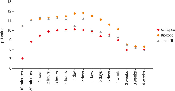
Objective This study aimed to examine the physical properties (pH and flow) of 2 novel bioceramic sealers.
Materials and Methods The tested sealers were a calcium hydroxide sealer (Sealapex) and 2 bioceramic sealers (BioRoot RCS and TotalFill BC Sealer). Flow measurements were conducted according to ISO 6876/2012, with a press method of 0.05 mL of sealer. The pH of fresh samples was tested immediately after manipulation, while set samples were stored for 3 times the recommended setting time. The predetermined time intervals ranged from 3 minutes to 24 hours for fresh samples and from 10 minutes to 7 days and 4 weeks for the set samples. Analysis of variance was performed, with
p = 0.05 considered indicating significance.Results The mean flow values were 26.99 mm for BioRoot, 28.19 for Sealapex, and 30.8 mm for TotalFill BC Sealer, satisfying the ISO standard. In the set samples, BioRoot RCS had higher pH values at 24 hours to 1 week after immersion in distilled water. At 2 weeks, both bioceramic sealers had similar pH values, greater than that of Sealapex. In the fresh samples, the bioceramic sealers had significantly higher initial pH values than Sealapex (
p < 0.05). At 24 hours post-immersion, all sealers showed an alkaline pH, with the highest pH observed for TotalFill.Conclusions The TotalFill BC Sealer demonstrated the highest flow. The bioceramic sealers initially presented higher alkaline activity than the polymeric calcium hydroxide sealer. However, at 3 and 4 weeks post-immersion, all sealers had similar pH values.
-
Citations
Citations to this article as recorded by- In vitro comparative evaluation of physicochemical and mechanical properties, cytocompatibility, and antimicrobial efficacy of various bioceramic root canal sealers
Fushi Wang, Jiaxing Li, Jingjing Wan, Siyuan Li, Shijia Tang, Li Wang, Liuyan Meng
Ceramics International.2026; 52(7): 9561. CrossRef - Comparative analysis between resin-based root canal sealer and recent bioceramic-based root canal sealers using MicroCT, film thickness, and solubility
Amira Galal Ismail, Manar M. Galal, Tamer M. Hamdy
Journal of Oral Biology and Craniofacial Research.2026; 16(2): 101400. CrossRef - Setting Characteristics, Solubility, Bioactivity and Interaction with Dentin of Four Calcium Silicate-Based Endodontic Sealers
Areti Dimitra Vrochari, Anastasia Agrafioti, Maria Dimitriadi, George Eliades
Journal of Functional Biomaterials.2026; 17(4): 192. CrossRef - Cryotherapy-Driven Modulation of Postoperative Pain in Single-Visit Endodontic Treatment Across Different Obturation Materials: A Retrospective Study
Kaan Ilıcalı, Ahter Şanal Çıkman, Özge Başar
Journal of Clinical Medicine.2026; 15(10): 3899. CrossRef - Adverse tissue reaction to a next-generation bioceramic sealer: a structured literature review and case report
Saverio Ceraulo, Antonio Barbarisi, Zhong Hao Hu, Ignazio Migliore, Dorina Lauritano, Gianluigi Caccianiga, Francesco Carinci
Frontiers in Dental Medicine.2026;[Epub] CrossRef - Effect of root canal moisture on intratubular penetration of bioceramic sealer using two obturation techniques: A confocal laser scanning microscopic study
Maryam Jasim Al-Qaralusi, Hikmet Abdul-Rahim Al-Gharrawi
Journal of Conservative Dentistry and Endodontics.2026; 29(7): 747. CrossRef - Functional and Bioactive Performance of Premixed Bioceramic Sealers with Warm Obturation: A Scoping Review
Patryk Wiśniewski, Stanisław Krokosz, Małgorzata Pietruska, Anna Zalewska
Gels.2025; 11(11): 932. CrossRef - Physicochemical properties of AH plus bioceramic sealer, Bio-C Sealer, and ADseal root canal sealer
Tamer M. Hamdy, Manar M. Galal, Amira Galal Ismail, Shehabeldin Saber
Head & Face Medicine.2024;[Epub] CrossRef - Characterization and Assessment of Physical Properties of 3 Single Syringe Hydraulic Cement–based Sealers
Veksina Raman, Josette Camilleri
Journal of Endodontics.2024; 50(3): 381. CrossRef - The Impact of Silver Nanoparticles on Dentinal Tubule Penetration of Endodontic Bioceramic Sealer
Sundus Bukhary, Sarah Alkahtany, Amal Almohaimede, Nourah Alkhayatt, Shahad Alsulaiman, Salma Alohali
Applied Sciences.2024; 14(24): 11639. CrossRef - Influence of root canal moisture on the penetration of TotalFill bioceramic sealer into the dentinal tubules: A confocal laser scanning microscopy study
Archika M Singh, Tarek M Elsewify, Walid S El-Sayed, Husam H Nuawafleh, Ranya F Elemam, Bassem M Eid
Saudi Endodontic Journal.2024; 14(2): 187. CrossRef - Unusual Canal Morphology in Mandibular Premolars With Two Distal and One Mesial Canal: A Case Series
Jinesh A, Sanjana Jayakumar Nair, Saurabh Gupta, Harsh Chansoria, Gaurav Rawat
Cureus.2024;[Epub] CrossRef - A scientometric, bibliometric, and thematic map analysis of hydraulic calcium silicate root canal sealers
Anastasios Katakidis, Konstantinos Kodonas, Anastasia Fardi, Christos Gogos
Restorative Dentistry & Endodontics.2023;[Epub] CrossRef - Thermal, chemical and physical analysis of VDW.1Seal, Fill Root ST, and ADseal root canal sealers
Shehabeldin Saber, Manar M. Galal, Amira Galal Ismail, Tamer M. Hamdy
Scientific Reports.2023;[Epub] CrossRef - α-tricalcium phosphate/fluorapatite-based cement - promising dental root canal filling material
Abdul Kazuz, Zeljko Radovanovic, Djordje Veljovic, Vesna Kojic, Dimitar Jakimov, Tamara Vlajic-Tovilovic, Vesna Miletic, Rada Petrovic, Djordje Janackovic
Processing and Application of Ceramics.2022; 16(1): 22. CrossRef
- In vitro comparative evaluation of physicochemical and mechanical properties, cytocompatibility, and antimicrobial efficacy of various bioceramic root canal sealers
- 3,926 View
- 49 Download
- 15 Crossref
-
A comparative evaluation of cytotoxicity of root canal sealers: an
in vitro study - Gautam Pyarelal Badole, Manjusha Madhukar Warhadpande, Ganesh Kothiramji Meshram, Rakesh Namdeoraoji Bahadure, Shubha Gopal Tawani, Gopal Tawani, Shital Gautam Badole
- Restor Dent Endod 2013;38(4):204-209. Published online November 12, 2013
- DOI: https://doi.org/10.5395/rde.2013.38.4.204
-
Abstract
PDFPubReaderePub
Objectives The objective of this
in vitro study was to evaluate and compare the cytotoxicity of four different root canal sealersi.e. Apexit Plus (Ivoclar Vivadent), Endomethasone N (Septodont), AH-26 (Dentsply) and Pulpdent Root Canal Sealer (Pulpdent), on a mouse fibroblast cell line (L929).Materials and Methods Thirty two discs for each sealer (5 mm in diameter and 2 mm in height) were fabricated in Teflon mould. The sealer extraction was made in cell culture medium (Dulbecco's Modified Eagle's Medium, DMEM) using the ratio 1.25 cm2/mL between the surface of the sealer samples and the volume of medium in a shaker incubator. Extraction of each sealer was obtained at 24 hr, 7th day, 14th day, and one month of interval. These extracts were incubated with L929 cell line and 3-(4,5-dimethylthiazol-2yl)-2,5-diphenyltetrazolium bromide (MTT) assay was done. Two-way ANOVA for interaction effects between sealer and time and Post-hoc multiple comparison using Tukey's test across all the 16 different groups were used for statistical analysis.
Results Apexit Plus root canal sealer was significantly less toxic than other sealers (
p < 0.05) and showed higher cellular growth than control. Endomethasone N showed mild cytotoxicity. AH-26 showed severe toxicity which became mild after one month while Pulpdent Root Canal Sealer showed severe to moderate toxicity.Conclusions Apexit Plus was relatively biocompatible sealer as compared to other three sealers which were cytotoxic at their initial stages, however, they became biocompatible with time.
-
Citations
Citations to this article as recorded by- The Evolution of In Vitro Toxicity Assessment Methods for Oral Cavity Tissues—From 2D Cell Cultures to Organ-on-a-Chip
Alexandra Jităreanu, Luminița Agoroaei, Ioana-Cezara Caba, Florina-Daniela Cojocaru, Liliana Vereștiuc, Mădălina Vieriu, Ioana Mârțu
Toxics.2025; 13(3): 195. CrossRef - Comparative analysis of the inflammatory response of human gingival fibroblasts to NeoSEALER Flo and CeraSeal bioceramic sealers: an in vitro study
Sarah Salah Gaafar, Abdel Rahman O. El Mekkawi, Rehab Ali Farag, Mohamed H. A. Gadelmawla, Ahmad Mostafa Hussein Mohamad Hussein, Mohamed Sayed, Mohammad Rayyan, Doaa Gamal AbdelMouez Basta
BMC Oral Health.2025;[Epub] CrossRef - Surface characteristics of nanostructured titanium surface modified via UV irradiation during SLA
S. Khorasani, G. Faraji
Applied Physics A.2025;[Epub] CrossRef - Cytotoxicity and Cell Viability of Two Bioactive Root Canal Sealers, Mineral Trioxide Aggregate, and BioRoot Root Canal Sealer: An In Vitro Study
Emmanuel Samson, Lata B Gangurde, Jaiprakash R Rathod, Pradnya S Jadhav, Sangeeta Ambhore, Pranav S Jadhav
CODS - Journal of Dentistry.2023; 14(2): 57. CrossRef - Human Gingival Fibroblasts Response to Different Endodontic Sealers: An In Vitro Study
Rita Noites, Inês Tavares, Miguel Cardoso, Isabel M. Carreira, Maria Bartolomeu, Ana S. Duarte, Ilda P. Ribeiro
Applied Sciences.2023; 13(19): 10976. CrossRef - Antibacterial and Cytotoxicity of Root Canal Sealer with the Addition of Chitosan Nanoparticle at Various Concentrations
Diatri Nari Ratih, Ema Mulyawati, Rika Kurnia Santi, Yulita Kristanti
European Journal of Dentistry.2023; 17(02): 398. CrossRef - Transforaminal and systemic diffusion of an active agent from a zinc oxide eugenol-based endodontic sealer containing hydrocortisone—in an in vivo model
Davy Aubeux, Anne Valot-Salengro, Gaelle Gautier, Arnaud Malet, Fabienne Pérez
Clinical Oral Investigations.2020; 24(12): 4395. CrossRef - PEGylated curcumin-loaded nanofibrous mats with controlled burst release through bead knot-on-spring design
Mahdi Saeed, Hamid Mirzadeh, Mojgan Zandi, Jalal Barzin
Progress in Biomaterials.2020; 9(4): 175. CrossRef - A New Calcium Silicate-Based Root Canal Dressing: Physical and Chemical Properties, Cytotoxicity and Dentinal Tubule Penetration
Natália Villa, Vanessa Valgas Dos Santos, Ubirajara Maciel da Costa, Aline Teixeira Mendes, Pedro Henrique Marks Duarte, Ricardo Abreu da Rosa, Jefferson Ricardo Pereira, Marcus Vinícius Reis Só
Brazilian Dental Journal.2020; 31(6): 598. CrossRef - Evaluation of the Antimicrobial Efficacy of Herbal Extracts Added to Root Canal Sealers of Different Bases: An In Vitro Study
Abhay M Tripathi, Minarani T Devi, Sonali K Kalra, Ujjala Ghoshal
International Journal of Clinical Pediatric Dentistry.2019; 12(5): 398. CrossRef - Endodontic-related inferior alveolar nerve injuries: A review and a therapeutic flow chart
R. Castro, M. Guivarc'h, J.M. Foletti, J.H. Catherine, C. Chossegros, L. Guyot
Journal of Stomatology, Oral and Maxillofacial Surgery.2018; 119(5): 412. CrossRef - Comparison between direct contact and extract exposure methods for PFO cytotoxicity evaluation
Girish K. Srivastava, Maria L. Alonso-Alonso, Ivan Fernandez-Bueno, Maria T. Garcia-Gutierrez, Fernando Rull, Jesús Medina, Rosa M. Coco, J. Carlos Pastor
Scientific Reports.2018;[Epub] CrossRef - Novel endodontic sealers induce cell cytotoxicity and apoptosis in a dose-dependent behavior and favorable response in mice subcutaneous tissue
L. A. B. Silva, L. U. Azevedo, A. Consolaro, F. Barnett, Y. Xu, R. A. Battaglino, P. S. Cañadas, Katharina Morant Holanda de Oliveira, R. A. B. Silva
Clinical Oral Investigations.2017; 21(9): 2851. CrossRef - Designing and fabrication of curcumin loaded PCL/PVA multi-layer nanofibrous electrospun structures as active wound dressing
Seyed Mahdi Saeed, Hamid Mirzadeh, Mojgan Zandi, Jalal Barzin
Progress in Biomaterials.2017; 6(1-2): 39. CrossRef
- The Evolution of In Vitro Toxicity Assessment Methods for Oral Cavity Tissues—From 2D Cell Cultures to Organ-on-a-Chip
- 2,876 View
- 7 Download
- 14 Crossref
 First
First Prev
Prev

