
Search
- Page Path
- HOME > Search
Research Articles
- Comparison of YouTube, TikTok, and Instagram as digital sources for obtaining information about pulp therapy in primary and permanent teeth
- Hüseyin Gürkan Güneç, Emine Kaya, Dila Nur Okumuş, Merve Gül Erence
- Restor Dent Endod 2025;50(3):e26. Published online July 24, 2025
- DOI: https://doi.org/10.5395/rde.2025.50.e26
-
 Abstract
 PDF
PDF PubReader
 ePub
ePub - Objectives
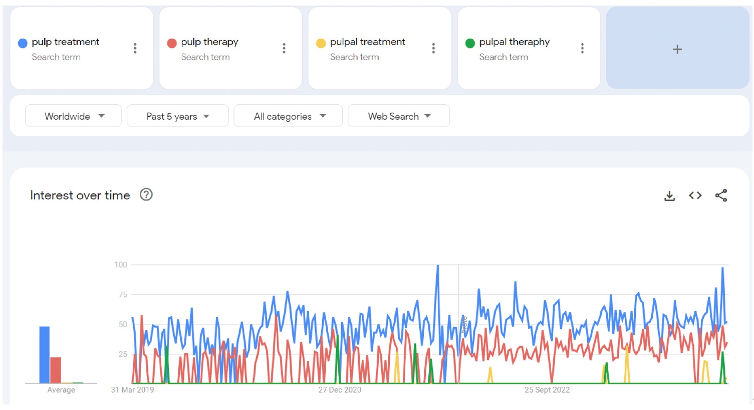
This study aimed to compare the content, educational quality, and dependability of videos on Instagram, TikTok, and YouTube about pulp therapy (PT) in pediatric dentistry and endodontics.
Methods
Three popular video sites, Instagram (Meta Platforms, Inc.,), TikTok (ByteDance Ltd.), and YouTube (Google LLC), were searched for PT content to analyze for compliance with the American Association of Endodontists and American Academy of Pediatric Dentistry guidelines for clinical endodontists and pediatric dentists. The searched hashtags were #pulpaltherapy, #pulpaltreatment, #pulptherapy, and #pulptreatment. The classification of 158 English-language videos was based on several variables: communication quality, duration, likes and dislikes, views, source, treatment, and genre. The videos were evaluated using a usefulness score and the Global Quality Scale (GQS), Video Information and Quality Index (VIQI), Journal of the American Medical Association (JAMA) score, and modified DISCERN score to rate their quality and reliability. The majority of the videos were published by healthcare professionals, dental clinics, and universities.
Results
Significant relationships existed between video length, source of upload, usefulness score, tooth type, pulp status, and VIQI, JAMA, GQS, and DISCERN scores for all three platforms (p<0 .05). A statistically significant relationship existed of YouTube, TikTok, and Instagram with the number of views, number of months since upload, view rates, comments and likes (p< 0.05).
Conclusions
TikTok and Instagram reel videos provided high- to moderate-quality information about PT, especially in children, but YouTube may provide more reliable information than other social media tools. -
Citations
Citations to this article as recorded by
- Evaluating the reliability and educational quality of YouTube™ and TikTok™ videos on custom subperiosteal implants: a cross-sectional methodological analysis
Göksel Tımarcıoğlu, Erdal Cem Kargu, Hülya Çerçi Akçay, Başak Tımarcıoğlu
BMC Oral Health.2026;[Epub] CrossRef - New Technologies and Materials in Oral Health and Dental Care of Pediatric Dentistry
Giuseppe Minervini
Children.2025; 12(10): 1310. CrossRef
- Evaluating the reliability and educational quality of YouTube™ and TikTok™ videos on custom subperiosteal implants: a cross-sectional methodological analysis
- 3,477 View
- 89 Download
- 2 Web of Science
- 2 Crossref

- Color discrepancy of single-shade composites at different distances from the interface measured using cell phone images
- Márcia Luciana Carregosa Santana, Gabriella de Jesus Santos Livi, André Luis Faria-e-Silva
- Restor Dent Endod 2024;49(1):e7. Published online January 24, 2024
- DOI: https://doi.org/10.5395/rde.2024.49.e7
-
Abstract
PDFPubReaderePub
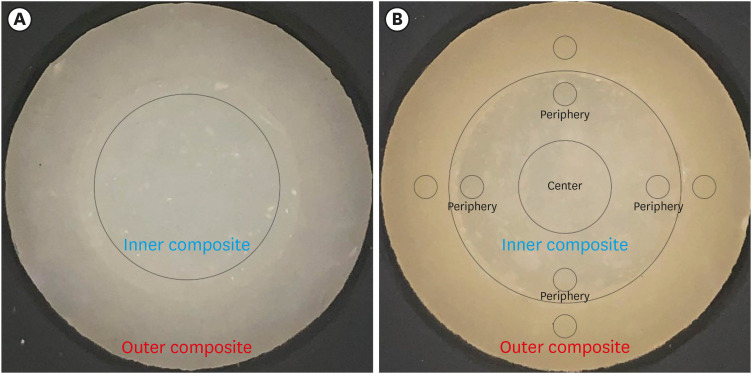
Objectives This study aimed to evaluate the impact of substrate color and interface distance on the color adjustment of 2 single-shade composites, Vittra APS Unique and Charisma Diamond One.
Materials and Methods Dual disc-shaped specimens were created using Vittra APS Unique or Charisma Diamond One as the center composite, surrounded by shaded composites (A1 or A3). Color measurements were taken with a spectrophotometer against a gray background, recording the color coordinates in the CIELAB color space. Illumination with a light-correcting device and image acquisition using a polarizing filter-equipped cell phone were performed on specimens over the same background. Image processing software was used to measure the color coordinates in the center and periphery of the inner composite and in the outer composite. The color data were then converted to CIELAB coordinates and adjusted using data from the spectrophotometer. Color differences (ΔE00) between the center/periphery of single-shade and outer composites were calculated, along with color changes in single-shade composites caused by different outer composites. Color differences for the inner composites surrounded by A1 and A3 were also calculated. Data were analyzed using repeated-measures analysis of variance (α = 0.05).
Results The results showed that color discrepancies were lowest near the interface and when the outer composite was whiter (A1). Additionally, Charisma Diamond One exhibited better color adjustment ability than Vittra APS Unique.
Conclusions Color discrepancies between the investigated single-shade composites diminished towards the interface with the surrounding composite, particularly when the latter exhibited a lighter shade.
-
Citations
Citations to this article as recorded by- Evaluation of color stability in single-shade composite resins using spectrophotometer and cross-polarized mobile photography
Hatice Tepe, Ozge Celiksoz, Batu Can Yaman
BMC Oral Health.2025;[Epub] CrossRef - Comparative Evaluation of the Staining Resistance of Two Single-Shade Composites in Coffee and Chlorhexidine: A Spectrophotometric Analysis
Unmesh Khanvilkar, Shrinath D Kulkarni, Siddhesh Bandekar, Ved M Talathi, Oshin Baghel, Priyanka Razdan, Seema Gupta
Cureus.2025;[Epub] CrossRef - Clinical Implications of Color Adjustment in Single-Shade Resins Post-Dental Bleaching: A Systematic Review
Samille Biasi Miranda, Caroline de Farias Charamba Leal, Rodrigo Barros Esteves Lins, Marcos Antonio Japiassu Resende Montes
Journal of Clinical Medicine.2025; 14(9): 3194. CrossRef - Accuracy and Reliability of Smartphone Versus Mirrorless Camera Images-Assisted Digital Shade Guides: An In Vitro Study
Soo Teng Chew, Suet Yeo Soo, Mohd Zulkifli Kassim, Khai Yin Lim, In Meei Tew
Applied Sciences.2025; 15(14): 8070. CrossRef
- Evaluation of color stability in single-shade composite resins using spectrophotometer and cross-polarized mobile photography
- 3,279 View
- 89 Download
- 3 Web of Science
- 4 Crossref
- Effects of surrounding and underlying shades on the color adjustment potential of a single-shade composite used in a thin layer
- Mariana Silva Barros, Paula Fernanda Damasceno Silva, Márcia Luciana Carregosa Santana, Rafaella Mariana Fontes Bragança, André Luis Faria-e-Silva
- Restor Dent Endod 2023;48(1):e7. Published online December 29, 2022
- DOI: https://doi.org/10.5395/rde.2023.48.e7
-
Abstract
PDFPubReaderePub
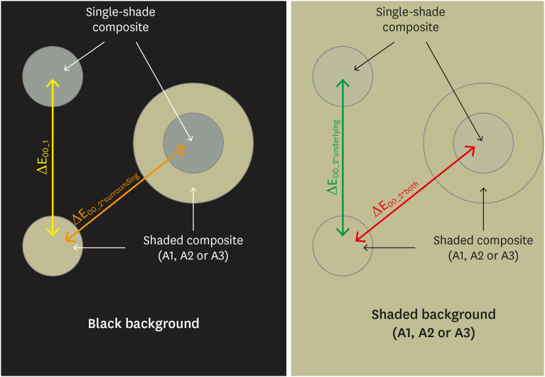
Objectives This study aimed to evaluate the surrounding and underlying shades’ effect on the color adjustment potential (CAP) of a single-shade composite used in a thin layer.
Materials and Methods Cylinder specimens (1.0 mm thick) were built with the Vittra APS Unique composite, surrounded (dual specimens) or not (simple specimens) by a control composite (shade A1, A2, or A3). Simple specimens were also built only with the control composites. Each specimen’s color was measured against white and black backgrounds or the simple control specimens with a spectrophotometer (CIELAB system). The whiteness index for dentistry (WID) and translucency parameters (TP00) were calculated for simple specimens. Differences (ΔE00) in color between the simple/dual specimens and the controls were calculated. The CAP was calculated based on the ratios between data from simple and dual specimens.
Results The Vittra APS Unique composite showed higher WID and TP00 values than the controls. The highest values of ΔE00 were observed among simple specimens. The color measurements of Vittra APS Unique (simple or dual) against the control specimens presented the lowest color differences. Only surrounding the single-shade composite with a shaded composite barely impacted the ΔE00. The highest CAP values were obtained using a shaded composite under simple or dual specimens.
Conclusions The CAP of Vittra APS Unique was strongly affected by the underlying shade, while surrounding this composite with a shaded one barely affected its color adjustment.
-
Citations
Citations to this article as recorded by- Impact of water absorption on the translucency of single-shade and conventional resin composites: an in vitro comparative study
Ceyda Sari, Elifnur Aydemir Aydın
Odontology.2026;[Epub] CrossRef - Baseline color-matching in anterior non-carious cervical lesions of patients of two single-shade resin composites: a randomized clinical trial
Ayşe Nur Doğan, Soley Arslan
Odontology.2026;[Epub] CrossRef - Evaluation of color blending effect of a single-shade resin composite with hybrid ceramics: an in vitro study
Jongchan Lee, Jinsoo Ahn, Sun-Young Kim
BMC Oral Health.2026;[Epub] CrossRef - Color Change and Compressive Strength of Novel Single-Shade Composite Resins Exposed to Staining Beverages
Juan P. Molina-Gantiva, Midian C. Castillo-Pedraza, Jorge H. Wilches-Visbal
Odovtos - International Journal of Dental Sciences.2026;[Epub] CrossRef - At‐Home and In‐Office Bleaching Protocols on the Color Match of Restorations Made With Single‐Shade Composites
Luciana Vasconcelos Ramos, Dayana Fernandes Rocha Aparicio, André Luis Faria‐e‐Silva, Maíra do Prado, Andréa Vaz Braga Pintor, Marcela Baraúna Magno
Journal of Esthetic and Restorative Dentistry.2025; 37(6): 1567. CrossRef - Evaluation of color matching of three single-shade composites employing simulated 3D printed cavities with different thicknesses using CIELAB and CIEDE2000 color difference formulae
Engin Kariper, Aylin Cilingir
REVIEWS ON ADVANCED MATERIALS SCIENCE.2025;[Epub] CrossRef - Impact of kombucha, coffee, and turmeric beverages on the color stability of a single-shade versus a multi-shade resin-based composite
Hanin E. Yeslam, Abdulaziz F. Bakhsh
PeerJ.2025; 13: e19759. CrossRef - Comparative Study of Esthetic Outcome of Pedo Shades of Composite Resin—A Randomized Controlled Trial: In Vivo and In Vitro Study
Priyanka Raj, Shikha Choubey, Divya Doneria, Diksha Bhat, Shivani Mathur, Shailja Sinha
International Journal of Clinical Pediatric Dentistry.2025; 18(S1): S22. CrossRef - Influence of cavity wall thickness on the color adjustment potential of single-shade resin composites
Fabrício Luscino Alves de Castro, Letícia Brandão Durand
The Journal of the American Dental Association.2024; 155(7): 605. CrossRef - Assessing color mismatch in single-shade composite resins for enamel replacement
Rafaella Mariana Fontes de Bragança, Diana Leyva Del Rio, Luiz Alves Oliveira-Neto, William Michael Johnston
The Journal of Prosthetic Dentistry.2024; 132(3): 613.e1. CrossRef - Color discrepancy of single-shade composites at different distances from the interface measured using cell phone images
Márcia Luciana Carregosa Santana, Gabriella de Jesus Santos Livi, André Luis Faria-e-Silva
Restorative Dentistry & Endodontics.2024;[Epub] CrossRef - Is It Possible for Single-shade Composites to Mimic the Color, Lightness, Chroma, and Hue of Other Single-shade Composites? An In Vitro Study
M Buldur, G Ayan
Operative Dentistry.2024; 49(6): 691. CrossRef - Color evaluation of a one-shade used for restoration of non-carious cervical lesions: an equivalence randomized clinical trial
Michael Willian Favoreto, Amanda de Oliveira de Miranda, Thalita P. Matos, Andrea dos Santos de Castro, Mylena de Abreu Cardoso, Julia Beatriz, Jenny Collantes-Acuña, Alessandra Reis, Alessandro Dourado Loguercio
BMC Oral Health.2024;[Epub] CrossRef - Influence of Thickness on the Translucency Parameter and Whiteness Index of Single-Shade Resin Composites
Ö Yağcı, M Fidan
Operative Dentistry.2024; 49(2): 189. CrossRef - A Comparative Study of the Sensitivity and Specificity of the Ishihara Test With Various Displays
Thomas Klinke, Wolfgang Hannak, Klaus Böning, Holger Jakstat
International Dental Journal.2024; 74(4): 892. CrossRef - Color match evaluation using instrumental method for three single-shade resin composites before and after in-office bleaching
Aylin Cilingir, Engin Kariper
REVIEWS ON ADVANCED MATERIALS SCIENCE.2023;[Epub] CrossRef - The role of interface distance and underlying substrate on the color adjustment potential of single‐shade composites
Gabriella Jesus Santos de Livi, Tauan Rosa Santana, Rafaella Mariana Fontes Bragança, Rosa Maria Viana de Bragança Garcez, André Luis Faria‐e‐Silva
Journal of Esthetic and Restorative Dentistry.2023; 35(8): 1279. CrossRef
- Impact of water absorption on the translucency of single-shade and conventional resin composites: an in vitro comparative study
- 5,960 View
- 134 Download
- 17 Web of Science
- 17 Crossref
- Errors in light-emitting diodes positioning when curing bulk fill and incremental composites: impact on properties after aging
- Abdulrahman A. Balhaddad, Isadora M. Garcia, Haifa Maktabi, Maria Salem Ibrahim, Qoot Alkhubaizi, Howard Strassler, Fabrício M. Collares, Mary Anne S. Melo
- Restor Dent Endod 2021;46(4):e51. Published online September 24, 2021
- DOI: https://doi.org/10.5395/rde.2021.46.e51

-
Abstract
PDFPubReaderePub
Objectives This study aimed to evaluate the effect of improper positioning single-peak and multi-peak lights on color change, microhardness of bottom and top, and surface topography of bulk fill and incremental composites after artificial aging for 1 year.
Materials and Methods Bulk fill and incremental composites were cured using multi-peak and single-peak light-emitting diode (LED) following 4 clinical conditions: (1) optimal condition (no angulation or tip displacement), (2) tip-displacement (2 mm), (3) slight tip angulation (α = 20°) and (4) moderate tip angulation (α = 35°). After 1-year of water aging, the specimens were analyzed for color changes (ΔE), Vickers hardness, surface topography (Ra, Rt, and Rv), and scanning electron microscopy.
Results For samples cured by single-peak LED, the improper positioning significantly increases the color change compared to the optimal position regardless of the type of composite (
p < 0.001). For multi-peak LED, the type of resin composite and the curing condition displayed a significant effect on ΔE (p < 0.001). For both LEDs, the Vickers hardness and bottom/top ratio of Vickers hardness were affected by the type of composite and the curing condition (p < 0.01).Conclusions The bulk fill composite presented greater resistance to wear, higher color stability, and better microhardness than the incremental composite when subjected to improper curing. The multi-peak LED improves curing under improper conditions compared to single-peak LED. Prevention of errors when curing composites requires the attention of all personnel involved in the patient's care once the clinical relevance of the appropriate polymerization reflects on reliable long-term outcomes.
-
Citations
Citations to this article as recorded by- A clinical survey of the output intensity of 50 light-curing units in dental clinics across Davangere and Mangalore region using a spectrometer system
Elizbeth Christy Jose, Sakshi Jha, Prema Shantagouda Biradar, J Arun, TN Nandini, Thushara Mohanan
International Journal of Oral Health Sciences.2025; 15(1): 41. CrossRef - The demineralization resistance and mechanical assessments of different bioactive restorative materials for primary and permanent teeth: an in vitro study
Maria Salem Ibrahim, Fahad Rakad Aldhafeeri, Abdullah Sami Banaemah, Mana S. Alhaider, Yousif A. Al-Dulaijan, Abdulrahman A. Balhaddad
BDJ Open.2024;[Epub] CrossRef - Inorganic Compounds as Remineralizing Fillers in Dental Restorative Materials: Narrative Review
Leena Ibraheem Bin-Jardan, Dalal Ibrahim Almadani, Leen Saleh Almutairi, Hadi A. Almoabid, Mohammed A. Alessa, Khalid S. Almulhim, Rasha N. AlSheikh, Yousif A. Al-Dulaijan, Maria S. Ibrahim, Afnan O. Al-Zain, Abdulrahman A. Balhaddad
International Journal of Molecular Sciences.2023; 24(9): 8295. CrossRef
- A clinical survey of the output intensity of 50 light-curing units in dental clinics across Davangere and Mangalore region using a spectrometer system
- 2,555 View
- 22 Download
- 2 Web of Science
- 3 Crossref
Case Reports
- A simple technique for impression taking of teeth and functionally generated paths
- Takatsugu Yamamoto, Yohei Sato, Hidehiko Watanabe, Amit Punj, Minoru Abe, Yasuko Momoi, Chikahiro Ohkubo
- Restor Dent Endod 2018;43(1):e9. Published online February 3, 2018
- DOI: https://doi.org/10.5395/rde.2018.43.e9
-
Abstract
PDFPubReaderePub
The objective of this case report is to introduce a simple technique for simultaneously taking a closed-mouth impression and functionally generated path (FGP) for a full coverage crown restoration. A monolithic zirconia crown was the restoration of choice. An alginate impression of the abutment tooth was taken to fabricate a custom-made closed-mouth impression tray covering the abutment tooth and the adjacent teeth. The tray had an FGP table and an abutment tray in cameo and intaglio surfaces, respectively. The impression was taken with silicone impression material after adjusting the abutment tray and inscribing the FGP using self-curing acrylic resins. Plaster casts were made from the impression, and a zirconia crown was fabricated. The crown was cemented to the abutment tooth with minimal adjustments. This simple technique resulted in a well-fitting crown that accounted for mandibular movements. Using the custom closed-mouth impression tray incorporating an FGP table simultaneously aids in fabricating an accurately fitting restoration that incorporates harmonious mandibular movements using a single impression capture.
-
Citations
Citations to this article as recorded by- Fabrication of fixed prosthesis by employing functionally generated path technique and dual scan technique in a tardive dyskinesia patient: a case report
Shilpa, Du-Hyeong Lee
The Journal of Korean Academy of Prosthodontics.2023; 61(3): 227. CrossRef - Morphological design of occlusal wear facets for the mandibular first molar crown using different bite registration methods
Hu Chen, Xu Yang, Linlin Li, Yong Wang, Yuchun Sun
Journal of Prosthodontics.2023; 32(5): 439. CrossRef
- Fabrication of fixed prosthesis by employing functionally generated path technique and dual scan technique in a tardive dyskinesia patient: a case report
- 3,092 View
- 23 Download
- 2 Crossref
- The use of platelet rich plasma in the treatment of immature tooth with periapical lesion: a case report
- Günseli Güven Polat, Ceren Yıldırım, Özlem Martı Akgün, Ceyhan Altun, Didem Dinçer, Cansel Köse Özkan
- Restor Dent Endod 2014;39(3):230-234. Published online June 2, 2014
- DOI: https://doi.org/10.5395/rde.2014.39.3.230
-
Abstract
PDFPubReaderePub
This study describes the treatment of an immature permanent tooth with periapical lesion which was treated with regenerative approach using platelet rich plasma (PRP). The root canal of immature human permanent tooth with periapical lesion was gently debrided of necrotic tissue and disinfected with 2.5% NaOCl, and then medicated with triple antibiotic paste comprised of ciprofloxacin, metronidazole, and tetracycline. When the tooth was asymptomatic, PRP and mineral trioxide aggregate (MTA) were placed into the root canal. Six months after PRP treatment, radiographical examination revealed resolution of the radiolucency and progressive thickening of the root wall and apical closure. Our findings suggest that PRP can be used for the treatment of immature permanent teeth with periapical lesion, as part of a regenerative endodontic treatment procedure.
-
Citations
Citations to this article as recorded by- Evaluation of postoperative pain and healing following regenerative endodontics using platelet‐rich plasma versus conventional endodontic treatment in necrotic mature mandibular molars with chronic periapical periodontitis. A randomized clinical trial
Yassmin Elsayed Ahmed, Geraldine Mohamed Ahmed, Angie Galal Ghoneim
International Endodontic Journal.2023; 56(4): 404. CrossRef - Different Approaches to the Regeneration of Dental Tissues in Regenerative Endodontics
Anna M. Krupińska, Katarzyna Skośkiewicz-Malinowska, Tomasz Staniowski
Applied Sciences.2021; 11(4): 1699. CrossRef - Coronal tooth discoloration induced by regenerative endodontic treatment using different scaffolds and intracanal coronal barriers: a 6-month ex vivo study
Noushin Shokouhinejad, Hassan Razmi, Maryam Farbod, Marzieh Alikhasi, Josette Camilleri
Restorative Dentistry & Endodontics.2019;[Epub] CrossRef - Efficacy of Autologous Platelet Concentrates in Regenerative Endodontic Treatment: A Systematic Review of Human Studies
Joanna Metlerska, Irini Fagogeni, Alicja Nowicka
Journal of Endodontics.2019; 45(1): 20. CrossRef - Bone, Periodontal and Dental Pulp Regeneration in Dentistry: A Systematic Scoping Review
Luiz Alexandre Chisini, Marcus Cristian Muniz Conde, Guillermo Grazioli, Alissa Schmidt San Martin, Rodrigo Varella de Carvalho, Letícia Regina Morello Sartori, Flávio Fernando Demarco
Brazilian Dental Journal.2019; 30(2): 77. CrossRef - Mineral trioxide aggregate and other bioactive endodontic cements: an updated overview – part II: other clinical applications and complications
M. Torabinejad, M. Parirokh, P. M. H. Dummer
International Endodontic Journal.2018; 51(3): 284. CrossRef - Alternative to Avoid Tooth Discoloration after Regenerative Endodontic Procedure: A Systematic Review
Luciane Geanini Pena dos Santos, Luiz Alexandre Chisini, Camila Guerner Springmann, Beatriz Dulcineia Mendes de Souza, Fernanda Geraldo Pappen, Flávio Fernando Demarco, Mara Cristina Santos Felippe, Wilson Tadeu Felippe
Brazilian Dental Journal.2018; 29(5): 409. CrossRef - Influence of Apical Diameter on the Outcome of Regenerative Endodontic Treatment in Teeth with Pulp Necrosis: A Review
Yanjun Fang, Xinhuan Wang, Jingjing Zhu, Chaonan Su, Ying Yang, Liuyan Meng
Journal of Endodontics.2018; 44(3): 414. CrossRef - A scoping review of root canal revascularization: relevant aspects for clinical success and tissue formation
M. C. M. Conde, L. A. Chisini, R. Sarkis‐Onofre, H. S. Schuch, J. E. Nör, F. F. Demarco
International Endodontic Journal.2017; 50(9): 860. CrossRef - Effects of Epigallocatechin Gallate, an Antibacterial Cross-linking Agent, on Proliferation and Differentiation of Human Dental Pulp Cells Cultured in Collagen Scaffolds
Young-Sun Kwon, Hee-Jin Kim, Yun-Chan Hwang, Vinicius Rosa, Mi-Kyung Yu, Kyung-San Min
Journal of Endodontics.2017; 43(2): 289. CrossRef - Regenerative Endodontics
Kristina Feigin, Bonnie Shope
Journal of Veterinary Dentistry.2017; 34(3): 161. CrossRef - Regenerative Endodontic Treatment or Mineral Trioxide Aggregate Apical Plug in Teeth with Necrotic Pulps and Open Apices: A Systematic Review and Meta-analysis
Mahmoud Torabinejad, Ali Nosrat, Prashant Verma, Oyoyo Udochukwu
Journal of Endodontics.2017; 43(11): 1806. CrossRef - Platelet concentrates for revitalization of immature necrotic teeth: a systematic review of the clinical studies
Alessandra Lolato, Cristina Bucchi, Silvio Taschieri, Ahmed El Kabbaney, Massimo Del Fabbro
Platelets.2016; 27(5): 383. CrossRef - Regenerative endodontics—Creating new horizons
Harnoor Dhillon, Mamta Kaushik, Roshni Sharma
Journal of Biomedical Materials Research Part B: Applied Biomaterials.2016; 104(4): 676. CrossRef - The impact of autologous platelet concentrates on endodontic healing: a systematic review
Nastaran Meschi, Ana B. Castro, Katleen Vandamme, Marc Quirynen, Paul Lambrechts
Platelets.2016; 27(7): 613. CrossRef - Pulp and Periodontal Regeneration of an Avulsed Permanent Mature Incisor Using Platelet-rich Plasma after Delayed Replantation: A 12-month Clinical Case Study
Harini Priya M, Pavan B. Tambakad, Jaya Naidu
Journal of Endodontics.2016; 42(1): 66. CrossRef - Platelet preparations in dentistry: How? Why? Where? When?
Luigi Fabrizio Rodella
World Journal of Stomatology.2015; 4(2): 39. CrossRef
- Evaluation of postoperative pain and healing following regenerative endodontics using platelet‐rich plasma versus conventional endodontic treatment in necrotic mature mandibular molars with chronic periapical periodontitis. A randomized clinical trial
- 2,527 View
- 11 Download
- 17 Crossref
 First
First Prev
Prev

