
Search
- Page Path
- HOME > Search
Research Articles
- Cone-beam computed tomography analysis of maxillary premolar canal anatomy: Ahmed’s versus Vertucci’s classifications in a Jordanian cohort
- Raidan Ba-Hattab, Muna M. Shaweesh, Nessrin A. Taha, Elham S. Abu Alhaija
- Restor Dent Endod 2026;51(1):e11. Published online February 26, 2026
- DOI: https://doi.org/10.5395/rde.2026.51.e11
-
 Abstract
 PDF
PDF PubReader
 ePub
ePub - Objectives
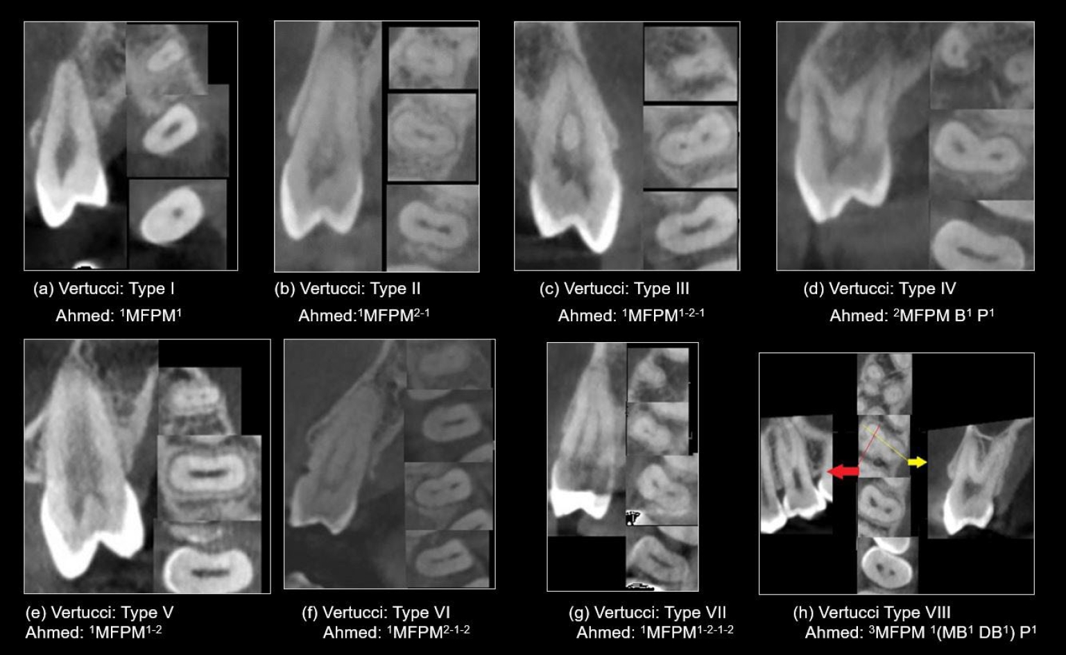
This study analyzed the root and canal configurations of maxillary premolars in a Jordanian subpopulation using cone-beam computed tomography (CBCT) and classified them based on Vertucci’s and Ahmed’s systems.
Methods
Two hundred CBCT scans of 800 maxillary premolars were retrospectively assessed for root morphology, canal configurations, and root canal divergence and merging. Data was statistically analyzed.
Results
The study included 70 males and 130 females. Most right and left maxillary first premolars (RFPM, LFPM) had two roots (59.0% and 58.5%), with a significant association between sex and root number for RFPM and LFPM (p < 0.05). In contrast, the right and left maxillary second premolars (RSPM, LSPM) mostly had a single root (87.5% and 88.5%), with no association with sex. Vertucci’s classification showed type IV as the predominant configuration in first premolars (RFPM, 65.0% and LFPM, 67.0%) and type I in second premolars (RSPM, 44.0% and LSPM, 49.0%). A significant sex association was found only with RSPM. Ahmed’s classification revealed that maxillary premolar with two separated roots and two separated canals (2MP B1 P1) was mostly found in first premolars (RFPM, 58.0% and LFPM, 56.0%), and maxillary premolar with one root and one canal (1MP1) in second premolars (RSPM, 44.0% and LSPM, 49.0%), with a significant sex association for RSPM and LSPM (p < 0.05). Age had no impact, and symmetry was observed between the right and left sides. Three-rooted premolars were identified in four cases. Almost all of Vertucci’s types and numerous codes from Ahmed’s classification were documented.
Conclusions
CBCT revealed diverse anatomical variations in the Jordanian subpopulation, with Ahmed’s classification providing more detailed canal configurations than Vertucci’s, uncovering previously overlooked variations.
- 831 View
- 48 Download

- Influence of inorganic composition and filler particle morphology on the mechanical properties of self-adhesive resin cements
- Marina Rodrigues Santi, Rodrigo Barros Esteves Lins, Beatriz Ometto Sahadi, Giovanna Corrêa Denucci, Gabriela Soffner, Luís Roberto Marcondes Martins
- Restor Dent Endod 2022;47(3):e32. Published online July 14, 2022
- DOI: https://doi.org/10.5395/rde.2022.47.e32
-
Abstract
PDFPubReaderePub
Objectives This study aimed to evaluate the influence of inorganic composition and filler particle morphology on the mechanical properties of different self-adhesive resin cements (SARCs).
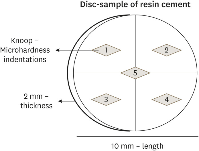
Materials and Methods Three SARCs including RelyX Unicem-2 (RUN), Maxcem Elite (MAX), and Calibra Universal (CAL) were tested. Rectangular bar-shaped specimens were prepared for flexural strength (FS) and flexural modulus (FM) and determined by a 3-point bending test. The Knoop microhardness (KHN) and top/bottom microhardness ratio (%KHN) were conducted on the top and bottom faces of disc-shaped samples. Sorption (Wsp) and solubility (Wsl) were evaluated after 24 hours of water immersion. Filler morphology was analyzed by scanning electron microscopy and X-ray energy dispersive spectroscopy (EDS). FS, FM, %KHN, Wsp, Wsl, and EDS results were submitted to 1-way analysis of variance and Tukey’s
post-hoc test, and KHN also to pairedt -test (α = 0.05).Results SARC-CAL presented the highest FS value, and SARC-RUN presented the highest FM. SARC-MAX and RUN showed the lowest Wsp and Wsl values. KHN values decreased from top to bottom and the SARCs did not differ statistically. Also, all resin cements presented carbon, aluminum, and silica in their composition. SARC-MAX and RUN showed irregular and splintered particles while CAL presented small and regular size particles.
Conclusions A higher mechanical strength can be achieved by a reduced spread in grit size and the filler morphology can influence the KHN, as well as photoinitiators in the composition. Wsp and Wsl can be correlated with ions diffusion of inorganic particles.
-
Citations
Citations to this article as recorded by
- Comparative Evaluation of Color Stability in Bioactive and Conventional Resin Cements Under Thermal Stress Conditions
Alaa Turkistani, Hanin E. Yeslam
Biomimetics.2025; 10(7): 432. CrossRef - Assessment of fit accuracy and retentive strength of additively manufactured zirconia crowns luted to Ti‐base abutments with different resin cements: An in vitro study
Rafat Sasany, Sultan Merve Uçar, Burak Yilmaz
Journal of Prosthodontics.2025;[Epub] CrossRef - Bioactive Resin Cement Color Stability and Restoration Thickness as Determinants of the Final Shade in a Glass–Ceramic CAD/CAM Material
Hanin E. Yeslam, Alaa Turkistani
Journal of Functional Biomaterials.2025; 16(9): 319. CrossRef - Light transmittance through resin-matrix composite onlays adhered to resin-matrix cements or flowable composites
Rita Fidalgo-Pereira, Susana O. Catarino, Óscar Carvalho, Nélio Veiga, Orlanda Torres, Annabel Braem, Júlio C.M. Souza
Journal of the Mechanical Behavior of Biomedical Materials.2024; 151: 106353. CrossRef - Effects of a relined fiberglass post with conventional and self-adhesive resin cement
Wilton Lima dos Santos Junior, Marina Rodrigues Santi, Rodrigo Barros Esteves Lins, Luís Roberto Marcondes Martins
Restorative Dentistry & Endodontics.2024;[Epub] CrossRef - Dental Resin-Based Luting Materials—Review
Aleksandra Maletin, Milica Jeremić Knežević, Daniela Đurović Koprivica, Tanja Veljović, Tatjana Puškar, Bojana Milekić, Ivan Ristić
Polymers.2023; 15(20): 4156. CrossRef - A Scoping Review on the Polymerization of Resin-Matrix Cements Used in Restorative Dentistry
Rita Fidalgo-Pereira, Orlanda Torres, Óscar Carvalho, Filipe S. Silva, Susana O. Catarino, Mutlu Özcan, Júlio C. M. Souza
Materials.2023; 16(4): 1560. CrossRef
- Comparative Evaluation of Color Stability in Bioactive and Conventional Resin Cements Under Thermal Stress Conditions
- 2,748 View
- 28 Download
- 7 Web of Science
- 7 Crossref
Review Article
- Age-dependent root canal instrumentation techniques: a comprehensive narrative review
- Michael Solomonov, Hyeon-Cheol Kim, Avi Hadad, Dan Henry Levy, Joe Ben Itzhak, Oleg Levinson, Hadas Azizi
- Restor Dent Endod 2020;45(2):e21. Published online March 4, 2020
- DOI: https://doi.org/10.5395/rde.2020.45.e21
-
Abstract
PDFPubReaderePub
The aim of this article was to review age-dependent clinical recommendations for appropriate root canal instrumentation techniques. A comprehensive narrative review of canal morphology, the structural characteristics of dentin, and endodontic outcomes at different ages was undertaken instead of a systematic review. An electronic literature search was carried out, including the Medline (Ovid), PubMed, and Web of Science databases. The searches used controlled vocabulary and free-text terms, as follows: ‘age-related root canal treatment,’ ‘age-related instrumentation,’ ‘age-related chemo-mechanical preparation,’ ‘age-related endodontic clinical recommendations,’ ‘root canal instrumentation at different ages,’ ‘geriatric root canal treatment,’ and ‘pediatric root canal treatment.’ Due to the lack of literature with practical age-based clinical recommendations for an appropriate root canal instrumentation technique, a narrative review was conducted to suggest a clinical algorithm for choosing the most appropriate instrumentation technique during root canal treatment. Based on the evidence found through the narrative review, an age-related clinical algorithm for choosing appropriate instrumentation during root canal treatment was proposed. Age affects the morphology of the root canal system and the structural characteristics of dentin. The clinician’s awareness of root canal morphology and dentin characteristics can influence the choice of instruments for root canal treatment.
-
Citations
Citations to this article as recorded by- Effect of calcium silicate and epoxy resin-based sealers on postoperative pain: a randomized clinical trial
Jessica Waayen Caleffi de Oliveira, Ingrid Luiza Mendonça Cunha, Aida Renée Assayag Hanan, André Augusto Franco Marques, Fernando José Herkrath, Emílio Carlos Sponchiado Júnior
BMC Oral Health.2026;[Epub] CrossRef - Başarısız Endodontik Tedavilerde Gözden Kaçan Kanalların İnsidansı
Okan Turgut, Melike Bayram, Emre Bayram
Acta Odontologica Turcica.2026; 43(2): 83. CrossRef - Cross-Sectional Analysis of the Challenges Faced by Undergraduate Dental Students During Root Canal Treatment (RCT) and the Oral Health-Related Quality of Life in Patients After RCT
Mubashir Baig Mirza, Abdullah Bajran Almuteb, Abdulaziz Tariq Alsheddi, Qamar Hashem, Mohammed Ali Abuelqomsan, Ahmed AlMokhatieb, Shahad AlBader, Abdullah AlShehri
Medicina.2025; 61(2): 215. CrossRef - OUTCOMES OF COMBINED ENDODONTIC TREATMENT AND APICAL SURGERY IN MANAGING LARGE PERIAPICAL CYSTS: A CLINICAL STUDY
Sapna Pandey, P Nihar, Amit Kumar, Nitin Bhagat, Vikram Karande, Zameer Pasha, Anukriti Kumari
BULLETIN OF STOMATOLOGY AND MAXILLOFACIAL SURGERY.2025; : 329. CrossRef - El Uso del hipoclorito de sodio en endodoncia: concentración, temperatura y activación
Ábilson Josue Fabiani Ticona, Fernanda Camargo Espejo
Revista de investigación e información en salud.2025;[Epub] CrossRef - Assessment of Anatomical Dentin Thickness in Mandibular First Molar: An In Vivo Cone‐Beam Computed Tomographic Study
Sahil Choudhari, Kavalipurapu Venkata Teja, Sindhu Ramesh, Jerry Jose, Mariangela Cernera, Parisa Soltani, Emmanuel João Nogueira Leal da Silva, Gianrico Spagnuolo, Ricardo Danil Guiraldo
International Journal of Dentistry.2024;[Epub] CrossRef - Oral Health Concerns of the ‘Sunset Age’
Pradnya V. Kakodkar, Amandeep Kaur, Shivasakthy Manivasakan, Sounyala Rayannavar, Revati Deshmukh, Smita Athavale
Journal of Medical Evidence.2023; 4(2): 141. CrossRef - Root canal treatment of a six-canal first mandibular molar with extensive periapical lesion: A case report
Xin Li, Shuyu Sun, Tengyi Zheng
Medicine.2023; 102(30): e34336. CrossRef - Endodontic Dentistry: Analysis of Dentinal Stress and Strain Development during Shaping of Curved Root Canals
Laura Iosif, Bogdan Dimitriu, Dan Florin Niţoi, Oana Amza
Healthcare.2023; 11(22): 2918. CrossRef - Mechanisms of age-related changes in the morphology of the pulp system of the first lower molars
N.B. Petrukhina, O.A. Zorina, V.A. Venediktova
Stomatologiya.2022; 101(2): 19. CrossRef
- Effect of calcium silicate and epoxy resin-based sealers on postoperative pain: a randomized clinical trial
- 4,024 View
- 40 Download
- 10 Crossref
Research Article
- The prevalence of radix molaris in the mandibular first molars of a Saudi subpopulation based on cone-beam computed tomography
- Hassan AL-Alawi, Saad Al-Nazhan, Nassr Al-Maflehi, Mazen A. Aldosimani, Mohammed Nabil Zahid, Ghadeer N. Shihabi
- Restor Dent Endod 2020;45(1):e1. Published online November 14, 2019
- DOI: https://doi.org/10.5395/rde.2020.45.e1
-
Abstract
PDFPubReaderePub
Objectives The purpose of this study was to determine the incidence of radix molaris (RM) (entomolaris and paramolaris) in the mandibular first permanent molars of a sample Saudi Arabian subpopulation using cone-beam computed tomography (CBCT).
Materials and Methods A total of 884 CBCT images of 427 male and 457 female Saudi citizens (age 16 to 70 years) were collected from the radiology department archives of 4 dental centers. A total of 450 CBCT images of 741 mature mandibular first molars that met the inclusion criteria were reviewed. The images were viewed at high resolution by 3 examiners and were analyzed with Planmeca Romexis software (version 5.2).
Results Thirty-three (4.5%) mandibular first permanent molars had RM, mostly on the distal side. The incidence of radix entomolaris (EM) was 4.3%, while that of radix paramolaris was 0.3%. The RM roots had one canal and occurred more unilaterally. No significant difference in root configuration was found between males and females (
p > 0.05). Types I and III EM root canal configurations were most common, while type B was the only RP configuration observed.Conclusions The incidence of RM in the mandibular first molars of this Saudi subpopulation was 4.5%. Identification of the supernumerary root can avoid missing the canal associated with the root during root canal treatment.
-
Citations
Citations to this article as recorded by- A comprehensive investigation into the prevalence of dental anomalies across Saudi Arabia, a systematic review
Abdulrahman K. Alshammari, Muteb A. Algharbi, Freah L. Alshammary, Nabeel S. Almotairy, Hatem D. Alshammari, Amjad M. Alsogaih, Ahmed A. Madfa
Critical Public Health.2026;[Epub] CrossRef - Anatomical Variations in Permanent Mandibular Molars in the Sharjah Population: A Cohort Study Using Cone‐Beam Computed Tomography
Shaima Dheyab, Entisar AlRasasi, Ahmed M. Aziz, Saad Albayatti, Mehmet Omer Gorduysus, Hannah Wesley
International Journal of Dentistry.2026;[Epub] CrossRef - Evaluation of the variations of mandibular molars and the distance from root apex to the inferior alveolar nerve in Saudi Sub-population: Three-dimensional radiographic evaluation
Tariq Mohammed Aqili, Esam Sami Almuzaini, Abdulbari Saleh Aljohani, Ahmed Khaled Al Saeedi, Hassan Abdulmuti Hammudah, Muath Alassaf, Muhannad M. Hakeem, Mohmed Isaqali Karobari
PLOS ONE.2025; 20(2): e0317053. CrossRef - Prevalence of radix molaris in mandibular molars of a subpopulation of Brazil’s Northeast region: a cross-sectional CBCT study
Yasmym Martins Araújo de Oliveira, Maria Clara Mendes Gomes, Maria Fernanda da Silva Nascimento, Ricardo Machado, Danna Mota Moreira, Hermano Camelo Paiva, George Táccio de Miranda Candeiro
Scientific Reports.2025;[Epub] CrossRef - Prevalence of radix entomolaris and distolingual canals and their association with the incidence of middle mesial canals in mandibular first molars of a Saudi subpopulation
Ahmed A. Madfa, Abdullah F. Alshammari, Eyad Almagadawyi, Afaf Al-Haddad, Ebtsam A. Aledaili
Scientific Reports.2025;[Epub] CrossRef - Assessment of the root and canal morphology in the permanent dentition of Saudi Arabian population using cone beam computed and micro-computed tomography – a systematic review
Mohammed Mustafa, Rumesa Batul, Mohmed Isaqali Karobari, Hadi Mohammed Alamri, Abdulaziz Abdulwahed, Ahmed A. Almokhatieb, Qamar Hashem, Abdullah Alsakaker, Mohammad Khursheed Alam, Hany Mohamed Aly Ahmed
BMC Oral Health.2024;[Epub] CrossRef - Prevalence of radix accesoria dentis in a northern Peruvian population evaluated by cone-beam tomography
Karla Renata León-Almanza, Anthony Adrián Jaramillo-Nuñez, Catherin Angélica Ruiz-Cisneros, Paul Martín Herrera-Plasencia
Heliyon.2024; 10(16): e35919. CrossRef - Radix molaris is a hidden truth of mandibular first permanent molars: A descriptive- analytic study using cone beam computed tomography
Mohammed A. Alobaid, Saurabh Chaturvedi, Ebtihal Mobarak S. Alshahrani, Ebtsam M. Alshehri, Amal S. Shaiban, Mohamed Khaled Addas, Giuseppe Minervini
Technology and Health Care.2023; 31(5): 1957. CrossRef - Prevalence of Radix Entomolaris in Mandibular Permanent Molars Analyzed by Cone-Beam CT in the Saudi Population of Ha'il Province
Moazzy I Almansour, Ahmed A Madfa, Adhwaa F Algharbi, Reem Almuslumani, Noeer K Alshammari, Ghufran M Al Hussain
Cureus.2023;[Epub] CrossRef - Prevalence of radix entomolaris in India and its comparison with the rest of the world
Sumit MOHAN, Jyoti THAKUR
Minerva Dental and Oral Science.2022;[Epub] CrossRef - Radix Paramolaris an Endodontic Challenge: A Case Report
Ashwini B Prasad, Deepak Raisingani, Ridhima Gupta, Rimjhim Jain
Journal of Mahatma Gandhi University of Medical Sciences and Technology.2022; 7(1): 32. CrossRef - Evaluation of Radix Entomolaris and Middle Mesial Canal in Mandibular Permanent First Molars in an Iraqi Subpopulation Using Cone‐Beam Computed Tomography
Ranjdar Mahmood Talabani, Kazhan Omer Abdalrahman, Rawa Jamal Abdul, Dlsoz Omer Babarasul, Sara Hilmi Kazzaz, Heng Bo Jiang
BioMed Research International.2022;[Epub] CrossRef - Evaluation of Root Canal Configuration of Maxillary and Mandibular First Molar by CBCT: A Retrospective Cross-Sectional Study
Rakan Rafdan Alhujhuj, Rizwan Jouhar, Muhammad Adeel Ahmed, Abdullatif Abdulrahman Almujhim, Mohammed Tariq Albutayh, Necdet Adanir
Diagnostics.2022; 12(9): 2121. CrossRef - Ethnical Anatomical Differences in Mandibular First Permanent Molars between Indian and Saudi Arabian Subpopulations: A Retrospective Cross-sectional Study
Abdulwahab Alamir, Mohammed Mashyakhy, Apathsakayan Renugalakshmi, Thilla S Vinothkumar, Anandhi S Arthisri, Ahmed Juraybi
The Journal of Contemporary Dental Practice.2021; 22(5): 484. CrossRef
- A comprehensive investigation into the prevalence of dental anomalies across Saudi Arabia, a systematic review
- 3,185 View
- 47 Download
- 14 Crossref
Case Report
- Endodontic management of a maxillary first molar with three roots and seven root canals with the aid of cone-beam computed tomography
- Gurudutt Nayak, Kamal Krishan Singh, Rhitu Shekhar
- Restor Dent Endod 2015;40(3):241-248. Published online June 3, 2015
- DOI: https://doi.org/10.5395/rde.2015.40.3.241
-
Abstract
PDFPubReaderePub
Variation in root canal morphology, especially in maxillary first molar presents a constant challenge for a clinician in their detection and management. This case report describes the successful root canal treatment of a three rooted right maxillary first molar presenting with three canals each in the mesiobuccal and distobuccal roots and one canal in the palatal root. The clinical detection of this morphologic aberration was made using a dental operating microscope, and the canal configuration was established after correlating and computing the clinical, radiographic and cone-beam computed tomography (CBCT) scan findings. CBCT images confirmed the configuration of the canals in the mesiobuccal and distobuccal roots to be Al-Qudah and Awawdeh type (3-2) and type (3-2-1), respectively, whereas the palatal root had a Vertucci type I canal pattern. This report reaffirms the importance of careful examination of the floor of the pulp chamber with a dental operating microscope and the use of multiangled preoperative radiographs along with advanced diagnostic aids such as CBCT in identification and successful management of aberrant canal morphologies.
-
Citations
Citations to this article as recorded by- Inhibition potential of rhamnolipid biosurfactant against Corynespora cassiicola – a phytopathogen of king chilli
Nilam Sarma, Suresh Deka, Hemen Deka
Studia Biologica.2025; 19(3): 153. CrossRef - Endodontic Management of Maxillary First Molar with Seven Root Canals Diagnosed Using Cone-beam Computed Tomography: A Case Report
Ravindranath Megha, Venkatachalam Prakash
World Journal of Dentistry.2021; 12(1): 89. CrossRef - The MB3 canal in maxillary molars: a micro-CT study
Ronald Ordinola-Zapata, Jorge N. R. Martins, Hugo Plascencia, Marco A. Versiani, Clovis M. Bramante
Clinical Oral Investigations.2020; 24(11): 4109. CrossRef - Maxillary first molar with 7 root canals diagnosed using cone-beam computed tomography
Evaldo Rodrigues, Antônio Henrique Braitt, Bruno Ferraz Galvão, Emmanuel João Nogueira Leal da Silva
Restorative Dentistry & Endodontics.2017; 42(1): 60. CrossRef - Endodontic management of a maxillary first molar with seven root canal systems evaluated using cone-beam computed tomography scanning
VijayReddy Venumuddala, Sridhar Moturi, SV Satish, BKalyan Chakravarthy, Sudhakar Malapati
Journal of International Society of Preventive and Community Dentistry.2017; 7(5): 297. CrossRef
- Inhibition potential of rhamnolipid biosurfactant against Corynespora cassiicola – a phytopathogen of king chilli
- 2,969 View
- 15 Download
- 5 Crossref
Original Articles
- Cross-sectional morphology and minimum canal wall widths in C-shaped root of mandibular molars
- Byung-Chul Song, Yong-Bum Cho
- J Korean Acad Conserv Dent 2007;32(1):37-46. Published online January 31, 2007
- DOI: https://doi.org/10.5395/JKACD.2007.32.1.037
-
Abstract
PDFPubReaderePub
The C-shaped canal system is an anatomical variation mostly seen in mandibular second molars, although it can also occur in maxillary and other mandibular molars. The main anatomical feature of C-shaped canals is the presence of fins or web connecting the individual root canals. The complexity of C-shaped canals prevents these canals from being cleaned, shaped, and obturated effectively during root canal therapy, and sometimes it leads to an iatrogenic perforation from the extravagant preparation.
The purpose of this study was to provide further knowledge of the anatomical configuration and the minimal thickness of dentinal wall according to the level of the root.
Thirty extracted mandibular second molars with fused roots and longitudinal grooves on lingual or buccal surface of the root were collected from a native Korean population. The photo images and radiographs from buccal, lingual, apical direction were taken. After access cavity was prepared, teeth were placed in 5.25% sodium hypochlorite solution for 2 hours to dissolve the organic tissue of the root surface and from the root canal system. After bench dried and all the teeth were embedded in a self-curing resin. Each block was sectioned using a microtome (Accutom-50, Struers, Denmark) at interval of 1 mm. The sectioned surface photograph was taken using a digital camera (Coolpix 995, Nikon, Japan) connected to the microscope. 197 images were evaluated for canal configurations and the minimal thickness of dentinal wall between canal and external wall using' Root Thickness Gauge Program' designed with Visual Basic.
The results were as follows:
1. At the orifice level of all teeth, the most frequent observed configuration was Melton's Type C I (73%), however the patterns were changed to type C II and C III when the sections were observed at the apical third. On the other hand, the type C III was observed at the orifice level of only 2 teeth but this type could be seen at apical region of the rest of the teeth.
2. The C-shaped canal showed continuous and semi-colon shape at the orifice level, but at the apical portion of the canal there was high possibility of having 2 or 3 canals.
3. Lingual wall was thinner than buccal wall at coronal, middle, apical thirds of root but there was no statistical differences.
- 1,364 View
- 1 Download
- Three dimensional reconstruction of teeth using x-ray microtomography
- Dong-Hoon Shin
- J Korean Acad Conserv Dent 2003;28(6):485-490. Published online November 30, 2003
- DOI: https://doi.org/10.5395/JKACD.2003.28.6.485
-
Abstract
PDFPubReaderePub
Complete understanding of the exterior and interior structure of the tooth would be prerequisite to the successful clinical results, especially in the restorative and endodontic treatment.
Although three-dimensional reconstruction method using x-ray microtomography could not be used in clinical cases, it may be the best way to reconstruct the morphologic characteristics of the tooth structure in detail without destructing the tooth itself. This study was done to three dimensionally reconstruct every teeth in the arch in order to increase the understanding about the endodontic treatment and to promote the effective restorative treatment by upgrading the knowledge of the tooth morphology.
After placing tooth between the microfocus x-ray tube and the image intensifier to obtain two-dimensional images of each level, scanning was done under the condition of 80 keV, 100 µA, 16.8 magnification with the spot size of 8 µm. Cross-section pixel size of 16.28 µm and 48.83 cross-section to cross-section distance were also used.
From the results of this study, precise three dimensional reconstructed images of every teeth could be obtained. Furthermore, it was possible to see image that showed interested area only, for example, enamel portion only, pulp and dentin area without enamel structure, pulp only, combination image of enamel and pulp, etc.
It was also possible to see transparent image without some part of tooth structure. This image might be used as a guide when restoring and preparing the full and partial crown by showing the positional and morphological relationship between the pulp and the outer tooth structure.
Another profit may be related with the fact that it would promote the understanding of the interior structure by making observation of the auto-rotating image of .AVI file from the various direction possible.
-
Citations
Citations to this article as recorded by- Fracture Flow of Radionuclides in Unsaturated Conditions at LILW Disposal Facility
Won-Seok Kim, Jungjin Kim, Jinmo Ahn, Seongsik Nam, Wooyong Um
Journal of Korean Society of Environmental Engineers.2015; 37(8): 465. CrossRef
- Fracture Flow of Radionuclides in Unsaturated Conditions at LILW Disposal Facility
- 1,478 View
- 1 Download
- 1 Crossref
 First
First Prev
Prev

