Warning: mkdir(): Permission denied in /home/virtual/lib/view_data.php on line 81

Warning: fopen(upload/ip_log/ip_log_2024-12.txt): failed to open stream: No such file or directory in /home/virtual/lib/view_data.php on line 83

Warning: fwrite() expects parameter 1 to be resource, boolean given in /home/virtual/lib/view_data.php on line 84
Endodontic management of a maxillary first molar with three roots and seven root canals with the aid of cone-beam computed tomography
Skip Navigation
Skip to contents

Restor Dent Endod : Restorative Dentistry & Endodontics

OPEN ACCESS

Articles

Page Path
HOME > Restor Dent Endod > Volume 40(3); 2015 > Article
Case Report Endodontic management of a maxillary first molar with three roots and seven root canals with the aid of cone-beam computed tomography
Gurudutt Nayak1, Kamal Krishan Singh2, Rhitu Shekhar2
Restorative Dentistry & Endodontics 2015;40(3):241-248.
DOI: https://doi.org/10.5395/rde.2015.40.3.241
Published online: June 3, 2015

1Department of Conservative Dentistry and Endodontics, Institute of Dental Sciences, Bareilly, UP, India.

2Department of Conservative Dentistry and Endodontics, Kanti Devi Dental College and Hospital, Mathura, UP, India.

Correspondence to Gurudutt Nayak, BDS, MDS. Professor, Department of Conservative Dentistry and Endodontics, Institute of Dental Sciences, Bareilly, Uttar Pradesh, India, 243006. TEL, +91-9997259742; FAX, +91-581-2526054; gurudutt_nayak@hotmail.com
• Received: February 27, 2015   • Accepted: April 13, 2015

©Copyrights 2015. The Korean Academy of Conservative Dentistry.

This is an Open Access article distributed under the terms of the Creative Commons Attribution Non-Commercial License (http://creativecommons.org/licenses/by-nc/3.0/) which permits unrestricted non-commercial use, distribution, and reproduction in any medium, provided the original work is properly cited.

  • 18 Views
  • 0 Download
prev next
  • Variation in root canal morphology, especially in maxillary first molar presents a constant challenge for a clinician in their detection and management. This case report describes the successful root canal treatment of a three rooted right maxillary first molar presenting with three canals each in the mesiobuccal and distobuccal roots and one canal in the palatal root. The clinical detection of this morphologic aberration was made using a dental operating microscope, and the canal configuration was established after correlating and computing the clinical, radiographic and cone-beam computed tomography (CBCT) scan findings. CBCT images confirmed the configuration of the canals in the mesiobuccal and distobuccal roots to be Al-Qudah and Awawdeh type (3-2) and type (3-2-1), respectively, whereas the palatal root had a Vertucci type I canal pattern. This report reaffirms the importance of careful examination of the floor of the pulp chamber with a dental operating microscope and the use of multiangled preoperative radiographs along with advanced diagnostic aids such as CBCT in identification and successful management of aberrant canal morphologies.
The anatomy of the tooth has long been an unresolved mystery. Successful root canal therapy depends upon identification and negotiation of all the canals, their subsequent cleaning and shaping and finally three-dimensional obturation of the entire root canal system. A major reason for endodontic failure is when extra roots or root canals are present are not detected.1 Traditionally, the maxillary first molars are generally described to have 3 roots, 1 palatal and 2 buccal, with each root having 1 canal. There is a wide range of variation in the literature with respect to the frequency and the number of canals in each root. The mesiobuccal root of the maxillary first molar contains a double root canal system more often than a single canal, and the incidence of second mesiobuccal canal (MB2) has been reported to be between 18.6% and 96.1%.2,3 The incidence of second distobuccal canal (DB2) has been reported to be between 1.6% and 9.5%.4,5 The incidence of two root canals in the palatal root has been found to be 0.2% to 7.0%.2,4 Apart from these anatomical aberrations, in extremely rare cases, the presence of a third canal in the mesiobuccal (MB3), distobuccal (DB3) and palatal roots has been reported.6, 7, 8, 9, 10, 11, 12, 13, 14, 15, 16, 17, 18, 19, 20, 21, 22, 23 Case reports of maxillary first molars presenting with 3 or more root canals in a single root are summarized in Table 1.
This case report describes the diagnosis and successful endodontic management of a three-rooted maxillary first molar presenting with seven root canals: three canals each in the mesiobuccal and distobuccal root and one in the palatal root.
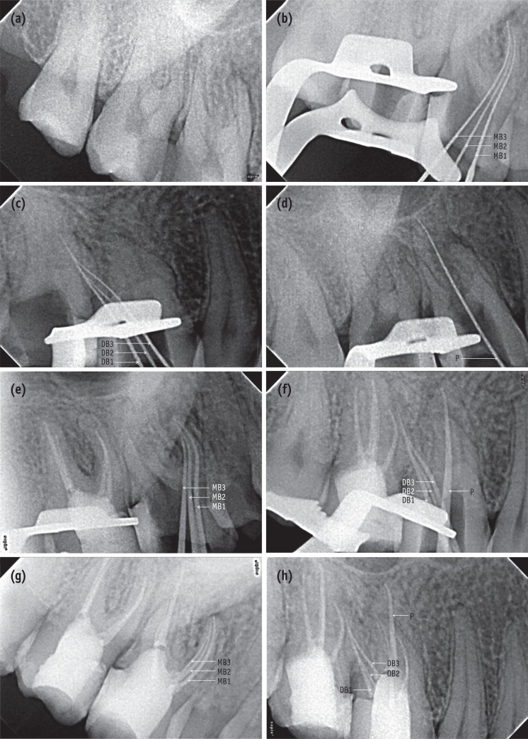
A 26 year old male patient reported to the Department of Conservative Dentistry and Endodontics with a chief complaint of pain in upper right posterior region of mouth for the past one week. The patient revealed a history of mild intermittent pain for the past 2 months, which had increased in intensity during the past one week. The patient reported subjective symptoms of prolonged sensitivity to hot and cold food and drinks. The pain was spontaneous and aggravated particularly at night, and the patient required to consume analgesic to get relief from pain. The general anamnesis contained no abnormal data. Clinical examination revealed a deep disto-occlusal and mesio-occlusal carious lesions on the right maxillary first (tooth 16) and second molar (tooth 17) respectively. Palpation of the buccal and palatal aspects of the involved teeth did not reveal any tenderness. However, the tooth 16 was tender to vertical percussion, whereas tooth 17 did not reveal any tenderness. The teeth were not mobile and periodontal probing around the teeth was within the physiological limits. Thermal testing of the involved tooth with heated gutta-percha (Dentsply Maillefer, Ballaigues, Switzerland) and cold spray (Roeko Endo-Frost spray, Coltene-Whaledent, Langenau, Germany) caused an intense lingering pain, whereas electronic pulp stimulation (Parkell Electronics Division, Farmingdale, NY, USA) showed pulpal vitality on both teeth. The preoperative radiograph revealed the carious lesions closely approximating the pulp with a widened periodontal ligament adjacent to the mesiobuccal root with respect to tooth 16 and no signs of periapical changes with respect to tooth 17 (Figure 1a). Based on sensitivity tests and radiographic findings, a diagnosis of symptomatic irreversible pulpitis with symptomatic apical periodontitis with respect to tooth 16 and symptomatic irreversible pulpitis with respect to tooth 17 were made and endodontic treatment was initiated.
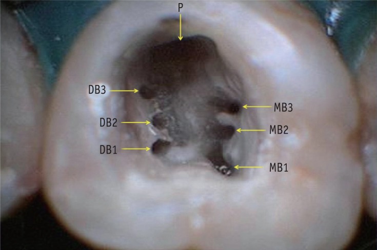
Radiographic evaluation of the involved teeth revealed three completely formed roots with no indication of any variation in the root canal anatomy (Figure 1a). The teeth were anesthetized using 1.8 mL 2% lidocaine with 1:200,000 epinephrine (Xylocaine, AstraZeneca Pharma Ind Ltd, Bangalore, India). The proximal surfaces of both the teeth were restored with composite resin (P60, 3M ESPE Dental Products, St Paul, MN, USA) after caries excavation to enable optimal isolation. Following rubber dam isolation, an endodontic access cavity was prepared on tooth 16 and 17. On inspection with a DG-16 endodontic explorer (Hu-Friedy, Chicago, IL, USA) under an operating microscope (Roslane Meditech, Haryana, India), the pulp chamber floor revealed two canal openings in the mesiobuccal root (MB1 and MB2), and one in the distobuccal (DB1) and palatal root on teeth 16 and 17. Anticipating the presence of extra canals, further exploration of the pulpal floor was carried out with a DG-16 endodontic explorer (Hu-Friedy) on tooth 16 on the line connecting the orifices of the buccal and palatal canal along the floor-wall junction. A "catch" just palatal to the orifice of the MB2 canal unveiled the third canal (MB3) in the mesiobuccal root. In addition, two sticky points were also detected on the line connecting the distobuccal and palatal canal orifices which led to the finding of two additional canals in the distobuccal root (DB2 and DB3, Figure 2). Inspection of the pulpal floor on tooth 17 was done for search of other orifices that were absent.
Even though initial visualization revealed additional canals present, however, several attempts to introduce a file into these orifices were unsuccessful. On further removal of dentinal shelf beginning from the orifice of the mesiobuccal and distobuccal canals and moving toward the orifice of the palatal canal with an ultrasonic micro-endodontic tip (ET 18D tip, Satelec Acteon, Merignac, France) clearly unveiled the orifice of additional canals. Negotiations of the canals were carried out with ISO size 06, 08, and 10 C-Pilot file (VDW GmbH, Munich, Germany) with clockwise and counter-clockwise rotational movement with apical pressure until the apex locator (Raypex5, VDW GmbH) reported that the apical patency was achieved. A glide path was established using size 15/0.01, 15/0.02 and 20/0.02 Hyflex Glide Path Files (Coltene-Whaledent) respectively. Coronal flaring was carried out using a NiTi size 25/0.08 Hyflex CM rotary instrument (Coltene-Whaledent) to improve the straight-line access. The working length which was initially determined with the help of an apex locator was later confirmed using individual radiographs for the mesiobuccal (Figure 1b), distobuccal (Figure 1c), and palatal root (Figure 1d) taken at different horizontal angulations. The working length radiographs showed that in the mesiobuccal root the MB2 canal merged with the MB3 canal at the apical third and exited through one apical foramen, whereas the MB1 canal had a separate canal orifice and exiting foramen. In the distobuccal root, the DB2 and DB3 canals merged with the DB1 canal at the middle and apical third respectively to form a single canal that exited through one foramen. Following which, the access cavities of both the teeth were temporized with a sterile cotton pellet and Cavit G (3M ESPE).
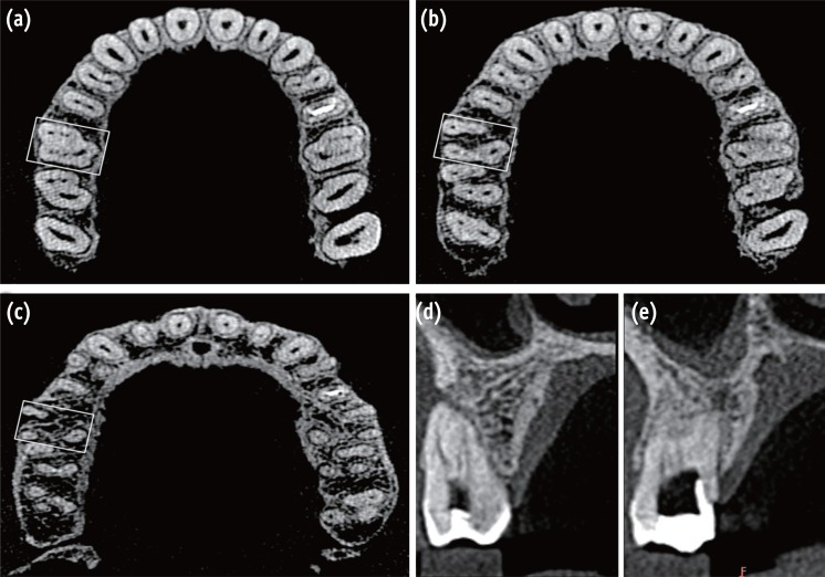
To confirm this rare and unusual morphology, dental imaging of the tooth with the help of CBCT (Carestream CS 9300, Carestream Dental LLC, Atlanta, GA, USA) was planned. An informed consent was obtained from the patient, and multi-slice scans of the maxilla was performed with a tube voltage of 90 KV and a tube current of 15 mA. All measures were taken to protect the patient from radiation. The involved tooth was focused, and the cross-sectional images were obtained in axial, transverse and sagittal planes (Figures 3a - 3e). All images were analyzed with the help of CS 3D imaging software (Carestream Dental LLC). Axial images were obtained at 180 µm thickness and were studied at cervical, middle and apical third of the roots to determine the canal morphology (Figures 3a - 3c). The images revealed that the tooth 16 had three roots and seven root canals (three mesiobuccal, three distobuccal, and one palatal). Transverse section CBCT scanned images of the mesiobuccal and distobuccal root confirmed the canal configurations that were seen in the working length radiographs (Figures 3d and 3e). In contrast, the contralateral tooth appeared to have a normal root canal anatomy (Figures 3a - 3e).
At the second appointment, the patient was asymptomatic. The tooth was anesthetized followed by rubber dam isolation. Cleaning and shaping was done using Hyflex CM rotary instruments along with lubricant (Glyde File Prep, Dentsply Maillefer, Tulsa, OK, USA) using the crown-down technique. All the mesiobuccal and distobuccal canals were enlarged to size 25/0.04, whereas the palatal canal was enlarged to size 40/0.04. Irrigation was performed using 3% sodium hypochlorite solution (Cmident, New Delhi, India) and 17% EDTA solution (Prevest Denpro Ltd, Jammu, India). The canals were finally flushed with sterile saline. Master cones were selected by placing gutta-percha cones (Dentsply Maillefer) corresponding with the size and taper of the last file used to the working length and was confirmed radiographically (Figures 1e and 1f). The canals were dried with absorbent points (Dentsply Maillefer), and obturation was performed using cold lateral compaction of gutta-percha and AH Plus resin sealer (Dentsply Maillefer). The tooth was then restored with a posterior composite resin core (P60, 3M ESPE). Final radiographs were taken to establish the quality of the obturation (Figures 1g and 1h). Tooth 17 was simultaneously treated using the same protocol as done for tooth 16. The patient remained asymptomatic during the 2 months of follow-up period.
Out of the various comprehensive maxillary first molars ex vivo studies in the dental literature, only Baratto Filho et al. reported a maxillary first molar with three roots and seven root canals.24 Of the 140 extracted maxillary first molars, only one tooth showed seven root canals in which 3 mesiobuccal canals, 3 distobuccal canals, and 1 palatal canal were identified. However, all roots showed one apical foramen each.24 Kottoor et al. were the first to report a case of endodontic management of a right maxillary first molar with three roots and seven root canals.12 The tooth presented with 3 canals in the mesiobuccal root, 2 in the distobuccal root and 2 in the palatal root. Both the palatal and distobuccal root had a Vertucci type II (2-1) canal pattern, while the mesiobuccal root showed a Sert and Bayirli type XV (3-2) canal configuration.15 Kottoor et al. later reported the endodontic management of a left maxillary first molar with three roots and eight root canals.12 The tooth presented with 3 canals each in the mesiobuccal and distobuccal roots, and 2 in the palatal root. Both the mesiobuccal and distobuccal roots contained a Sert and Bayirli type XV (3-2) canal configuration, whereas the palatal root showed a Vertucci type II (2-1) canal configuration.12 More recently, Kumar and Badole et al. reported a case of endodontic management of a left maxillary first molar with three roots and seven root canals.6,7 The tooth presented with 3 canals in the mesiobuccal root, 2 in the distobuccal root and 2 in the palatal root. In the case reported by Kumar, both the palatal and distobuccal roots showed a Vertucci type II (2-1) canal pattern, whereas the mesiobuccal root canal showed a Sert and Bayirli type XV (3-2) configuration.6 In the case reported by Badole et al., both the palatal and distobuccal root showed a Vertucci type IV (2) canal pattern while the mesiobuccal root showed a Vertucci type VIII (3) canal configuration.7 In the cases reported by Kumar and Badole et al., the contralateral tooth also exhibited an identical morphology.6,7 Martins reported a unique case of endodontic management of a right maxillary first molar with three roots and seven root canals.25 The tooth presented with 4 canals in the mesiobuccal root, 2 in the distobuccal root and 1 in the palatal root. The mesiobuccal root showed a Sert and Bayirli type XXI (4-1) canal configuration, whereas the distobuccal and the palatal roots showed Vertucci type II (2-1) and type I (1) configuration, respectively.25
A literature review of the case reports presenting 3 canals in the mesiobuccal root of the maxillary first molar revealed a common Sert and Bayirli type XV (3-2) canal configuration.6,8,9,10,12,14,15,16,17,18,20 An exception seen was in the cases reported by Ayranci et al. and Du et al. who found a Sert and Bayirli type XVIII (3-1) and type XVI (2-3) canal configuration respectively.11,13 On the other hand, Badole et al. and Beatty reported a Vertucci type VIII (3-3) canal pattern in the mesiobuccal root presenting with 3 canals.7,19 Karthikeyan and Mahalaxmi reported a case of endodontic management of a right maxillary first molar with six canals, 3 of which were located in the distobuccal root.21 The distobuccal root presented a Gulabivala's supplemental type III (3-2) canal configuration.21 Maggiore et al. and Wong reported a Sert and Bayirli type XVIII (3-1) canal configuration in the palatal root presenting with 3 canals.22,23 In the present case, after correlating and computing the clinical, radiographic and CBCT scan findings, the configurations of the canals in the mesiobuccal and distobuccal roots were perceived to be Al-Qudah and Awawdeh type (3-2) and type (3-2-1), respectively. Figure 4 illustrates Al-Qudah and Awawdeh supplemental canal configurations.26
In 2006, Cleghorn et al. did a comprehensive review of the root and root canal morphology of the maxillary first molar from the data obtained from various anatomical studies.27 The internal canal morphology of the mesiobuccal root in 8,399 teeth was assessed in 34 laboratory and clinical studies. In this large sample, the prevalence of one canal (MB) was 43.1%, and the incidence of two canals (MB1 and MB2) was 56.8%. The incidence of one apical foramen in the mesiobuccal root was 61.6%, while two separate apical foramina were present in 38.3%. The canal morphology of the distobuccal and palatal roots was assessed in 14 laboratory and clinical studies that included 2,576 teeth. In the distobuccal root, the prevalence of one canal (DB) was 98.3%, and the incidence of two canals (DB1 and DB2) was 1.7%. A single apical foramen was present 98% of the time. In the palatal root, the prevalence of a single canal and a single foramen were 99% and 98.8%, respectively. One common finding in this extensive review was the absence of any study reporting the presence of the third canal in any of the three roots.27
However, more recent anatomical studies were able to establish the prevalence of the third canal in the mesiobuccal root. Degerness et al. sectioned and examined 90 uninstrumented mesiobuccal roots of maxillary first molars under stereomicroscope, and found that MB3 has an incidence of 1.1%.28 Using CBCT, Lee et al. and Kim et al. analysed 458 and 814 maxillary first molars, respectively, and reported a prevalence of MB3 of 1.3% and 0.1%, respectively.29,30 Verma et al. and Kim et al. documented an incidence of 10% and 12%, respectively.31,32 The study from Verma et al. had a small sample of 20 roots, and the results may not have a very strong statistical value.31 However, the study from Kim et al. had a sample of 154 roots and must be considered as a frame of reference.32
The higher incidence of multiple canals in the mesiobuccal root as compared to the distobuccal root could possibly be due to the mesiobuccal root being broad buccolingually while the distobuccal root is round or ovoid in cross-section.27 Sometimes fusion of distobuccal root with palatal root may occur due to distobuccal root being broad buccolingually and may result in additional canals in the distal root. In the present case, axial and transverse section of CBCT scanned images clearly depicted that the mesiobuccal and distobuccal roots were broad buccolingually with distobuccal root fused with the palatal root (Figure 3b), confirming that this variation in the external root morphology may account for the multiple canals found in them.
This report serves to remind the dental practitioners of the complexities in root canal morphologies that the maxillary first molars can exhibit. A clinician should have a thorough knowledge of all the diversities and should perform careful examination of the floor of the pulp chamber with a dental operating microscope, and use advanced diagnostic aids such as CBCT for the success in endodontic treatment.

Conflict of Interest: No potential conflict of interest relevant to this article was reported.

  • 1. Slowey RR. Radiographic aids in the detection of extra root canals. Oral Surg Oral Med Oral Pathol 1974;37:762-772. ArticlePubMed
  • 2. Hartwell G, Bellizzi R. Clinical investigation of in vivo endodontically treated mandibular and maxillary molars. J Endod 1982;8:555-557. PubMed
  • 3. Kulild JC, Peters DD. Incidence and configuration of canal systems in the mesiobuccal root of maxillary first and second molars. J Endod 1990;16:311-317. ArticlePubMed
  • 4. Calişkan MK, Pehlivan Y, Sepetçioçlu F, Türkün M, Tuncer SS. Root canal morphology of human permanent teeth in a Turkish population. J Endod 1995;21:200-204. ArticlePubMed
  • 5. Sert S, Bayirli GS. Evaluation of the root canal configurations of the mandibular and maxillary permanent teeth by gender in the Turkish population. J Endod 2004;30:391-398. ArticlePubMed
  • 6. Kumar R. Report of a rare case: a maxillary first molar with seven canals confirmed with cone-beam computed tomography. Iran Endod J 2014;9:153-157. PubMedPMC
  • 7. Badole GP, Warhadpande MM, Shenoi PR, Lachure C, Badole SG. A rare root canal configuration of bilateral maxillary first molar with 7 root canals diagnosed using cone-beam computed tomographic scanning: a case. J Endod 2014;40:296-301. PubMed
  • 8. Kaushik M, Mehra N. Maxillary first molars with six canals diagnosed with the aid of cone beam computed tomography: a report of two cases. Case Rep Dent 2013;2013:406923. ArticlePubMedPMCPDF
  • 9. Kakkar P, Singh A. Maxillary first molar with three mesiobuccal canals confirmed with spiral computer tomography. J Clin Exp Dent 2012;4:e256-e259. ArticlePubMedPMC
  • 10. Zhang P, Mao LS. Left maxillary first molar with three mesiobuccal canals: a case report. Beijing Da Xue Xue Bao 2011;43:919-920. PubMed
  • 11. Ayranci LB, Arslan H, Topcuoglu HS. Maxillary first Molar with three canal orifices in MesioBuccal root. J Conserv Dent 2011;14:436-437. ArticlePubMedPMC
  • 12. Kottoor J, Velmurugan N, Surendran S. Endodontic management of a maxillary first molar with eight root canal systems evaluated using cone-beam computed tomography scanning: a case report. J Endod 2011;37:715-719. ArticlePubMed
  • 13. Du Y, Soo I, Zhang CF. A case report of six canals in a maxillary first molar. Chin J Dent Res 2011;14:151-153. PubMed
  • 14. Ma L, Yu J, Sun JJ. Maxillary first molar with three mesiobuccal root canals: a case report. Hua Xi Kou Qiang Yi Xue Za Zhi 2011;29:102-103. PubMed
  • 15. Kottoor J, Velmurugan N, Sudha R, Hemamalathi S. Maxillary first molar with seven root canals diagnosed with cone-beam computed tomography scanning: a case report. J Endod 2010;36:915-921. ArticlePubMed
  • 16. Garg AK, Tewari RK, Kumar A, Agrawal N. Endodontic treatment of a maxillary first molar having three mesiobuccal canals with the aid of spiral computed tomography: a case report. J Oral Sci 2010;52:495-499. ArticlePubMed
  • 17. Favieri A, Barros FG, Campos LC. Root canal therapy of a maxillary first molar with five root canals: case report. Braz Dent J 2006;17:75-78. ArticlePubMed
  • 18. Ferguson DB, Kjar KS, Hartwell GR. Three canals in the mesiobuccal root of a maxillary first molar: a case report. J Endod 2005;31:400-402. PubMed
  • 19. Beatty RG. A five-canal maxillary first molar. J Endod 1984;10:156-157. ArticlePubMed
  • 20. Martínez-Berná A, Ruiz-Badanelli P. Maxillary first molars with six canals. J Endod 1983;9:375-381. ArticlePubMed
  • 21. Karthikeyan K, Mahalaxmi S. New nomenclature for extra canals based on four reported cases of maxillary first molars with six canals. J Endod 2010;36:1073-1078. ArticlePubMed
  • 22. Maggiore F, Jou YT, Kim S. A six-canal maxillary first molar: case report. Int Endod J 2002;35:486-491. PubMed
  • 23. Wong M. Maxillary first molar with three palatal canals. J Endod 1991;17:298-299. ArticlePubMed
  • 24. Baratto Filho F, Zaitter S, Haragushiku GA, de Campos EA, Abuabara A, Correr GM. Analysis of the internal anatomy of maxillary first molars by using different methods. J Endod 2009;35:337-342. ArticlePubMed
  • 25. Martins JN. Endodontic treatment of a maxillary first molar with seven root canals confirmed with cone beam computer tomography - case report. J Clin Diagn Res 2014;8:ZD13-ZD15.
  • 26. Al-Qudah AA, Awawdeh LA. Root and canal morphology of mandibular first and second molar teeth in a Jordanian population. Int Endod J 2009;42:775-784. PubMed
  • 27. Cleghorn BM, Christie WH, Dong CC. Root and root canal morphology of the human permanent maxillary first molar: a literature review. J Endod 2006;32:813-821. ArticlePubMed
  • 28. Degerness RA, Bowles WR. Dimension, anatomy and morphology of the mesiobuccal root canal system in maxillary molars. J Endod 2010;36:985-989. ArticlePubMed
  • 29. Lee JH, Kim KD, Lee JK, Park W, Jeong JS, Lee Y, Gu Y, Chang SW, Son WJ, Lee WC, Baek SH, Bae KS, Kum KY. Mesiobuccal root canal anatomy of Korean maxillary first and second molars by cone-beam computed tomography. Oral Surg Oral Med Oral Pathol Oral Radiol Endod 2011;111:785-791. ArticlePubMed
  • 30. Kim Y, Lee SJ, Woo J. Morphology of maxillary first and second molars analyzed by cone-beam computed tomography in a Korean population: variations in the number of roots and canals and the incidence of fusion. J Endod 2012;38:1063-1068. PubMed
  • 31. Verma P, Love RM. A Micro CT study of the mesiobuccal root canal morphology of the maxillary first molar tooth. Int Endod J 2011;44:210-217. ArticlePubMed
  • 32. Kim Y, Chang SW, Lee JK, Chen IP, Kaufman B, Jiang J, Cha BY, Zhu Q, Safavi KE, Kum KY. A micro-computed tomography study of canal configuration of multiple-canalled mesiobuccal root of maxillary first molar. Clin Oral Investig 2013;17:1541-1546.ArticlePubMedPDF
Figure 1

(a) A preoperative radiograph of teeth 16 and 17; (b - d) Working length radiographs of mesiobuccal (b), distobuccal (c), and palatal (d) roots of tooth 16 from distal, mesial, and straight angulations, respectively; (e - f) Master cone radiographs of mesiobuccal (e), distobuccal, and palatal roots (f) of tooth 16 from distal and mesial angulations, respectively; (g - h) Post-obturation radiographs of mesiobuccal (g), distobuccal, and palatal roots (h) of tooth 16 from distal and mesial angulations, respectively. MB, mesiobuccal; DB, distobuccal.
rde-40-241-g001.jpg
Figure 2

Access chamber showing seven root canal orifices of tooth 16. MB, mesiobuccal; DB, distobuccal; P, palatal.
rde-40-241-g002.jpg
Figure 3

CBCT images of tooth 16. (a - c) Axial sections. CBCT scan images of the maxillary arch at the cervical (a), middle (b), and apical third (c) levels showing three roots and seven root canals (squared area); (d - e) Transverse sections. CBCT scan images of the mesiobuccal (d) and distobuccal (e) roots.
rde-40-241-g003.jpg
Figure 4

Diagrammatic representation, modified from Al-Qudah and Awawdeh supplemental canal configurations.26

rde-40-241-g004.jpg
Table 1

List of case reports of maxillary first molars presenting with 3 or more root canals in a single root

Investigator Methodology Year Ethnicity Sex Age Root Canal
MB DB P
Martins25 CBCT 2014 Indian Male 30 4 2 1
Kumar6 CBCT 2014 Indian Male 20 3 2 2
Kaushik and Mehra8 CBCT 2013 Indian Female 28 3 1 2
Kakkar and Singh9 SCT 2012 Indian Male 19 3 1 1
Zhang and Mao10 IOPA 2011 Chinese Male 42 3 1 1
Ayranci et al.11 IOPA 2011 Turkish Male 22 3 1 1
Kottoor et al.12 CBCT 2011 Indian Male 30 3 3 2
Du et al.13 IOPA 2011 Chinese Female 21 3 1 2
Ma et al.14 CBCT 2011 Chinese Male 46 3 1 1
Kottoor et al.15 CBCT 2010 Indian Male 37 3 2 2
Garg et al.16 SCT 2010 Indian Male 41 3 1 1
Karthikeyan and Mahalaxmi21 IOPA 2010 Indian Female 25 1 3 1
Favieri17 IOPA 2006 Brazilian Male 15 3 1 1
Ferguson et al.18 IOPA 2005 American Male 18 3 1 1
Maggiore et al.22 IOPA 2002 American Male 19 2 1 3
Wong23 IOPA 1991 American Female 22 1 1 3
Beatty19 IOPA 1984 American Male 14 3 1 1
Martínez-Berná and Ruiz-Badanelli (3 case reports)20 IOPA 1983 Spanish Male 10 3 2 1
IOPA 1983 Spanish Male 17 3 2 1
IOPA 1983 Spanish - - 3 2 1

MB, mesiobuccal; DB, distobuccal; P, palatal; CBCT, cone beam computed tomography; IOPA, intraoral periapical radiograph; SCT, spiral computed tomography.

Tables & Figures

REFERENCES

    Citations

    Citations to this article as recorded by  

      • ePub LinkePub Link
      • Cite
        CITE
        export Copy Download
        Close
        Download Citation
        Download a citation file in RIS format that can be imported by all major citation management software, including EndNote, ProCite, RefWorks, and Reference Manager.

        Format:
        • RIS — For EndNote, ProCite, RefWorks, and most other reference management software
        • BibTeX — For JabRef, BibDesk, and other BibTeX-specific software
        Include:
        • Citation for the content below
        Endodontic management of a maxillary first molar with three roots and seven root canals with the aid of cone-beam computed tomography
        Restor Dent Endod. 2015;40(3):241-248.   Published online June 3, 2015
        Close
      • XML DownloadXML Download
      Figure
      • 0
      • 1
      • 2
      • 3
      Endodontic management of a maxillary first molar with three roots and seven root canals with the aid of cone-beam computed tomography
      Image Image Image Image
      Figure 1 (a) A preoperative radiograph of teeth 16 and 17; (b - d) Working length radiographs of mesiobuccal (b), distobuccal (c), and palatal (d) roots of tooth 16 from distal, mesial, and straight angulations, respectively; (e - f) Master cone radiographs of mesiobuccal (e), distobuccal, and palatal roots (f) of tooth 16 from distal and mesial angulations, respectively; (g - h) Post-obturation radiographs of mesiobuccal (g), distobuccal, and palatal roots (h) of tooth 16 from distal and mesial angulations, respectively. MB, mesiobuccal; DB, distobuccal.
      Figure 2 Access chamber showing seven root canal orifices of tooth 16. MB, mesiobuccal; DB, distobuccal; P, palatal.
      Figure 3 CBCT images of tooth 16. (a - c) Axial sections. CBCT scan images of the maxillary arch at the cervical (a), middle (b), and apical third (c) levels showing three roots and seven root canals (squared area); (d - e) Transverse sections. CBCT scan images of the mesiobuccal (d) and distobuccal (e) roots.
      Figure 4 Diagrammatic representation, modified from Al-Qudah and Awawdeh supplemental canal configurations.26
      Endodontic management of a maxillary first molar with three roots and seven root canals with the aid of cone-beam computed tomography

      List of case reports of maxillary first molars presenting with 3 or more root canals in a single root

      InvestigatorMethodologyYearEthnicitySexAgeRoot Canal
      MBDBP
      Martins25CBCT2014IndianMale30421
      Kumar6CBCT2014IndianMale20322
      Kaushik and Mehra8CBCT2013IndianFemale28312
      Kakkar and Singh9SCT2012IndianMale19311
      Zhang and Mao10IOPA2011ChineseMale42311
      Ayranci et al.11IOPA2011TurkishMale22311
      Kottoor et al.12CBCT2011IndianMale30332
      Du et al.13IOPA2011ChineseFemale21312
      Ma et al.14CBCT2011ChineseMale46311
      Kottoor et al.15CBCT2010IndianMale37322
      Garg et al.16SCT2010IndianMale41311
      Karthikeyan and Mahalaxmi21IOPA2010IndianFemale25131
      Favieri17IOPA2006BrazilianMale15311
      Ferguson et al.18IOPA2005AmericanMale18311
      Maggiore et al.22IOPA2002AmericanMale19213
      Wong23IOPA1991AmericanFemale22113
      Beatty19IOPA1984AmericanMale14311
      Martínez-Berná and Ruiz-Badanelli (3 case reports)20IOPA1983SpanishMale10321
      IOPA1983SpanishMale17321
      IOPA1983Spanish--321

      MB, mesiobuccal; DB, distobuccal; P, palatal; CBCT, cone beam computed tomography; IOPA, intraoral periapical radiograph; SCT, spiral computed tomography.

      Table 1 List of case reports of maxillary first molars presenting with 3 or more root canals in a single root

      MB, mesiobuccal; DB, distobuccal; P, palatal; CBCT, cone beam computed tomography; IOPA, intraoral periapical radiograph; SCT, spiral computed tomography.


      Restor Dent Endod : Restorative Dentistry & Endodontics
      Close layer
      TOP