Previous issues
- Page Path
- HOME > Browse articles > Previous issues
-
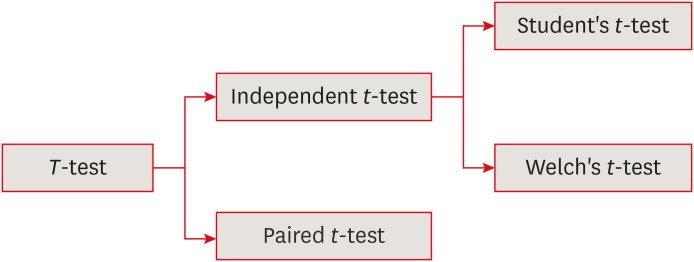
Statistical notes for clinical researchers: the independent samples
t -test - Hae-Young Kim
- Restor Dent Endod 2019;44(3):e26. Published online July 17, 2019
- DOI: https://doi.org/10.5395/rde.2019.44.e26
-
 PDF
PDF PubReader
 ePub
ePub -
Citations
Citations to this article as recorded by- Miyawaki ‘Mini-Forests’ – The New Kids On The Block? Exploring perceptions of the miyawaki forest method among UK landscape professionals
Hanyu Qi, Nicola Dempsey, Ross Cameron
Urban Forestry & Urban Greening.2026; 116: 129238. CrossRef - Examining Gender Differences in Aggression as a Predictor of Anxiety, Depression, and Suicide in a Cross‐Sectional French Sample
Sylvia Martin
Health Science Reports.2025;[Epub] CrossRef - Structural and topological analysis of thiosemicarbazone-based metal complexes: computational and experimental study of bacterial biofilm inhibition and antioxidant activity
Doaa S. El‑Sayed, Shaymaa S. Hassan, Liblab S. Jassim, Ali Abdullah Issa, Firas AL-Oqaili, Mustafa k. Albayaty, Buthenia A. Hasoon, Majid S. Jabir, Khetam H. Rasool, Hemmat A. Elbadawy
BMC Chemistry.2025;[Epub] CrossRef - xeredar: An open-source R package for the statistical analysis of endocrine new approach methodologies using fish and amphibian eleutheroembryos
Inka Marie Spyridonov, Lijuan Yan, Eduard Szöcs, Ana Filipa Pereira Miranda, Carsten Lange, Andrew Tindall, David Du Pasquier, Gregory Lemkine, Lennart Weltje, Maike Habekost, Pernille Thorbek
Environmental Toxicology and Chemistry.2025; 44(9): 2673. CrossRef - Dealing with lipoedema: women’s experiences of healthcare, self-care, and treatments—a mixed-methods study
Johanna Falck, Annette Nygårdh, Bo Rolander, Lise-Lotte Jonasson, Jan Mårtensson
BMC Women's Health.2025;[Epub] CrossRef - Learning From Principals: Exploring Malaysian Primary Deputy Principals’ Informal Mentoring Experiences
Lokman Mohd Tahir, Roslizam Hassan, Mohammed Borhandden Musah
Sage Open.2025;[Epub] CrossRef - Transcriptomic studies on the product stress response revealed that YCF1 is a beneficial factor for progesterone production in Yarrowia lipolytica
Ying Wang, Ruosi Zhang, Mingdong Yao, Wenhai Xiao, Ying Wang, Ying-Jin Yuan
Synthetic and Systems Biotechnology.2025; 10(4): 1087. CrossRef - Remote Monitoring of Psoriasis: Comparing Care Models and Evaluating Quality of Life Outcomes: Mixed Methods Study
Jana Arsenjeva, Priit Kruus, Riina Hallik, Secil Matasova, Laura Prett, Katrin Kaarna, Liisi Raam, Oliver Taul, Liis Ilves, Kaisa Viljar, Pille Konno, Peeter Ross, Külli Kingo
Journal of Medical Internet Research.2025; 27: e73664. CrossRef - Preoperative lower limb kinematic analysis in spinal sagittal imbalance using machine learning and IMU sensors
Sadegh Madadi, Mostafa Rostami, Hadi Farahani, Farshad Nikouee, Mohammad Samadian, Ram Haddas
Signal, Image and Video Processing.2025;[Epub] CrossRef - Predictive for patients with pneumonia in pediatric intensive care unit
Mingxuan Jia, Xiyan Hu, Lin Ji, Jiawen Lin, Jialin Liu, Yong Wang
Frontiers in Pediatrics.2025;[Epub] CrossRef - Enjoyment, Empathy, and Environmental Behavior: A Study of Young Children’s Nature Connection in Iceland
Meghan C. Orman, Shannon B. Wanless, Freyja Birgisdóttir, Kristín Norðdahl
Journal of Cognition and Development.2025; : 1. CrossRef - Source discrimination of colchicine based on carbon stable isotope analysis
Hanyang Zheng, Jun Zhu, Zhuotong Cai, Zhaowei Jie, Wei Wang, Hanyu Zhang, Can Hu, Hongling Guo, Hongcheng Mei
Journal of Forensic Sciences.2025; 70(6): 2227. CrossRef - Empirical Investigation of the Motivation and Perceptions of Tourists Visiting Spa Resorts in the Vâlcea Subcarpathians, Romania
Amalia Niță, Ionuț-Adrian Drăguleasa
Sustainability.2025; 17(14): 6590. CrossRef - Barriers and drivers of digitalization of technical products: a survey in industry
Michel Fett, Richard Breimann, Adrian Mustafa, Angelina Hirsch, Chentao Cai, Matthias Striebel, Jonathan Dagnew, Eckhard Kirchner
Forschung im Ingenieurwesen.2025;[Epub] CrossRef - Late secondary alveolar bone grafting using autologous versus alloplastic material for treating patients with cleft lip and palate with one year follow-up—A retrospective comparative study
Amrit Thapa, Saugat Ray, B. Jayan, S.S. Chopra, B.S. Walia, Abhijeet Kadu, Kapil Tomar
Journal of Plastic, Reconstructive & Aesthetic Surgery.2025; 110: 35. CrossRef - Las inyecciones de plasma rico en plaquetas mejoran los resultados funcionales en comparación con la radiofrecuencia pulsada mediante bloqueo del ganglio impar en el tratamiento de la coxigodinia
J. Pilco Inga, A. Fervienza Sánchez, J.J. Velázquez Fragoso, M. Fa-Binefa, I. Moya Molinas
Revista Española de Anestesiología y Reanimación.2025; 72(10): 501929. CrossRef - The Role of Character Strengths in Employment Among People With Multiple Sclerosis
Beatrice Lee
Rehabilitation Counseling Bulletin.2025;[Epub] CrossRef - Students’ attitudes toward LLMs and its association with metacognitive abilities: A cross-sectional study
Shihui Natalie Yeh, Chiew-Jiat Rosalind Siah
Nurse Education in Practice.2025; 88: 104567. CrossRef - Preparing for the Future: Evaluating the Employability Skills of 4-H Extension Agents
William Norris, Adebayo Adeoti, Jake Devine
Journal of Agricultural Education.2025; 66(4): 4. CrossRef - Platelet-rich plasma injections improve functional results over pulsed radiofrequency in ganglion impar treatments for coccydynia
J. Pilco Inga, A. Fervienza Sánchez, J.J. Velázquez Fragoso, M. Fa-Binefa, I. Moya Molinas
Revista Española de Anestesiología y Reanimación (English Edition).2025; 72(10): 501929. CrossRef - Network architecture optimization for ophthalmic ultrasound image detection based on modular ablation of multi-version YOLO
Zemeng Li, Xiaochun Wang, Xinqi Yu, Zhiyuan Zhao, Yan Wang, Sheng Zhou
BioMedical Engineering OnLine.2025;[Epub] CrossRef - Delays in diagnosis and treatment of lymphoma in Nepal: A cross-sectional study
Jasmine Gurung, Bishesh S Poudyal, Bishnu D Poudel, Bibek Acharya, Sandhya Chapagain, Saugat Poudyal, Pradeep Thapa, Shama Pandey, Ramila Shilpakar
Cancer Research, Statistics, and Treatment.2025; 8(4): 269. CrossRef - THE RELATIONSHIP BETWEEN FINANCIAL BEHAVIOR AND DETERMINANTS OF FINANCIAL EDUCATION DEMAND: AN APPLICATION TO UNIVERSITY STUDENTS
Kutlu Ergün
Uluslararası İktisadi ve İdari İncelemeler Dergisi.2025; (49): 429. CrossRef - Genome-Wide Identification, Characterization, and Expression Profiles of TLR Genes in Darkbarbel Catfish (Pelteobagrus vachelli) Following Aeromonas hydrophila Infection
Zhengyong Wen, Lisha Guo, Jianchao Chen, Qiyu Chen, Yanping Li, Yunyun Lv, Qiong Shi, Shengtao Guo
Biology.2025; 14(12): 1724. CrossRef - Assessing the contribution of impoundment–induced climate change to vegetation growth in the Xiluodu reservoir area of the Jinsha River
Xinyan Wu, Meng Hao, Jingya Wen, Pengyuan Wang, Chenxu Ji, Yong Huang
Ecological Frontiers.2025;[Epub] CrossRef - Improving the efficacy of functional endoscopic sinus surgery in chronic rhinosinusitis with nasal polyps through the combination of budesonide infiltration therapy
Jing-Jing Cun, Jian-Hui Wu, Qian Pan, Xin Li, Qi Cui, Zhong Pan, Rong Chen, Min-Yi Fu
BMC Surgery.2025;[Epub] CrossRef - Comparison of the effects of video conference and video-based home exercise on physical performance and body composition in older adult individuals
Özgün Elmas, Mustafa Cemali, Ayşe Livanelioğlu
Medicine.2024; 103(44): e40329. CrossRef - Deep Residual Learning-Based Classification with Identification of Incorrect Predictions and Quantification of Cellularity and Nuclear Morphological Features in Digital Pathological Images of Common Astrocytic Tumors
Yen-Chang Chen, Shinn-Zong Lin, Jia-Ru Wu, Wei-Hsiang Yu, Horng-Jyh Harn, Wen-Chiuan Tsai, Ching-Ann Liu, Ken-Leiang Kuo, Chao-Yuan Yeh, Sheng-Tzung Tsai
Cancers.2024; 16(13): 2449. CrossRef - The Impact of Stress First Aid on Perceived Stress Levels of New Graduate Nurses
John R. Balcuk
Nursing Economic$.2024; 42(4): 191. CrossRef - CloudBrain-MRS: An intelligent cloud computing platform for in vivo magnetic resonance spectroscopy preprocessing, quantification, and analysis
Xiaodie Chen, Jiayu Li, Dicheng Chen, Yirong Zhou, Zhangren Tu, Meijin Lin, Taishan Kang, Jianzhong Lin, Tao Gong, Liuhong Zhu, Jianjun Zhou, Ou-yang Lin, Jiefeng Guo, Jiyang Dong, Di Guo, Xiaobo Qu
Journal of Magnetic Resonance.2024; 358: 107601. CrossRef - Bactericidal activities and biochemical features of 16 antimicrobial peptides against bovine-mastitis causative pathogens
Hye-sun Cho, Dohun Kim, Hyoim Jeon, Prathap Somasundaram, Nagasundarapandian Soundrarajan, Chankyu Park
Veterinary Research.2024;[Epub] CrossRef - Expressions of Interleukin-4 and Interleukin-5 in Nodular Prurigo and Pruritic Papular Lesions
Ayu Wikan Sayekti, Ann Kautsaria Putri, Dwi Retno Adi Winarni, Satiti Retno Pudjiati
Folia Medica Indonesiana.2024; 60(1): 47. CrossRef - Challenges and barriers to e-leadership participation: Examining the perspectives of Malaysian secondary school teachers
Cha Shi Ping, Lokman Mohd Tahir, Mohd Shafie Rosli, Noor Azean Atan, Mohd Fadzli Ali
Education and Information Technologies.2024; 29(8): 10329. CrossRef - One-Leg Standing Test with Eyes Open as a Screening Tool for Prefrailty in Community-Dwelling Older Japanese Women
Zhenyue Liu, Shuji Sawada, Hisashi Naito, Shuichi Machida
Healthcare.2024; 12(23): 2378. CrossRef - Detection of cardiovascular disease cases using advanced tree-based machine learning algorithms
Fariba Asadi, Reza Homayounfar, Yaser Mehrali, Chiara Masci, Samaneh Talebi, Farid Zayeri
Scientific Reports.2024;[Epub] CrossRef - Recent Advancements in Subcellular Proteomics: Growing Impact of Organellar Protein Niches on the Understanding of Cell Biology
Vanya Bhushan, Aleksandra Nita-Lazar
Journal of Proteome Research.2024; 23(8): 2700. CrossRef - A comparison study: The use of digital and conventional impression techniques in dental hygiene education
Raha K. Naderi, Tulsi J. Patel, Michelle A. Thompson
Journal of Dental Education.2024; 88(5): 518. CrossRef - An Evaluation of the Nunez Community College Quality Enhancement Program: Assessing the Impact of Embedded Conover Workplace Readiness® Modules on Students’ Work Readiness
Sandra Aurand Bosch, Donna Mowery Rice
Sage Open.2024;[Epub] CrossRef - Integrating electric vehicles into hybrid microgrids: A stochastic approach to future-ready renewable energy solutions and management
Aykut Fatih Güven
Energy.2024; 303: 131968. CrossRef - Comparison of postural assessment and awareness in individuals receiving posture training using the digital AI posture assessment and correction system
Musa Çankaya, Fatma Nur Takı
International Journal of Occupational Safety and Ergonomics.2024; 30(4): 1311. CrossRef - Detection and Analysis of Fake News Users’ Communities in Social Media
Abdelouahab Amira, Abdelouahid Derhab, Samir Hadjar, Mustapha Merazka, Md. Golam Rabiul Alam, Mohammad Mehedi Hassan
IEEE Transactions on Computational Social Systems.2024; 11(4): 5050. CrossRef - How do fish miss? Attack strategies of threespine stickleback capturing non-evasive prey
Seth Shirazi, Timothy E. Higham
Journal of Experimental Biology.2024;[Epub] CrossRef - Effects of Occlusal Contact on Maxillary Alveolar Bone Morphology in Patients with and without Anterior Open Bite: A Cross-Sectional Study
Chiyo Shimizu-Tomoda, Yuji Ishida, Aiko Ishizaki-Terauchi, Yukari Mizoguchi, Shuji Oishi, Takashi Ono
Journal of Clinical Medicine.2024; 13(11): 3061. CrossRef - Bioceramic modular tissue-engineered bone with rapid vascularization for large bone defects
Siwei Luo, Zhen Wang, Jialin He, Geng Tang, Daizhu Yuan, Zhanyu Wu, Zihao Zou, Long Yang, Tao Lu, Chuan Ye
Ceramics International.2024; 50(11): 18275. CrossRef - Effect of Arogya Raksha Panchatantra (five lifestyle principles) on heart rate variability, menstrual symptoms, health-related quality of life, performance and self-efficacy in Young female adults with primary dysmenorrhea: protocol for an exploratory ran
Karishma Silwal, Prakash Babu Kodali, Vakeel Khan, Hemanshu Sharma, Gulab Rai Tewani, Pradeep M. K. Nair
CCRYN Indian Journal of Yoga & Naturopathy.2024; 1(1): 15. CrossRef - Letter to the editor on: “Calcaneal positioning in equinus immobilization of the ankle joint: A comparison of common orthoses used in the treatment of acute Achilles tendon ruptures”
Rahul Kumar, Diggaj Shrestha, Kunal Setia, Sunita Sharma
Foot and Ankle Surgery.2023; 29(6): 498. CrossRef - Irradiation damage reduces alloy corrosion rate via oxide space charge compensation effects
Zefeng Yu, Elizabeth Kautz, Hongliang Zhang, Anton Schneider, Taeho Kim, Yongfeng Zhang, Sten Lambeets, Arun Devaraj, Adrien Couet
Acta Materialia.2023; 253: 118956. CrossRef - Predictors of Relevant Changes in Pain and Function for Adolescents With Idiopathic Scoliosis Following Surgery
Samia Alamrani, Adrian Gardner, Alison B. Rushton, Deborah Falla, Nicola R. Heneghan
Spine.2023; 48(16): 1166. CrossRef - A multicenter feasibility study on implementing a brief mindful breathing exercise into regular university courses
Annika C. Konrad, Veronika Engert, Reyk Albrecht, Christian Dobel, Nicola Döring, Jens Haueisen, Olga Klimecki, Mike Sandbothe, Philipp Kanske
Scientific Reports.2023;[Epub] CrossRef - Adult children of parents with mental illness: Family stigma and coping on sense of self
Chynna Campbell, Pamela Patrick
Child & Family Social Work.2023; 28(3): 622. CrossRef - Veri Madenciliğine Dayalı Olarak Çalışanların Örgütsel Bağlılık Düzeyinin Belirlenmesi: İstanbul ve Kocaeli Örneği
Nadir ERSEN, Timuçin BARDAK, Uğur Can USTA
Bartın Orman Fakültesi Dergisi.2023; 25(3): 398. CrossRef - Pooled rates and demographics of POTS following SARS-CoV-2 infection versus COVID-19 vaccination: Systematic review and meta-analysis
Shin Jie Yong, Alice Halim, Shiliang Liu, Michael Halim, Ahmad A. Alshehri, Mohammed A. Alshahrani, Mohammed M. Alshahrani, Amal H. Alfaraj, Lamees M. Alburaiky, Faryal Khamis, Muzaheed, Bashayer M. AlShehail, Mubarak Alfaresi, Reyouf Al Azmi, Hawra Alba
Autonomic Neuroscience.2023; 250: 103132. CrossRef - Rapid suture-free repair of arterial bleeding: A novel approach with ultra-thin bioadhesive hydrogel membrane
Siwei Luo, Long Yang, Qiang Zou, Daizhu Yuan, Shunen Xu, Yanchi Zhao, Xin Wu, Zhen Wang, Chuan Ye
Chemical Engineering Journal.2023; 472: 144865. CrossRef - The Relationship Between Obsessive-Compulsive Disorder and Gaming Disorder
Nazir Hawi, Maya Samaha
International Journal of Cyber Behavior, Psychology and Learning.2023; 13(1): 1. CrossRef - Microbiological Quality and Presence of Foodborne Pathogens in Raw and Extruded Canine Diets and Canine Fecal Samples
Doina Solís, Magaly Toro, Paola Navarrete, Patricio Faúndez, Angélica Reyes-Jara
Frontiers in Veterinary Science.2022;[Epub] CrossRef - Stapler Assisted Total Laryngectomy: A Prospective Randomized Clinical Study
Omar Ahmed, Hesham Mustafa Abdel-Fattah, Hisham E. M. Elbadan
Indian Journal of Otolaryngology and Head & Neck Surgery.2022; 74(S2): 2205. CrossRef - Project VLOGI (Video Lectures on Giving Instructions): Effects on Learners’ Performance in Probability and Statistics
Sherwin BATİLANTES
International Journal of Educational Studies in Mathematics.2021; 8(4): 299. CrossRef
- Miyawaki ‘Mini-Forests’ – The New Kids On The Block? Exploring perceptions of the miyawaki forest method among UK landscape professionals
- 11,361 View
- 249 Download
- 57 Crossref

- Effect of calcium hydroxide on inflammatory root resorption and ankylosis in replanted teeth compared with other intracanal materials: a review
- Maryam Zare Jahromi, Mahmood Reza Kalantar Motamedi
- Restor Dent Endod 2019;44(3):e32. Published online August 1, 2019
- DOI: https://doi.org/10.5395/rde.2019.44.e32
-
 Abstract
 PDF
PDF PubReader
 ePub
ePub Calcium hydroxide (CH) is the gold-standard intracanal dressing for teeth subjected to traumatic avulsion. A common complication after the replantation of avulsed teeth is root resorption (RR). The current review was conducted to compare the effect of CH with that of other intracanal medications and filling materials on inflammatory RR and replacement RR (ankylosis) in replanted teeth. The PubMed and Scopus databases were searched through June 2018 using specific keywords related to the title of the present article. The materials that were compared to CH were in 2 categories: 1) mineral trioxide aggregate (MTA) and endodontic sealers as permanent filling materials for single-visit treatment, and 2) Ledermix, bisphosphonates, acetazolamide, indomethacin, gallium nitrate, and enamel matrix-derived protein (Emdogain) as intracanal medicaments for multiple-visit management of avulsed teeth prior to the final obturation. MTA can be used as a single-visit root filling material; however, there are limited data on its efficacy due to a lack of clinical trials. Ledermix and acetazolamide were comparable to CH in reducing RR. Emdogain seems to be an interesting material, but the data supporting its use as an intracanal medication remain very limited. The conclusions drawn in this study were limited by the insufficiency of clinical trials.
-
Citations
Citations to this article as recorded by- Endodontic Intracanal Medicaments and Agents
Anu Priya Guruswamy Pandian, Depti Bellani, Ritya Mary Jibu, Varsha Agnihotri
Dental Clinics of North America.2026; 70(1): 45. CrossRef - Efficacy of Simvastatin in Inhibiting Bone Resorption and Promoting Healing in Delayed Tooth Avulsion: A Case Series
Rajesh Kumar, Supraja N Atluri, Alekhya Achanta, Chittaranjan Bogishetty, Tejaswini R Chunduri, Tejaswini PSS, Ramakrishna Ravi
Cureus.2025;[Epub] CrossRef - Interdisziplinäre Lösung nach dentalem Trauma mit Avulsion und Wurzelresorption
Eva Maier, Julia Lubauer, Kerstin M. Galler
Oralprophylaxe & Kinderzahnmedizin.2025; 47(3): 161. CrossRef - Bioactive potential of Bio-C Temp demonstrated by systemic mineralization markers and immunoexpression of bone proteins in the rat connective tissue
Camila Soares Lopes, Mateus Machado Delfino, Mário Tanomaru-Filho, Estela Sasso-Cerri, Juliane Maria Guerreiro-Tanomaru, Paulo Sérgio Cerri
Journal of Materials Science: Materials in Medicine.2024;[Epub] CrossRef - The use of mineral trioxide aggregate for treatment of children with complications of dental trauma
L.Yu. Kharkova, M.V. Korolenkova
Stomatology.2024; 103(4): 59. CrossRef - Instant Re-Implantation of Avulsed Teeth
Smita Paul, Sambarta Das, Nirmal Debbarma, Barun Dasgupta, Bidyut Seal, Ayesha Satapathy
Journal of Pharmacy and Bioallied Sciences.2024; 16(Suppl 4): S3461. CrossRef - Interpretation by literature review of the use of calcium hydroxide as an intra-ductal medication
María Belén Muñoz Padilla, Verónica Alicia Vega Martínez, Camila Alejandra Villafuerte Moya
Salud, Ciencia y Tecnología.2024; 4: 924. CrossRef - Evaluation of the physicochemical properties of intracanal medications used in traumatized teeth
Patricia Almeida da Silva de Macedo, Walbert de Andrade Vieira, Paulo Henrique Gabriel, Karla de Faria Vasconcelos, Francisco Haiter Neto, Ana Carolina Correia Laurindo de Cerqueira Neto, Brenda Paula Figueiredo de Almeida Gomes, Marcos Roberto dos Santo
Brazilian Journal of Oral Sciences.2024; 23: e242997. CrossRef - Treatment of Teeth with Root Resorptions: A Case Report and Systematic Review
Damla Erkal, Abdullah Başoğlu, Damla Kırıcı, Nezahat Arzu Kayar, Simay Koç, Kürşat Er
Galician Medical Journal.2023;[Epub] CrossRef - Successful outcome of permanent maxillary incisor reimplanted after 30 hours of extra‐oral time—a case report with 5‐year follow‐up
Ibadat Preet Kaur, Ashok Kumar, Mukul Kumar, Kanistika Jha
Clinical Case Reports.2023;[Epub] CrossRef - Replantation of an Avulsed Tooth: A Case Report
Nishad Kadulkar, Rubi Kataki, Adrija Deka, Salouno Thonai
Cureus.2023;[Epub] CrossRef - Avulsion of Permanent Mandibular Incisors: A Report of Two Cases with Pertinent Literature
Ibadat Preet Kaur, Jitendra Sharan, Pallawi Sinha, Ashok Kumar, Anand Marya, Leandro Napier de Souza
Case Reports in Dentistry.2023; 2023: 1. CrossRef - The Impact of Autologous Platelet Concentrates on the Periapical Tissues and Root Development of Replanted Teeth: A Systematic Review
Zohaib Khurshid, Faris Yahya I. Asiri, Shariq Najeeb, Jithendra Ratnayake
Materials.2022; 15(8): 2776. CrossRef
- Endodontic Intracanal Medicaments and Agents
- 3,582 View
- 73 Download
- 13 Crossref

- Influence of different universal adhesives on the repair performance of hybrid CAD-CAM materials
- Gülbike Demirel, İsmail Hakkı Baltacıoğlu
- Restor Dent Endod 2019;44(3):e23. Published online May 20, 2019
- DOI: https://doi.org/10.5395/rde.2019.44.e23
-
 Abstract
 PDF
PDF PubReader
 ePub
ePub Objectives The aim of this study was to investigate the microshear bond strength (μSBS) of different universal adhesive systems applied to hybrid computer-aided design/computer-aided manufacturing (CAD-CAM) restorative materials repaired with a composite resin.
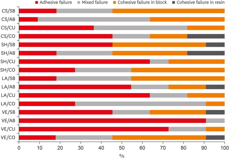
Materials and Methods Four types of CAD-CAM hybrid block materials—Lava Ultimate (LA), Vita Enamic (VE), CeraSmart (CS), and Shofu Block HC (SH)—were used in this study, in combination with the following four adhesive protocols: 1) control: porcelain primer + total etch adhesive (CO), 2) Single Bond Universal (SB), 3) All Bond Universal (AB), and 4) Clearfil Universal Bond (CU). The μSBS of the composite resin (Clearfil Majesty Esthetic) was measured and the data were analyzed using two-way analysis of variance and the Tukey test, with the level of significance set at
p < 0.05.Results The CAD-CAM block type and block-adhesive combination had significant effects on the bond strength values (
p < 0.05). Significant differences were found between the following pairs of groups: VE/CO and VE/AB, CS/CO and CS/AB, VE/CU and CS/CU, and VE/AB and CS/AB (p < 0.05).Conclusions The μSBS values were affected by hybrid block type. All tested universal adhesive treatments can be used as an alternative to the control treatment for repair, except the AB system on VE blocks (the VE/AB group). The μSBS values showed variation across different adhesive treatments on different hybrid CAD-CAM block types.
-
Citations
Citations to this article as recorded by- Effect of surface treatments on the bond strength of resin-repaired resin matrix CAD-CAM ceramic: A scoping review
Ana Beatriz de Souza Albergardi, João Pedro Justino de Oliveira Limírio, Jéssica Marcela de Luna Gomes, Aldiéris Alves Pesqueira, Eduardo Piza Pellizzer
Journal of Dentistry.2025; 154: 105594. CrossRef - Bond strength to aged CAD/CAM composites and polymer-infiltrated ceramic network using a universal adhesive with or without previous application of a universal primer
Clemens Lechte, Lisa Sophia Faesser, Jana Biermann, Alexandra Schmidt, Philipp Kanzow, Annette Wiegand
International Journal of Adhesion and Adhesives.2025; 140: 104017. CrossRef - The Influence of Thermocycling and Ultraviolet Aging on Surface Characteristics and the Repair Bond Strength of CAD/CAM Resin Nanoceramics
Beyza Unalan Degirmenci, Alperen Degirmenci, Zelal Seyfioglu Polat
Journal of Functional Biomaterials.2025; 16(5): 156. CrossRef - Impact of in vitro findings on clinical protocols for the adhesion of CAD-CAM blocks: A systematic integrative review and meta-analysis
Maria João Calheiros-Lobo, Ricardo Carbas, Lucas F.M. da Silva, Teresa Pinho
The Journal of Prosthetic Dentistry.2024; 131(6): 1051. CrossRef - Repair protocols for indirect monolithic restorations: a literature review
Lucas Saldanha da Rosa, Rafaela Oliveira Pilecco, Pablo Machado Soares, Marília Pivetta Rippe, Gabriel Kalil Rocha Pereira, Luiz Felipe Valandro, Cornelis Johannes Kleverlaan, Albert J. Feilzer, João Paulo Mendes Tribst
PeerJ.2024; 12: e16942. CrossRef - Bonding performance of universal adhesives with concomitant use of silanes to CAD/CAM blocks
Marina AMARAL, Jaqueline Maria Brandão RIZZATO, Victoria Caroline Souza de ALMEIDA, Priscila Christiane Suzy LIPORONI, Rayssa Ferreira ZANATTA
RGO - Revista Gaúcha de Odontologia.2023;[Epub] CrossRef - Resistencia a la fractura de una nanocerámica CAD/CAM reparada con dos tratamientos de superficie: estudio in vitro
Marcelo Geovanny Cascante-Calderón, Kevin Alejandro Reascos Flores, Inés María Villacís-Altamirano, Anggely Maite Bayas Salinas, Jessica Elizabeth Taraguay Galindo
Universitas Odontologica.2023;[Epub] CrossRef - Influence of surface treatments and adhesive protocols on repair bond strength of glass‐matrix and resin‐matrix CAD/CAM ceramics
Rana Turunç‐Oğuzman, Soner Şişmanoğlu
Journal of Esthetic and Restorative Dentistry.2023; 35(8): 1322. CrossRef - Effect of Anti-COVID-19 Mouthwashes on Shear Bond Strength of Resin-Matrix Ceramics Repaired with Resin Composite Using Universal Adhesive: An In Vitro Study
Wichuda Limsiriwong, Awiruth Klaisiri, Nantawan Krajangta
Journal of Functional Biomaterials.2023; 14(3): 158. CrossRef - Effect of ceramic primers with different chemical contents on the shear bond strength of CAD/CAM ceramics with resin cement after thermal ageing
Mehmet Uğur, İdris Kavut, Özgür Ozan Tanrıkut, Önder Cengiz
BMC Oral Health.2023;[Epub] CrossRef - Effect of microstructure of reinforced CAD/CAM hybrid composite resin block on shear bond strength of composite resin
Sung-Ho Um, Minjeong Shin, Shin-hye Chung, Young-Seok Park, Bum-Soon Lim
Korean Journal of Dental Materials.2023; 50(1): 29. CrossRef - Dentin contamination during repair procedures: A threat to universal adhesives?
Anne‐Katrin Lührs, Cosima Brachmann, Silke Jacker‐Guhr
Clinical and Experimental Dental Research.2022; 8(3): 771. CrossRef - Influence of mechanical and chemical pre-treatments on the repair of a hybrid ceramic
Sascha Niklas Jung, Stefan Rüttermann
Dental Materials.2022; 38(7): 1140. CrossRef - Influence of different repair protocols and artificial aging on bond strength of composite to a CAD/CAM polymer-infiltrated ceramic
Ece İrem OĞUZ, Gökhan ÇİÇEKCİ
Cumhuriyet Dental Journal.2021; 24(1): 37. CrossRef - REZİN MATRİKS SERAMİKLER-DERLEME
Elif Melike AKARCA, Dilara ŞAHİN, Ragibe Şenay CANAY
Atatürk Üniversitesi Diş Hekimliği Fakültesi Dergisi.2021; : 1. CrossRef - REZİN MATRİKS SERAMİKLER-DERLEME
Elif Melike AKARCA, Dilara ŞAHİN, Ragibe Şenay CANAY
Atatürk Üniversitesi Diş Hekimliği Fakültesi Dergisi.2021; : 1. CrossRef - Microshear bond strength of contemporary self-adhesive resin cements to CAD/CAM restorative materials: effect of surface treatment and aging
Soner Şişmanoğlu, Rana Turunç-Oğuzman
Journal of Adhesion Science and Technology.2020; 34(22): 2484. CrossRef - Influence of different surface treatments and universal adhesives on the repair of CAD-CAM composite resins: An in vitro study
Soner Sismanoglu, Zuhal Yildirim-Bilmez, Aysegul Erten-Taysi, Pınar Ercal
The Journal of Prosthetic Dentistry.2020; 124(2): 238.e1. CrossRef
- Effect of surface treatments on the bond strength of resin-repaired resin matrix CAD-CAM ceramic: A scoping review
- 2,138 View
- 12 Download
- 18 Crossref

-
Coronal tooth discoloration induced by regenerative endodontic treatment using different scaffolds and intracanal coronal barriers: a 6-month
ex vivo study - Noushin Shokouhinejad, Hassan Razmi, Maryam Farbod, Marzieh Alikhasi, Josette Camilleri
- Restor Dent Endod 2019;44(3):e25. Published online July 16, 2019
- DOI: https://doi.org/10.5395/rde.2019.44.e25
-
 Abstract
 PDF
PDF PubReader
 ePub
ePub Objective The aim of this study was to evaluate discoloration of teeth undergoing regenerative endodontic procedures (REPs) using blood clot or platelet-rich fibrin (PRF) as the scaffolds and different calcium silicate-based materials as the intracanal coronal barriers in an
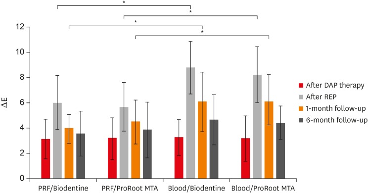
ex vivo model.Materials and Methods Forty-eight bovine incisors were prepared and disinfected using 1 mg/mL double antibiotic paste (DAP). The specimens were then randomly divided into 2 groups (
n = 24) according to the scaffolds (blood or PRF). After placement of scaffolds each group was divided into 2 subgroups (n = 12) according to the intracanal coronal barriers (ProRoot MTA or Biodentine). The pulp chamber walls were sealed with dentin bonding agent before placement of DAP and before placement of scaffolds. The color changes (∆E) were measured at different steps. The data were analyzed using 2-way analysis of variance.Results Coronal discoloration induced by DAP was not clinically perceptible (ΔE ≤ 3.3). Regarding the type of the scaffold, coronal discoloration was significantly higher in blood groups compared with PRF groups at the end of REP and after 1 month (
p < 0.05). However, no significant difference was found between PRF and blood clot after 6 months (p > 0.05). Considering the type of intracanal coronal barrier, no significant difference existed between ProRoot MTA and Biodentine (p > 0.05).Conclusions With sealing the dentinal tubules of pulp chamber with a dentin bonding agent and application of DAP as an intracanal medicament, coronal color change of the teeth following the use of PRF and blood sealed with either ProRoot MTA or Biodentine was not different at 6-month follow-up.
-
Citations
Citations to this article as recorded by- Impact of Biodentine Placement on Fracture Resistance and its Influence on Discoloration with Different Scaffolds
Evren Sarıyılmaz, Öznur Sarıyılmaz, Burak Çarıkçıoğlu, Gülşah Uslu, Raif Alan
Journal of Endodontics.2025; 51(9): 1199. CrossRef - Effect of pH on the solubility and volumetric change of ready-to-use Bio-C Repair bioceramic material
Luana Raphael da SILVA, Jader Camilo PINTO, Juliane Maria GUERREIRO-TANOMARU, Mário TANOMARU-FILHO
Brazilian Oral Research.2024;[Epub] CrossRef - Efficacy of Potassium Iodide and Glutathione for Correlation of Dentin Discoloration Caused by Silver Diamine Fluoride
Mahsa Samani, Hamid Majzoub, Faramarz Zakavi, Ayyub Mojaddami
Cureus.2024;[Epub] CrossRef - Intracanal medicaments and coronal sealing materials influence on root fracture resistance and coronal discoloration: An in vitro study
Rasoul Sahebalam, Marzie Boskabady, Maryam Naghavi, Samira Dehghanitafti
Saudi Endodontic Journal.2024; 14(2): 199. CrossRef - Potential Crown Discoloration Induced by the Combination of Various Intracanal Medicaments and Scaffolds Applied in Regenerative Endodontic Therapy
NB Altun, A Turkyilmaz
Nigerian Journal of Clinical Practice.2024; 27(7): 897. CrossRef - Evaluation of the effectiveness of different treatment approaches in preventing coronal discoloration caused by regenerative endodontic treatment
Melis Oya Ateş, Zeliha Uğur Aydın
Clinical Oral Investigations.2023; 27(8): 4595. CrossRef - Evaluation of the Effectiveness of Laser‐Assisted Bleaching of the Teeth Discolored due to Regenerative Endodontic Treatment
Noushin Shokouhinejad, Mehrfam Khoshkhounejad, Fatemeh Hamidzadeh, Murilo Baena Lopes
International Journal of Dentistry.2022;[Epub] CrossRef - Effectiveness of Teeth Whitening after Regenerative Endodontics Procedures: An In Vitro Study
Irini Fagogeni, Joanna Metlerska, Tomasz Falgowski, Maciej Górski, Mariusz Lipski, Alicja Nowicka
Journal of Clinical Medicine.2022; 11(23): 7016. CrossRef - Microstructure and color stability of calcium silicate-based dental materials exposed to blood or platelet-rich fibrin
Noushin Shokouhinejad, Ibrahim Abu Tahun, Shima Saber Tahan, Fatemeh Mohandes, Mohammad H. Nekoofar, Paul M. H. Dummer
Clinical Oral Investigations.2022; 27(3): 1193. CrossRef - Spectrophotometric analysis of internal bleaching of traumatized teeth with coronal discoloration following regenerative endodontic procedures
Jaqueline Lazzari, Walbert Vieira, Vanessa Pecorari, Brenda Paula Figueiredo de Almeida Gomes, José Flávio Affonso de Almeida, Adriana De-Jesus-Soares
Brazilian Journal of Oral Sciences.2021;[Epub] CrossRef - Biological parameters, discolouration and radiopacity of calcium silicate‐based materials in a simulated model of partial pulpotomy
Lilian Vieira Oliveira, Gabriela Leite de Souza, Gisele Rodrigues da Silva, Thamara Eduarda Alves Magalhães, Gabrielle Alves Nunes Freitas, Ana Paula Turrioni, Gabriella Lopes de Rezende Barbosa, Camilla Christian Gomes Moura
International Endodontic Journal.2021; 54(11): 2133. CrossRef - Effect of hydrogel-based antibiotic intracanal medicaments on crown discoloration
Rayan B. Yaghmoor, Jeffrey A. Platt, Kenneth J. Spolnik, Tien Min Gabriel Chu, Ghaeth H. Yassen
Restorative Dentistry & Endodontics.2021;[Epub] CrossRef - The effect of different calcium silicate-based pulp capping materials on tooth discoloration: an in vitro study
Ahmad S. Al-Hiyasat, Dana M. Ahmad, Yousef S. Khader
BMC Oral Health.2021;[Epub] CrossRef - Knowledge, attitudes, and practices of undergraduate students concerning Regenerative Endodontics
Ligia B. da Silva, Mariana Gabriel, Márcia M. Marques, Fernanda C. Carrer, Flávia Gonçalves, Giovanna Sarra, Giovanna L. Carvalho, Ana Armas-Vega, Maria S. Moreira
Minerva Stomatologica.2020;[Epub] CrossRef - Coronal Discoloration Related to Bioceramic and Mineral Trioxide Aggregate Coronal Barrier in Non-vital Mature Teeth Undergoing Regenerative Endodontic Procedures
Mazen Doumani, Mohammad Yaman Seirawan, Kinda Layous, Mohammad Kinan Seirawan
World Journal of Dentistry.2020; 11(1): 52. CrossRef
- Impact of Biodentine Placement on Fracture Resistance and its Influence on Discoloration with Different Scaffolds
- 1,954 View
- 22 Download
- 15 Crossref

- The influence of nanofillers on the properties of ethanol-solvated and non-solvated dental adhesives
- Leonardo Bairrada Tavares da Cruz, Marcelo Tavares Oliveira, Cintia Helena Coury Saraceni, Adriano Fonseca Lima
- Restor Dent Endod 2019;44(3):e28. Published online July 24, 2019
- DOI: https://doi.org/10.5395/rde.2019.44.e28
-
 Abstract
 PDF
PDF PubReader
 ePub
ePub Objectives The aim of this study was to evaluate the influence of different concentrations of nanofillers on the chemical and physical properties of ethanol-solvated and non-solvated dental adhesives.
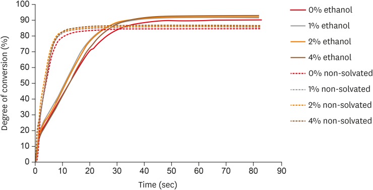
Materials and Methods Eight experimental adhesives were prepared with different nanofiller concentrations (0, 1, 2, and 4 wt%) and 2 solvent concentrations (0% and 10% ethanol). Several properties of the experimental adhesives were evaluated, such as water sorption and solubility (
n = 5, 20 seconds light activation), real-time degree of conversion (DC;n = 3, 20 and 40 seconds light activation), and stability of cohesive strength at 6 months (CS;n = 20, 20 seconds light activation) using the microtensile test. A light-emitting diode (Bluephase 20i, Ivoclar Vivadent) with an average light emittance of 1,200 mW/cm2 was used.Results The presence of solvent reduced the DC after 20 seconds of curing, but increased the final DC, water sorption, and solubility of the adhesives. Storage in water reduced the strength of the adhesives. The addition of 1 wt% and 2 wt% nanofillers increased the polymerization rate of the adhesives.
Conclusions The presence of nanofillers and ethanol improved the final DC, although the DC of the solvated adhesives at 20 seconds was lower than that of the non-solvated adhesives. The presence of ethanol reduced the strength of the adhesives and increased their water sorption and solubility. However, nanofillers did not affect the water sorption and strength of the tested adhesives.
-
Citations
Citations to this article as recorded by- Effect of boric acid on the color stability and mechanical properties of 3D-printed permanent resins
Dalndushe Abdulai, Rafat Sasany, Raghib Suradi, Mehran Moghbel, Seyed Ali Mosaddad
Scientific Reports.2025;[Epub] CrossRef - Development of a Boron Nitride-Filled Dental Adhesive System
Senthilguru Kulanthaivel, Jeremiah Poppen, Sandra Ribeiro Cunha, Benjamin Furman, Kyumin Whang, Erica C. Teixeira
Polymers.2023; 15(17): 3512. CrossRef - Analyses of Experimental Dental Adhesives Based on Zirconia/Silver Phosphate Nanoparticles
Abdul Khan, Yasmin Alhamdan, Hala Alibrahim, Khalid Almulhim, Muhammad Nawaz, Syed Ahmed, Khalid Aljuaid, Ijlal Ateeq, Sultan Akhtar, Mohammad Ansari, Intisar Siddiqui
Polymers.2023; 15(12): 2614. CrossRef - Mechanical characterization and adhesive properties of a dental adhesive modified with a polymer antibiotic conjugate
Camila Sabatini, Russell J. Aguilar, Ziwen Zhang, Steven Makowka, Abhishek Kumar, Megan M. Jones, Michelle B. Visser, Mark Swihart, Chong Cheng
Journal of the Mechanical Behavior of Biomedical Materials.2022; 129: 105153. CrossRef
- Effect of boric acid on the color stability and mechanical properties of 3D-printed permanent resins
- 1,319 View
- 8 Download
- 4 Crossref

-
Dentinal defects induced by 6 different endodontic files when used for oval root canals: an
in vitro comparative study - Ajinkya M Pawar, Bhagyashree Thakur, Anda Kfir, Hyeon-Cheol Kim
- Restor Dent Endod 2019;44(3):e31. Published online July 29, 2019
- DOI: https://doi.org/10.5395/rde.2019.44.e31
-
 Abstract
 PDF
PDF PubReader
 ePub
ePub Objectives To compare the formation of dentinal defects using stainless-steel hand K-files (HFs), rotary files, reciprocating files, and Self-Adjusting File (SAF), when used for oval root canals.
Materials and Methods One hundred and forty extracted human mandibular premolar with single root and oval canal were selected for this study. Oval canals were confirmed by exposing to mesio-distal and bucco-lingual radiographs. Teeth with open apices or anatomic irregularities were excluded. All selected teeth were de-coronated perpendicular to the long axis of the tooth, leaving roots segments approximately of 16 mm in length. Twenty teeth were left unprepared (control), and the remaining 120 teeth were divided into 6 groups (
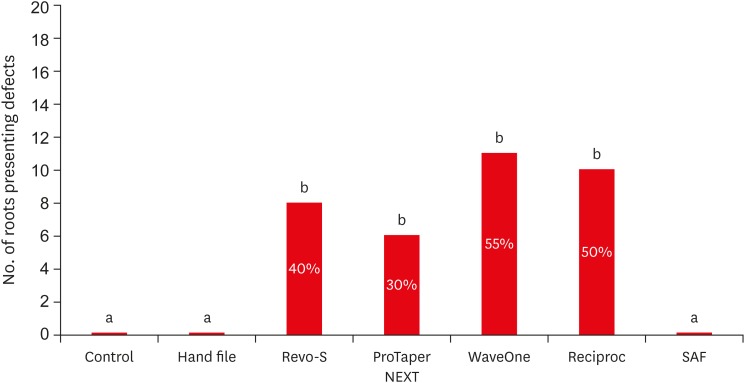
n = 20) and instrumented using HF (size 40/0.02), Revo-S (RS; size 40/0.06), ProTaper NEXT (PTN; size 40/0.06), WaveOne (WO; size 40/0.09), RECIPROC (RC; size 40/0.06), and the SAF (2 mm). Roots were then sectioned 3, 6, and 9 mm from the apex, and observed under stereomicroscope, for presence of dentinal defects. “No defect” was defined as root dentin that presented with no visible microcracks or fractures. “Defect” was defined by microcracks or fractures in the root dentin.Results The control, HF, and SAF did not exhibit any dentinal defects. In roots instrumented by RS, PTN, WO, and RC files exhibited microcracks (incomplete or complete) in 40%, 30%, 55%, and 50%, respectively.
Conclusions The motor-driven root canal instrumentation with rotary and reciprocating files may create microcracks in radicular dentine, whereas the stainless-steel hand file instrumentation, and the SAF produce minimal or less cracks.
-
Citations
Citations to this article as recorded by- Evaluation of dentinal crack formation during post space preparation using different fiber post systems with micro-computed tomography
Ayşe Nur Kuşuçar, Damla Kırıcı
BMC Oral Health.2025;[Epub] CrossRef - Computational Insights into Root Canal Treatment: A Survey of Selected Methods in Imaging, Segmentation, Morphological Analysis, and Clinical Management
Jianning Li, Kerstin Bitter, Anh Duc Nguyen, Hagay Shemesh, Paul Zaslansky, Stefan Zachow
Dentistry Journal.2025; 13(12): 579. CrossRef - Comparative evaluation of incidence of dentinal defects after root canal preparation using three different endodontic retreatment systems – An in vitro study
S. Aarthi, J. S. Sivakumar, A. Andamuthu Sivakumar, J. Saravanapriyan Soundappan, M. Chittrarasu, G. Jayanthi
Journal of Conservative Dentistry and Endodontics.2024; 27(3): 262. CrossRef - Evaluation of Dentin Cracks by Stereomicroscope after Preparation of Mesiobuccal Canal of Maxillary First Molars Using Edge Taper Platinum and ProTaper Gold Rotary Files: A Laboratory Study
Narjes Hoshyari, Seyedali Seyedmajidi, Anahita Lotfizadeh, Eghlima Malakan, Abolfazl Hosseinnataj, Azam Haddadi Kohsar
Avicenna Journal of Dental Research.2023; 15(4): 167. CrossRef - A comparative evaluation of dentinal defects after root canal preparation with different rotary and reciprocal systems
Ece Yakın, Berna Aslan, Emine Odabaşı Tezer
Northwestern Medical Journal.2023; 3(3): 147. CrossRef - Comparison of Dentinal Defects Induced by Rotary, Reciprocating, and Hand Files in Oval Shaped Root Canal - An In-Vitro Study
Harakh Chand Branawal, Neelam Mittal, Prachi Rani, Aiyman Ayubi, Silviya Samad
Indian Journal of Dental Research.2023; 34(4): 433. CrossRef - Comparative evaluation of incidence of dentinal defects after root canal preparation using hand, rotary, and reciprocating files: An ex vivo study
Debanjan Das, Sudipto Barai, Rohit Kumar, Sourav Bhattacharyya, AsimB Maity, Pushpa Shankarappa
Journal of International Oral Health.2022; 14(1): 78. CrossRef - Effect of XP‐endo Shaper versus conventional rotary files on postoperative pain and bacterial reduction in oval canals with necrotic pulps: a randomized clinical study
R. S. Emara, S. I. Gawdat, H. M. M. El‐Far
International Endodontic Journal.2021; 54(7): 1026. CrossRef - Comparative Evaluation of Dentinal Microcrack Formation by Single Reciprocating File Systems: An In Vitro Study
Baby James, A Devadathan, Manuja Nair, Ashitha T Kulangara, Jose Jacob
Conservative Dentistry and Endodontic Journal.2020; 5(1): 1. CrossRef - The potential effect of instrumentation with different nickel titanium rotary systems on dentinal crack formation—An in vitro study
Márk Fráter, András Jakab, Gábor Braunitzer, Zsolt Tóth, Katalin Nagy, Andrej M. Kielbassa
PLOS ONE.2020; 15(9): e0238790. CrossRef
- Evaluation of dentinal crack formation during post space preparation using different fiber post systems with micro-computed tomography
- 1,755 View
- 23 Download
- 10 Crossref

- Effect of the restorative technique on load-bearing capacity, cusp deflection, and stress distribution of endodontically-treated premolars with MOD restoration
- Daniel Maranha da Rocha, João Paulo Mendes Tribst, Pietro Ausiello, Amanda Maria de Oliveira Dal Piva, Milena Cerqueira da Rocha, Rebeca Di Nicoló, Alexandre Luiz Souto Borges
- Restor Dent Endod 2019;44(3):e33. Published online August 7, 2019
- DOI: https://doi.org/10.5395/rde.2019.44.e33
-
 Abstract
 PDF
PDF PubReader
 ePub
ePub Objectives To evaluate the influence of the restorative technique on the mechanical response of endodontically-treated upper premolars with mesio-occluso-distal (MOD) cavity.
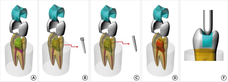
Materials and Methods Forty-eight premolars received MOD preparation (4 groups,
n = 12) with different restorative techniques: glass ionomer cement + composite resin (the GIC group), a metallic post + composite resin (the MP group), a fiberglass post + composite resin (the FGP group), or no endodontic treatment + restoration with composite resin (the CR group). Cusp strain and load-bearing capacity were evaluated. One-way analysis of variance and the Tukey test were used with α = 5%. Finite element analysis (FEA) was used to calculate displacement and tensile stress for the teeth and restorations.Results MP showed the highest cusp (
p = 0.027) deflection (24.28 ± 5.09 µm/µm), followed by FGP (20.61 ± 5.05 µm/µm), CR (17.72 ± 6.32 µm/µm), and GIC (17.62 ± 7.00 µm/µm). For load-bearing, CR (38.89 ± 3.24 N) showed the highest, followed by GIC (37.51 ± 6.69 N), FGP (29.80 ± 10.03 N), and MP (18.41 ± 4.15 N) (p = 0.001) value. FEA showed similar behavior in the restorations in all groups, while MP showed the highest stress concentration in the tooth and post.Conclusions There is no mechanical advantage in using intraradicular posts for endodontically-treated premolars requiring MOD restoration. Filling the pulp chamber with GIC and restoring the tooth with only CR showed the most promising results for cusp deflection, failure load, and stress distribution.
-
Citations
Citations to this article as recorded by- How to adaptively balance ‘classic’ or ‘conservative’ approaches in tooth defect management: a 3D-finite element analysis study
Jiani Xu, Xu Liang, Lili Hu, Chen Sun, Zhipeng Zhang, Jiawei Yang, Jie Wang
BMC Oral Health.2025;[Epub] CrossRef - Inkjet-printed strain gauge sensors: Materials, manufacturing, and emerging applications
Lara Abdel Salam, Samir Mustapha, Alexandra Mikhael, Nisrine Bakri, Sahera Saleh, Massoud L. Khraiche
Sensors and Actuators A: Physical.2025; 394: 116934. CrossRef - Influence of endodontic access cavity design on mechanical properties of a first mandibular premolar tooth: a finite element analysis study
Taha Özyürek, Gülşah Uslu, Burçin Arıcan, Mustafa Gündoğar, Mohammad Hossein Nekoofar, Paul Michael Howell Dummer
Clinical Oral Investigations.2024;[Epub] CrossRef - Comparison of the Effect of Different Cavity Designs and Temporary Restoration Materials on the Fracture Resistance of Upper Premolars, Undergone Re-treatment: An In-Vitro Study
Parnian Alavinejad, Mohammad Yazdizadeh, Ali Mombeinipour, Ebrahim Karimzadeh
Proceedings of the National Academy of Sciences, India Section B: Biological Sciences.2024; 94(3): 677. CrossRef - Fracture resistance and failure mode of endodontically treated premolars reconstructed by different preparation approaches: Cervical margin relocation and crown lengthening with complete and partial ferrule with three different post and core systems
Mehran Falahchai, Naghmeh Musapoor, Soroosh Mokhtari, Yasamin Babaee Hemmati, Hamid Neshandar Asli
Journal of Prosthodontics.2024; 33(8): 774. CrossRef - Comparison of the stress distribution in base materials and thicknesses in composite resin restorations
Min-Kwan Jung, Mi-Jeong Jeon, Jae-Hoon Kim, Sung-Ae Son, Jeong-Kil Park, Deog-Gyu Seo
Heliyon.2024; 10(3): e25040. CrossRef -
Fracture resistance and failure pattern of endodontically treated maxillary premolars restored with transfixed glass fiber post: an
in vitro
and finite element analysis
Saleem Abdulrab, Greta Geerts, Ganesh Thiagarajan
Computer Methods in Biomechanics and Biomedical Engineering.2024; 27(4): 419. CrossRef - Influence of size-anatomy of the maxillary central incisor on the biomechanical performance of post-and-core restoration with different ferrule heights
Domingo Santos Pantaleón, João Paulo Mendes Tribst, Franklin García-Godoy
The Journal of Advanced Prosthodontics.2024; 16(2): 77. CrossRef - Influence of internal angle and shape of the lining on residual stress of Class II molar restorations
Qianqian Zuo, Annan Li, Haidong Teng, Zhan Liu
Computer Methods in Biomechanics and Biomedical Engineering.2024; 27(5): 680. CrossRef - Evaluation of stress distribution in coronal base and restorative materials: A narrative review of finite element analysis studies
Yelda Polat, İzzet Yavuz
Conservative Dentistry Journal.2024; 14(2): 47. CrossRef - The influence of horizontal glass fiber posts on fracture strength and fracture pattern of endodontically treated teeth: A systematic review and meta‐analysis of in vitro studies
Saleem Abdulrab, Greta Geerts, Sadeq Ali Al‐Maweri, Mohammed Nasser Alhajj, Hatem Alhadainy, Raidan Ba‐Hattab
Journal of Prosthodontics.2023; 32(6): 469. CrossRef - Stress distribution of a novel bundle fiber post with curved roots and oval canals
Deniz Yanık, Nurullah Turker
Journal of Esthetic and Restorative Dentistry.2022; 34(3): 550. CrossRef - The Effect of Endodontic Treatment and Thermocycling on Cuspal Deflection of Teeth Restored with Different Direct Resin Composites
Cansu Atalay, Ayse Ruya Yazici, Aynur Sidika Horuztepe, Emre Nagas
Conservative Dentistry and Endodontic Journal.2022; 6(2): 38. CrossRef - The use of different adhesive filling material and mass combinations to restore class II cavities under loading and shrinkage effects: a 3D-FEA
P. Ausiello, S. Ciaramella, A. De Benedictis, A. Lanzotti, J. P. M. Tribst, D. C. Watts
Computer Methods in Biomechanics and Biomedical Engineering.2021; 24(5): 485. CrossRef - Biomechanical Analysis of a Custom-Made Mouthguard Reinforced With Different Elastic Modulus Laminates During a Simulated Maxillofacial Trauma
João Paulo Mendes Tribst, Amanda Maria de Oliveira Dal Piva, Pietro Ausiello, Arianna De Benedictis, Marco Antonio Bottino, Alexandre Luiz Souto Borges
Craniomaxillofacial Trauma & Reconstruction.2021; 14(3): 254. CrossRef - Mechanical Assessment of Glass Ionomer Cements Incorporated with Multi-Walled Carbon Nanotubes for Dental Applications
Manuela Spinola, Amanda Maria Oliveira Dal Piva, Patrícia Uchôas Barbosa, Carlos Rocha Gomes Torres, Eduardo Bresciani
Oral.2021; 1(3): 190. CrossRef - Stress Concentration of Endodontically Treated Molars Restored with Transfixed Glass Fiber Post: 3D-Finite Element Analysis
Alexandre Luiz Souto Borges, Manassés Tercio Vieira Grangeiro, Guilherme Schmitt de Andrade, Renata Marques de Melo, Kusai Baroudi, Laís Regiane Silva-Concilio, João Paulo Mendes Tribst
Materials.2021; 14(15): 4249. CrossRef - Computer Aided Design Modelling and Finite Element Analysis of Premolar Proximal Cavities Restored with Resin Composites
Amanda Guedes Nogueira Matuda, Marcos Paulo Motta Silveira, Guilherme Schmitt de Andrade, Amanda Maria de Oliveira Dal Piva, João Paulo Mendes Tribst, Alexandre Luiz Souto Borges, Luca Testarelli, Gabriella Mosca, Pietro Ausiello
Materials.2021; 14(9): 2366. CrossRef - Effect of Shrinking and No Shrinking Dentine and Enamel Replacing Materials in Posterior Restoration: A 3D-FEA Study
Pietro Ausiello, Amanda Maria de Oliveira Dal Piva, Alexandre Luiz Souto Borges, Antonio Lanzotti, Fausto Zamparini, Ettore Epifania, João Paulo Mendes Tribst
Applied Sciences.2021; 11(5): 2215. CrossRef - Effect of Fiber-Reinforced Composite and Elastic Post on the Fracture Resistance of Premolars with Root Canal Treatment—An In Vitro Pilot Study
Jesús Mena-Álvarez, Rubén Agustín-Panadero, Alvaro Zubizarreta-Macho
Applied Sciences.2020; 10(21): 7616. CrossRef
- How to adaptively balance ‘classic’ or ‘conservative’ approaches in tooth defect management: a 3D-finite element analysis study
- 1,969 View
- 21 Download
- 20 Crossref

-
Publication patterns in
Restorative Dentistry and Endodontics - Sherin Jose Chockattu, Byathnal Suryakant Deepak
- Restor Dent Endod 2019;44(3):e34. Published online August 8, 2019
- DOI: https://doi.org/10.5395/rde.2019.44.e34
-
 Abstract
 PDF
PDF PubReader
 ePub
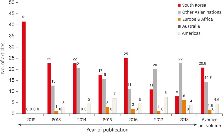
ePub Objectives Restorative Dentistry and Endodontics (Restor Dent Endod ;RDE ) is an English-language journal published by the Korean Academy of Conservative Dentistry, and it has been online since 2012 with quarterly publications. The purpose of this paper was to review and analyze the publications in this journal since its inception and over the 7-year period from 2012 to 2018.Materials and Methods This paper assessed the number, type, and subject of articles published, as well as authorship patterns and article citations of the journal over a 7-year period. The citation indicator for the journal (h-index) was assessed using Google Scholar.
Results The number of articles per issue has remained relatively consistent in the 7 years that were analyzed. An analysis of the article types revealed various categories of review articles. Original research articles accounted for the most articles per volume. Twice as many articles per volume were on endodontic topics than on restorative subjects. Articles published in
RDE have been widely cited in Synapse, Crossref, and PubMed Central. A country-wise mapping of authors' institutions revealed significant contributions from authors around the world. With an h-index of 24,RDE ranks third among journals in its specialty. The most cited articles were open lectures on statistics and research articles on recent concepts, technology, and materials.Conclusion Over the last 7 years,
RDE has served as a platform for a large number of manuscripts in the field of restorative dentistry and endodontics.-
Citations
Citations to this article as recorded by- The trend of publications in the Journal of Conservative Dentistry and Endodontics: A 6-year systematic survey of the journal
Muhammad Hamdan, Ganesh Ranganath Jadhav, Prerna Verma
Journal of Conservative Dentistry and Endodontics.2025; 28(2): 126. CrossRef - Effects of Photobiomodulation on Oral Mucositis: Visualization and Analysis of Knowledge
Wallacy Watson Pereira Melo, Walessa Alana Bragança Aragão, Daiane Claydes Baia-da-Silva, Priscila Cunha Nascimento, Rafael Rodrigues Lima, Renata Duarte de Souza-Rodrigues
Life.2022; 12(11): 1940. CrossRef
- The trend of publications in the Journal of Conservative Dentistry and Endodontics: A 6-year systematic survey of the journal
- 1,639 View
- 11 Download
- 2 Crossref

- A CAD/CAM-based strategy for concurrent endodontic and restorative treatment
- Patricia Maria Escobar, Anil Kishen, Fabiane Carneiro Lopes, Caroline Cristina Borges, Eugenio Gabriel Kegler, Manoel Damião Sousa-Neto
- Restor Dent Endod 2019;44(3):e27. Published online July 24, 2019
- DOI: https://doi.org/10.5395/rde.2019.44.e27
-
 Abstract
 PDF
PDF PubReader
 ePub
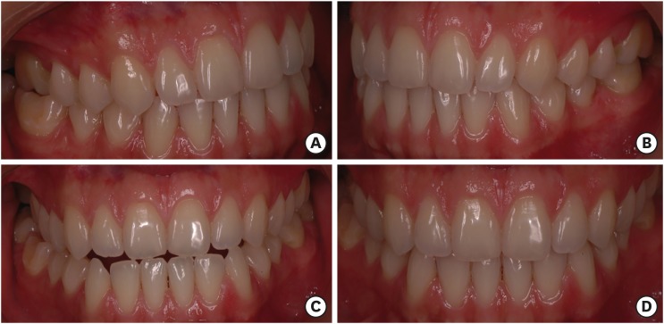
ePub This case report describes a technique in which endodontic treatment and permanent indirect restoration were completed in the same clinical appointment with the aid of a computer-aided design/computer-aided manufacturing (CAD/CAM) system. Two patients were diagnosed with irreversible pulpitis of the mandibular first molar. After access preparation, root canals were located, irrigation was performed until bleeding ceased, and the coronal tooth structure was prepared for indirect restoration. Then, utilizing an interim 3-mm build-up of the endodontic access cavity, a hemi-arch digital scan was performed with an intraoral scanner. Subsequent to digital scanning, restoration design was performed simultaneously with the endodontic procedure. The root canals were shaped using the Race system under irrigation with 2.5% sodium hypochlorite followed by root canal filling. The pulp chamber was subsequently filled with a 3-mm-thick composite resin restoration mimicking the interim build-up previously utilized to facilitate block milling in the CAD/CAM system. Clinical try-in of the permanent onlay restoration was followed by acid etching, application of a 5th generation adhesive, and cementation of the indirect restoration. Once the restoration was cemented, rubber dam isolation was removed, followed by occlusal adjustment and polishing. After 2 years of follow-up, the restorations were esthetically and functionally satisfactory, without complications.
-
Citations
Citations to this article as recorded by- Endodontic Management with Posterior Indirect Adhesive Restoration by CAD-CAM Procedure - Case Report
Intan Syuhada, Wandania Farahanny, Widi Prasetia
Journal of Evolution of Medical and Dental Sciences.2025; : 88. CrossRef - Evaluation of Marginal Adaptation of Three Different Materials Restored in Class II Inlay Cavity Preparations: An In Vitro Study
Rajasekhar Vemareddy, Someshwar Battu, Jyotsnanjali Thati, Sudhakar Naidu, Balaraju Korrai, Akhila Nalli
World Journal of Dentistry.2024; 15(5): 411. CrossRef
- Endodontic Management with Posterior Indirect Adhesive Restoration by CAD-CAM Procedure - Case Report
- 3,749 View
- 32 Download
- 2 Crossref

-
A new minimally invasive guided endodontic microsurgery by cone beam computed tomography and 3-dimensional printing technology

- Jong-Eun Kim, June-Sung Shim, Yooseok Shin
- Restor Dent Endod 2019;44(3):e29. Published online July 25, 2019
- DOI: https://doi.org/10.5395/rde.2019.44.e29
-
 Abstract
 PDF
PDF Supplementary Material
 PubReader
 ePub
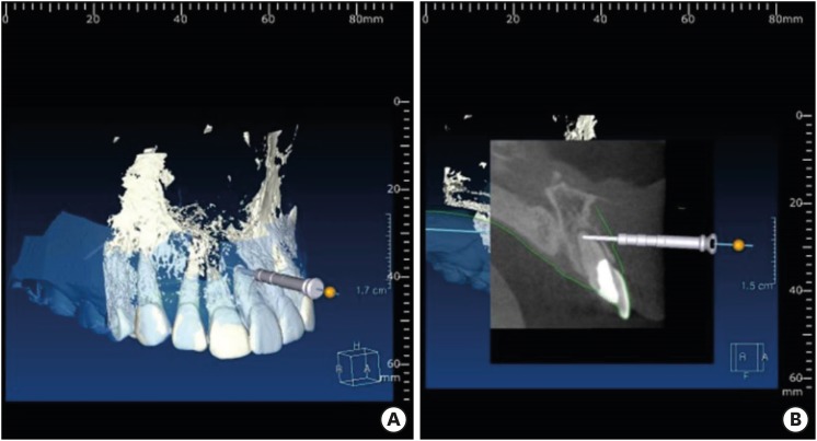
ePub Endodontic microsurgery is defined as the treatment performed on the root apices of an infected tooth, which was unresolved with conventional root canal therapy. Recently, the advanced technology in 3-dimensional model reconstruction based on computed tomography such as cone beam computed tomography has opened a new avenue in application of personalized, accurate diagnosis and has been increasingly used in the field of dentistry. Nevertheless, direct intra-oral localization of root apex based on the 3-dimensional information is extremely difficult and significant amount of bone removal is inevitable when freehand surgical procedure was employed. Moreover, gingival flap and alveolar bone fenestration are usually required, which leads to prolonged time of surgery, thereby increasing the chance of trauma as well as the risk of infection. The purpose of this case report is to present endodontic microsurgery using the guide template that can accurately target the position of apex for the treatment of an anterior tooth with calcified canal which was untreatable with conventional root canal therapy and unable to track the position of the apex due to the absence of fistula.
-
Citations
Citations to this article as recorded by- A narrative review of papilla preservation techniques in clinical dentistry
Yinghua Fu, Zhixin Zhang, Xiaoping Tang, Jiangling Su
Medicine.2025; 104(3): e41033. CrossRef - Segmentation algorithms of dental CT images: A comprehensive review from classical to deep learning trend
Dianhao Wu, Jingang Jiang, Jinke Wang, Zhuming Bi, Guang Yu
Expert Systems with Applications.2025; 275: 126853. CrossRef - Endodontic Microsurgery of Mandibular Molars with an Autonomous Robotic System
Haiying Zhang, Zi Yang, Mangnan Liu, Yaoxin Wang, Mei Fu, Benxiang Hou, Chen Zhang
Journal of Endodontics.2025; 51(12): 1830. CrossRef - Removal of Extraradicular Separated Instrument by Targeted Endodontic Microsurgery Using the 3D‐Printed Guide and Trephine: A Case Report
Lin Yang, Liang Chen
Clinical Case Reports.2025;[Epub] CrossRef - Augmented Reality-Assisted Micro-Invasive Apicectomy with Markerless Visual–Inertial Odometry: An In Vivo Pilot Study
Marco Farronato, Davide Farronato, Federico Michelini, Giulio Rasperini
Applied Sciences.2025; 15(23): 12588. CrossRef - 3D finite element analysis of stress distribution on the shape of resected root-end or with/without bone graft of a maxillary premolar during endodontic microsurgery
Aein Mon, Mi-El Kim, Kee-Yeon Kum, Ho-Beom Kwon
Journal of Dental Sciences.2024; 19(2): 837. CrossRef - TREATMENT OF YATROGENIC POST-TRAUMATIC NEUROPATHY ASSOCIATED WITH
ENDODONTIC THERAPY USING 3D TECHNOLOGIES
Karen Sevterteryan, Vladislav Tarasenok, Lyudmila Tatintsyan
BULLETIN OF STOMATOLOGY AND MAXILLOFACIAL SURGERY.2024; : 73. CrossRef - Advancements in guided surgical endodontics: A scoping review of case report and case series and research implications
Giusy Rita Maria La Rosa, Matteo Peditto, Andrea Venticinque, Antonia Marcianò, Alberto Bianchi, Eugenio Pedullà
Australian Endodontic Journal.2024; 50(2): 397. CrossRef - Comparison of a Novel Static Computer-aided Surgical and Freehand Techniques for Osteotomy and Root-end Resection
Kyle Westbrook, Corey Rollor, Sara A. Aldahmash, Guadalupe G. Fay, Elias Rivera, Jeffery B. Price, Ina Griffin, Patricia A. Tordik, Frederico C. Martinho
Journal of Endodontics.2023; 49(5): 528. CrossRef - Comparison of the Three-Dimensional Accuracy of Guided Apicoectomy Performed with a Drill or a Trephine: An In Vitro Study
Ramóna Kiscsatári, Eszter Nagy, Máté Szabó, Gábor Braunitzer, József Piffkó, Márk Fráter, Márk Ádám Antal
Applied Sciences.2023; 13(17): 9642. CrossRef - Review of “Outcome of Endodontic Surgery: A Meta- Analysis of the Literature—Part 1: Comparison
of Traditional Root-End Surgery and Endodontic Microsurgery” by Setzer and Colleagues in J Endod 36(11):1757-1765, 2010
Oleksandr Nozhenko
Journal of Endodontic Microsurgery.2023; 2: 41. CrossRef - The Impact of the Preferred Reporting Items for Case Reports in Endodontics (PRICE) 2020 Guidelines on the Reporting of Endodontic Case Reports
Sofian Youssef, Phillip Tomson, Amir Reza Akbari, Natalie Archer, Fayjel Shah, Jasmeet Heran, Sunmeet Kandhari, Sandeep Pai, Shivakar Mehrotra, Joanna M Batt
Cureus.2023;[Epub] CrossRef - New-designed 3D printed surgical guide promotes the accuracy of endodontic microsurgery: a study of 14 upper anterior teeth
Dan Zhao, Weige Xie, Tianguo Li, Anqi Wang, Li Wu, Wen Kang, Lu Wang, Shiliang Guo, Xuna Tang, Sijing Xie
Scientific Reports.2023;[Epub] CrossRef - An Exploratory In Vitro Microcomputed Tomographic Investigation of the Efficacy of Semicircular Apicoectomy Performed with Trephine Bur
Eszter Nagy, Brigitta Vőneki, Lívia Vásárhelyi, Imre Szenti, Márk Fráter, Ákos Kukovecz, Márk Ádám Antal
Applied Sciences.2023; 13(16): 9431. CrossRef - The Time Has Come: Journal of Endodontic Microsurgery: A First Peer-Reviewed Open Access Publication Focused on Microsurgery in Endodontics
Ievgen Fesenko
Journal of Endodontic Microsurgery.2022;[Epub] CrossRef - Prefabricated Grid-guided Endodontic Microsurgery: A Pilot Study
Cruz Nishanthine, Manali Ramakrishnan Srinivasan, Ravi Devi, Kadhar Begam Farjana, Dasarathan Duraivel
Journal of Operative Dentistry & Endodontics.2022; 6(2): 58. CrossRef - Guided osteotomy
Saini Rashmi, Saini V Kr
Tanta Dental Journal.2022; 19(3): 172. CrossRef - Accuracy of digitally planned, guided apicoectomy with a conventional trephine and a custom-made endodontic trephine: An in vitro comparative study
Eszter Nagy, Gábor Braunitzer, Dániel Gerhard Gryschka, Ibrahim Barrak, Mark Adam Antal
Journal of Stomatology, Oral and Maxillofacial Surgery.2022; 123(4): 388. CrossRef - Stress Distribution on Trephine-Resected Root-end in Targeted Endodontic Microsurgery: A Finite Element Analysis
Yeon-Jee Yoo, Hiran Perinpanayagam, Miel Kim, Qiang Zhu, Seung-Ho Baek, Ho-Beom Kwon, Kee-Yeon Kum
Journal of Endodontics.2022; 48(12): 1517. CrossRef - An Update on Endodontic Microsurgery of Mandibular Molars: A Focused Review
Sun Mi Jang, Euiseong Kim, Kyung-San Min
Medicina.2021; 57(3): 270. CrossRef - When to consider the use of CBCT in endodontic treatment planning in adults
Nisha Patel, Andrew Gemmell, David Edwards
Dental Update.2021; 48(11): 932. CrossRef
- A narrative review of papilla preservation techniques in clinical dentistry
- 2,215 View
- 27 Download
- 21 Crossref

- Surgical management of an accessory canal in a maxillary premolar: a case report
- Hee-Jin Kim, Mi-Kyung Yu, Kwang-Won Lee, Kyung-San Min
- Restor Dent Endod 2019;44(3):e30. Published online July 29, 2019
- DOI: https://doi.org/10.5395/rde.2019.44.e30
-
 Abstract
 PDF
PDF PubReader
 ePub
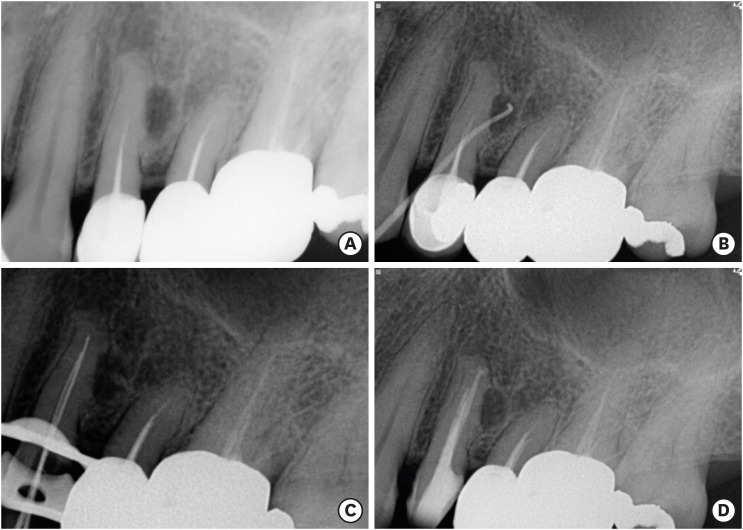
ePub We report the surgical endodontic treatment of a maxillary first premolar with a lateral lesion that originated from an accessory canal. Although lesions originating from accessory canals frequently heal with simple conventional endodontic therapy, some lesions may need additional and different treatment. In the present case, conventional root canal retreatment led to incomplete healing with the need for further treatment (
i.e. , surgery). Surgical endodontic management with a fast-setting calcium silicate cement was performed on the accessory canal using a dental operating microscope. At the patient's 9-month recall visit, the lesion was resolved upon radiography.-
Citations
Citations to this article as recorded by- Predictive analysis of root canal morphology in relation to root canal treatment failures: a retrospective study
Mohmed Isaqali Karobari, Vishnu Priya Veeraraghavan, P. J. Nagarathna, Sudhir Rama Varma, Jayaraj Kodangattil Narayanan, Santosh R. Patil
Frontiers in Dental Medicine.2025;[Epub] CrossRef - Endodontic management of internal replacement resorption of two maxillary central incisors with the aid of cone-beam computed tomography as the diagnostic tool: a case report and review of literature
Fatemeh Eskandari, Safoora Sahebi, Negar Ghorbani Jahandizi, Hossein Mofidi
Journal of Medical Case Reports.2025;[Epub] CrossRef - The Impact of the Preferred Reporting Items for Case Reports in Endodontics (PRICE) 2020 Guidelines on the Reporting of Endodontic Case Reports
Sofian Youssef, Phillip Tomson, Amir Reza Akbari, Natalie Archer, Fayjel Shah, Jasmeet Heran, Sunmeet Kandhari, Sandeep Pai, Shivakar Mehrotra, Joanna M Batt
Cureus.2023;[Epub] CrossRef - Main and Accessory Canal Filling Quality of a Premixed Calcium Silicate Endodontic Sealer According to Different Obturation Techniques
Su-Yeon Ko, Hae Won Choi, E-Deun Jeong, Vinicius Rosa, Yun-Chan Hwang, Mi-Kyung Yu, Kyung-San Min
Materials.2020; 13(19): 4389. CrossRef
- Predictive analysis of root canal morphology in relation to root canal treatment failures: a retrospective study
- 1,460 View
- 15 Download
- 4 Crossref

- Erratum: Correction of Data in Table 3. Effect of casein phosphopeptide-amorphous calcium phosphate on fluoride release and micro-shear bond strength of resin-modified glass ionomer cement in caries-affected dentin
- Jamila Nuwayji Agob, Neven Saad Aref, Essam El Saeid Al-Wakeel
- Restor Dent Endod 2019;44(3):e24. Published online July 9, 2019
- DOI: https://doi.org/10.5395/rde.2019.44.e24

- 648 View
- 13 Download


 KACD
KACD


 First
First Prev
Prev


