
Previous issues
- Page Path
- HOME > Browse articles > Previous issues
- Volume 48 (2); May 2023
-


Review Article
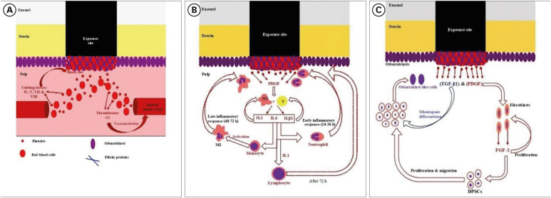
- Stem cell-derived exosomes for dentin-pulp complex regeneration: a mini-review
- Dina A. Hammouda, Alaa M Mansour, Mahmoud A. Saeed, Ahmed R. Zaher, Mohammed E. Grawish
- Restor Dent Endod 2023;48(2):e20. Published online May 3, 2023
- DOI: https://doi.org/10.5395/rde.2023.48.e20
-
 Abstract
 PDF
PDF PubReader
 ePub
ePub This mini-review was conducted to present an overview of the use of exosomes in regenerating the dentin-pulp complex (DPC). The PubMed and Scopus databases were searched for relevant articles published between January 1, 2013 and January 1, 2023. The findings of basic
in vitro studies indicated that exosomes enhance the proliferation and migration of mesenchymal cells, as human dental pulp stem cells, via mitogen-activated protein kinases and Wingless-Int signaling pathways. In addition, they possess proangiogenic potential and contribute to neovascularization and capillary tube formation by promoting endothelial cell proliferation and migration of human umbilical vein endothelial cells. Likewise, they regulate the migration and differentiation of Schwann cells, facilitate the conversion of M1 pro-inflammatory macrophages to M2 anti-inflammatory phenotypes, and mediate immune suppression as they promote regulatory T cell conversion. Basicin vivo studies have indicated that exosomes triggered the regeneration of dentin-pulp–like tissue, and exosomes isolated under odontogenic circumstances are particularly strong inducers of tissue regeneration and stem cell differentiation. Exosomes are a promising regenerative tool for DPC in cases of small pulp exposure or for whole-pulp tissue regeneration.-
Citations
Citations to this article as recorded by
- Extracellular vesicles derived from dental mesenchymal stem cells for regenerative medicine: a scoping review
Maria Emília Mota, Márcia Martins Marques, Thaís Gimenez, Suely Kunimi Kubo Ariga, Tiago Góss dos Santos, Fábio Abreu Alves, Maria Stella Moreira
Molecular Biology Reports.2026;[Epub] CrossRef - Stem cell-derived exosomes in tissue regeneration of oral and maxillofacial region: A systematic review
Qianlei Dai, Qianfen Dai
Medicine.2026; 105(1): e46948. CrossRef - Stem Cell-Derived Exosomes in Wound Healing and Skin Regeneration: Emerging Therapeutic Strategies and Mechanisms
Nithin Vidiyala, Pavani Sunkishala, Prashanth Reddy Parupathi, Dinesh Nyavanandi
Cells.2026; 15(10): 872. CrossRef - REGENERATIVE POTENTIAL OF DENTAL STEM CELLS IN DENTISTRY: FROM BIOCHEMICAL MECHANISMS TO CLINICAL APPLICATIONS
A.Ye. Demkovych, O.Z. Yaremchuk, M.I. Kulitska, N.B. Hlyvka, V.Ya. Krupei, O.Ya. Lavrin
Ukrainian Dental Almanac.2026; (2): 5. CrossRef - Dental stem cell-derived extracellular vesicles for oral tissue regeneration: Mechanisms, engineering strategies, and clinical translation
Yang Liu, Peiyao Ge, Yuzhuo Ma, Haizhou Fu
Archives of Oral Biology.2026; 190: 106676. CrossRef - Cell Homing Strategies in Regenerative Endodontic Therapy
David Kim, Sahng G. Kim
Cells.2025; 14(3): 201. CrossRef - Impact of dental pulp cells-derived small extracellular vesicles on the properties and behavior of dental pulp cells: an in-vitro study
Dina A. Hammouda, Alaa M. Mansour, Ahmed R. Zaher, Mohammed E. Grawish
BMC Oral Health.2025;[Epub] CrossRef - Methodological Approaches for Economic Comparison of Mesenchymal Stem Cell and Exosome-based Therapies with Conventional Endodontic Treatments in Regenerative Endodontics
Madina A. Kurmanalina Kurmanalina, Nadiar M. Mussin, Aigul M. Sumanova, Violetta R. Detochkina, Maryam Mardani, Nader Tanideh, Amin Tamadon
West Kazakhstan Medical Journal.2025; 67(2): 188. CrossRef - Exosomal circ_0003057 promotes osteo/odontogenic differentiation of hDPSCs by binding with EIF4A3 through upregulated parental gene ANKH
Bingtao Wang, Yuanyuan Kong, Huixian Dong, Feng Lai, Zixin Guo, Liecong Lin, Jingyi Xu, Jingkun Zhang, Yiguo Jiang, Qianzhou Jiang
International Endodontic Journal.2025; 58(9): 1433. CrossRef - Mechanistic insights into dental stem cells‐derived exosomes in regenerative endodontics
Paras Ahmad, Nathan Estrin, Nima Farshidfar, Yufeng Zhang, Richard J. Miron
International Endodontic Journal.2025; 58(9): 1384. CrossRef - Development and characterization of an exosome-loaded biomimetic hydroxyapatite/gelatin scaffold for enhanced dental pulp regeneration
Yuen-Shan Tsai, Shih-Jung Cheng, Tsao-Li Chuang, Shu-Fang Chang, Feng-Huei Lin, Chun-Pin Lin
Journal of Dental Sciences.2025;[Epub] CrossRef - Exosomes as Promising Therapeutic Tools for Regenerative Endodontic Therapy
Qingyue Kong, Yujie Wang, Nan Jiang, Yifan Wang, Rui Wang, Xiaohan Hu, Jing Mao, Xin Shi
Biomolecules.2024; 14(3): 330. CrossRef - Role and Molecular Mechanism of miR-586 in the Differentiation of Dental Pulp Stem Cells into Odontoblast-like Cells
Gang Pan, Qianwen Zhou, Chenhua Pan, Yingxue Zhang
Cell Biochemistry and Biophysics.2024; 83(1): 507. CrossRef
- Extracellular vesicles derived from dental mesenchymal stem cells for regenerative medicine: a scoping review
- 11,401 View
- 150 Download
- 10 Web of Science
- 13 Crossref

Research Articles
- Effect of medium or high concentrations of in-office dental bleaching gel on the human pulp response in the mandibular incisors
- Douglas Augusto Roderjan, Rodrigo Stanislawczuk, Diana Gabriela Soares, Carlos Alberto de Souza Costa, Michael Willian Favoreto, Alessandra Reis, Alessandro D. Loguercio
- Restor Dent Endod 2023;48(2):e12. Published online March 8, 2023
- DOI: https://doi.org/10.5395/rde.2023.48.e12
-
Abstract
PDFPubReaderePub
Objectives The present study evaluated the pulp response of human mandibular incisors subjected to in-office dental bleaching using gels with medium or high concentrations of hydrogen peroxide (HP).
Materials and Methods The following groups were compared: 35% HP (HP35;
n = 5) or 20% HP (HP20;n = 4). In the control group (CONT;n = 2), no dental bleaching was performed. The color change (CC) was registered at baseline and after 2 days using the Vita Classical shade guide. Tooth sensitivity (TS) was also recorded for 2 days post-bleaching. The teeth were extracted 2 days after the clinical procedure and subjected to histological analysis. The CC and overall scores for histological evaluation were evaluated by the Kruskal-Wallis and Mann-Whitney tests. The percentage of patients with TS was evaluated by the Fisher exact test (α = 0.05).Results The CC and TS of the HP35 group were significantly higher than those of the CONT group (
p < 0.05) and the HP20 group showed an intermediate response, without significant differences from either the HP35 or CONT group (p > 0.05). In both experimental groups, the coronal pulp tissue exhibited partial necrosis associated with tertiary dentin deposition. Overall, the subjacent pulp tissue exhibited a mild inflammatory response.Conclusions In-office bleaching therapies using bleaching gels with 20% or 35% HP caused similar pulp damage to the mandibular incisors, characterized by partial necrosis, tertiary dentin deposition, and mild inflammation.
-
Citations
Citations to this article as recorded by- DENTA: A Dual Enzymatic Nanoagent for Self‐Activating Tooth Whitening and Biofilm Disruption
Junseok Kim, Dai‐Hwan Kim, Priyannth R. Sundharbaabu, Chae Yeon Lee, Jina Bae, Jiyu Hyun, Young‐Ju Jang, Haeni Kim, Min‐Ho Hong, Juewen Liu, Tobias Fey, Suk Ho Bhang, Jun Hyuk Heo, Jung Heon Lee
Advanced Functional Materials.2026;[Epub] CrossRef - Effects of a bioactive desensitizing material on in-office bleaching–induced tooth sensitivity: A randomized double-blind controlled trial
Ghada A. Maghaireh, Hanan Alzraikat, Majd Y. Altarazi
Journal of Dentistry.2026; 166: 106326. CrossRef - Application of the Er:YAG laser in pulpotomy for mature permanent teeth with pulpitis: An animal study
Zeqi Li, Pengfei Xin, Siyao Yang, Xiaoxing Hao, Jun Wang, Kuanshou Zhang, Qingmei Liu, Mohmed Isaqali Karobari
PLOS One.2026; 21(1): e0341017. CrossRef - Influence of tooth sensitivity on shortening the interval between in-office bleaching sessions with acidic pH gel
Sarah Arruda Gonçalves Ferraz da Costa, Carolina da Costa Ferreira Silva, Tâmmile Vallentine Soares Amado, Alessandro D. Loguercio, Michael Willian Favoreto, Alessandra Reis, Fernanda Signorelli Calazans, Marcos Oliveira Barceleiro
The Journal of the American Dental Association.2026;[Epub] CrossRef - Can pigments of different natures interfere with the cytotoxicity from in-office bleaching?
Rafael Antonio de Oliveira Ribeiro, Beatriz Voss Martins, Marlon Ferreira Dias, Victória Peruchi, Caroline Anselmi, Igor Paulino Mendes Soares, Josimeri Hebling, Vanessa Cavalli, Carlos Alberto de Souza Costa
Odontology.2025; 113(4): 1447. CrossRef - Does Patient Age Impact In-Office Tooth Bleaching Outcomes? A Parallel Clinical Trial
JL Martins, IS Araújo, JF Rabelo, CJ Soares, AL Faria-e-Silva, AD Loguercio, PCFS Filho, HL Carlo, GR da Silva
Operative Dentistry.2025; 50(3): 251. CrossRef - The pH of Bleaching Gels on the Structural and Biological Response of Dental Tissues: A Scoping Review
Jamile Menezes de Souza, Maria Olimpia Paz Alvarenga, Ana Luisa Cassiano Alves Bezerra, Gabriela Queiroz de Melo Monteiro
Journal of Esthetic and Restorative Dentistry.2025; 37(10): 2193. CrossRef - Efficacy of 35 % self-mixed hydrogen peroxide In-office bleaching with reduced application time: A single-blind randomized controlled trial
Gabrielle Gomes Centenaro, Deisy Cristina Ferreira Cordeiro, Maria Alice de Matos Rodrigues, Mariah Maluf Lenhani, Roberta Micheten Dias, Cristina Gómez Polo, Alessandra Reis, Alessandro D. Loguercio
Journal of Dentistry.2025; 163: 106178. CrossRef - Evaluation of cytotoxicity and bleaching efficacy of gels with calcium polyphosphate and violet LED
Larissa de Jesus Gomes, Rafael Antonio de Oliveira Ribeiro, Mariangela Ivette Guanipa Ortiz, Klaus Rischka, Carlos Alberto de Souza Costa, Débora Alves Nunes Leite Lima
Brazilian Dental Journal.2025;[Epub] CrossRef - Combined catalytic strategies applied to in-office tooth bleaching: whitening efficacy, cytotoxicity, and gene expression of human dental pulp cells in a 3D culture model
Rafael Antonio de Oliveira Ribeiro, Victória Peruchi, Igor Paulino Mendes Soares, Filipe Koon Wu Mon, Diana Gabriela Soares, Josimeri Hebling, Carlos Alberto de Souza Costa
Clinical Oral Investigations.2024;[Epub] CrossRef - Low and high hydrogen peroxide concentrations of in-office dental bleaching associated with violet light: an in vitro study
Isabela Souza Vardasca, Michael Willian Favoreto, Mylena de Araujo Regis, Taynara de Souza Carneiro, Emanuel Adriano Hul, Christiane Philippini Ferreira Borges, Alessandra Reis, Alessandro D. Loguercio, Carlos Francci
Clinical Oral Investigations.2024;[Epub] CrossRef - Evaluation of hydrogen peroxide permeability, color change, and physical–chemical properties on the in‐office dental bleaching with different mixing tip
Michael Willian Favoreto, Sibelli Olivieri Parreiras, Michel Wendlinger, Taynara De Souza Carneiro, Mariah Ignez Lenhani, Christiane Phillipini Ferreira Borges, Alessandra Reis, Alessandro D. Loguercio
Journal of Esthetic and Restorative Dentistry.2024; 36(3): 460. CrossRef - Catalysis-based approaches with biopolymers and violet LED to improve in-office dental bleaching
Rafael Antonio de Oliveira Ribeiro, Beatriz Voss Martins, Marlon Ferreira Dias, Victória Peruchi, Igor Paulino Mendes Soares, Caroline Anselmi, Josimeri Hebling, Carlos Alberto de Souza Costa
Lasers in Medical Science.2024;[Epub] CrossRef - Feasibility and Safety of Adopting a New Approach in Delivering a 450 nm Blue Laser with a Flattop Beam Profile in Vital Tooth Whitening. A Clinical Case Series with an 8-Month Follow-Up
Reem Hanna, Ioana Cristina Miron, Stefano Benedicenti
Journal of Clinical Medicine.2024; 13(2): 491. CrossRef - Hydrogen Peroxide in the Pulp Chamber and Color Change in Maxillary Anterior Teeth After In-Office Bleaching
Alexandra Mena-Serrano, Sandra Sanchez, María G. Granda-Albuja, Michael Willian Favoreto, Taynara de Souza Carneiro, Deisy Cristina Ferreira Cordeiro, Alessandro D. Loguercio, Alessandra Reis
Brazilian Dental Journal.2024;[Epub] CrossRef - Influence of coating dental enamel with a TiF4-loaded polymeric primer on the adverse effects caused by a bleaching gel with 35% H2O2
Victória Peruchi, Rafael Antonio de Oliveira Ribeiro, Igor Paulino Mendes Soares, Lídia de Oliveira Fernandes, Juliana Rios de Oliveira, Maria Luiza Barucci Araújo Pires, Josimeri Hebling, Diana Gabriela Soares, Carlos Alberto de Souza Costa
Journal of the Mechanical Behavior of Biomedical Materials.2024; 153: 106497. CrossRef
- DENTA: A Dual Enzymatic Nanoagent for Self‐Activating Tooth Whitening and Biofilm Disruption
- 4,773 View
- 114 Download
- 14 Web of Science
- 16 Crossref
- Epigallocatechin-3-gallate prior to composite resin in abfraction lesions: a split-mouth randomized clinical trial
- Luísa Valente Gotardo Lara Alves, Lisiane Martins Fracasso, Thiago Vinicius Cortez, Aline Evangelista Souza-Gabriel, Silmara Aparecida Milori Corona
- Restor Dent Endod 2023;48(2):e13. Published online March 20, 2023
- DOI: https://doi.org/10.5395/rde.2023.48.e13
-
Abstract
PDFPubReaderePub
Objectives Natural extracts have been investigated as a biomimetic strategy to mechanically strengthen the collagen network and control the biodegradation of extracellular matrix. This study evaluated the effect of epigallocatechin-3-gallate (EGCG) on abfraction lesions prior to the composite resin.
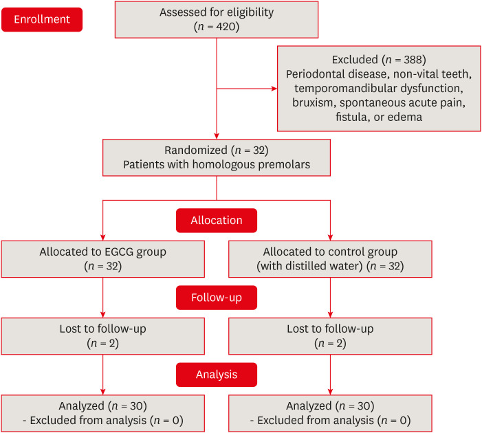
Materials and Methods The sample consisted of 30 patients (aged between 28 and 60 years) with abfraction lesions located in 2 homologous premolars. The teeth were randomly assigned according to dentin treatment: 0.02% EGCG solution or distilled water (control). After enamel acid etching, the solutions were applied immediately for 1 minute. The teeth were restored with Universal Adhesive (3M) and Filtek Z350 XT (3M). Analyzes were done by 2 independent examiners using modified USPHS (retention, secondary caries, marginal adaptation, and postoperative sensitivity) and photographic (color, marginal pigmentation, and anatomical form) criteria at baseline (7 days) and final (18 months). The data analysis used Friedman and Wilcoxon signed-rank tests (α = 0.05).
Results At baseline, all restorations were evaluated as alpha for all criteria. After 18 months, restorations were evaluated as alpha for secondary caries, color, and marginal pigmentation. There was significant difference between baseline and 18 months (
p = 0.009) for marginal adaptation and postoperative sensitivity (p = 0.029), but no significant difference were verified between treatments (p = 0.433). The EGCG group had a restoration retention rate of 93.3%, while the control group had 96.7%.Conclusions The application of EGCG solution on abfraction lesions did not significantly influence the survival of the restorations based on clinical and photographic criteria.
-
Citations
Citations to this article as recorded by- Therapeutic potential of flavonoids in erosive tooth wear management: a scoping review
Gabriel Pereira Nunes, Renata de Oliveira Alves, Geórgia Rondó Peres, Priscila Toninatto Alves de Toledo, Aline Rogéria Freire de Castilho
Clinical Oral Investigations.2025;[Epub] CrossRef
- Therapeutic potential of flavonoids in erosive tooth wear management: a scoping review
- 2,477 View
- 56 Download
- 1 Web of Science
- 1 Crossref
- Effect of an aluminum chloride hemostatic agent on the dentin shear bond strength of a universal adhesive
- Sujin Kim, Yoorina Choi, Sujung Park
- Restor Dent Endod 2023;48(2):e14. Published online March 22, 2023
- DOI: https://doi.org/10.5395/rde.2023.48.e14
-
Abstract
PDFPubReaderePub
Objectives This study investigated the effect of an aluminum chloride hemostatic agent on the shear bond strength (SBS) of a universal adhesive to dentin.
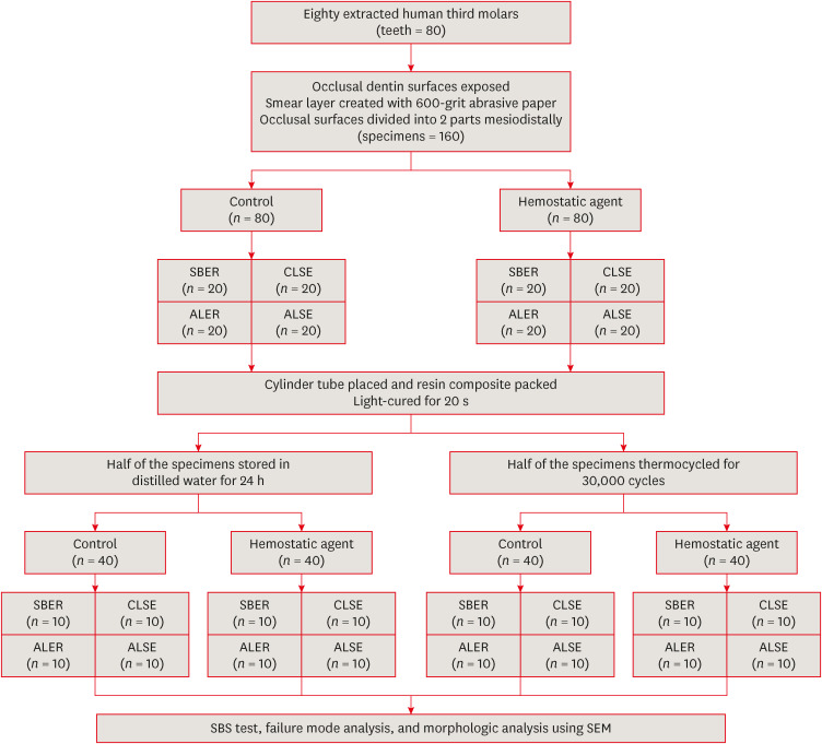
Materials and Methods Eighty extracted human molars were trimmed at the occlusal dentin surfaces and divided mesiodistally. According to hemostatic agent application, specimens were randomly allocated into control (C) and hemostatic agent (Traxodent; H) groups. Each group was divided into 4 subgroups according to the adhesive system (
n = 20): Scotchbond Multi-Purpose (SBER), Clearfil SE Bond (CLSE), All-Bond Universal etch-and-rinse mode (ALER), and All-Bond Universal self-etch mode (ALSE). SBS was measured for half of the specimens at 24 hours, and the other half were thermocycled in water baths (group T). Fracture surfaces were examined to determine the failure mode. The SBS was measured, and data were analyzed using 1-way analysis of variance, the Student’st -test, and the Tukey honestly significant difference test (p = 0.05).Results No significant differences in SBS were found between groups C and H for any adhesive system at 24 hours. After thermocycling, a statistically significant difference was observed between CT+ALSE and HT+ALSE (
p < 0.05). When All-Bond Universal was applied to hemostatic agent-contaminated dentin, the SBS of H+ALSE was significantly lower than that of H+ALER (p < 0.05). The SBER subgroups showed no significant differences in SBS regardless of treatment and thermocycling.Conclusions When exposed dentin was contaminated by an aluminum chloride hemostatic agent before dentin adhesive treatment, application of All-Bond Universal in etch-and-rinse mode was superior to self-etch mode.
-
Citations
Citations to this article as recorded by- Effect of a Hemostatic Agent on Bond Strength and Durability of Bonding in Three Different Adhesive Systems
Saeedeh Ebrahimgol, Saeed Nemati Anaraki, Mahsa Ghanaat, Emad Ramouz, Maryam Hoorizad ganjkar
Journal of Research in Dental and Maxillofacial Sciences.2026; 11(2): 164. CrossRef - Nature-driven blue-emissive N, S-CDs: Harnessing sequential "switch-off-on" fluorescence signals for detection of chrysin and Al³⁺ along with cellular imaging versatility
Maha Mohammad Abdel-Monem, Mohamed I. Walash, Asmaa Kamal El-Deen
Talanta Open.2025; : 100466. CrossRef - Comparative Evaluation of the Shear Bond Strength of Self-Adhesive and Glass Ionomer Cement to Dentin After Removal of Hemostatic Agents Using Different Cleansing Protocols: An In Vitro Study
Hemashree Namburajan, Mathew Chalakuzhiyil Abraham, Vidhyasankari N, Rajkumar K, Abhinayaa Suthagar, Vishnupriya Venkatasubramanian, Sindhuja Nagarajan
Cureus.2025;[Epub] CrossRef - Emalje- og dentinadhesiver: Avgjørende faser i klinisk behandling
Torgils Lægreid, Tom Paulseth, Arne Lund
Den norske tannlegeforenings Tidende.2024; 134(8): 604. CrossRef
- Effect of a Hemostatic Agent on Bond Strength and Durability of Bonding in Three Different Adhesive Systems
- 4,061 View
- 84 Download
- 3 Web of Science
- 4 Crossref
- Effectiveness of endodontic retreatment using WaveOne Primary files in reciprocating and rotary motions
- Patricia Marton Costa, Renata Maíra de Souza Leal, Guilherme Hiroshi Yamanari, Bruno Cavalini Cavenago, Marco Antônio Húngaro Duarte
- Restor Dent Endod 2023;48(2):e15. Published online April 25, 2023
- DOI: https://doi.org/10.5395/rde.2023.48.e15
-
Abstract
PDFPubReaderePub
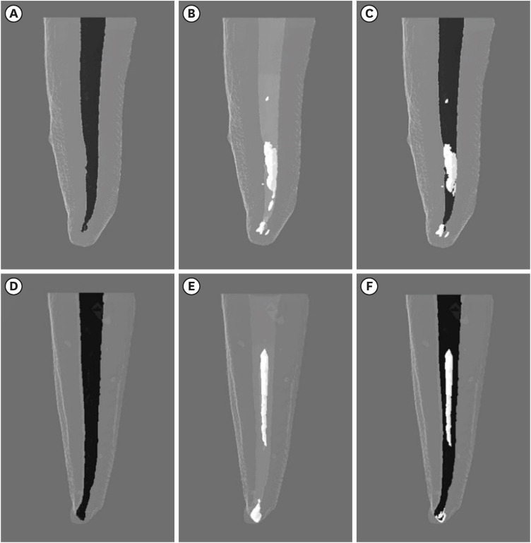
Objectives This study evaluated the efficiency of WaveOne Primary files (Dentsply Sirona) for removing root canal fillings with 2 types of movement: reciprocating (RCP) and continuous counterclockwise rotation (CCR).
Materials and Methods Twenty mandibular incisors were prepared with a RCP instrument (25.08) and filled using the Tagger hybrid obturation technique. The teeth were retreated with a WaveOne Primary file and randomly allocated to 2 experimental retreatment groups (
n = 10) according to movement type: RCP and CCR. The root canals were emptied of filling material in the first 3 steps of insertion, until reaching the working length. The timing of retreatment and procedure errors were recorded for all samples. The specimens were scanned before and after the retreatment procedure with micro-computed tomography to calculate the percentage and volume (mm3) of the residual filling material. The results were statistically evaluated using paired and independentt -tests, with a significance level set at 5%.Results No significant difference was found in the timing of filling removal between the groups, with a mean of 322 seconds (RCP) and 327 seconds (CCR) (
p < 0.05). There were 6 instrument fractures: 1 in a RCP motion file and 5 in continuous rotation files. The volumes of residual filling material were similar (9.94% for RCP and 15.94% for CCR;p > 0.05).Conclusions The WaveOne Primary files used in retreatment performed similarly in both RCP and CCR movements. Neither movement type completely removed the obturation material, but the RCP movement provided greater safety.
-
Citations
Citations to this article as recorded by- Micro-CT evaluation of the removal of root fillings using rotary and reciprocating systems supplemented by XP-Endo Finisher, the Self-Adjusting File, or Er,Cr:YSGG laser
Gülsen Kiraz, Bulem Üreyen Kaya, Mert Ocak, Muhammet Bora Uzuner, Hakan Hamdi Çelik
Restorative Dentistry & Endodontics.2023;[Epub] CrossRef
- Micro-CT evaluation of the removal of root fillings using rotary and reciprocating systems supplemented by XP-Endo Finisher, the Self-Adjusting File, or Er,Cr:YSGG laser
- 3,590 View
- 76 Download
- 1 Crossref
-
Influence of CBCT parameters on image quality and the diagnosis of vertical root fractures in teeth with metallic posts: an
ex vivo study - Larissa Pereira Lagos de Melo, Polyane Mazucatto Queiroz, Larissa Moreira-Souza, Mariana Rocha Nadaes, Gustavo Machado Santaella, Matheus Lima Oliveira, Deborah Queiroz Freitas
- Restor Dent Endod 2023;48(2):e16. Published online April 27, 2023
- DOI: https://doi.org/10.5395/rde.2023.48.e16
-
Abstract
PDFPubReaderePub
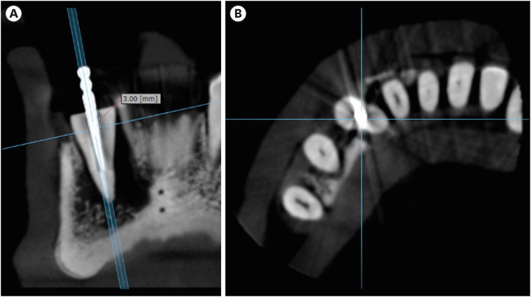
Objectives The aim of this study was to evaluate the influence of peak kilovoltage (kVp) and a metal artifact reduction (MAR) tool on image quality and the diagnosis of vertical root fracture (VRF) in cone-beam computed tomography (CBCT).
Materials and Methods Twenty single-rooted human teeth filled with an intracanal metal post were divided into 2 groups: control (
n = 10) and VRF (n = 10). Each tooth was placed into the socket of a dry mandible, and CBCT scans were acquired using a Picasso Trio varying the kVp (70, 80, 90, or 99), and the use of MAR (with or without). The examinations were assessed by 5 examiners for the diagnosis of VRF using a 5-point scale. A subjective evaluation of the expression of artifacts was done by comparing random axial images of the studied protocols. The results of the diagnoses were analyzed using 2-way analysis of variance and the Tukeypost hoc test, the subjective evaluations were compared using the Friedman test, and intra-examiner reproducibility was evaluated using the weighted kappa test (α = 5%).Results The kVp and MAR did not influence the diagnosis of VRF (
p > 0.05). According to the subjective classification, the 99 kVp protocol with MAR demonstrated the least expression of artifacts, while the 70 kVp protocol without MAR led to the most artifacts.Conclusions Protocols with higher kVp combined with MAR improved the image quality of CBCT examinations. However, those factors did not lead to an improvement in the diagnosis of VRF.
-
Citations
Citations to this article as recorded by- Digital Dentistry Society Quality Forum: Clinical recommendations on cone-beam computed tomography for the digital dentistry workflow
Hugo Gaêta-Araujo, Rocharles Cavalcante Fontenele, Reinhilde Jacobs
Digital Dentistry Journal.2026; 3(1): 100065. CrossRef - Photon‐Counting CT for Diagnosing Vertical Root Fractures in Teeth With Metal Posts: An Ex Vivo Comparative Analysis With Four CBCT Devices
Renata M. S. Leal, Fernanda B. Fagundes, Maria F. S. A. Bortoletto, Samuel C. Kluthcovsky, Walter Coudyzer, Bruno C. Cavenago, Reinhilde Jacobs, Rocharles Cavalcante Fontenele
International Endodontic Journal.2026; 59(4): 718. CrossRef - Cone‑beam computed tomography in endodontics: Historical development, technical principles, clinical applications and operative outcomes (Review)
Anshuman Shetty, Shivprasad Rai
World Academy of Sciences Journal.2026; 8(4): 1. CrossRef - Diagnostic Performance of Iterative Reconstruction of Cone-beam Computed Tomography for Detecting Vertical Root Fractures in the Presence of Metal Artifacts
Matheus Barros-Costa, Gustavo Santaella, Christiano Oliveira-Santos, Deborah Queiroz Freitas, William C. Scarfe, Francisco Carlos Groppo
Journal of Endodontics.2025; 51(6): 715. CrossRef - Radiographic and Clinical Outcomes of Laser-Enhanced Disinfection in Endodontic Therapy
Janos Kantor, Sorana Maria Bucur, Eugen Silviu Bud, Victor Nimigean, Ioana Maria Crișan, Mariana Păcurar
Journal of Clinical Medicine.2025; 14(12): 4055. CrossRef - Exploring Diagnostic Reliability of CBCT for Vertical Root Fractures: A Systematic Review and Meta‐Analytical Approach
Luiz Carlos de Lima Dias-Junior, Diego Leonardo de Souza, Adriana Pinto Bezerra, Marcio Correa, Cleonice da Silveira Teixeira, Eduardo Antunes Bortoluzzi, Lucas da Fonseca Roberti Garcia, Stefano Corbella
International Journal of Dentistry.2025;[Epub] CrossRef - Deep learning for dentomaxillofacial cone-beam computed tomography image quality enhancement: A pilot study
Ali Nazari, Seyed Mohammad Yousef Najafi, Reza Abbasi, Hossein Mohammad-Rahimi, Parisa Motie, Mina Iranparvar Alamdari, Mehdi Hosseinzadeh, Ruben Pauwels, Falk Schwendicke
Imaging Science in Dentistry.2025; 55(3): 271. CrossRef - Diagnostic Accuracy of Intraoral, Extraoral and Cone Beam Computed Tomography (CBCT)-Generated Bitewings for Detecting Approximal Caries and Periodontal Bone Loss
Jyoti Mago, Alan G Lurie, Aadarsh Gopalakrishna, Aditya Tadinada
Cureus.2025;[Epub] CrossRef - Vertical root fracture diagnosis in teeth with metallic posts: Impact of metal artifact reduction and sharpening filters
Débora Costa Ruiz, Lucas P. Lopes Rosado, Rocharles Cavalcante Fontenele, Amanda Farias-Gomes, Deborah Queiroz Freitas
Imaging Science in Dentistry.2024; 54(2): 139. CrossRef - Comparing standard- and low-dose CBCT in diagnosis and treatment decisions for impacted mandibular third molars: a non-inferiority randomised clinical study
Kuo Feng Hung, Andy Wai Kan Yeung, May Chun Mei Wong, Michael M. Bornstein, Yiu Yan Leung
Clinical Oral Investigations.2024;[Epub] CrossRef
- Digital Dentistry Society Quality Forum: Clinical recommendations on cone-beam computed tomography for the digital dentistry workflow
- 4,306 View
- 73 Download
- 7 Web of Science
- 10 Crossref
- Effects of CTHRC1 on odontogenic differentiation and angiogenesis in human dental pulp stem cells
- Jong-soon Kim, Bin-Na Lee, Hoon-Sang Chang, In-Nam Hwang, Won-Mann Oh, Yun-Chan Hwang
- Restor Dent Endod 2023;48(2):e18. Published online April 28, 2023
- DOI: https://doi.org/10.5395/rde.2023.48.e18
-
Abstract
PDFPubReaderePub
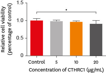
Objectives This study aimed to determine whether collagen triple helix repeat containing-1 (CTHRC1), which is involved in vascular remodeling and bone formation, can stimulate odontogenic differentiation and angiogenesis when administered to human dental pulp stem cells (hDPSCs).
Materials and Methods The viability of hDPSCs upon exposure to CTHRC1 was assessed with the WST-1 assay. CTHRC1 doses of 5, 10, and 20 µg/mL were administered to hDPSCs. Reverse-transcription polymerase reaction was used to detect dentin sialophosphoprotein, dentin matrix protein 1, vascular endothelial growth factor, and fibroblast growth factor 2. The formation of mineralization nodules was evaluated using Alizarin red. A scratch wound assay was conducted to evaluate the effect of CTHRC1 on cell migration. Data were analyzed using 1-way analysis of variance followed by the Tukey
post hoc test. The threshold for statistical significance was set atp < 0.05.Results CTHRC1 doses of 5, 10, and 20 µg/mL had no significant effect on the viability of hDPSCs. Mineralized nodules were formed and odontogenic markers were upregulated, indicating that CTHRC1 promoted odontogenic differentiation. Scratch wound assays demonstrated that CTHRC1 significantly enhanced the migration of hDPSCs.
Conclusions CTHRC1 promoted odontogenic differentiation and mineralization in hDPSCs.
- 2,501 View
- 40 Download
- The clinical success of ART restorations and Hall technique in primary molars: a randomized 18-month follow-up study
- Esra Oz, Zuhal Kırzıoglu, Canan Kale
- Restor Dent Endod 2023;48(2):e19. Published online May 1, 2023
- DOI: https://doi.org/10.5395/rde.2023.48.e19
-
Abstract
PDFPubReaderePub
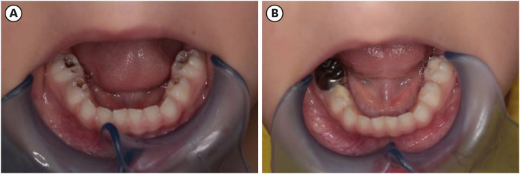
Objectives The aim of the study was to evaluate the clinical and radiographic success of the Hall technique (HT) and atraumatic restorative treatment (ART) restorations using high-viscosity glass-ionomer cement for the management of occlusal carious lesions in primary molars.
Materials and Methods This randomized clinical study observed 40 children (aged 5–6 years). For each child, one tooth was treated with HT and one with ART. The primary outcome measures for HT restorations were successful, minor, and major failure rates. Clinical evaluations of ART restorations were performed according to the modified United States Public Health Service criteria during 18-month follow-up. McNemar test was used for statistical analysis.
Results Thirty of 40 (75%) participants returned for 18 months of follow-up. In the clinical evaluations of teeth that were treated with HT, the patients did not have complaints of pain or other symptoms, all crowns remained in the oral cavity, the gums were healthy, and the teeth were functional in all evaluations. At the end of the 18-month follow-up, the surface texture and marginal integrity criteria of ART restorations were recorded as 26.7% and 33.3%, respectively. In the radiographic evaluation of 30 patients treated with ART and HT, all restorations were considered successful.
Conclusions The 18-month clinical and radiographic results after treatments applied to single-surface cavities in anxious children showed that both treatment methods were successful.
-
Citations
Citations to this article as recorded by- Two-year outcomes of hall technique and modified hall technique in deep carious lesions of primary molars: a randomized clinical trial
Sumeyye Konukman Turker, Ayse Isil Cihan
BMC Oral Health.2026;[Epub] CrossRef - Clinical Performance of High-Viscosity Glass Hybrid Restorative Systems in Posterior Teeth: A Scoping Review
Antonia Theodora Vrabie, Tinela Panaite, Bogdan Radu Dragomir, Simona Stoleriu, Angela Ghiorghe, Irina Nica, Alice Murariu, Sorin Andrian, Gianina Iovan
Prosthesis.2026; 8(7): 71. CrossRef - Success rate of Hall Technique for restoring carious primary molars - systematic review and meta-analysis
Tamara Kerber Tedesco, Nicola Patricia Innes, Claudia Lopez Gallegos, Gabriela Seabra Silva, Thais Gimenez, Mariana Minatel Braga, Mariana Pinheiro Araujo, Jayakumar Jayaraman, Waraf Al-yaseen, Daniela Prócida Raggio
Evidence-Based Dentistry.2025; 26(1): 65. CrossRef - Effectiveness of Dental Restorative Materials in the Atraumatic Treatment of Carious Primary Teeth in Pediatric Dentistry: A Systematic Review
Gianna Dipalma, Angelo Michele Inchingolo, Lucia Casamassima, Paola Nardelli, Danilo Ciccarese, Paolo De Sena, Francesco Inchingolo, Andrea Palermo, Marco Severino, Cinzia Maria Norma Maspero, Alessio Danilo Inchingolo
Children.2025; 12(4): 511. CrossRef - Clinical, radiographic, and microhardness evaluation of caries in primary molars managed with modified Hall technique
Eman El Sayed El Bedewy, Nahed A.A. Abohamila, Shereen A. M. Ali, Shimaa M.M. Hadwa
Tanta Dental Journal.2025; 22(1): 133. CrossRef - Pain Perception During Minimally Invasive Caries Removal in Children: A Randomized Clinical Trial Comparing Chemo-Mechanical Caries Removal
Dhirja Goel, Neha Awasthi, Yanina Singh, Sukhdeep Singh, Nenung Yirang
Journal of International Society of Preventive and Community Dentistry.2025; 15(4): 348. CrossRef - Clinical and histological evaluation of hall technique with and without silver diamine fluoride in the treatment of carious primary molars
Marwa M.A.Z. Abd-Elhaleium, Fatma A.-A. El-Hendawy, Lamis A. El-Ghareb, Sara Y. AboAli
Tanta Dental Journal.2025; 22(2): 351. CrossRef - Comparative success of minimally invasive treatments for cavitated caries in primary teeth: a network meta-analysis
Rasoul Sahebalam, Mahsa Ghorbani, Alireza Sarraf Shirazi, Motahareh Khosrojerdi, Mana Mowji
BMC Oral Health.2025;[Epub] CrossRef
- Two-year outcomes of hall technique and modified hall technique in deep carious lesions of primary molars: a randomized clinical trial
- 8,088 View
- 146 Download
- 6 Web of Science
- 8 Crossref
- Effects of different calcium-silicate based materials on fracture resistance of immature permanent teeth with replacement root resorption and osteoclastogenesis
- Gabriela Leite de Souza, Gabrielle Alves Nunes Freitas, Maria Tereza Hordones Ribeiro, Nelly Xiomara Alvarado Lemus, Carlos José Soares, Camilla Christian Gomes Moura
- Restor Dent Endod 2023;48(2):e21. Published online May 5, 2023
- DOI: https://doi.org/10.5395/rde.2023.48.e21
-
Abstract
PDF
 Supplementary MaterialPubReaderePub
 Objectives This study evaluated the effects of Biodentine (BD), Bio-C Repair (BCR), and mineral trioxide aggregate (MTA) plug on the fracture resistance of simulated immature teeth with replacement root resorption (RRR) and
in vitro -induced osteoclastogenesis.Materials and Methods Sixty bovine incisors simulating immature teeth and RRR were divided into 5 groups: BD and BCR groups, with samples completely filled with the respective materials; MTA group, which utilized a 3-mm apical MTA plug; RRR group, which received no root canal filling; and normal periodontal ligament (PL) group, which had no RRR and no root canal filling. All the teeth underwent cycling loading, and compression strength testing was performed using a universal testing machine. RAW 264.7 macrophages were treated with 1:16 extracts of BD, BCR, and MTA containing receptor activator of nuclear factor-kappa B ligand (RANKL) for 5 days. RANKL-induced osteoclast differentiation was assessed by staining with tartrate-resistant acid phosphatase. The fracture load and osteoclast number were analyzed using 1-way ANOVA and Tukey’s test (α = 0.05).
Results No significant difference in fracture resistance was observed among the groups (
p > 0.05). All materials similarly inhibited osteoclastogenesis (p > 0.05), except for BCR, which led to a lower percentage of osteoclasts than did MTA (p < 0.0001).Conclusions The treatment options for non-vital immature teeth with RRR did not strengthen the teeth and promoted a similar resistance to fractures in all cases. BD, MTA, and BCR showed inhibitory effects on osteoclast differentiation, with BCR yielding improved results compared to the other materials.
-
Citations
Citations to this article as recorded by- Effect of Restoration Strategy and Cavity Location on the Fracture Resistance of Teeth with External Cervical Resorption
Saadet Elpe, Öznur Sarıyılmaz
Journal of Endodontics.2026; 52(6): 980. CrossRef - Influence of Different Post-core Restorative Modalities on Fracture Characteristics of Immature Endodontically Treated Premolars
Wafa H Alaajam, Khalid M Abdelaziz, Malaz M Mustafa, Mohammed S Al-Ak'hali, Ashraf A Khalil, Mohammed M Al Moaleem, Hoda L Abouzeid
The Journal of Contemporary Dental Practice.2026; 27(4): 399. CrossRef - In vitro comparison of fracture strength of maxillary incisors with the simulated external root resorption cavities repaired with BioMTA or Biodentine
Tufan Ozasir, Birgul Ozasir, Nagihan Aribal, Derin Bugu Yuzer, Baris Kandemir, Kamran Gulsahi
Journal of Dental Sciences.2025; 20(3): 1532. CrossRef - Comparative Analysis of Gene Expression in Periodontal Ligament Stem Cells Exposed to Biodentine and Bio-C Repair: Implications for Cementogenesis—An In Vitro Study
Mahmoud M. Bakr, Mahmoud Al Ankily, Mohammed Meer, Mohamed Shamel
Oral.2025; 5(1): 19. CrossRef - Efficacy of Mineral Trioxide Aggregate Versus Biodentine as a Direct Pulp Capping Material in Carious Human Mature Permanent Teeth: A Systematic Review
Rashmi Misra, Nikita Toprani, Sumita Bhagwat, Aashaka Vaishnav, Aastha Dureja, Omkar Bhosale
Cureus.2025;[Epub] CrossRef - Evaluation of Different Techniques and Materials for Filling in 3-dimensional Printed Teeth Replicas with Perforating Internal Resorption by Means of Micro–Computed Tomography
Angelo J.S. Torres-Carrillo, Helena C. Assis, Rodrigo E. Salazar-Gamarra, Leonardo Moreira Teodosio, Alice C. Silva-Sousa, Jardel F. Mazzi-Chaves, Priscila B. Ferreira-Soares, Manoel D. Sousa-Neto, Fabiane C. Lopes-Olhê
Journal of Endodontics.2024; 50(2): 205. CrossRef
- Effect of Restoration Strategy and Cavity Location on the Fracture Resistance of Teeth with External Cervical Resorption
- 3,701 View
- 82 Download
- 4 Web of Science
- 6 Crossref
Case Report
- Successful nonsurgical treatment of type II dens invaginatus with 5 root canals using a self-adjusting file: a case report
- George Táccio de Miranda Candeiro, Antônio Sérgio Teixeira de Menezes, Ana Carolina Saldanha de Oliveira, Flávio Rodrigues Ferreira Alves
- Restor Dent Endod 2023;48(2):e17. Published online April 27, 2023
- DOI: https://doi.org/10.5395/rde.2023.48.e17
-
Abstract
PDFPubReaderePub
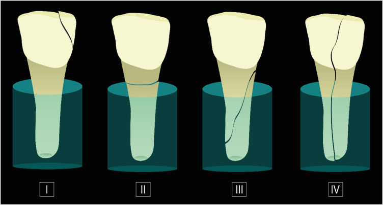
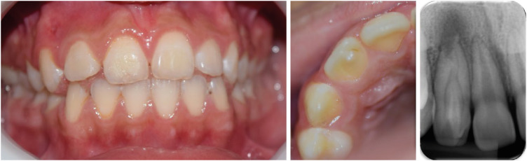
The present report describes the endodontic treatment of an Oehlers type II dens invaginatus in a maxillary lateral incisor with 5 root canals, an extremely rare condition. Apical periodontitis and related symptoms were noted. Cone-beam computed tomography was used to aid the diagnosis, reveal tooth morphology, and assist in canal location. The pulp chamber was carefully accessed, and the root canals were explored under magnification. All root canals were prepared with an R25 Reciproc Blue system and sodium hypochlorite (NaOCl) irrigation. After initial preparation, a self-adjusting file (SAF) with NaOCl and ethylenediaminetetraacetic acid was used to complement the disinfection. Additionally, calcium hydroxide medication was applied. Vertical compaction was used to fill the canals with a calcium silicate-based endodontic sealer and gutta-percha. After 12 months, the patient exhibited healing of the periapical region, absence of symptoms, and normal dental function. In conclusion, this nonsurgical treatment protocol was successful in promoting the cure of apical periodontitis. Both complementary disinfection with an SAF and use of calcium hydroxide medication should be considered when choosing the best treatment approach for dens invaginatus with very complex anatomy.
-
Citations
Citations to this article as recorded by- Non-surgical endodontic management of dens invaginatus type II in an immature maxillary lateral incisor using a bioceramic apical plug: A 3-year follow-up case report
Yahya Raja Alharbi, Qayed Saad Alharbi, Shaul Hameed Kolarkodi
Saudi Endodontic Journal.2026; 16(1): 115. CrossRef
- Non-surgical endodontic management of dens invaginatus type II in an immature maxillary lateral incisor using a bioceramic apical plug: A 3-year follow-up case report
- 3,037 View
- 89 Download
- 1 Crossref
 First
First Prev
Prev

