Previous issues
- Page Path
- HOME > Browse articles > Previous issues
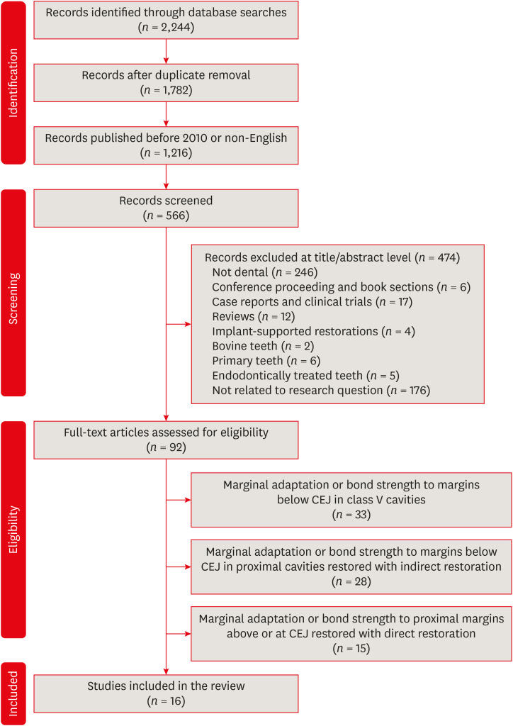
- Deep proximal margin rebuilding with direct esthetic restorations: a systematic review of marginal adaptation and bond strength
- Hoda S. Ismail, Ashraf I. Ali, Rabab El. Mehesen, Jelena Juloski, Franklin Garcia-Godoy, Salah H. Mahmoud
- Restor Dent Endod 2022;47(2):e15. Published online March 4, 2022
- DOI: https://doi.org/10.5395/rde.2022.47.e15
-
 Abstract
 PDF
PDF PubReader
 ePub
ePub This review aimed to characterize the effect of direct restorative material types and adhesive protocols on marginal adaptation and the bond strength of the interface between the material and the proximal dentin/cementum. An electronic search of 3 databases (the National Library of Medicine [MEDLINE/PubMed], Scopus, and ScienceDirect) was conducted. Studies were included if they evaluated marginal adaptation or bond strength tests for proximal restorations under the cementoenamel junction. Only 16 studies met the inclusion criteria and were included in this review. These studies presented a high degree of heterogeneity in terms of the materials used and the methodologies and evaluation criteria of each test; therefore, only a descriptive analysis could be conducted. The included studies were individually evaluated for the risk of bias following predetermined criteria. To summarize the results of the included studies, the type of restorative material affected the test results, whereas the use of different adhesive protocols had an insignificant effect on the results. It could be concluded that various categories of resin-based composites could be a suitable choice for clinicians to elevate proximal dentin/cementum margins, rather than the open sandwich technique with resin-modified glass ionomers. Despite challenges in bonding to proximal dentin/cementum margins, different adhesive protocols provided comparable outcomes.
-
Citations
Citations to this article as recorded by- An In Vitro Evaluation of Novel Bioactive Liner's Effect on Marginal Adaptation of Class II Composite Restorations: A Scanning Electron Microscope Analysis
Girija S Sajjan, Naveena Ponnada, Praveen Dalavai, Madhu Varma Kanumuri, Venkata Karteek Varma Penmatsa, B V Sindhuja
World Journal of Dentistry.2025; 15(9): 749. CrossRef - Effect of Cervical Margin Relocation With Different Injectable Restorative Materials on Fracture Resistance of Molars Received MOD CAD/CAM Onlay Restorations
Basema N. Roshdy, Radwa I. Eltoukhy, Ashraf I. Ali, Salah Hasab Mahmoud
Journal of Esthetic and Restorative Dentistry.2025; 37(6): 1522. CrossRef - Short dentin etching with universal adhesives: effect on bond strength and gingival margin adaptation
Hoda Saleh Ismail, Hanan Ahmed Nabil Soliman
BMC Oral Health.2025;[Epub] CrossRef - Awareness and Practice of Deep Margin Elevation among Dental Practitioners in India: A Cross-Sectional Survey
Mythri Padaru, Preethesh Shetty, Namith Rai, Raksha Bhat
Pesquisa Brasileira em Odontopediatria e Clínica Integrada.2025;[Epub] CrossRef - Effect of surface treatment on glass ionomers in sandwich restorations: a systematic review and meta-analysis of laboratory studies
Hoda S. Ismail, Ashraf Ibrahim Ali, Franklin Garcia-Godoy
Restorative Dentistry & Endodontics.2025; 50(2): e13. CrossRef - Do irrigation solutions effect bond strength of composite resin to deep margin elevation material? An in-vitro study
Şeref Nur Mutlu, Yasemin Derya Fidancıoğlu, Hatice Büyüközer Özkan, Hayriye Esra Ülker
BMC Oral Health.2025;[Epub] CrossRef - Two-year evaluation of periodontal parameters following deep-margin-elevation and CAD/CAM partial lithium disilicate restorations – a prospective controlled clinical trial
Tim Hausdörfer, Philipp Kanzow, Tina Rödig, Annette Wiegand, Clemens Lechte
Journal of Dentistry.2025; 160: 105901. CrossRef - Deep Margin Elevation: Current Evidence and a Critical Approach to Clinical Protocols—A Narrative Review
Athanasios Karageorgiou, Maria Fostiropoulou, Maria Antoniadou, Eftychia Pappa
Adhesives.2025; 1(3): 10. CrossRef - Comparative Micro-CT Analysis of Internal Adaptation and Closed Porosity of Conventional Layered and Thermoviscous Bulk-Fill Resin Composites Using Total-Etch or Universal Adhesives
Dóra Jordáki, Virág Veress, Tamás Kiss, József Szalma, Márk Fráter, Edina Lempel
Polymers.2025; 17(15): 2049. CrossRef - Effect of different restorative systems and aging on marginal adaptation of resin composites to deep proximal margins
Hoda S. Ismail, Ashraf I. Ali
Journal of Esthetic and Restorative Dentistry.2024; 36(2): 346. CrossRef - Management of subgingival proximal defects
Jagruti Mutalikdesai, K. C. Dhaniba, Supriya Choudhary, Promila Verma, Rhythm Bains
Asian Journal of Oral Health and Allied Sciences.2024; 14: 15. CrossRef - Effect of Deep Margin Elevation on the Pulpal and Periodontal Health of Teeth: A Systematic Review
S Srirama, S Jain, B Arul, K Prabakar, V Natanasabapathy
Operative Dentistry.2024; 49(4): 388. CrossRef - Alternative Direct Restorative Materials for Dental Amalgam: A Concise Review Based on an FDI Policy Statement
Gottfried Schmalz, Falk Schwendicke, Reinhard Hickel, Jeffrey A. Platt
International Dental Journal.2024; 74(4): 661. CrossRef - Comparison of the stress distribution in base materials and thicknesses in composite resin restorations
Min-Kwan Jung, Mi-Jeong Jeon, Jae-Hoon Kim, Sung-Ae Son, Jeong-Kil Park, Deog-Gyu Seo
Heliyon.2024; 10(3): e25040. CrossRef - Influence of curing mode and aging on the bonding performance of universal adhesives in coronal and root dentin
Hoda Saleh Ismail, Ashraf Ibrahim Ali, Mohamed Elshirbeny Elawsya
BMC Oral Health.2024;[Epub] CrossRef - CLINICAL ASSESSMENT OF THE EFFECTIVENESS OF ESTHETIC RESTORATION OF ANTERIOR TEETH
Lyudmila Tatintsyan, Minas Poghosyan, Armen Shaginyan, Hovhannes Gevorgyan, Biayna Hoveyan, Tatevik Margaryan, Arsen Kupelyan
BULLETIN OF STOMATOLOGY AND MAXILLOFACIAL SURGERY.2023; : 16. CrossRef - Deep margin elevation—Present status and future directions
Florin Eggmann, Jose M. Ayub, Julián Conejo, Markus B. Blatz
Journal of Esthetic and Restorative Dentistry.2023; 35(1): 26. CrossRef
- An In Vitro Evaluation of Novel Bioactive Liner's Effect on Marginal Adaptation of Class II Composite Restorations: A Scanning Electron Microscope Analysis
- 4,615 View
- 100 Download
- 15 Web of Science
- 17 Crossref

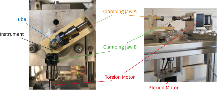
- Comparison of the cyclic fatigue resistance of One Curve, F6 Skytaper, Protaper Next, and Hyflex CM endodontic files
- Charlotte Gouédard, Laurent Pino, Reza Arbab-Chirani, Shabnam Arbab-Chirani, Valérie Chevalier
- Restor Dent Endod 2022;47(2):e16. Published online March 4, 2022
- DOI: https://doi.org/10.5395/rde.2022.47.e16
-
 Abstract
 PDF
PDF PubReader
 ePub
ePub Objectives This study compared the cyclic fatigue resistance of One Curve (C wire) and F6 Skytaper (conventional austenite nickel-titanium [NiTi]), and 2 instruments with thermo-mechanically treated NiTi: Protaper Next X2 (M wire) and Hyflex CM (CM wire).
Materials and Methods Ten new instruments of each group (size: 0.25 mm, 6% taper in the 3 mm tip region) were tested using a rotary bending machine with a 60° curvature angle and a 5 mm curvature radius, at room temperature. The number of cycles until fracture was recorded. The length of the fractured instruments was measured. The fracture surface of each fragment was examined with a scanning electron microscope (SEM). The data were analyzed using one-way analysis of variance and the
post hoc Tukey test. The significance level was set at 0.05.Results At 60°, One Curve, F6 Skytaper and Hyflex CM had significantly longer fatigue lives than Protaper Next X2 (
p < 0.05). No statistically significant differences were found in the cyclic fatigue lives of One Curve, F6 Skytaper, and Hyflex CM (p > 0.05). SEM images of the fracture surfaces of the different instruments showed typical features of fatigue failure.Conclusions Within the conditions of this study, at 60° and with a 5 mm curvature radius, the cyclic fatigue life of One Curve was not significantly different from those of F6 Skytaper and Hyflex CM. The cyclic fatigue lives of these 3 instruments were statistically significantly longer than that of Protaper Next.
-
Citations
Citations to this article as recorded by- Evaluation of cyclic fatigue in three pediatric endodontic rotary file systems in root canals of primary molars: A finite element analysis (FEA)
Monika sri S.S., K.C. Vignesh, K. Vivek, Kavitha Swaminathan, Selvakumar Haridoss
Journal of Oral Biology and Craniofacial Research.2025; 15(2): 310. CrossRef - Stress analysis of different experimental finite element models of rotary endodontic instruments
Manar M. Galal, Amira Galal Ismail, Nada Omar
Bulletin of the National Research Centre.2025;[Epub] CrossRef - Understanding Cyclic Fatigue in Three Nickel–Titanium Pediatric Files: An In Vitro Study for Enhanced Patient Care
Alwaleed Abushanan, Rajashekhara Bhari Sharanesha, Fahd Aljarbou, Hadi Alamri, Mohammed Hamad Almasud, Abdulfatah AlAzmah, Sara Alghamdi, Mubashir Baig Mirza
Medicina.2025; 61(5): 830. CrossRef - Analyzing Surface Morphology Changes Induced by Cyclic Fatigue in Three Different Nickel–Titanium Rotary Files Using Scanning Electron Microscopy Analysis
Chintan Joshi, Mahima P Jain, Sweety M Thumar, Jay H Dave, Applu R Bhatt, Juhi I Dholani
World Journal of Dentistry.2024; 15(7): 579. CrossRef - Nickel ion release and surface analyses on instrument fragments fractured beyond the apex: a laboratory investigation
Sıdıka Mine Toker, Ekim Onur Orhan, Arzu Beklen
BMC Oral Health.2023;[Epub] CrossRef
- Evaluation of cyclic fatigue in three pediatric endodontic rotary file systems in root canals of primary molars: A finite element analysis (FEA)
- 3,201 View
- 42 Download
- 2 Web of Science
- 5 Crossref

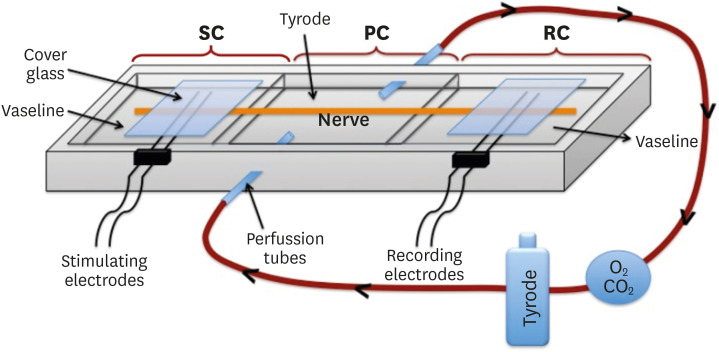
- Effects of calcium silicate cements on neuronal conductivity
- Derya Deniz-Sungur, Mehmet Ali Onur, Esin Akbay, Gamze Tan, Fügen Daglı-Comert, Taner Cem Sayın
- Restor Dent Endod 2022;47(2):e18. Published online March 7, 2022
- DOI: https://doi.org/10.5395/rde.2022.47.e18
-
 Abstract
 PDF
PDF PubReader
 ePub
ePub Objectives This study evaluated alterations in neuronal conductivity related to calcium silicate cements (CSCs) by investigating compound action potentials (cAPs) in rat sciatic nerves.
Materials and Methods Sciatic nerves were placed in a Tyrode bath and cAPs were recorded before, during, and after the application of test materials for 60-minute control, application, and recovery measurements, respectively. Freshly prepared ProRoot MTA, MTA Angelus, Biodentine, Endosequence RRM-Putty, BioAggregate, and RetroMTA were directly applied onto the nerves. Biopac LabPro version 3.7 was used to record and analyze cAPs. The data were statistically analyzed.
Results None of the CSCs totally blocked cAPs. RetroMTA, Biodentine, and MTA Angelus caused no significant alteration in cAPs (
p > 0.05). Significantly lower cAPs were observed in recovery measurements for BioAggregate than in the control condition (p < 0.05). ProRoot MTA significantly but transiently reduced cAPs in the application period compared to the control period (p < 0.05). Endosequence RRM-Putty significantly reduced cAPs.Conclusions Various CSCs may alter cAPs to some extent, but none of the CSCs irreversibly blocked them. The usage of fast-setting CSCs during apexification or regeneration of immature teeth seems safer than slow-setting CSCs due to their more favorable neuronal effects.
-
Citations
Citations to this article as recorded by- Endodontic Sealers and Innovations to Enhance Their Properties: A Current Review
Anna Błaszczyk-Pośpiech, Natalia Struzik, Maria Szymonowicz, Przemysław Sareło, Maria Wiśniewska-Wrona, Kamila Wiśniewska, Maciej Dobrzyński, Magdalena Wawrzyńska
Materials.2025; 18(18): 4259. CrossRef
- Endodontic Sealers and Innovations to Enhance Their Properties: A Current Review
- 1,372 View
- 19 Download
- 1 Web of Science
- 1 Crossref

- Morphotypes of the apical constriction of maxillary molars: a micro-computed tomographic evaluation
- Jeffrey Wen-Wei Chang, Kuzhanchinathan Manigandan, Lakshman Samaranayake, Chellapandian NandhaKumar, Pazhamalai AdhityaVasun, Johny Diji, Angambakkam Rajasekharan PradeepKumar
- Restor Dent Endod 2022;47(2):e19. Published online March 24, 2022
- DOI: https://doi.org/10.5395/rde.2022.47.e19

-
 Abstract
 PDF
PDF PubReader
 ePub
ePub Objectives The aim of this study was to evaluate and compare the apical constriction (AC) and apical canal morphology of maxillary first and second molars, using micro-computed tomography (micro-CT).
Materials and Methods The anatomical features of 313 root canals from 41 maxillary first molars and 57 maxillary second molars of patients with known age and sex were evaluated using micro-CT, with a resolution of 26.7 µm. The factors evaluated were the presence or absence of AC, the morphotypes, bucco-lingual dimension, mesio-distal dimension, and the profile (shape) of AC and the apical root canal. The apical root canal dimensions, location of the apical foramen (AF), AC to AF distance, and presence of accessory canals in the apical 5 mm were also assessed. Descriptive and analytical statistics were used for data evaluation.
Results AC was present in all 313 root canals. Patients’ age and sex did not significantly impact either AC or the apical canal dimensions. The most common AC morphotype detected was the traditional (single) constriction (52%), followed by the parallel (29%) morphotype. The mean AC dimensions in maxillary first molars were not significantly different from those in maxillary second molars. Sixty percent of AF were located within 0.5 mm from the anatomic apex.
Conclusions The most common morphotype of AC detected was the traditional constriction. Neither patients’ age nor sex had a significant impact on the dimensions of the AC or the apical root canal. The majority of AF (60%) were located within 0.5 mm from the anatomic apex.
-
Citations
Citations to this article as recorded by- In Vivo and In Vitro Accuracy and Precision Evaluations of Mini Electronic Apex Locators
Özlem Kara, Rüstem Kemal Sübay
Australian Endodontic Journal.2025; 51(2): 329. CrossRef - Effect of Coronal Flaring on Initial Apical File Size Estimation in Curved Canals Using Three Distinct Rotary Instruments: A Comparative In Vitro Study
Vinodhini Varatharajan, Muhammed Abdul Rahman Thazhathveedan, Mohammed Salman Kuttikkodan, Ismail Puzhangaraillath Mundanatayil, Amrutha Ravindran Thazhe Mangool, Ashraf Karumbil
Cureus.2024;[Epub] CrossRef - In Vitro Evaluation of the Accuracy of Three Electronic Apex Locators Using Different Sodium Hypochlorite Concentrations
Sanda Ileana Cîmpean, Radu Marcel Chisnoiu, Adela Loredana Colceriu Burtea, Rareș Rotaru, Marius Gheorghe Bud, Ada Gabriela Delean, Ioana-Sofia Pop-Ciutrilă
Medicina.2023; 59(5): 918. CrossRef - Cone beam computed tomography analysis of the root and canal morphology of the maxillary second molars in a Hail province of the Saudi population
Ahmed A. Madfa, Moazzy I. Almansour, Saad M. Al-Zubaidi, Albandari H. Alghurayes, Safanah D. AlDAkhayel, Fatemah I. Alzoori, Taif F. Alshammari, Abrar M. Aldakhil
Heliyon.2023; 9(9): e19477. CrossRef
- In Vivo and In Vitro Accuracy and Precision Evaluations of Mini Electronic Apex Locators
- 2,154 View
- 44 Download
- 6 Web of Science
- 4 Crossref

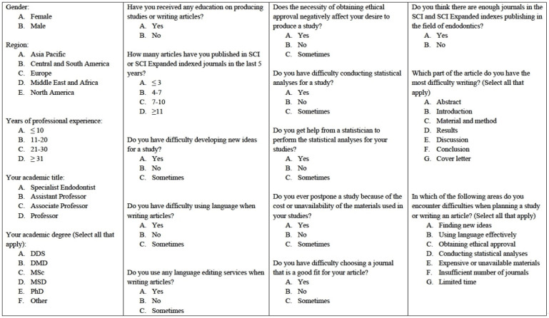
- Difficulties experienced by endodontics researchers in conducting studies and writing papers
- Betul Aycan Alim-Uysal, Selin Goker-Kamali, Ricardo Machado
- Restor Dent Endod 2022;47(2):e20. Published online March 15, 2022
- DOI: https://doi.org/10.5395/rde.2022.47.e20
-
 Abstract
 PDF
PDF PubReader
 ePub
ePub Objectives The study investigated the difficulties experienced by endodontics researchers around the world in conducting studies and writing papers.
Materials and Methods A survey consisting of 18 questions on the difficulties experienced by endodontics researchers in performing studies and writing papers was e-mailed to academics in the field of endodontics working at 202 universities. The independent risk factors were analyzed using binary logistic regression at a significance level of 0.05.
Results A total of 581 individuals (10.7%) agreed to participate in the study. Almost half the participants (48.2%) reported that they had received some type of training in conducting studies and writing papers. In response to the question, “Do you get help from a statistician to perform the statistical analyses of your studies?,” 77.1% answered “yes.” Around 40% of the participants stated that the need to obtain ethical approval negatively affected their desire to conduct studies. The participants’ regions had no effect on the reported difficulties associated with writing papers in English or conducting statistical analyses (
p > 0.05). Most participants (81.8%) reported difficulties in writing the Discussion section, regardless of their region, academic degrees, or years of experience.Conclusions The participants stated they experienced difficulties in many areas, such as conducting statistical analyses, finding new ideas, and writing in English. Engaging in a detailed examination of ethics committee rules, expanding biostatistics education, increasing the number of institutions providing research funding, and increasing the number of endodontics journals can increase the enthusiasm of endodontics researchers to publish papers.
-
Citations
Citations to this article as recorded by- Prevalence of radix molaris in mandibular molars of a subpopulation of Brazil’s Northeast region: a cross-sectional CBCT study
Yasmym Martins Araújo de Oliveira, Maria Clara Mendes Gomes, Maria Fernanda da Silva Nascimento, Ricardo Machado, Danna Mota Moreira, Hermano Camelo Paiva, George Táccio de Miranda Candeiro
Scientific Reports.2025;[Epub] CrossRef - Statistical pitfalls in endodontic research
Nandini Suresh
Endodontology.2023; 35(1): 1. CrossRef
- Prevalence of radix molaris in mandibular molars of a subpopulation of Brazil’s Northeast region: a cross-sectional CBCT study
- 1,823 View
- 27 Download
- 1 Web of Science
- 2 Crossref

- Bonding effects of cleaning protocols and time-point of acid etching on dentin impregnated with endodontic sealer
- Tatiane Miranda Manzoli, Joissi Ferrari Zaniboni, João Felipe Besegato, Flávia Angélica Guiotti, Andréa Abi Rached Dantas, Milton Carlos Kuga
- Restor Dent Endod 2022;47(2):e21. Published online April 6, 2022
- DOI: https://doi.org/10.5395/rde.2022.47.e21
-
 Abstract
 PDF
PDF PubReader
 ePub
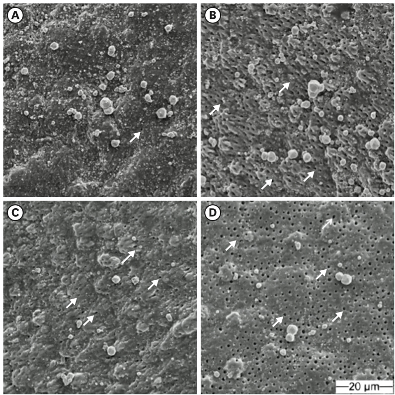
ePub Objectives This study aimed to investigate the bonding effects of cleaning protocols on dentin impregnated with endodontic sealer residues using ethanol (E) or xylol (X). The effects of dentin acid etching immediately (I) or 7 days (P) after cleaning were also evaluated. For bonding to dentin, universal adhesive (Scotchbond Universal; 3M ESPE) was used. The persistence of sealer residues, hybrid layer formation and microshear bond strength were the performed analysis.
Materials and Methods One hundred and twenty bovine dentin specimens were allocated into 4 groups (
n = 10): G1 (E+I); G2 (X+I); G3 (E+P); and G4 (X+P). The persistence of sealer residues was evaluated by SEM. Confocal laser scanning microscopy images were taken to measure the formed hybrid layer using the Image J program. For microshear bond strength, 4 resin composite cylinders were placed over the dentin after the cleaning protocols. ANOVA followed by Tukey test and Kruskal-Wallis followed by Dunn test were used for parametric and non-parametric data, respectively (α = 5%).Results G2 and G4 groups showed a lower persistence of residues (
p < 0.05) and thicker hybrid layer than the other groups (p < 0.05). No bond strength differences among all groups were observed (p > 0.05).Conclusions Dentin cleaning using xylol, regardless of the time-point of acid etching, provided lower persistence of residues over the surface and thicker hybrid layer. However, the bond strength of the universal adhesive system in etch-and-rinse strategy was not influenced by the cleaning protocols or time-point of acid etching.
-
Citations
Citations to this article as recorded by- Efficacy of Post-Endodontic Access Cavity Cleaning Techniques: A Randomized Clinical Study
Ayse Karadayi, Elif Irem Altintas, Ezgi Tüter Bayraktar, Bora Korkut
Journal of Endodontics.2025;[Epub] CrossRef - Does cleaning of post space before cementation of fiber reinforced post affect the push-out bond strength to resin cement?
Maher S. Hajjaj, Khalid A. Alghamdi, Abdulrahman A. Alshehri, Hassan A. Almusallam, Nabeel M. Munshi, Osamah A. Alsulimani, Naseeba H. Khouja, Yousef A. Alnowailaty, Saeed J. Alzahrani
BMC Oral Health.2025;[Epub] CrossRef - Influence of the Use of a Mixed Solution of Equal Amounts of Amyl Acetate, Acetone, and Ethanol on the Cleaning of Endodontic Sealer Residues on the Bond Strength of the Fiber Post Cementation System: A Laboratory Investigation
Antonia Patricia Oliveira Barros, Ana Paula Aparecida Raimundo Alves Freitas, Frederico Guilherme Otto Kokol, Elizangela Maria Pereira de Souza, Adirson Jorge Junior, Cristiane de Melo Alencar, Marcelo Ferrarezi de Andrade, Milton Carlos Kuga
The Open Dentistry Journal.2024;[Epub] CrossRef - Effects of the application protocol and bonding strategy of the universal adhesive on dentin previously impregnated with bioceramic sealer
Antonia Patricia Oliveira Barros, Joatan Lucas de Sousa Gomes Costa, Jardel Camilo do Carmo Monteiro, Lucas David Galvani, Marcelo Ferrarezi de Andrade, José Roberto Cury Saad, Milton Carlos Kuga
International Journal of Adhesion and Adhesives.2024; 134: 103765. CrossRef - Influência do protocolo de remoção de resíduos de cimentos à base de resina epóxi sobre a interface de adesão com o adesivo universal, utilizado na estratégia condiciona-e-lava
Paulo Firmino Da Costa Neto, Mariana Bena Gelio, Elisângela Maria Pereira De Souza, Jardel Camilo do Carmo Monteiro, Adirson Jorge Júnior, Thais Piragine Leandrin, José Roberto Cury Saad, Milton Carlos Kuga
Cuadernos de Educación y Desarrollo.2023; 15(5): 4802. CrossRef
- Efficacy of Post-Endodontic Access Cavity Cleaning Techniques: A Randomized Clinical Study
- 2,070 View
- 38 Download
- 3 Web of Science
- 5 Crossref

-
Effectiveness and safety of rotary and reciprocating kinematics for retreatment of curved root canals: a systematic review of
in vitro studies - Lucas Pinho Simões, Alexandre Henrique dos Reis-Prado, Carlos Roberto Emerenciano Bueno, Ana Cecília Diniz Viana, Marco Antônio Húngaro Duarte, Luciano Tavares Angelo Cintra, Cleidiel Aparecido Araújo Lemos, Francine Benetti
- Restor Dent Endod 2022;47(2):e22. Published online April 6, 2022
- DOI: https://doi.org/10.5395/rde.2022.47.e22
-
 Abstract
 PDF
PDF PubReader
 ePub
ePub Objectives This systematic review (register-osf.io/wg7ba) compared the efficacy and safety of rotary and reciprocating kinematics in the removal of filling material from curved root canals.
Materials and Methods Only
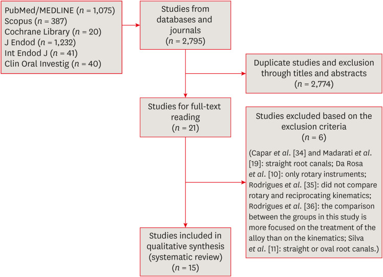
in vitro studies evaluating both kinematics during retreatment were included. A systematic search (PubMed/MEDLINE, Scopus, and other databases, until January 2021), data extraction, and risk of bias analysis (Joanna Briggs Institute checklist) were performed. Efficacy in filling removal was the primary outcome.Results The search resulted in 2,795 studies, of which 15 were included. Efficacy was measured in terms of the remaining filling material and the time required for this. Nine studies evaluated filling material removal, of which 7 found no significant differences between rotary and reciprocating kinematics. Regarding the time for filling removal, 5 studies showed no difference between both kinematics, 2 studies showed faster results with rotary systems, and other 2 showed the opposite. No significant differences were found in apical transportation, centering ability, instrument failure, dentin removed and extruded debris. A low risk of bias was observed.
Conclusions This review suggests that the choice of rotary or reciprocating kinematics does not influence the efficacy of filling removal from curved root canals. Further studies are needed to compare the kinematics safety in curved root canals.
-
Citations
Citations to this article as recorded by- EVALUATION OF MICROLEAKAGE AFTER ENDODONTIC FILLING IN TEETH WITH APICAL WIDENING: A SYSTEMATIC REVIEW
Isabella da Costa Ferreira, Gabriela da Costa Ferreira, Isabella Figueiredo de Assis Macedo, Gustavo Oliveira Campos, Isabella Faria da Cunha Peixoto, Ana Cecília Diniz Viana, Rodrigo Rodrigues Amaral, Warley Luciano Fonseca Tavares
ARACÊ .2025; 7(10): e8792. CrossRef - Fifteen years of engine‐driven nickel–titanium reciprocating instruments, what do we know so far? An umbrella review
Felipe Immich, Lucas Peixoto de Araújo, Rafaella Rodrigues da Gama, Wellington Luiz de Oliveira da Rosa, Evandro Piva, Giampiero Rossi‐Fedele
Australian Endodontic Journal.2024; 50(2): 409. CrossRef - Efficacy of Various Heat-treated Retreatment File Systems on the Apical Deformity and Canal Centering Ability in a Single-rooted Teeth using Nano CT
Swathi S, Pradeep Solete, Ganesh Jeevanandan, Delphine Priscilla Antony S, Kavalipurapu Venkata Teja, Dona Sanju
The Open Dentistry Journal.2024;[Epub] CrossRef - Micro-CT evaluation of the removal of root fillings using rotary and reciprocating systems supplemented by XP-Endo Finisher, the Self-Adjusting File, or Er,Cr:YSGG laser
Gülsen Kiraz, Bulem Üreyen Kaya, Mert Ocak, Muhammet Bora Uzuner, Hakan Hamdi Çelik
Restorative Dentistry & Endodontics.2023;[Epub] CrossRef - Influence of sodium hypochlorite on cyclic fatigue resistance of nickel–titanium instruments: A systematic review and meta-analysis of in vitro studies
Alexandre Henrique dos Reis-Prado, Lucas Guimarães Abreu, Lara Cancella de Arantes, Kiani dos Santos de Paula, Sabrina de Castro Oliveira, Juliana Goto, Ana Cecília Diniz Viana, Francine Benetti
Clinical Oral Investigations.2023; 27(11): 6291. CrossRef - Retreatment of XP-endo Shaper and R-Endo files in curved root canals
Hayam Y. Hassan, Fahd M. Hadhoud, Ayman Mandorah
BMC Oral Health.2023;[Epub] CrossRef - Advancing Endodontics through Kinematics
Shilpa Bhandi, Dario Di Nardo, Francesco Pagnoni, Rosemary Abbagnale
World Journal of Dentistry.2023; 14(6): 479. CrossRef
- EVALUATION OF MICROLEAKAGE AFTER ENDODONTIC FILLING IN TEETH WITH APICAL WIDENING: A SYSTEMATIC REVIEW
- 3,128 View
- 57 Download
- 5 Web of Science
- 7 Crossref

- Is dentin biomodification with collagen cross-linking agents effective for improving dentin adhesion? A systematic review and meta-analysis
- Julianne Coelho Silva, Edson Luiz Cetira Filho, Paulo Goberlânio de Barros Silva, Fábio Wildson Gurgel Costa, Vicente de Paulo Aragão Saboia
- Restor Dent Endod 2022;47(2):e23. Published online May 6, 2022
- DOI: https://doi.org/10.5395/rde.2022.47.e23
-
 Abstract
 PDF
PDF Supplementary Material
 PubReader
 ePub
ePub Objectives The aim of this investigation was to evaluate the effectiveness of collagen cross-linking agents (CCLAs) used in combination with the adhesive technique in restorative procedures.
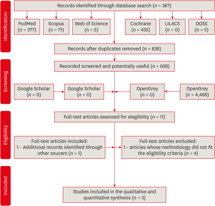
Materials and Methods In this systematic review, the authors followed the Preferred Reporting Items for Systematic Reviews and Meta-Analyses checklist. An electronic search was performed using PubMed, Scopus, Web of Science, Cochrane Library, LILACS, and DOSS, up to October 2020. The gray literature was also researched. Only randomized clinical trials were selected.
Results The selection process yielded 3 studies from the 838 retrieved. The addition of CCLAs in the retention of restorations increased the number of events. The postoperative sensitivity scores and marginal adaptation scores showed no significant difference between the CCLA and control groups, and the marginal pigmentation scores showed a significant increase in the CCLA group. There were no caries events in any group throughout the evaluation period.
Conclusions This systematic review showed that there is no clinical efficacy to justify the use of CCLAs in the protocols performed.
-
Citations
Citations to this article as recorded by- Riboflavin-ultraviolet-A collagen crosslinking treatments in improving dentin bonding and resistance to enzymatic digestion
Yung-Show Chiang, Ping-Ju Chen, Chun-Chan Ting, Yuh-Ling Chen, Shu-Fen Chuang
Journal of Dental Sciences.2025; 20(1): 109. CrossRef - Effect of dentin bio modifications and matrix metalloproteinase activity on bond strength – A systematic review and meta-analysis
D. Agarwal, S. R. Srinidhi, S. D. Aggarwal, P. Ingle, S. Tandon
Endodontics Today.2025; 23(1): 71. CrossRef - O USO DE ADESIVO AUTOCONDICIONANTE E RESINA FLOW COMO INTERFACE ADESIVA PROTETORA DA DENTINA FRENTE À IRRIGAÇÃO COM NaClO NO TRATAMENTO ENDODÔNTICO: ESTUDO IN-VITRO
Luís Daniel Ramos de Oliveira, Leandro Botelho Hanna, José Augusto Rodrigues
RECIMA21 - Revista Científica Multidisciplinar - ISSN 2675-6218.2025; 6(12): e6127063. CrossRef - Stability of dentin matrix treated with caffeic acid phenethyl ester at different concentrations
Aline Honorato Damázio, Rosanna Tarkany Basting, Enrico Coser Bridi, Fabiana Mantovani Gomes França, Flávia Lucisano Botelho do Amaral, Cecilia Pedroso Turssi, Waldemir Francisco Vieira Junior, Roberta Tarkany Basting
Brazilian Journal of Oral Sciences.2024; 23: e244006. CrossRef - Effect of Collagen Crosslinkers on Dentin Bond Strength of Adhesive Systems: A Systematic Review and Meta-Analysis
Louis Hardan, Umer Daood, Rim Bourgi, Carlos Enrique Cuevas-Suárez, Walter Devoto, Maciej Zarow, Natalia Jakubowicz, Juan Eliezer Zamarripa-Calderón, Mateusz Radwanski, Giovana Orsini, Monika Lukomska-Szymanska
Cells.2022; 11(15): 2417. CrossRef
- Riboflavin-ultraviolet-A collagen crosslinking treatments in improving dentin bonding and resistance to enzymatic digestion
- 1,976 View
- 45 Download
- 3 Web of Science
- 5 Crossref

- Clinical and radiographic outcomes of regenerative endodontic treatment performed by endodontic postgraduate students: a retrospective study
- Hadi Rajeh Alfahadi, Saad Al-Nazhan, Fawaz Hamad Alkazman, Nassr Al-Maflehi, Nada Al-Nazhan
- Restor Dent Endod 2022;47(2):e24. Published online May 9, 2022
- DOI: https://doi.org/10.5395/rde.2022.47.e24
-
 Abstract
 PDF
PDF PubReader
 ePub
ePub Objectives Regenerative endodontic treatment is a clinical procedure aimed at biologically regenerating damaged root canal tissue of immature permanent teeth. This study aimed to report the outcomes of regenerative endodontic treatment performed by endodontic postgraduate students.
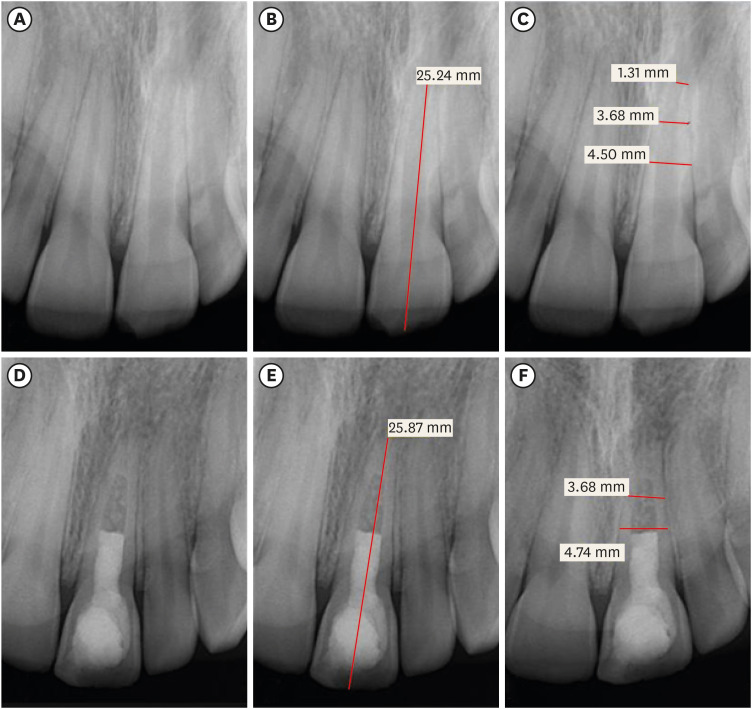
Materials and Methods Clinical and radiographic data of 27 patients, aged 10–22 years, who underwent regenerative treatment of immature permanent teeth from 2015 to 2019 were followed up, wherein clinical and radiographic examinations were performed for each patient. Postoperative success rate and tooth survival were analyzed, and the postoperative radiographic root area changes were quantified.
Results A total of 23 patients attended the dental appointments, showing that all teeth survived and were asymptomatic. Specifically, 7 periapical pathosis cases were completely healed, 12 were incompletely healed, and 4 cases failed. Moreover, significant differences were found between discolored and non-discolored teeth, and between the presence or absence of periapical radiolucency. Additionally, 3 anterior teeth showed complete closure of the apical foramen, while the apical foramen width was reduced in 17 teeth and failed in 3 teeth. Root length was also found to have been increased in 7 anterior and 4 posterior teeth, and the average length ranged from 4.00–0.63 mm in the anterior teeth, 2.85–1.48 mm of the mesial root, and 2.73–2.16 mm of the molar teeth distal root. Furthermore, calcified tissue deposition was observed in 7 teeth.
Conclusions A favorable outcome of regenerative endodontic treatment of immature permanent teeth with necrotic pulp was achieved with a high survival rate.
-
Citations
Citations to this article as recorded by- Pre‐Operative Factors on Prognosis of Regenerative Endodontic Procedures: A Systematic Review and Meta‐Analysis
Filipe Colombo Vitali, Alexandre Henrique dos Reis‐Prado, Pablo Silveira Santos, Ana Paula Portes Zeno, Patrícia de Andrade de Risso, Lucianne Cople Maia, Francine Benetti, Cleonice da Silveira da Teixeira
International Endodontic Journal.2025; 58(12): 1814. CrossRef - Clinical, radiographic, and biomarker perspectives of low-level laser therapy during regenerative endodontic procedures in necrotic immature young teeth: a randomized clinical study
Pragya Pandey, Neha Jasrasaria, Ramesh Bharti, Rakesh Kumar Yadav, Monika Kumari, Abinia Vaishnavi, Rahul Pandey
Lasers in Medical Science.2025;[Epub] CrossRef - Allogeneic Bone Marrow Mesenchymal Stromal Cell Transplantation Induces Dentin Pulp Complex-like Formation in Immature Teeth with Pulp Necrosis and Apical Periodontitis
Jose Francisco Gomez-Sosa, José E. Cardier, Olga Wittig, Dylana Díaz-Solano, Eloisa Lara, Kharelys Duque, Giselle Ramos-González
Journal of Endodontics.2024; 50(4): 483. CrossRef - Radiographic assessment of dental post and core placement at different educational levels in an undergraduate student clinic: a 4-year retrospective study
Turki Alshehri, Nourhan M. Aly, Raand Altayyar, Deena Alghamdi, Shahad Alotaibi, Passent Ellakany
F1000Research.2024; 12: 976. CrossRef - Evaluation of the efficacy of injectable platelet‐rich fibrin versus platelet‐rich plasma in the regeneration of traumatized necrotic immature maxillary anterior teeth: A randomized clinical trial
Maha Mohamed Abo‐Heikal, Jealan M. El‐Shafei, Samia A. Shouman, Nehal N. Roshdy
Dental Traumatology.2024; 40(1): 61. CrossRef - Radiographical assessment of post and core placement errors encountered by Saudi dental students at different educational levels
Turki Alshehri, Nourhan M. Aly, Raand Altayyar, Deena Alghamdi, Shahad Alotaibi, Passent Ellakany
F1000Research.2023; 12: 976. CrossRef
- Pre‐Operative Factors on Prognosis of Regenerative Endodontic Procedures: A Systematic Review and Meta‐Analysis
- 3,863 View
- 69 Download
- 6 Web of Science
- 6 Crossref

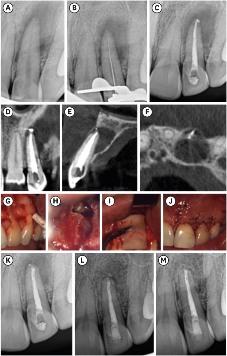
- Leukocyte platelet-rich fibrin in endodontic microsurgery: a report of 2 cases
- Mariana Domingos Pires, Jorge N. R. Martins, Abayomi Omokeji Baruwa, Beatriz Pereira, António Ginjeira
- Restor Dent Endod 2022;47(2):e17. Published online March 4, 2022
- DOI: https://doi.org/10.5395/rde.2022.47.e17
-
 Abstract
 PDF
PDF PubReader
 ePub
ePub Endodontic microsurgery is a predictable treatment option when orthograde treatment or retreatment is unsuccessful or unfeasible. However, when there is a gross compromise of periapical bone, achievement of bone regeneration after the surgical procedure may be hampered. In such cases, the application of guided tissue regeneration principles, with adjunctive use of leukocyte platelet-rich fibrin to fill the bone defect as a bone substitute and as a membrane to cover the site, provides a cost-effective solution with the benefits of accelerated physiological healing and reduced post-surgical pain and discomfort. This case report presents 2 cases of endodontic microsurgery of the upper lateral incisors with loss of buccal cortical plate, where platelet-rich fibrin was successfully applied.
-
Citations
Citations to this article as recorded by- Focuses and Trends of Research on Platelet-Rich Fibrin: A Bibliometric and Visual Analysis
Ying Zhao, Chen Dong, Liumeizi Fan, Ting Lei, Xin Ge, Zhou Yu, Sheng Hu
Indian Journal of Plastic Surgery.2024; 57(05): 356. CrossRef
- Focuses and Trends of Research on Platelet-Rich Fibrin: A Bibliometric and Visual Analysis
- 1,558 View
- 30 Download
- 1 Web of Science
- 1 Crossref


 KACD
KACD


 First
First Prev
Prev


