
Previous issues
- Page Path
- HOME > Browse articles > Previous issues
- Volume 51 (1); February 2026
-


Editorials
- Restorative Dentistry and Endodontics news: celebrating the 50th anniversary of the journal
- Yeon-Jee Yoo
- Restor Dent Endod 2026;51(1):e13. Published online February 26, 2026
- DOI: https://doi.org/10.5395/rde.2026.51.e13
- 1,423 View
- 52 Download

- Restorative Dentistry and Endodontics 2025 annual highlights: gratitude to our reviewers and achievement of the first journal impact factor
- Kyung-San Min
- Restor Dent Endod 2026;51(1):e14. Published online February 26, 2026
- DOI: https://doi.org/10.5395/rde.2026.51.e14
- 1,537 View
- 55 Download
Research Articles
- Enhancing antimicrobial properties of a resin-based material via incorporation of a powdered phytotherapeutic extract: an in vitro experimental study
- Rodolfo Xavier de Sousa-Lima, Maria Eduarda Lima do Nascimento Marinho, Janielly Cristina Costa da Silva, Moan Jéfter Fernandes Costa, Pedro Henrique Sette-de-Souza, Giana da Silveira Lima, Boniek Castillo Dutra Borges
- Restor Dent Endod 2026;51(1):e2. Published online January 20, 2026
- DOI: https://doi.org/10.5395/rde.2026.51.e2
-
 Abstract
 PDF
PDF PubReader
 ePub
ePub - Objectives
This study aimed to evaluate the degree of conversion (DC), immediate enamel bond strength (IEBS), antimicrobial activity, and release of the active principle of a resin-based material (RBM) enriched with the powdered Schinopsis brasiliensis (Braúna) stem antibacterial extract.
Methods
The RBM was enriched with 0, 1.25, 2.5, 5, 10, and 20 wt% powdered Braúna extract. The DC (n = 7) was assessed using micro-Raman spectroscopy. The IEBS (n = 7) was determined through the microshear test until failure, and failure modes were examined under a stereomicroscope. The antimicrobial activity (n = 15) was assessed by quantifying colony-forming units, and the release of the active principle was determined using ultra-high-performance liquid chromatography. One-way analysis of variance/Tukey and Kruskal-Wallis/Dunn tests were utilized to analyze the data (p < 0.05).
Results
Materials with 10 wt% and 20 wt% extract showed the lowest DC statistically. However, for IEBS, there were no statistically significant differences among the different groups. All materials released the active principle, but only those with 20 wt% and 10 wt% extract could inhibit biofilm formation similarly to 0.12% chlorhexidine.
Conclusions
Adding powdered Braúna extract between 10 wt% and 20 wt% is a promising alternative to provide an antimicrobial function to RBMs.
- 2,251 View
- 232 Download
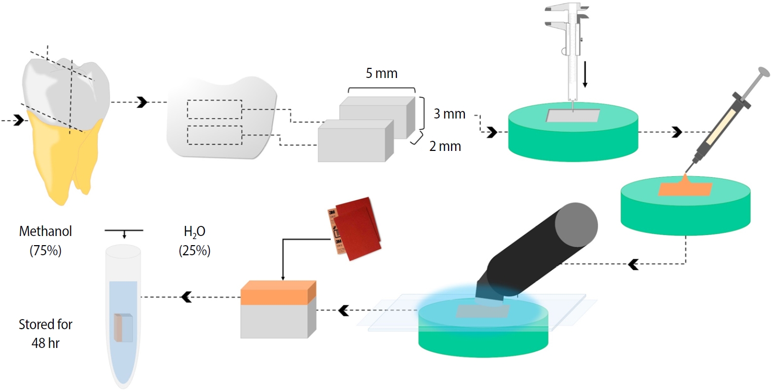
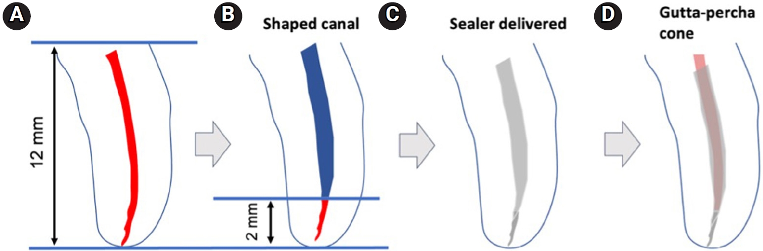
- Ex vivo comparative analysis of retrievability among four calcium silicate-based sealers for regaining apical patency
- Darian Shomali, Timothy Kirkpatrick, Sang Won Kwak, Hyeon-Cheol Kim, Ji Wook Jeong
- Restor Dent Endod 2026;51(1):e3. Published online January 14, 2026
- DOI: https://doi.org/10.5395/rde.2026.51.e3
-
Abstract
PDFPubReaderePub
- Objectives
Efficient retrievability is a key requirement for endodontic sealers. This study evaluated the retrievability of four different calcium silicate-based sealers (CSS).
Methods
A total of 153 single-rooted human teeth with straight canals were decoronated to a standardized working length of 12 mm. The canals were negotiated to working length using K files up to size 15/.02, followed by rotary instrumentation up to 35/.04, 2 mm short of working length. The teeth were randomly assigned to five groups: NeoSEALER Flo (NEO; Avalon Biomed), Ceraseal (CS; Meta Biomed), Endosequence BC Sealer (BC; Brasseler USA), AH Plus Bioceramic Sealer (AHB; Dentsply Sirona), and a negative control group. Sealer application and obturation with a 35/.04 gutta-percha cone were performed. After incubation at 37°C in 100% humidity for 7 days, retreatment was performed until apical patency was obtained, with retrievability assessed by regaining apical patency. One-way analysis of variance and Tukey contrast test were used to determine whether there was a significant difference among the four different CSS (p < 0.05).
Results
Success rates in regaining apical patency were NEO (79.4%), CS (37.0%), BC (50.0%), and AHB (69.7%). NEO demonstrated the highest retrievability, while CS had the lowest (p < 0.01).
Conclusions
The type of CSS used has a considerable impact on retreatment difficulty. Among the tested sealers, Neo- SEALER Flo showed the highest retrievability, making it the most retrievable CSS in terms of retreatment efficacy.
- 2,137 View
- 178 Download
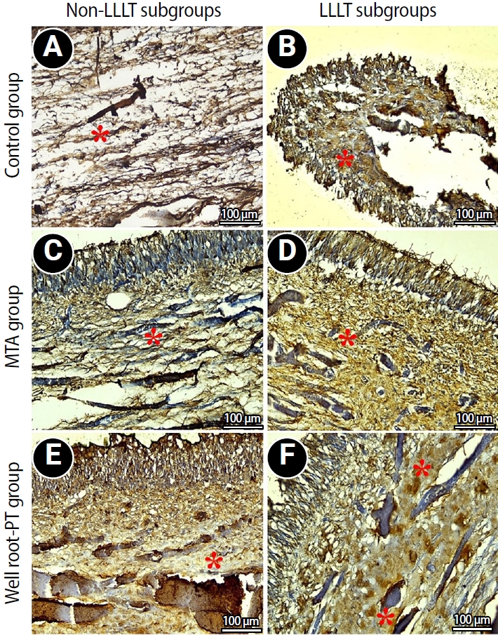
- Effect of combined application of premixed bioceramic paste and diode laser in vital pulp therapy: an immunohistochemical randomized controlled split-mouth in vivo animal experiment
- Mo’men A. Salama, Dalia M. Fayyad, Mohamed I. Rabie, Manar A. A. Selim, Mahmoud F. Ahmed
- Restor Dent Endod 2026;51(1):e4. Published online January 20, 2026
- DOI: https://doi.org/10.5395/rde.2026.51.e4
-
Abstract
PDFPubReaderePub
- Objectives
This study aimed to evaluate the effect of premixed bioceramic paste (Well-Root PT; Vericom) compared to mineral trioxide aggregate (MTA) on the expression of the mineralization-related marker dentin sialoprotein (DSP) in dental pulp following direct pulp capping, with or without prior diode laser application.
Methods
Direct pulp exposures were performed in the upper and lower incisors of eight dogs (n = 96 teeth). Cavities (Class V) were created and received pulp capping with either Well-Root PT (n = 32), MTA (n = 32), or no capping material (polytetrafluoroethylene disc only) (n = 32), with or without the application of a diode laser. Immunohistochemical analysis of DSP expression was conducted and quantified as the mean area percentage using ImageJ software at 2 and 8 weeks posttreatment.
Results
Both the Well-Root PT and MTA groups showed significantly increased DSP expression compared to the control group at both 2 and 8 weeks (p < 0.05). No significant difference in the mean area percentage of DSP expression was found between the Well-Root PT and MTA groups. The diode laser application did not produce a significant effect on DSP expression. Within-group comparison revealed a significant increase in DSP expression between the 2- and 8-week follow-up periods (p < 0.05).
Conclusions
Well-Root PT demonstrated comparable efficacy to MTA in promoting DSP expression, supporting its use as an effective direct pulp capping material. Diode laser application prior to capping had no effect on DSP expression in this experimental model.
- 2,026 View
- 141 Download
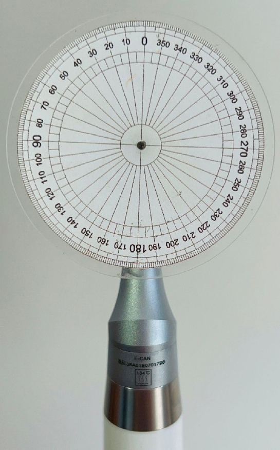
- Analysis of the reciprocating kinematics of the VDW Silver Reciproc, E-Connect Pro, Ecom, and Endopen endodontic motors: an in vitro experimental study
- Cristielly França, Juliana D. Bronzato, Dieimes Braambati, Adriana de-Jesus-Soares, Carla C. R. B. Félix, Michelle A. N. S. Ferreira, Marcos Frozoni
- Restor Dent Endod 2026;51(1):e5. Published online January 20, 2026
- DOI: https://doi.org/10.5395/rde.2026.51.e5
-
Abstract
PDFPubReaderePub
- Objectives
This study aimed to evaluate the actual parameters of four endodontic motors, each adjusted for reciprocating motion, and compare them to the manufacturers’ declared values.
Methods
The motors used were the VDW Silver Reciproc (VDW GmbH), E-Connect Pro (MK Life), Ecom (Woodpecker), and Endopen (Schuster Woodpecker). A custom optical target was attached to the motor contra-angle, the movements were recorded with a high-resolution camera, and the images were analyzed. Engagement, disengagement, net angles, and speed for each operation cycle, duration of clockwise (CW) and counter-clockwise (CCW) movement, duration of standstill after CW and CCW movement, and the number of cycles to complete a full rotation were analyzed. The data were statistically analyzed at a significance level of 5%. The replicability of all reciprocal parameters analyzed was statistically different from that reported by the manufacturers.
Results
There was no statistically significant difference between the VDW Silver Reciproc, Ecom, and Endopen for the engagement angle. The E-Connect Pro was the least reliable at the 150°/30° settings for both angle parameters. There was no significant difference between the set and actual cycle net angles for the VDW Silver Reciproc (p = 0.493). While the actual values for the Ecom and E-Connect Pro were significantly higher than the set (p < 0.001), the actual values for the Endopen were significantly lower than the set (p < 0.001).
Conclusions
Experiments on four commercially available reciprocating endodontic motors revealed that the actual motor values differed significantly from the set values.
- 2,021 View
- 87 Download
- Influence of adjacent restorative material and distance on the accuracy of inlay cavity impressions with intraoral scanner: an in vitro study
- So-Yeon Lee, Sung-Ae Son, Jae-Hoon Kim, Deog-Gyu Seo, Jeong-Kil Park
- Restor Dent Endod 2026;51(1):e6. Published online January 23, 2026
- DOI: https://doi.org/10.5395/rde.2026.51.e6
-
Abstract
PDFPubReaderePub
- Objectives
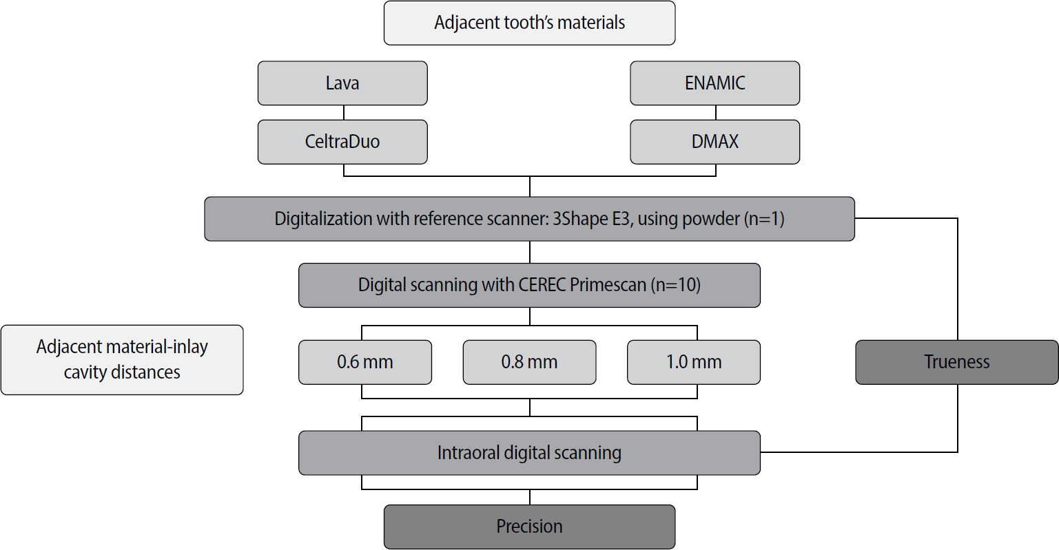
This study aimed to evaluate the influence of adjacent restorative material and interproximal distance on the accuracy of digital impressions of inlay cavities obtained using an intraoral scanner.
Methods
A disto-occlusal inlay cavity was prepared on a mandibular right first molar model, and digital scans were performed using a CEREC Primescan (Dentsply Sirona). The adjacent restorative materials used were Lava (3M ESPE), ENAMIC (VITA Zahnfabrik), Celtra Duo (Dentsply Sirona), and DMAX (DMAX), and the interproximal distances were set to 0.6 mm, 0.8 mm, and 1.0 mm. The obtained scan data were analyzed using GOM Inspect software (GOM GmbH).
Results
Trueness, maximum positive and negative deviations, and precision were significantly influenced by both the adjacent restorative material and the interproximal distance, while their interaction showed a significant effect only on precision. Celtra Duo demonstrated the highest trueness, with mean deviation values decreasing from 7.8 μm at a 0.6 mm interproximal distance to 7.3 μm at 1.0 mm. ENAMIC showed the best precision, presenting mean deviations of 2.6 μm at 0.6 mm, 2.9 μm at 0.8 mm, and 2.4 μm at 1.0 mm. A narrow interproximal distance of 0.6 mm resulted in lower trueness, measured at 8.3 μm, and the highest precision deviation of 3.4 μm. In contrast, an interproximal distance of 1.0 mm yielded improved scan accuracy, with increased trueness and reduced precision variation.
Conclusions
Digital impression accuracy of inlay cavities was influenced by adjacent restorative material and interproximal distance, suggesting clinical consideration is needed in CAD/CAM workflows to optimize restoration fit. -
Citations
Citations to this article as recorded by
- 3D-SCANNING IN PROSTHETIC DENTISTRY: ADVANTAGES, DISADVANTAGES, AND DEVELOPMENT PROSPECTS
V. S. Kuz, O. I. Teslenko, H. M. Kuz, H. M. Balia, Yu. S. Lunkova, O. V. Shemetov, I. M. Martynenko
Bulletin of Problems Biology and Medicine.2026; 1(1): 98. CrossRef
- 3D-SCANNING IN PROSTHETIC DENTISTRY: ADVANTAGES, DISADVANTAGES, AND DEVELOPMENT PROSPECTS
- 2,115 View
- 119 Download
- 1 Crossref
- Biological mechanisms underlying the inflammatory radicular cyst formation-focus on epithelial proliferation: a systematic review of experimental cell and tissue models
- Néstor Ríos-Osorio, Sandra Briñez-Rodríguez, David Betancur-Calle, Marggie Grajales, Óscar Mauricio Jiménez-Peña, Mario Guerrero-Torres, Rafael Fernández-Grisales
- Restor Dent Endod 2026;51(1):e7. Published online February 12, 2026
- DOI: https://doi.org/10.5395/rde.2026.51.e7
-
Abstract
PDFPubReaderePub
- Objectives
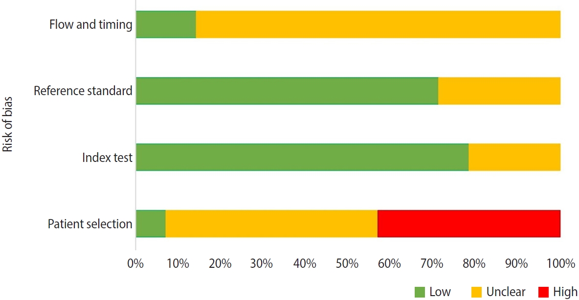
This study aimed to assess the molecular and cellular mechanisms involved in the epithelial proliferation that leads to the transformation of periapical granulomas (PGs) into inflammatory radicular cysts (IRCs).
Methods
A comprehensive search was conducted in three databases. Experimental, observational, or descriptive studies using human or animal tissue samples, or epithelial cell cultures that assessed the molecular and/or cellular mechanisms driving the proliferation of epithelial rests of Malassez and their role in the transformation of PGs into IRCs were included. The risk of bias and applicability of the included studies were assessed using the QUADAS-2.
Results
Fourteen studies (including 399 samples) met the inclusion criteria for qualitative synthesis. The studies highlight the role of pro-inflammatory cytokines (IL-1β, IL-6), growth factors (EGF, KGF, TGF-β, and IGF), and signaling pathways (NF-κB, MAPK/ERK, PI3K/AKT, and Smad) in the progression of PG to IRC. Biomarkers of epithelial proliferation, such as Ki-67, PCNA, and CD34, are consistently associated with this process, while MMP-13 emerges as a key regulator of epithelial behavior and matrix remodeling.
Conclusions
IRC development arises from a transition from homeostatic to pathological signaling, in which pro-inflammatory mediator levels inside the periapical chronic inflammation override regulatory checkpoints.
- 2,592 View
- 97 Download
- Comparative evaluation of dentinal tubule occlusion by desensitizing agents after tooth bleaching: an in vitro study
- Dimitrios Dionysopoulos, Petros Mourouzis, Spyros Papageorgiou, Kosmas Tolidis
- Restor Dent Endod 2026;51(1):e8. Published online February 10, 2026
- DOI: https://doi.org/10.5395/rde.2026.51.e8
-
Abstract
PDFPubReaderePub
- Objectives
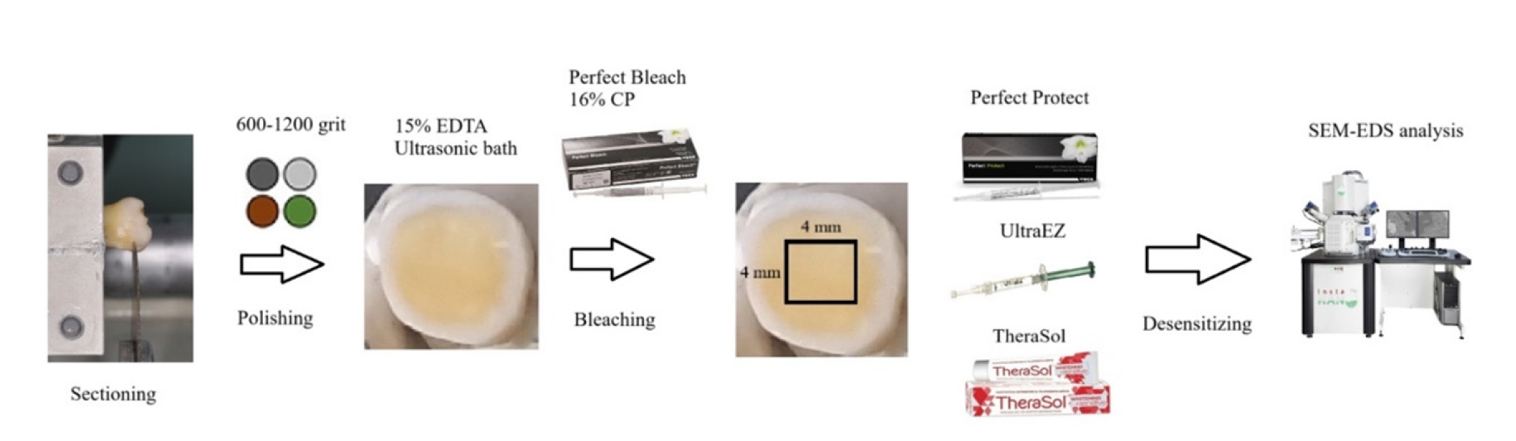
This study aimed to evaluate the efficacy of three commercially available desensitizing agents in occluding dentinal tubules, which may help reduce tooth sensitivity following a bleaching treatment.
Methods
Twenty healthy human third molars were utilized in this investigation. The samples were prepared by transversely sectioning 2.5 mm of the crowns to expose the dentin. They were initially treated with 15% ethylenediaminetetraacetic acid gel for 4 minutes, followed by application of Perfect Bleach (VOCO GmbH) bleaching agent (16% carbamide peroxide) for 2 hours. The samples were randomly allocated into four groups (n = 5), each receiving one of the following treatments: group 1: No treatment (control), group 2: treated with UltraEZ (Ultradent Products Inc.,), containing potassium nitrate and sodium fluoride, group 3: treated with Perfect Protect (VOCO GmbH), also containing potassium nitrate and sodium fluoride and group 4: treated with TheraSol Whitening & Sensitive (ABC Kinitron IKE), containing strontium acetate and sodium monofluorophosphate. Subsequently, the specimens were examined using scanning electron microscopy (SEM) and energy-dispersive X-ray spectroscopy to evaluate dentin tubule occlusion.
Results
SEM observations showed no occlusion of dentin tubules in the control group, whereas groups 2 to 4 exhibited significant occlusion. The most effective treatment was Perfect Protect (p < 0.05), while UltraEZ and TheraSol Whitening & Sensitive demonstrated similar effectiveness, with no statistically significant difference between them (p > 0.05).
Conclusions
The tested desensitizing agents effectively occluded dentin tubules to a considerable extent. Differences in their effectiveness were attributed to variations in their formulations.
- 1,572 View
- 181 Download
- Determination of optimal horizontal beam angulations for canal separation in mandibular molars using cone-beam computed tomography: a retrospective image-based analysis
- Benedikt Schneider, Tamina Tepe, Daniel Rapp, Wilhelm Frank, Maria Lessani, Constantin von See, Sebastian Fitzek, Jörg Philipp Tchorz
- Restor Dent Endod 2026;51(1):e9. Published online February 26, 2026
- DOI: https://doi.org/10.5395/rde.2026.51.e9
-
Abstract
PDFPubReaderePub
- Objectives
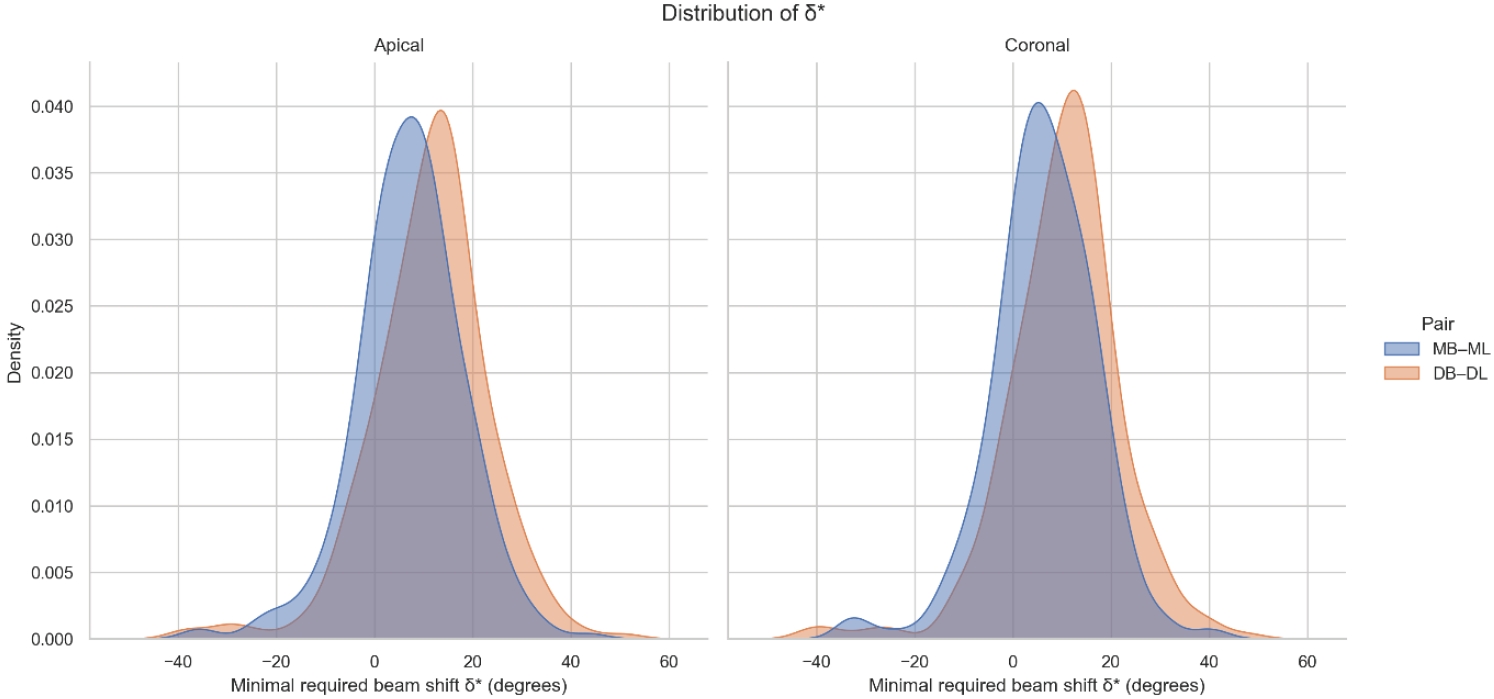
Two-dimensional intraoral radiographs often obscure canals due to superimposition, especially in mandibular molars with complex anatomy. This cone-beam computed tomography (CBCT) study identified the horizontal beam angles at which first and second molar canals overlap and derived clinically applicable angulations for enhanced canal separation.
Methods
Eighty-five CBCT datasets from 100 patients met the inclusion criteria, yielding 318 mandibular molars (160 first, 158 second). Using ImageJ, absolute horizontal overlap angles (α) were measured to determine the corresponding theoretical separation angles defined as δ* = 90° – α. Separability was modeled across horizontal beam angulation increments from −45° to +45° in five steps, and Wilson’s 95% confidence intervals were computed. Group comparisons used the Mann-Whitney U and independent t-tests (p ≤ 0.05)
Results
Minimal mesial beam angulations for effective canal separability (δ* = 90° − α) ranged from approximately 7° to 15° for mesial roots and approximately 10° to 13° for distal roots. No significant mesial differences were observed between first and second molars (p > 0.30). Distal roots of second molars exhibited significantly higher angulations (p = 0.003 coronal, p < 0.001 apical). Mesial canals achieved ≥95% separability at approximately 25° and ≥99% at approximately 35°; distal canals required approximately 30° and approximately 40°.
Conclusions
A mesial beam angulation of 30° to 35° provides probable canal differentiation in mandibular molars, separating mesial canals in ≥99% and distal canals in ≥95% of cases. This range refines previous recommendations and supports the as low as reasonably achievable (ALARA) principle.
- 1,338 View
- 34 Download
- Neuropeptide Y regulation of dental pulp neurogenic inflammation provoked by tooth bleaching agents: a descriptive comparative clinical study
- Javier Caviedes-Bucheli, Néstor Ríos-Osorio, Mario Pérez-Villota, Karolina Aucú-Miño, Diana Escobar-Mafla, Hernán Darío Muñoz-Alvear, José Francisco Gomez-Sosa, Luis Diaz-Barrera, Edgar Güiza – Cristancho, Hugo Roberto Munoz
- Restor Dent Endod 2026;51(1):e10. Published online February 13, 2026
- DOI: https://doi.org/10.5395/rde.2026.51.e10
-
Abstract
PDFPubReaderePub
- Objectives
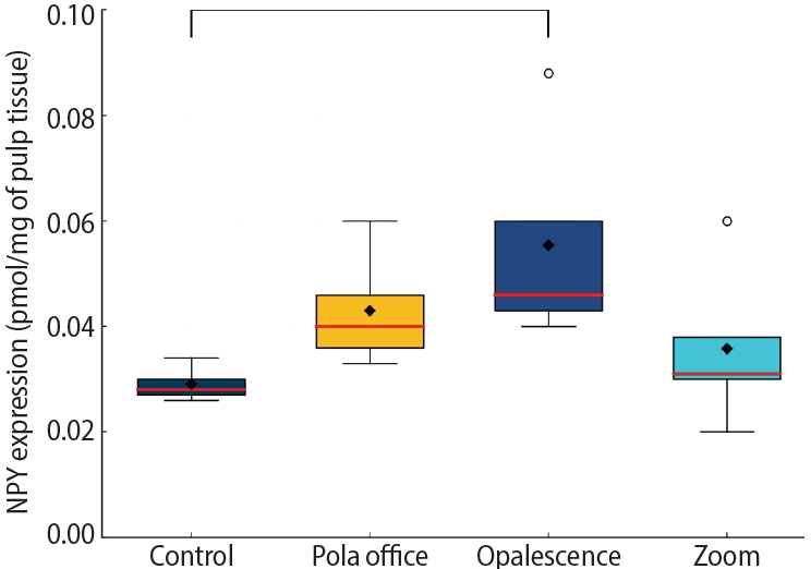
This study aimed to assess the expression of neuropeptide Y (NPY) in human dental pulp after tooth bleaching with three in-office hydrogen peroxide (H2O2)-based systems.
Methods
Forty pulps were collected from premolars scheduled for extraction and divided into four groups (n = 10): Control (no bleaching; basal NPY values); Pola Office (35% H2O2, 8 minutes); Opalescence Boost (40% H2O2, 20 minutes); and Zoom (25% H2O2 + cold blue light, 15 minutes). After extraction, pulps were fixed in 4% formaldehyde and processed. NPY levels were quantified using enzyme-linked immunosorbent assay. Data distribution was assessed with the Shapiro-Wilk test. One-way analysis of variance and Tukey post-hoc test with Bonferroni correction were applied (p < 0.05).
Results
NPY expression differed significantly among groups (p = 0.0097). The control group showed the lowest mean expression (0.026 ± 0.002 pmol/mg of pulp tissue), followed by Zoom (0.031 ± 0.005 pmol/mg), Pola Office (0.040 ± 0.004 pmol/mg), and Opalescence Boost, which exhibited the highest NPY expression (0.044 ± 0.004 pmol/mg). Post-hoc analysis revealed a statistically significant difference between the control and Opalescence Boost groups (p = 0.0122).
Conclusions
The increase in NPY expression—particularly with Opalescence Boost—indicates that in-office bleaching agents can elicit measurable neurobiological responses in pulp tissue after a single application. The significant difference between the control and Opalescence Boost groups suggests a possible H2O2 concentration- or formulation-dependent effect on pulpal neuropeptide activity, underscoring the need for further research on the biological impact of bleaching treatments.
- 1,528 View
- 87 Download
- Cone-beam computed tomography analysis of maxillary premolar canal anatomy: Ahmed’s versus Vertucci’s classifications in a Jordanian cohort
- Raidan Ba-Hattab, Muna M. Shaweesh, Nessrin A. Taha, Elham S. Abu Alhaija
- Restor Dent Endod 2026;51(1):e11. Published online February 26, 2026
- DOI: https://doi.org/10.5395/rde.2026.51.e11
-
Abstract
PDFPubReaderePub
- Objectives
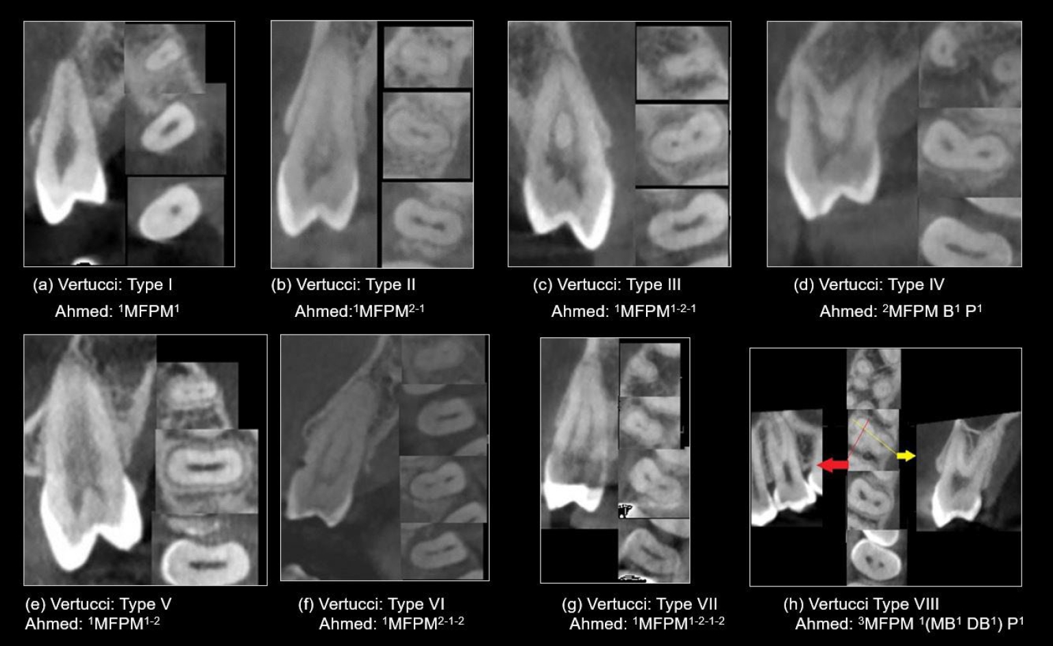
This study analyzed the root and canal configurations of maxillary premolars in a Jordanian subpopulation using cone-beam computed tomography (CBCT) and classified them based on Vertucci’s and Ahmed’s systems.
Methods
Two hundred CBCT scans of 800 maxillary premolars were retrospectively assessed for root morphology, canal configurations, and root canal divergence and merging. Data was statistically analyzed.
Results
The study included 70 males and 130 females. Most right and left maxillary first premolars (RFPM, LFPM) had two roots (59.0% and 58.5%), with a significant association between sex and root number for RFPM and LFPM (p < 0.05). In contrast, the right and left maxillary second premolars (RSPM, LSPM) mostly had a single root (87.5% and 88.5%), with no association with sex. Vertucci’s classification showed type IV as the predominant configuration in first premolars (RFPM, 65.0% and LFPM, 67.0%) and type I in second premolars (RSPM, 44.0% and LSPM, 49.0%). A significant sex association was found only with RSPM. Ahmed’s classification revealed that maxillary premolar with two separated roots and two separated canals (2MP B1 P1) was mostly found in first premolars (RFPM, 58.0% and LFPM, 56.0%), and maxillary premolar with one root and one canal (1MP1) in second premolars (RSPM, 44.0% and LSPM, 49.0%), with a significant sex association for RSPM and LSPM (p < 0.05). Age had no impact, and symmetry was observed between the right and left sides. Three-rooted premolars were identified in four cases. Almost all of Vertucci’s types and numerous codes from Ahmed’s classification were documented.
Conclusions
CBCT revealed diverse anatomical variations in the Jordanian subpopulation, with Ahmed’s classification providing more detailed canal configurations than Vertucci’s, uncovering previously overlooked variations.
- 1,014 View
- 55 Download
- Bonding and fractographic characterization of universal adhesives applied to dentin in multimode strategies: an in vitro study
- Samaa M. Morsy, Rim Bourgi, Louis Hardan, Carlos Enrique Cuevas-Suárez, Naji Kharouf, Ahmed A. Holiel
- Restor Dent Endod 2026;51(1):e12. Published online February 26, 2026
- DOI: https://doi.org/10.5395/rde.2026.51.e12
-
Abstract
PDFPubReaderePub
- Objectives
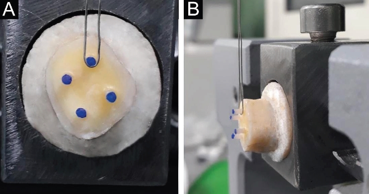
Universal adhesives (UAs) are marketed as versatile systems for both self-etch (SE) and total-etch (TE) modes. While their bond strength has been widely investigated, evidence linking fracture characteristics to bonding performance remains limited. This study evaluated the micro-shear bond strength (μSBS) and failure patterns of three UAs applied in SE and TE modes, complemented by fractographic scanning electron microscopy (SEM) analysis.
Methods
Eighteen extracted human molars were sectioned to expose mid-coronal dentin and randomly allocated to SE or TE application. Three UAs were tested: Tetric N-Bond Universal, All-Bond Universal, and Single Bond Universal (SBU). Composite micro-rods (n = 72) were bonded, thermocycled for 500 cycles between 5°C and 55°C, and subjected to μSBS testing. Fracture surfaces were examined under SEM and classified as adhesive, cohesive, or mixed. Data were analyzed using two-way analysis of variance, Tukey post hoc test, and Spearman correlation (α = 0.05).
Results
In TE mode, SBU demonstrated the highest μSBS (p < 0.001), whereas no significant differences were observed among adhesives in SE mode (p > 0.05). SEM analysis revealed adhesive failures as interfacial fractures, cohesive failures with beach marks, and mixed failures involving crack propagation through both dentin and composite. Adhesive failures correlated negatively with μSBS (rs = –0.77), while mixed failures correlated positively (rs = 0.81).
Conclusions
Both the etching strategy and adhesive formulation significantly affect bond strength and fracture behavior. Fractographic SEM analysis provides critical insights into the mechanical reliability of UAs and informs their clinical application.
- 1,259 View
- 106 Download
Case Report
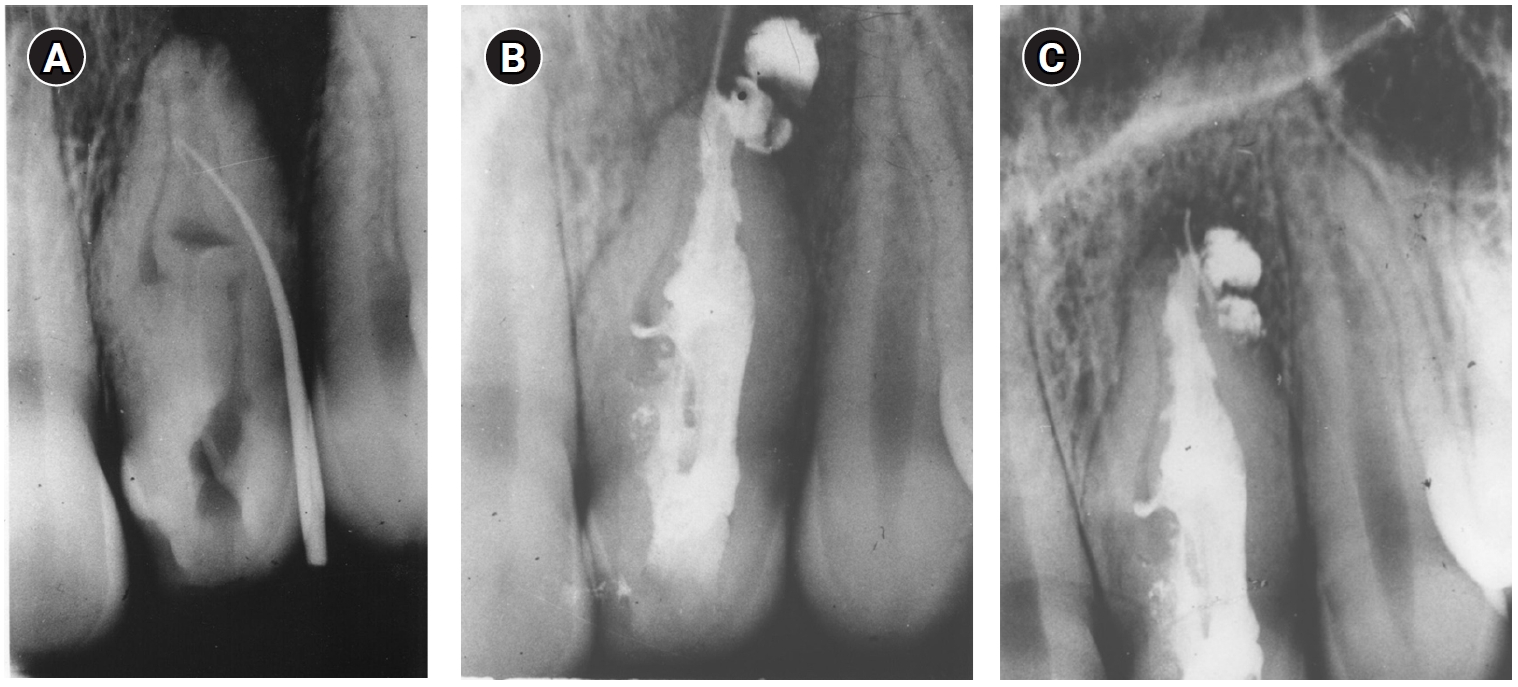
- Fifty-year follow-up of dens invaginatus treated by nonsurgical and surgical endodontic treatments: a case report
- Qais Arow, Eyal Rosen, Galit Sela, Shlomo Elbahary, Igor Tsesis
- Restor Dent Endod 2026;51(1):e1. Published online December 18, 2025
- DOI: https://doi.org/10.5395/rde.2026.51.e1
-
Abstract
PDFPubReaderePub
- This case report presents a lateral maxillary incisor with dens invaginatus (DI) type IIIb that was treated both nonsurgically and surgically over 50 years. Treatment of teeth with DI can be challenging. Suggested options may include nonsurgical root canal treatment, endodontic surgery, or extraction. In this case report, a 13-year-old patient with a lateral maxillary incisor with DI type IIIb was treated by nonsurgical root canal treatment, modern endodontic surgery, and reoperation over the course of 50 years. There was complete healing at the last follow-up, 11 years after the reoperation. Correct diagnosis and proper treatment using modern endodontic techniques can enable teeth with DI to survive throughout the life span of the patient.
- 1,900 View
- 158 Download
- 1 Web of Science
 First
First Prev
Prev

