
Previous issues
- Page Path
- HOME > Browse articles > Previous issues
- Volume 48 (1); February 2023
-


Editorial
- Appreciation to reviewers and announcement of a new editorial board
- Kyung-San Min
- Restor Dent Endod 2023;48(1):e11. Published online February 23, 2023
- DOI: https://doi.org/10.5395/rde.2023.48.e11
- 1,365 View
- 25 Download

Review Article
- Effect of endodontic sealer on postoperative pain: a network meta-analysis
- Cynthia Maria Chaves Monteiro, Ana Cristina Rodrigues Martins, Alessandra Reis, Juliana Larocca de Geus
- Restor Dent Endod 2023;48(1):e5. Published online December 29, 2022
- DOI: https://doi.org/10.5395/rde.2023.48.e5
-
 Abstract
 PDF
PDF PubReader
 ePub
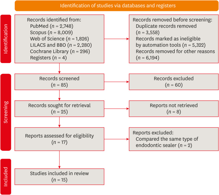
ePub This systematic review and network meta-analysis aimed to answer the following focused research question: “Does the type of endodontic sealer affect the postoperative pain in patients who received endodontic treatment?” Different databases and grey literature were surveyed. Only one randomized controlled trial were included. The risk of bias in the studies was evaluated by using the Cochrane Collaboration’s tool. A random-effects meta-analysis was conducted to compare the risk and intensity of postoperative pain. The quality of the body of evidence was assessed using the Grading of Recommendations Assessment, Development, and Evaluation approach. Out of 11,601 studies, 15 remained for qualitative analyses and 12 for meta-analysis. Seven studies were classified at high risk of bias, and 8 studies raised some concerns. No significant differences between the endodontic materials were observed in the direct comparisons, both in risk and in intensity of postoperative pain (pairwise comparisons with 2 studies: I2 = 0%;
p > 0.05 and 8 studies: I2 = 23%;p > 0.05, respectively). The certainty of the evidence was graded as low or moderate. There was no difference in the risk and intensity of postoperative pain after filling with different endodontic sealers. Further systematic reviews should be conducted.Trial Registration PROSPERO Identifier:
CRD42020215314 -
Citations
Citations to this article as recorded by
- Does the Use of a Bioceramic Sealer Reduce Postoperative Pain Compared With an Epoxy Resin‐Based Sealer After Primary Root Canal Treatment and Retreatment?—An Umbrella Review
Lokhasudhan Govindaraju, Rajeswari Kalaiselvam, Mathan Rajan Rajendran, Aleksandar Jakovljevic, Jelena Jacimovic, Henry F. Duncan, Venkateshbabu Nagendrababu
International Endodontic Journal.2026; 59(3): 341. CrossRef - Evidence synthesis of postoperative pain with bioceramic vs. epoxy resin sealers: umbrella review of randomized trials within existing systematic reviews
Mrunali Dahikar, Ashish Mandwe, Kulvinder Singh Banga, Alexander Maniangat Luke, Suraj Arora, Unmesh Khanvilkar, Ajinkya M. Pawar
Frontiers in Dental Medicine.2026;[Epub] CrossRef - Factors Influencing Apical Extrusion of 2 Types of Endodontic Sealers with Different Delivery Systems
Shumaila Iqbal, Nicholas S. Adams, Josette Camilleri
Journal of Endodontics.2026; 52(5): 806. CrossRef - Evaluation of Postoperative Pain Frequency in Single‐Session Endodontic Treatments With Patency and Foraminal Enlargement
Viviane Barbosa Godoy, Ana Grasiela Limoeiro, Vanessa Sandini, Vini Mehta, Wayne Martins Nascimento, Marilia Fagury Videira Marceliano‐Alves, Marcos Frozoni
Clinical and Experimental Dental Research.2026;[Epub] CrossRef - Silicone vs. Silicon/Silica in Intraoral Healing: A Systematic Review
David Parker, Aditi Bopardikar, Georgios E. Romanos
Materials.2026; 19(7): 1425. CrossRef - Post-Operative Pain After Endodontic Instrumentation, Irrigation and Obturation: An Umbrella Review of Systematic Reviews Published from 2016 to 2025
Fausto Zamparini, Andrea Spinelli, Gioia Quadrini, Maria Giovanna Gandolfi, Carlo Prati
Journal of Clinical Medicine.2026; 15(12): 4775. CrossRef - Comparative Evaluation of Postoperative Pain Following Nonsurgical Endodontic Therapy with Calcium Silicate-Based Sealer and Traditional Sealers: A Systematic Review and Meta-Analysis
Guha Poulomi, Solete Pradeep, Antony Delphine, Arun Nishitha, Surendar Ramamoorthi, Choudhari Sahil, Hima Sandeep Adimulapu
Pesquisa Brasileira em Odontopediatria e Clínica Integrada.2026;[Epub] CrossRef - Effect of occlusal reduction on post-operative pain of symptomatic and asymptomatic molar teeth
Aysenur Kamacı Esen, Fatma Furuncuoğlu, Fatima Betul Basturk, Muhammet Nuri Taşcıoğlu, Masoud Parirokh
Acta Odontologica Scandinavica.2025; 84: 371. CrossRef - An Observational Study on Pain Occurrence After Root Canal Treatment: Role of Operator Experience When Using a Bioceramic Sealer
Mihai Merfea, Ioana Sofia Pop-Ciutrila, Mindra Eugenia Badea, Ada Gabriela Delean, Oana Cimponeriu, Razvan Corneliu Pop, Maria Peter, Iulia Clara Badea, Sanda Ileana Cimpean
Journal of Clinical Medicine.2025; 14(13): 4558. CrossRef - Assessment of Postoperative Pain After Single‐ or Multiple‐Visit Endodontic Therapy and Its Molecular Aspects: A Randomised Controlled Study
Larissa Nunes Rosa Bedene, Denise Piotto Leonardi, Joana Santana Couto, Bruno Marques‐da‐Silva, Marilisa Carneiro Leão Gabardo, João Arnando Brancher, Flávia Sens Fagundes Tomazinho
Australian Endodontic Journal.2025; 51(3): 668. CrossRef - Clinical and Radiographic Outcomes of Root Canal Obturation with Hydraulic Condensation and Tricalcium Silicate Bioceramic Sealer: A 12-Month Observational Study on Periapical Healing
Kostadin Zhekov, Vesela Stefanova
Journal of Functional Biomaterials.2025; 16(11): 412. CrossRef - Comparative evaluation of postoperative pain and periapical healing after root canal treatment using three different endodontic sealers: A randomized controlled clinical trial
Ruchika Pandey, Nitin Kararia, Deepak Kumar Sharma, Vishal Rathod, Anand Vilas Bansod, Dhaval Desai
Journal of Conservative Dentistry and Endodontics.2024; 27(9): 962. CrossRef - Effect of bioceramic-based and resin-based sealers on postoperative discomfort following root canal therapy: a systematic review and meta-analysis
Mansi Supare, Ajinkya M. Pawar, Kashmira Sawant, Dian Agustin Wahjuningrum, Suraj Arora, Firas Elmsmari, Mohmed Isaqali Karobari, Bhagyashree Thakur
PeerJ.2024; 12: e18198. CrossRef - Comparative Evaluation of Incidences of Post Operative Pain in Patient Treated in Single Visit Root Canal Treatment by Using Different Sealers: - An in-Vivo Study
Sadashiv Daokar, Aishwarya Ranjalkar, Kalpana Pawar, Komal Potfode, Dhanashri Padwal, Sana Khan
International Journal of Innovative Science and Research Technology (IJISRT).2024; : 2743. CrossRef
- Does the Use of a Bioceramic Sealer Reduce Postoperative Pain Compared With an Epoxy Resin‐Based Sealer After Primary Root Canal Treatment and Retreatment?—An Umbrella Review
- 8,442 View
- 164 Download
- 12 Web of Science
- 14 Crossref
Research Articles
- Dentinal tubule penetration of sodium hypochlorite in root canals with and without mechanical preparation and different irrigant activation methods
- Renata Aqel de Oliveira, Theodoro Weissheimer, Gabriel Barcelos Só, Ricardo Abreu da Rosa, Matheus Albino Souza, Rodrigo Gonçalves Ribeiro, Marcus Vinicius Reis Só
- Restor Dent Endod 2023;48(1):e1. Published online December 1, 2022
- DOI: https://doi.org/10.5395/rde.2023.48.e1
-
Abstract
PDFPubReaderePub
Objectives This study evaluated the dentinal penetration depth of 2.5% sodium hypochlorite (NaOCl) in root canals with and without preparation and different irrigant activation protocols.
Materials and Methods Sixty-three bovine mandibular incisors were randomly allocated to 6 groups (
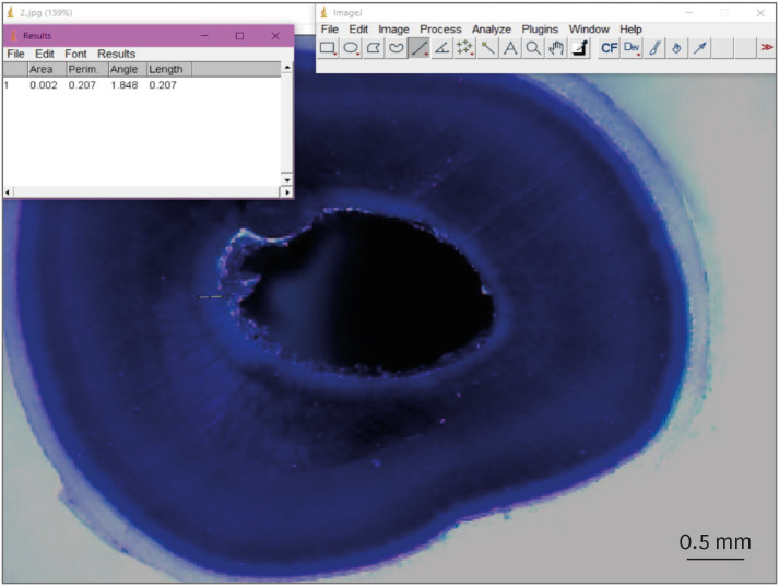
n = 10): G1, preparation + conventional needle irrigation (CNI); G2, preparation + passive ultrasonic irrigation (PUI); G3, preparation + Odous Clean (OC); G4, no preparation + CNI; G5, no preparation + PUI; G6, no preparation + OC; and CG (negative control;n = 3). Samples were filled with crystal violet for 72 hours. Irrigant activation was performed. Samples were sectioned perpendicularly along the long axis, 3 mm and 7 mm from the apex. Images of the root thirds of each block were captured with a stereomicroscope and analyzed with an image analysis software. One-way analysis of variance, followed by the Tukeypost hoc test, and the Student’st -test were used for data analysis, with a significance level of 5%.Results The NaOCl penetration depth was similar when preparation was performed, regardless of the method of irrigation activation (
p > 0.05). In the groups without preparation, G6 showed greater NaOCl penetration depth (p < 0.05). The groups without preparation had a greater NaOCl penetration depth than those with preparation (p = 0.0019).Conclusions The NaOCl penetration depth was similar in groups with root canal preparation. Without root canal preparation, OC allowed deeper NaOCl penetration. The groups without preparation had greater NaOCl penetration than those undergoing root canal preparation.
-
Citations
Citations to this article as recorded by- Novel approaches involving curcumin in endodontic and periodontal diseases: a scoping review
Yuxi Xing, Yanbing Zhu, Yukai Shen, Yuou Xu, Ziman Xu, Mengxue Wang, Xudong Ma, Lehua Liu, Shu Chen
BMC Oral Health.2026;[Epub] CrossRef - Influence of passive ultrasonic irrigation cycles on the penetration depth of sodium hypochlorite into root dentin
Hüseyin Gündüz, Esin Özlek, Züleyha Baş
Scientific Reports.2025;[Epub] CrossRef - Evaluating the Effects of Various Antioxidants on Dentinal Tubule Penetrability of a Resin-Based Sealer: A Confocal Laser Microscopic Study
Sanjeev Srivastava, Shijita Sinha, Abhishek Singh, Aditya Singh, Pragyan Paliwal, Syed H Mehdii
Cureus.2025;[Epub] CrossRef - Impact of different activation procedures on sodium hypochlorite penetration into dentinal tubules after endodontic retreatment via confocal laser scanning microscopy
Betul Gunes, Kübra Yeşildal Yeter, Yasin Altay
BMC Oral Health.2024;[Epub] CrossRef - Debridement ability of the WaveOne Gold and TruNatomy systems in the apical third of root canals: ex vivo assessment
Sara Carvalho Avelar de Oliveira, Carlos Eduardo da Silveira Bueno, Rina Andréa Pelegrine, Carlos Eduardo Fontana, Alexandre Sigrist de Martin, Carolina Pessoa Stringheta
Brazilian Dental Journal.2024;[Epub] CrossRef - Combined effect of electrical energy and graphene oxide on Enterococcus faecalis biofilms
Myung-Jin LEE, Mi-Ah KIM, Kyung-San MIN
Dental Materials Journal.2023; 42(6): 844. CrossRef
- Novel approaches involving curcumin in endodontic and periodontal diseases: a scoping review
- 3,670 View
- 86 Download
- 5 Web of Science
- 6 Crossref
- Antimicrobial and cytotoxic properties of calcium-enriched mixture cement, Iranian propolis, and propolis with herbal extracts in primary dental pulp stem cells
- Mohammad Esmaeilzadeh, Shirin Moradkhani, Fahimeh Daneshyar, Mohammad Reza Arabestani, Sara Soleimani Asl, Soudeh Tayebi, Maryam Farhadian
- Restor Dent Endod 2023;48(1):e2. Published online December 1, 2022
- DOI: https://doi.org/10.5395/rde.2023.48.e2
-
Abstract
PDFPubReaderePub
Objectives In this study, natural substances were introduced as primary dental pulp caps for use in pulp therapy, and the antimicrobial and cytotoxic properties of these substances were investigated.
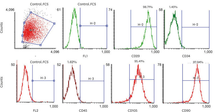
Materials and Methods In this
in vitro study, the antimicrobial properties of calcium-enriched mixture (CEM) cement, propolis, and propolis individually combined with the extracts of several medicinal plants were investigated againstEnterococcus faecalis ,Escherichia coli ,Pseudomonas aeruginosa , andStaphylococcus aureus . Then, the cytotoxicity of each substance or mixture against pulp stem cells extracted from 30 primary healthy teeth was evaluated at 4 concentrations. Data were gathered via observation, and optical density values were obtained using the 3-(4,5-dimethylthiazol-2-yl)-2,5-diphenyl-2H-tetrazolium bromide (MTT) test and recorded. SPSS software version 23 was used to analyze the data. Data were evaluated using 2-way analysis of variance and the Tukey test.Results Regarding antimicrobial properties, thyme alone and thyme + propolis had the lowest minimum inhibitory concentrations (MICs) against the growth of
S. aureus ,E. coli , andP. aeruginosa bacteria. ForE. faecalis , thyme + propolis had the lowest MIC, followed by thyme alone. At 24 and 72 hours, thyme + propolis, CEM cement, and propolis had the greatest bioviability in the primary dental pulp stem cells, and lavender + propolis had the lowest bioviability.Conclusions Of the studied materials, thyme + propolis showed the best results in the measures of practical performance as a dental pulp cap.
-
Citations
Citations to this article as recorded by- Self-Adapting Mouthguard with Nano-Hydroxyapatite and Propolis for Early Childhood Caries: Preclinical Safety and Efficacy
Mata Soslanbekovna Mustapaeva, Khadizhat Adamovna Sataeva, Elina Ilyasovna Zhabrailova, Alina Mairbekovna Sidakova, Karina Maharbekovna Mukagova, Umar Said-Asanovich Magomadov, Muslim Usmanovich Dunaev, Mehdi Usmanovich Dunaev, Khava Khuseynovna Amaeva, V
Asian Journal of Periodontics and Orthodontics.2026; 6(1): 32. CrossRef - Comprehensive review of composition, properties, clinical applications, and future perspectives of calcium-enriched mixture (CEM) cement: a systematic analysis
Saeed Asgary, Mahtab Aram, Mahta Fazlyab
BioMedical Engineering OnLine.2024;[Epub] CrossRef - Effects of aqueous and ethanolic extracts of Chinese propolis on dental pulp stem cell viability, migration and cytokine expression
Ha Bin Park, Yen Dinh, Pilar Yesares Rubi, Jennifer L. Gibbs, Benoit Michot
PeerJ.2024; 12: e18742. CrossRef
- Self-Adapting Mouthguard with Nano-Hydroxyapatite and Propolis for Early Childhood Caries: Preclinical Safety and Efficacy
- 3,487 View
- 59 Download
- 2 Web of Science
- 3 Crossref
- Physicochemical properties of a calcium aluminate cement containing nanoparticles of zinc oxide
- Amanda Freitas da Rosa, Thuany Schmitz Amaral, Maria Eduarda Paz Dotto, Taynara Santos Goulart, Hebert Luís Rossetto, Eduardo Antunes Bortoluzzi, Cleonice da Silveira Teixeira, Lucas da Fonseca Roberti Garcia
- Restor Dent Endod 2023;48(1):e3. Published online December 8, 2022
- DOI: https://doi.org/10.5395/rde.2023.48.e3
-
Abstract
PDFPubReaderePub
Objectives This study evaluated the effect of different nanoparticulated zinc oxide (nano-ZnO) and conventional-ZnO ratios on the physicochemical properties of calcium aluminate cement (CAC).
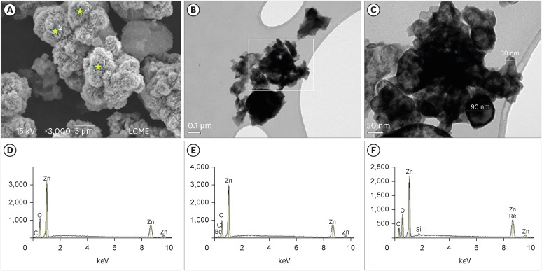
Materials and Methods The conventional-ZnO and nano-ZnO were added to the cement powder in the following proportions: G1 (20% conventional-ZnO), G2 (15% conventional-ZnO + 5% nano-ZnO), G3 (12% conventional-ZnO + 3% nano-ZnO) and G4 (10% conventional-ZnO + 5% nano-ZnO). The radiopacity (Rad), setting time (Set), dimensional change (Dc), solubility (Sol), compressive strength (Cst), and pH were evaluated. The nano-ZnO and CAC containing conventional-ZnO were also assessed using scanning electron microscopy, transmission electron microscopy, and energy-dispersive X-ray spectroscopy. Radiopacity data were analyzed by the 1-way analysis of variance (ANOVA) and Bonferroni tests (
p < 0.05). The data of the other properties were analyzed by the ANOVA, Tukey, and Fisher tests (p < 0.05).Results The nano-ZnO and CAC containing conventional-ZnO powders presented particles with few impurities and nanometric and micrometric sizes, respectively. G1 had the highest Rad mean value (
p < 0.05). When compared to G1, groups containing nano-ZnO had a significant reduction in the Set (p < 0.05) and lower values of Dc at 24 hours (p < 0.05). The Cst was higher for G4, with a significant difference for the other groups (p < 0.05). The Sol did not present significant differences among groups (p > 0.05).Conclusions The addition of nano-ZnO to CAC improved its dimensional change, setting time, and compressive strength, which may be promising for the clinical performance of this cement.
-
Citations
Citations to this article as recorded by- Histopathologic responses of the dental pulp to an experimental calcium aluminate–calcium silicate based capping material in comparison to mineral trioxide aggregate
Reham S. Saleh, Eyad A. Elbattawy, Shymaa Hamza, Yousra Aly
Scientific Reports.2026;[Epub] CrossRef - Calcium aluminate cement: a study on the effect of additives for dental applications
Sara Ghorbani, Rahim Naghizadeh, Ebrahim Ghasemi, Hamidreza Rezaie
Advances in Cement Research.2025; 37(4): 269. CrossRef - Experimental Study on Cement-Based Materials Modified by Nano-Zinc Oxide and Nano-Zirconia Based on Response Surface Optimization Design
Hongyin Hu, Fufei Wu, Jiao Chen, Shuangshuang Guan, Peng Qu, Hongqin Zhang, Yuyi Chen, Zirun Xu, Chuanteng Huang, Shuang Pu
Materials.2025; 18(7): 1515. CrossRef - Radiographic, mechanical, and chemical properties of mineral trioxide aggregate from nanosilica and clam shell calcium carbonate
Leny Yuliatun, Muhammad Adly Rahandi Lubis, Muhammad Khaliim Jati Kusala, Lia Destiarti, Ratna Betriani, Jolang Budiarta, Mariyam Mariyam
Polyhedron.2025; 278: 117590. CrossRef - Application of Calcium Aluminate-Based Materials for Direct Pulp Capping – In Vivo Study
Ognjenka Janković, Smiljana Paraš, Tijana Adamović, Ljiljana Tadić Latinović, Radmila Arbutina, Igor Đukić, Saša Marin, Marko Bulajić, Karolina Vukoje, Vukoman Jokanović, Verica Pavlić
Acta Veterinaria.2025; 75(2): 212. CrossRef - Nanotechnology for calcium aluminate cement: thematic analysis
Lapyote Prasittisopin
REVIEWS ON ADVANCED MATERIALS SCIENCE.2025;[Epub] CrossRef
- Histopathologic responses of the dental pulp to an experimental calcium aluminate–calcium silicate based capping material in comparison to mineral trioxide aggregate
- 2,969 View
- 55 Download
- 7 Web of Science
- 6 Crossref
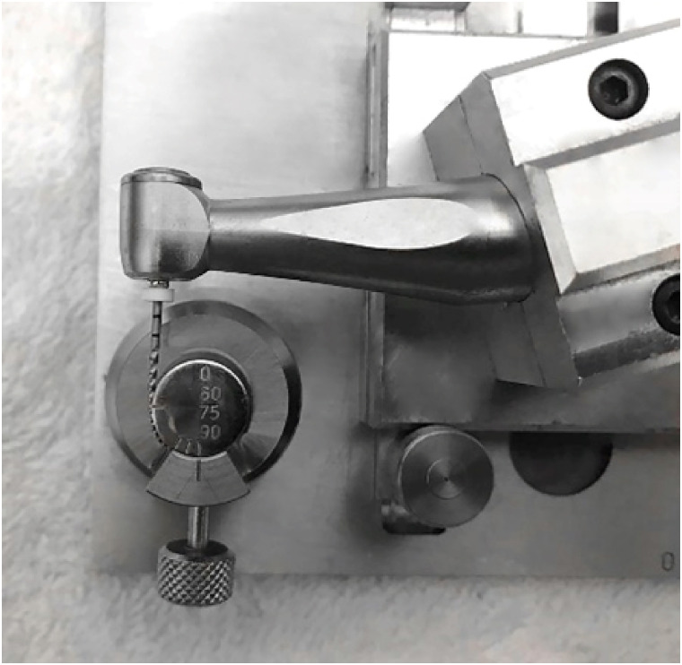
- Comparative analysis of torsional and cyclic fatigue resistance of ProGlider, WaveOne Gold Glider, and TruNatomy Glider in simulated curved canal
- Pedro de Souza Dias, Augusto Shoji Kato, Carlos Eduardo da Silveira Bueno, Rodrigo Ricci Vivan, Marco Antonio Hungaro Duarte, Pedro Henrique Souza Calefi, Rina Andréa Pelegrine
- Restor Dent Endod 2023;48(1):e4. Published online December 8, 2022
- DOI: https://doi.org/10.5395/rde.2023.48.e4
-
Abstract
PDFPubReaderePub
Objectives This study aimed to compare the torsional and cyclic fatigue resistance of ProGlider (PG), WaveOne Gold Glider (WGG), and TruNatomy Glider (TNG).
Materials and Methods A total of 15 instruments of each glide path system (
n = 15) were used for each test. A custom-made device simulating an angle of 90° and a radius of 5 millimeters was used to assess cyclic fatigue resistance, with calculation of number of cycles to failure. Torsional fatigue resistance was assessed by maximum torque and angle of rotation. Fractured instruments were examined by scanning electron microscopy (SEM). Data were analyzed with Shapiro-Wilk and Kruskal-Wallis tests, and the significance level was set at 5%.Results The WGG group showed greater cyclic fatigue resistance than the PG and TNG groups (
p < 0.05). In the torsional fatigue test, the TNG group showed a higher angle of rotation, followed by the PG and WGG groups (p < 0.05). The TNG group was superior to the PG group in torsional resistance (p < 0.05). SEM analysis revealed ductile morphology, typical of the 2 fracture modes: cyclic fatigue and torsional fatigue.Conclusions Reciprocating WGG instruments showed greater cyclic fatigue resistance, while TNG instruments were better in torsional fatigue resistance. The significance of these findings lies in the identification of the instruments’ clinical applicability to guide the choice of the most appropriate instrument and enable the clinician to provide a more predictable glide path preparation.
-
Citations
Citations to this article as recorded by- Mechanical Behaviour of Three Heat‐Treated NiTi Glide Path Systems at Simulated Intracanal Temperature
Ahmad Jamleh, Taher Omari, Abdulmohsen Alfadley, Abdullah Alqedairi, Hussam Alfawaz
Australian Endodontic Journal.2026;[Epub] CrossRef - Buckling resistance of various pathfinding endodontic instruments: An in vitro study
Ujjwal Das, Rajesh Kumar Das, Kallol Kumar Saha, Lugu Buru Murmu, Srimanta Banerjee, Rishila Nag
Journal of Conservative Dentistry and Endodontics.2025; 28(4): 384. CrossRef - Comparative evaluation of the remaining dentin volume following instrumentation with rotary, reciprocating, and hand files during root canal treatment in primary molars: An ex vivo study
İrem Eren, Berkant Sezer
Journal of Dental Sciences.2024; 19(4): 2126. CrossRef - Screw-in force, torque generation, and performance of glide-path files with three rotation kinetics
Jee-Yeon Woo, Ji-Hyun Jang, Seok Woo Chang, Soram Oh
Odontology.2024; 112(3): 761. CrossRef - Evaluation of shaping ability of different glide path instruments: a micro-computed tomography study
Merve Yeniçeri Özata, Seda Falakaloğlu, Ali Keleş, Özkan Adıgüzel, Mustafa Gündoğar
BMC Oral Health.2023;[Epub] CrossRef
- Mechanical Behaviour of Three Heat‐Treated NiTi Glide Path Systems at Simulated Intracanal Temperature
- 3,982 View
- 82 Download
- 4 Web of Science
- 5 Crossref
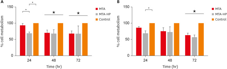
- High-plasticity mineral trioxide aggregate and its effects on M1 and M2 macrophage viability and adherence, phagocyte activity, production of reactive oxygen species, and cytokines
- Betânia Canal Vasconcellos, Layara Cristine Tomaz Tavares, Danilo Couto da Silva, Francielen Oliveira Fonseca, Francine Benetti, Antônio Paulino Ribeiro Sobrinho, Warley Luciano Fonseca Tavares
- Restor Dent Endod 2023;48(1):e6. Published online December 29, 2022
- DOI: https://doi.org/10.5395/rde.2023.48.e6
-
Abstract
PDFPubReaderePub
Objectives This study evaluated the effects of high-plasticity mineral trioxide aggregate (MTA-HP) on the activity of M1 and M2 macrophages, compared to white MTA (Angelus).
Materials and Methods Peritoneal inflammatory M1 (from C57BL/6 mice) and M2 (from BALB/c mice) macrophages were cultured in the presence of the tested materials. Cell viability (MTT and trypan blue assays), adhesion, phagocytosis, reactive oxygen species (ROS) production, and tumor necrosis factor (TNF)-α and transforming growth factor (TGF)-β production were evaluated. Parametric analysis of variance and the non-parametric Kruskal-Wallis test were used. Results were considered significant when
p < 0.05.Results The MTT assay revealed a significant decrease in M1 metabolism with MTA-HP at 24 hours, and with MTA and MTA-HP later. The trypan blue assay showed significantly fewer live M1 at 48 hours and live M2 at 48 and 72 hours with MTA-HP, compared to MTA. M1 and M2 adherence and phagocytosis showed no significant differences compared to control for both materials. Zymosan A stimulated ROS production by macrophages. In the absence of interferon-γ, TNF-α production by M1 did not significantly differ between groups. For M2, both materials showed higher TNF-α production in the presence of the stimulus, but without significant between-group differences. Likewise, TGF-β production by M1 and M2 macrophages was not significantly different between the groups.
Conclusions M1 and M2 macrophages presented different viability in response to MTA and MTA-HP at different time points. Introducing a plasticizer into the MTA vehicle did not interfere with the activity of M1 and M2 macrophages.
-
Citations
Citations to this article as recorded by- Local Immune Response to Mineral Trioxide Aggregate: A Narrative Review
Shankargouda Patil, Shilpa Bhandi, Oladapo T Okareh
World Journal of Dentistry.2023; 14(4): 382. CrossRef
- Local Immune Response to Mineral Trioxide Aggregate: A Narrative Review
- 2,968 View
- 40 Download
- 1 Crossref
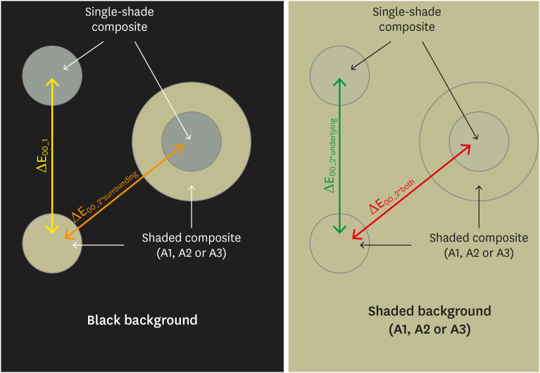
- Effects of surrounding and underlying shades on the color adjustment potential of a single-shade composite used in a thin layer
- Mariana Silva Barros, Paula Fernanda Damasceno Silva, Márcia Luciana Carregosa Santana, Rafaella Mariana Fontes Bragança, André Luis Faria-e-Silva
- Restor Dent Endod 2023;48(1):e7. Published online December 29, 2022
- DOI: https://doi.org/10.5395/rde.2023.48.e7
-
Abstract
PDFPubReaderePub
Objectives This study aimed to evaluate the surrounding and underlying shades’ effect on the color adjustment potential (CAP) of a single-shade composite used in a thin layer.
Materials and Methods Cylinder specimens (1.0 mm thick) were built with the Vittra APS Unique composite, surrounded (dual specimens) or not (simple specimens) by a control composite (shade A1, A2, or A3). Simple specimens were also built only with the control composites. Each specimen’s color was measured against white and black backgrounds or the simple control specimens with a spectrophotometer (CIELAB system). The whiteness index for dentistry (WID) and translucency parameters (TP00) were calculated for simple specimens. Differences (ΔE00) in color between the simple/dual specimens and the controls were calculated. The CAP was calculated based on the ratios between data from simple and dual specimens.
Results The Vittra APS Unique composite showed higher WID and TP00 values than the controls. The highest values of ΔE00 were observed among simple specimens. The color measurements of Vittra APS Unique (simple or dual) against the control specimens presented the lowest color differences. Only surrounding the single-shade composite with a shaded composite barely impacted the ΔE00. The highest CAP values were obtained using a shaded composite under simple or dual specimens.
Conclusions The CAP of Vittra APS Unique was strongly affected by the underlying shade, while surrounding this composite with a shaded one barely affected its color adjustment.
-
Citations
Citations to this article as recorded by- Impact of water absorption on the translucency of single-shade and conventional resin composites: an in vitro comparative study
Ceyda Sari, Elifnur Aydemir Aydın
Odontology.2026;[Epub] CrossRef - Baseline color-matching in anterior non-carious cervical lesions of patients of two single-shade resin composites: a randomized clinical trial
Ayşe Nur Doğan, Soley Arslan
Odontology.2026;[Epub] CrossRef - Evaluation of color blending effect of a single-shade resin composite with hybrid ceramics: an in vitro study
Jongchan Lee, Jinsoo Ahn, Sun-Young Kim
BMC Oral Health.2026;[Epub] CrossRef - Color Change and Compressive Strength of Novel Single-Shade Composite Resins Exposed to Staining Beverages
Juan P. Molina-Gantiva, Midian C. Castillo-Pedraza, Jorge H. Wilches-Visbal
Odovtos - International Journal of Dental Sciences.2026;[Epub] CrossRef - At‐Home and In‐Office Bleaching Protocols on the Color Match of Restorations Made With Single‐Shade Composites
Luciana Vasconcelos Ramos, Dayana Fernandes Rocha Aparicio, André Luis Faria‐e‐Silva, Maíra do Prado, Andréa Vaz Braga Pintor, Marcela Baraúna Magno
Journal of Esthetic and Restorative Dentistry.2025; 37(6): 1567. CrossRef - Evaluation of color matching of three single-shade composites employing simulated 3D printed cavities with different thicknesses using CIELAB and CIEDE2000 color difference formulae
Engin Kariper, Aylin Cilingir
REVIEWS ON ADVANCED MATERIALS SCIENCE.2025;[Epub] CrossRef - Impact of kombucha, coffee, and turmeric beverages on the color stability of a single-shade versus a multi-shade resin-based composite
Hanin E. Yeslam, Abdulaziz F. Bakhsh
PeerJ.2025; 13: e19759. CrossRef - Comparative Study of Esthetic Outcome of Pedo Shades of Composite Resin—A Randomized Controlled Trial: In Vivo and In Vitro Study
Priyanka Raj, Shikha Choubey, Divya Doneria, Diksha Bhat, Shivani Mathur, Shailja Sinha
International Journal of Clinical Pediatric Dentistry.2025; 18(S1): S22. CrossRef - Influence of cavity wall thickness on the color adjustment potential of single-shade resin composites
Fabrício Luscino Alves de Castro, Letícia Brandão Durand
The Journal of the American Dental Association.2024; 155(7): 605. CrossRef - Assessing color mismatch in single-shade composite resins for enamel replacement
Rafaella Mariana Fontes de Bragança, Diana Leyva Del Rio, Luiz Alves Oliveira-Neto, William Michael Johnston
The Journal of Prosthetic Dentistry.2024; 132(3): 613.e1. CrossRef - Color discrepancy of single-shade composites at different distances from the interface measured using cell phone images
Márcia Luciana Carregosa Santana, Gabriella de Jesus Santos Livi, André Luis Faria-e-Silva
Restorative Dentistry & Endodontics.2024;[Epub] CrossRef - Is It Possible for Single-shade Composites to Mimic the Color, Lightness, Chroma, and Hue of Other Single-shade Composites? An In Vitro Study
M Buldur, G Ayan
Operative Dentistry.2024; 49(6): 691. CrossRef - Color evaluation of a one-shade used for restoration of non-carious cervical lesions: an equivalence randomized clinical trial
Michael Willian Favoreto, Amanda de Oliveira de Miranda, Thalita P. Matos, Andrea dos Santos de Castro, Mylena de Abreu Cardoso, Julia Beatriz, Jenny Collantes-Acuña, Alessandra Reis, Alessandro Dourado Loguercio
BMC Oral Health.2024;[Epub] CrossRef - Influence of Thickness on the Translucency Parameter and Whiteness Index of Single-Shade Resin Composites
Ö Yağcı, M Fidan
Operative Dentistry.2024; 49(2): 189. CrossRef - A Comparative Study of the Sensitivity and Specificity of the Ishihara Test With Various Displays
Thomas Klinke, Wolfgang Hannak, Klaus Böning, Holger Jakstat
International Dental Journal.2024; 74(4): 892. CrossRef - Color match evaluation using instrumental method for three single-shade resin composites before and after in-office bleaching
Aylin Cilingir, Engin Kariper
REVIEWS ON ADVANCED MATERIALS SCIENCE.2023;[Epub] CrossRef - The role of interface distance and underlying substrate on the color adjustment potential of single‐shade composites
Gabriella Jesus Santos de Livi, Tauan Rosa Santana, Rafaella Mariana Fontes Bragança, Rosa Maria Viana de Bragança Garcez, André Luis Faria‐e‐Silva
Journal of Esthetic and Restorative Dentistry.2023; 35(8): 1279. CrossRef
- Impact of water absorption on the translucency of single-shade and conventional resin composites: an in vitro comparative study
- 5,960 View
- 134 Download
- 17 Web of Science
- 17 Crossref
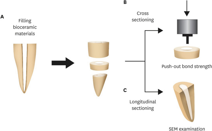
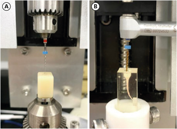
- Push-out bond strength and intratubular biomineralization of a hydraulic root-end filling material premixed with dimethyl sulfoxide as a vehicle
- Ju-Ha Park, Hee-Jin Kim, Kwang-Won Lee, Mi-Kyung Yu, Kyung-San Min
- Restor Dent Endod 2023;48(1):e8. Published online January 20, 2023
- DOI: https://doi.org/10.5395/rde.2023.48.e8
-
Abstract
PDFPubReaderePub
Objectives This study was designed to evaluate the parameters of bonding performance to root dentin, including push-out bond strength and dentinal tubular biomineralization, of a hydraulic bioceramic root-end filling material premixed with dimethyl sulfoxide (Endocem MTA Premixed) in comparison to a conventional powder-liquid–type cement (ProRoot MTA).
Materials and Methods The root canal of a single-rooted premolar was filled with either ProRoot MTA or Endocem MTA Premixed (
n = 15). A slice of dentin was obtained from each root. Using the sliced specimen, the push-out bond strength was measured, and the failure pattern was observed under a stereomicroscope. The apical segment was divided into halves; the split surface was observed under a scanning electron microscope, and intratubular biomineralization was examined by observing the precipitates formed in the dentinal tubule. Then, the chemical characteristics of the precipitates were evaluated with energy-dispersive X-ray spectroscopic (EDS) analysis. The data were analyzed using the Student’st -test followed by the Mann-WhitneyU test (p < 0.05).Results No significant difference was found between the 2 tested groups in push-out bond strength, and cohesive failure was the predominant failure type. In both groups, flake-shaped precipitates were observed along dentinal tubules. The EDS analysis indicated that the mass percentage of calcium and phosphorus in the precipitate was similar to that found in hydroxyapatite.
Conclusions Regarding bonding to root dentin, Endocem MTA Premixed may have potential for use as an acceptable root-end filling material.
-
Citations
Citations to this article as recorded by- Comparison of intratubular biomineralization between in vivo and in vitro conditions
Sieun Nam, Yeon-Jee Yoo, Mi-Kyung Yu, Kyung-San Min
Journal of Oral Science.2026; 68(1): 30. CrossRef - Effectiveness of Sectioning Method and Filling Materials on Roughness and Cell Attachments in Root Resection Procedure
Tarek Ashi, Naji Kharouf, Olivier Etienne, Bérangère Cournault, Pierre Klienkoff, Varvara Gribova, Youssef Haikel
European Journal of Dentistry.2025; 19(01): 240. CrossRef - Bond Strength and Adhesive Interface Quality of New Pre‐Mixed Bioceramic Root Canal Sealer
Gustavo Creazzo, Bruna Monteiro de Barros Ciribelli Alves, Helena Cristina de Assis, Karen Gisselle Garay Villamayor, Manoel Damião de Sousa‐Neto, Jardel Francisco Mazzi‐Chaves, Fabiane Carneiro Lopes‐Olhê
Microscopy Research and Technique.2025; 88(7): 1989. CrossRef - Evaluation of clinical and radiographic outcome of premixed injectable mineral trioxide aggregate and conventional mineral trioxide aggregate as pulpotomy medicaments in primary molars – A split-mouth randomized control trial
U. S. Aiswarya, Sharan S. Sargod, Sundeep K. Hegde, H. T. Ajay Rao, Nanditha Hegde
Journal of Indian Society of Pedodontics and Preventive Dentistry.2025; 43(4): 559. CrossRef - Evaluation of the root dentin bond strength and intratubular biomineralization of a premixed calcium aluminate-based hydraulic bioceramic endodontic sealer
Yu-Na Lee, Min-Kyeong Kim, Hee-Jin Kim, Mi-Kyung Yu, Kwang-Won Lee, Kyung-San Min
Journal of Oral Science.2024; 66(2): 96. CrossRef - Removal efficiency of a fast setting pozzalan-based bioactive cement: a micro CT study
Feyza Çetinkaya, Ahter Şanal Çıkman, Ali Keleş, Banu Arıcıoğlu
BMC Oral Health.2024;[Epub] CrossRef - Antibacterial Activity and Sustained Effectiveness of Calcium Silicate-Based Cement as a Root-End Filling Material against Enterococcus faecalis
Seong-Hee Moon, Seong-Jin Shin, Seunghan Oh, Ji-Myung Bae
Materials.2023; 16(18): 6124. CrossRef
- Comparison of intratubular biomineralization between in vivo and in vitro conditions
- 4,174 View
- 112 Download
- 7 Web of Science
- 7 Crossref
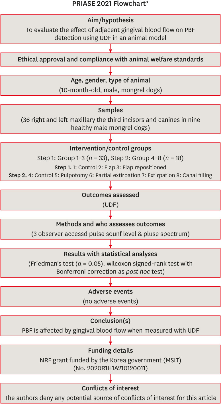
- The effects of gingival blood flow on pulpal blood flow detection using ultrasound Doppler flowmetry: animal study
- Dohyun Kim, Hyoung-Seok Ko, Soo-Yeon Park, Seung-Yeon Ryu, Sung-ho Park
- Restor Dent Endod 2023;48(1):e9. Published online January 30, 2023
- DOI: https://doi.org/10.5395/rde.2023.48.e9
-
Abstract
PDFPubReaderePub
Objectives This study evaluated the effect of adjacent gingival blood flow on detection of pulpal blood flow (PBF) using ultrasound Doppler flowmetry (UDF) through animal study.
Materials and Methods The study included 36 right and left maxillary the third incisors and canines in 9 experimental dogs. The study included 2 main steps: In the first step, the pulse sound level (PSL) was recorded on the cervical part of each tooth without flap elevation (Group 1), with flap elevation (Group 2), and after it was repositioned in place (Group 3). In the second step, the PSL was recorded on the cervical part of each tooth (Group 4), after pulpotomy (Group 5), after partial pulp extirpation (Group 6), after complete extirpation (Group 7), and after canal filling (Group 8). In Groups 5–8, the study was performed with and without flap elevation in the left and right teeth, respectively. The PSL was graded as follows: 0, inaudible; 1, heard faintly; and 2, heard well. The difference between each group was analyzed using Friedman’s test with Wilcoxon signed-rank tests (α = 0.05).
Results In step 1, the PSL results were Group 1 > 2 and 3. In step 2, there was no significant difference between the groups when the flap was not elevated, while PSL results were Group 4 > 5 ≥ 6 and 7 ≥ 8 when the flap was elevated.
Conclusions PBF is affected by gingival blood flow when measured with UDF. UDF measurements require isolation of gingiva from the tooth.
-
Citations
Citations to this article as recorded by- Modern aspects of the use of hardware methods for diagnosing pulp vitality (Part 2. Non-traditional diagnostic methods)
K. V. Shadrina, L. Yu. Orekhova, V. D. Goncharov, V. Yu. Vashneva, E. S. Silina, E. V. Kosova, A. A. Petrov
Endodontics Today.2025; 23(3): 423. CrossRef - Exploring approaches to pulp vitality assessment: A scoping review of nontraditional methods
Farzaneh Afkhami, Patricia Paule Wright, Philip Yuan‐Ho Chien, Chun Xu, Laurence James Walsh, Ove Andreas Peters
International Endodontic Journal.2024; 57(8): 1065. CrossRef
- Modern aspects of the use of hardware methods for diagnosing pulp vitality (Part 2. Non-traditional diagnostic methods)
- 3,428 View
- 49 Download
- 1 Web of Science
- 2 Crossref
- Buckling resistance, torque, and force generation during retreatment with D-RaCe, HyFlex Remover, and Mtwo retreatment files
- Yoojin Kim, Seok Woo Chang, Soram Oh
- Restor Dent Endod 2023;48(1):e10. Published online February 6, 2023
- DOI: https://doi.org/10.5395/rde.2023.48.e10
-
Abstract
PDFPubReaderePub
Objectives This study compared the buckling resistance of 3 nickel-titanium (NiTi) retreatment file systems and the torque/force generated during retreatment.
Materials and Methods The buckling resistance was compared among the D-RaCe (DR2), HyFlex Remover, and Mtwo R25/05 retreatment systems. J-shaped canals within resin blocks were prepared with ProTaper NEXT X3 and obturated by the single-cone technique with AH Plus. After 4 weeks, 4 mm of gutta-percha in the coronal aspect was removed with Gates-Glidden drills. Retreatment was then performed using DR1 (size 30, 10% taper) followed by DR2 (size 25, 4% taper), HyFlex Remover (size 30, 7% taper), or Mtrwo R25/05 (size 25, 5% taper) (15 specimens in each group). Further apical preparation was performed with WaveOne Gold Primary. The clockwise torque and upward force generated during retreatment were recorded. After retreatment, resin blocks were examined using stereomicroscopy, and the percentage of residual filling material in the canal area was calculated. Data were analyzed using 1-way analysis of variance with the Tukey test.
Results The HyFlex Remover files exhibited the greatest buckling resistance (
p < 0.05), followed by the Mtwo R25/05. The HyFlex Remover and Mtwo R25/05 files generated the highest maximum clockwise torque and upward force, respectively (p < 0.05). The DR1 and DR2 files generated the least upward force and torque (p < 0.05). The percentage of residual filling material after retreatment was not significantly different between file systems (p > 0.05).Conclusions NiTi retreatment instruments with higher buckling resistance generated greater clockwise torque and upward force.
-
Citations
Citations to this article as recorded by- Time Required to Retreat Carrier-Based Obturation: Comparison Between Two Techniques at Two Levels of Experience
Matteo Salvadori, Elisabetta Audino, Miriam Facchinetti, Vikas Kumar, Mario Alovisi, Luca Visconti, Stefano Salgarello
Dentistry Journal.2026; 14(3): 173. CrossRef - Comparative Evaluation of Rotary and Reciprocating Systems in Root Canal Retreatment with Subsequent Apical Enlargement
Burçin Arıcan, Ayşe Tuba Özalp Koca, Taha Özyürek, Fatma Macit Ermiş
European Journal of General Dentistry.2026;[Epub] CrossRef - Comparative evaluation of the efficacy of three different retreatment files in removing root canal filling material: An In vitro confocal microscopy study
Meghna Sarah Abraham, Aravind R. Kudva, Prathap M. S. Nair, Shravan Kini, Samreena Kalander, Faseeh Muhammed Bin Farookh
Endodontology.2025; 37(2): 136. CrossRef - Efficacy of Endodontic Files in Root Canal Retreatment: A Systematic Review of In Vitro Studies
Anna Soler-Doria, José Luis Sanz, Marcello Maddalone, Leopoldo Forner
Journal of Functional Biomaterials.2025; 16(8): 293. CrossRef - Postoperative Pain Following Single‐Visit Nonsurgical Retreatment Using Minimally Invasive Rotary vs. Reciprocating Nickel‐Titanium File Systems: A Two‐Arm Parallel Randomized Clinical Trial
Hüseyin Gürkan Güneç, Büşra Pehlivan, Celalettin Topbaş, Abdurrahman Kerim Kul, Dursun Ali Şirin, Sivakumar Nuvvula
Pain Research and Management.2025;[Epub] CrossRef - Comparative Evaluation of Canal Centering Ability of Single-file Retreatment System vs Multiple-file Retreatment System, with and without Gutta-Percha Solvent: An In Vitro Study
Sangkeetha Gnanasekaran, Arasappan Rajakumaran, Rajeswari Kalaiselvam, Mathan Rajan Rajendran, Seshan Rakkesh Ramesh, Manigandan Kuzhanchinathan
The Journal of Contemporary Dental Practice.2025; 26(9): 898. CrossRef - Effect of Different Downward Loads and Rotational Speeds on the Removal of Gutta-Percha and Root Canal Sealer Using a Nickel-Titanium Rotary Gutta-Percha Removal System: An Ex Vivo Study
Koki Toyoda, Shunsuke Kimura, Keiichiro Maki, Satoshi Omori, Keiko Hirano, Arata Ebihara, Takashi Okiji
Applied Sciences.2025; 16(1): 446. CrossRef - Cone-beam computed tomographic evaluation and fracture resistance of endodontically retreated teeth using hyflex remover, Mtwo, and ProTaper retreatment file systems: An in vitro study
Isha Singh, Dakshita Joy Sinha, Pallavi Sharma, Kunal Bedi, Priyanka Rani, Swapnil Vats
Saudi Endodontic Journal.2024; 14(1): 56. CrossRef - Comparison of torsional, bending, and buckling resistances of different nickel-titanium glide path files
Feyyaz Çeliker, İrem Çetinkaya
Matéria (Rio de Janeiro).2024;[Epub] CrossRef - Assessing the impact of obturation techniques, kinematics and irrigation protocols on apical debris extrusion and time required in endodontic retreatment
Eugenio Pedullà, Francesco Iacono, Martina Pitrolo, Giovanni Barbagallo, Giusy Rita Maria La Rosa, Chiara Pirani
Australian Endodontic Journal.2023; 49(3): 623. CrossRef
- Time Required to Retreat Carrier-Based Obturation: Comparison Between Two Techniques at Two Levels of Experience
- 3,624 View
- 67 Download
- 6 Web of Science
- 10 Crossref
 First
First Prev
Prev

