-
Persistent pain after successful endodontic treatment in a patient with Wegener’s granulomatosis: a case report
-
Ricardo Machado, Jorge Aleixo Pereira, Filipe Colombo Vitali, Michele Bolan, Elena Riet Correa Rivero
-
Restor Dent Endod 2022;47(3):e26. Published online June 9, 2022
-
DOI: https://doi.org/10.5395/rde.2022.47.e26
-
-
 Abstract Abstract
 PDF PDF PubReader PubReader ePub ePub
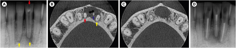
Wegener’s granulomatosis (WG) is a condition with immune-mediated pathogenesis that can present oral manifestations. This report describes the case of a patient diagnosed with WG 14 years previously, who was affected by persistent pain of non-odontogenic origin after successful endodontic treatment. A 39-year-old woman with WG was diagnosed with pulp necrosis and apical periodontitis of teeth #31, #32, and #41, after evaluation through a clinical examination and cone-beam computed tomography (CBCT). At the first appointment, these teeth were subjected to conventional endodontic treatment. At 6- and 12-month follow-up visits, the patient complained of persistent pain associated with the endodontically treated teeth (mainly in tooth #31), despite complete remission of the periapical lesions shown by radiographic and CBCT exams proving the effectiveness of the endodontic treatments, thus indicating a probable diagnostic of persistent pain of non-odontogenic nature. After the surgical procedure was performed to curette the lesion and section 3 mm of the apical third of tooth #31, the histopathological analysis suggested that the painful condition was likely associated with the patient's systemic condition. Based on clinical, radiographic, and histopathological findings, this unusual case report suggests that WG may be related to non-odontogenic persistent pain after successful endodontic treatments. -
Citations
Citations to this article as recorded by  - Toothaches of Non-odontogenic Origin
Davis C. Thomas, Tanvee Somaiya, Ahana Ajayakumar, Vaishnavi Prabhakar
Dental Clinics of North America.2026; 70(1): 209. CrossRef
-
6,197
View
-
76
Download
-
1
Crossref
-
Difficulties experienced by endodontics researchers in conducting studies and writing papers
-
Betul Aycan Alim-Uysal, Selin Goker-Kamali, Ricardo Machado
-
Restor Dent Endod 2022;47(2):e20. Published online March 15, 2022
-
DOI: https://doi.org/10.5395/rde.2022.47.e20
-
-
Abstract
PDFPubReaderePub
- Objectives
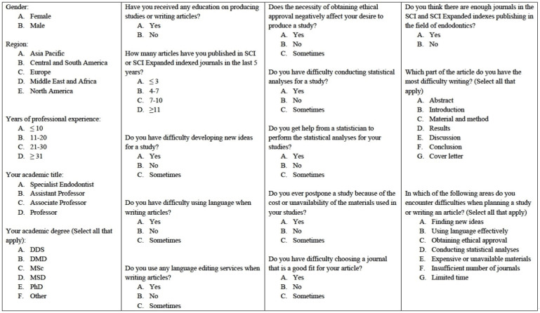
The study investigated the difficulties experienced by endodontics researchers around the world in conducting studies and writing papers. Materials and MethodsA survey consisting of 18 questions on the difficulties experienced by endodontics researchers in performing studies and writing papers was e-mailed to academics in the field of endodontics working at 202 universities. The independent risk factors were analyzed using binary logistic regression at a significance level of 0.05. ResultsA total of 581 individuals (10.7%) agreed to participate in the study. Almost half the participants (48.2%) reported that they had received some type of training in conducting studies and writing papers. In response to the question, “Do you get help from a statistician to perform the statistical analyses of your studies?,” 77.1% answered “yes.” Around 40% of the participants stated that the need to obtain ethical approval negatively affected their desire to conduct studies. The participants’ regions had no effect on the reported difficulties associated with writing papers in English or conducting statistical analyses (p > 0.05). Most participants (81.8%) reported difficulties in writing the Discussion section, regardless of their region, academic degrees, or years of experience. ConclusionsThe participants stated they experienced difficulties in many areas, such as conducting statistical analyses, finding new ideas, and writing in English. Engaging in a detailed examination of ethics committee rules, expanding biostatistics education, increasing the number of institutions providing research funding, and increasing the number of endodontics journals can increase the enthusiasm of endodontics researchers to publish papers.
-
Citations
Citations to this article as recorded by - Prevalence of radix molaris in mandibular molars of a subpopulation of Brazil’s Northeast region: a cross-sectional CBCT study
Yasmym Martins Araújo de Oliveira, Maria Clara Mendes Gomes, Maria Fernanda da Silva Nascimento, Ricardo Machado, Danna Mota Moreira, Hermano Camelo Paiva, George Táccio de Miranda Candeiro
Scientific Reports.2025;[Epub] CrossRef - Statistical pitfalls in endodontic research
Nandini Suresh
Endodontology.2023; 35(1): 1. CrossRef
-
2,720
View
-
31
Download
-
1
Web of Science
-
2
Crossref
-
Postoperative pain after endodontic treatment of necrotic teeth with large intentional foraminal enlargement
-
Ricardo Machado, Daniel Comparin, Sérgio Aparecido Ignácio, Ulisses Xavier da Silva Neto
-
Restor Dent Endod 2021;46(3):e31. Published online May 31, 2021
-
DOI: https://doi.org/10.5395/rde.2021.46.e31
-
-
Abstract
PDFPubReaderePub
- Objectives
To evaluate postoperative pain after endodontic treatment of necrotic teeth using large intentional foraminal enlargement (LIFE). Materials and MethodsThe sample included 60 asymptomatic necrotic teeth (with or without chronic apical periodontitis), and a periodontal probing depth of 3 mm, previously accessed and referred to perform endodontic treatment. After previous procedures, the position and approximate size of the apical foramen (AF) were determined by using an apex locator and K flexo-files, respectively. The chemomechanical preparation was performed with Profile 04 files 2 mm beyond the AF to achieve the LIFE, using 2.5 mL of 2.5% NaOCl at each file change. The filling was performed by Tagger's hybrid technique and EndoFill sealer. Phone calls were made to all the patients at 24, 48 and 72 hours after treatment, to classify postoperative pain. Statistical analysis was performed by different tests with a significance level of 5%. ResultsAge, gender, periradicular status and tooth type did not influence postoperative pain (p > 0.05). Only 1 patient (1.66%) reported severe pain after 72 hours. Moderate pain was reported by 7, 4 and 3 patients after 24, 48 and 72 hours, respectively (p = 0.0001). However, paired analyses showed a statistically significant difference only between 24 and 72 hours (p = 0.04). Sealer extrusion did not influence the postoperative pain (p > 0.05). ConclusionsAcute or moderate postoperative pain was uncommon after endodontic treatment of necrotic teeth with LIFE. Trial RegistrationThe Brazilian Clinical Trials Registry Identifier: RBR-3r967t
-
Citations
Citations to this article as recorded by - Postoperative Pain After Endodontic Treatment in HIV‐Positive Patients Under HAART: A Prospective Observational Cohort Study
Marcos Felipe Iparraguirre Nuñovero, Marco Antonio Hungaro Duarte, Luciana Reis Azevedo Alanis, Bruno Cavalini Cavenago, Ulisses Xavier da Silva Neto, Everdan Carneiro
International Endodontic Journal.2026; 59(5): 788. CrossRef - Does maintaining apical patency reduce early postoperative pain after root canal treatment? A randomized controlled trial in asymptomatic vital single-rooted teeth
Ozan Arda Deger, Sehnaz Yilmaz, Kübra Gürler
Clinical Oral Investigations.2026;[Epub] CrossRef - Evaluation of Postoperative Pain Frequency in Single‐Session Endodontic Treatments With Patency and Foraminal Enlargement
Viviane Barbosa Godoy, Ana Grasiela Limoeiro, Vanessa Sandini, Vini Mehta, Wayne Martins Nascimento, Marilia Fagury Videira Marceliano‐Alves, Marcos Frozoni
Clinical and Experimental Dental Research.2026;[Epub] CrossRef - Assessment of apical extrusion in regenerative endodontics: a comparative study of different irrigation methods using three-dimensional immature tooth models
Vahide Hazal Abat, Gökçen Deniz Bayrak, Mustafa Gündoğar
Odontology.2025; 113(1): 213. CrossRef - Clinical Advances in Calcium Phosphate for Maxillomandibular Bone Regeneration: From Bench to Bedside
Seyed Ali Mostafavi Moghaddam, Hamid Mojtahedi, Amirhossein Bahador, Lotfollah Kamali Hakim, Hamid Tebyaniyan
Ceramics.2025; 8(4): 129. CrossRef - Postoperative pain after single-visit root canal treatments in necrotic teeth comparing instruments’ kinematics and apical instrumentation limits – a prospective randomized multicenter clinical trial
Ricardo Machado, Guilherme Moreira, Daniel Comparin, Arthur Pimentel Barroso, Jaqueline Nascimento, Caio Cézar Randi Ferraz, Sérgio Aparecido Ignácio, Lucas da Fonseca Roberti Garcia, Rodrigo Rodrigues Amaral, David Shadid, Ulisses Xavier da Silva Neto
BMC Oral Health.2024;[Epub] CrossRef - Assessment of mechanical allodynia in healthy teeth adjacent and contralateral to endodontically diseased teeth: a clinical study
Vaishnavi Ratnakar Patankar, Ashish K Jain, Rahul D Rao, Prajakta R Rao
Restorative Dentistry & Endodontics.2024;[Epub] CrossRef - A systematic review and meta-analysis on the effects of phototherapy on postoperative pain in conventional endodontic reintervention
Larissa Pereira Nunes, Gabriel Pereira Nunes, Túlio Morandin Ferrisse, Henrico Badaoui Strazzi-Sahyon, Eloi Dezan-Júnior, Luciano Tavares Angelo Cintra, Gustavo Sivieri-Araujo
Clinical Oral Investigations.2024;[Epub] CrossRef - The effect of intracanal cryotherapy with and without foraminal enlargement on pain prevention after endodontic treatment: a randomized clinical trial
Marcos Felipe Iparraguirre Nuñovero, Marco Antonio Hungaro Duarte, André Vinícius Kaled Segato, Ulisses Xavier da Silva Neto, Vania Portela Ditzel Westphalen, Everdan Carneiro
Scientific Reports.2024;[Epub] CrossRef - Clinical determination of anatomical diameter in different dental groups correlating them with gender, age, tooth/canal and pulpoperiradicular diagnosis: an observational clinical study
Ricardo Machado, Gabriel Filipe Pamplona, Claudemir de Souza Júnior, Jaqueline Nascimento, Eduardo Donato Eing Elgelke Back, Daniel Comparin, Sérgio Aparecido Ignácio, Stella Maria Glaci Reinke, Ana Cristina Kovalik, Ulisses Xavier da Silva Neto
Scientific Reports.2023;[Epub] CrossRef - How much to enlarge? A letter to the editor
Krishnamachari Janani, Kavalipurapu Venkata Teja, Kumar Chandan Srivatsava
Saudi Endodontic Journal.2023; 13(3): 288. CrossRef - Efficiency of diode laser in control of post-endodontic pain: a randomized controlled trial
Hend H. Ismail, Maram Obeid, Ehab Hassanien
Clinical Oral Investigations.2023; 27(6): 2797. CrossRef - Periapical Healing following Root Canal Treatment Using Different Endodontic Sealers: A Systematic Review
Akshay Khandelwal, Krishnamachari Janani, KavalipurapuVenkata Teja, Jerry Jose, Gopi Battineni, Francesco Riccitiello, Alessandra Valletta, Ajitha Palanivelu, Gianrico Spagnuolo, Vincenzo Grassia
BioMed Research International.2022;[Epub] CrossRef
-
6,094
View
-
65
Download
-
11
Web of Science
-
13
Crossref
-
Smear layer removal by passive ultrasonic irrigation and 2 new mechanical methods for activation of the chelating solution
-
Ricardo Machado, Isadora da Silva, Daniel Comparin, Bianca Araujo Marques de Mattos, Luiz Rômulo Alberton, Ulisses Xavier da Silva Neto
-
Restor Dent Endod 2021;46(1):e11. Published online January 26, 2021
-
DOI: https://doi.org/10.5395/rde.2021.46.e11
-
-
Abstract
PDFPubReaderePub
- Objectives
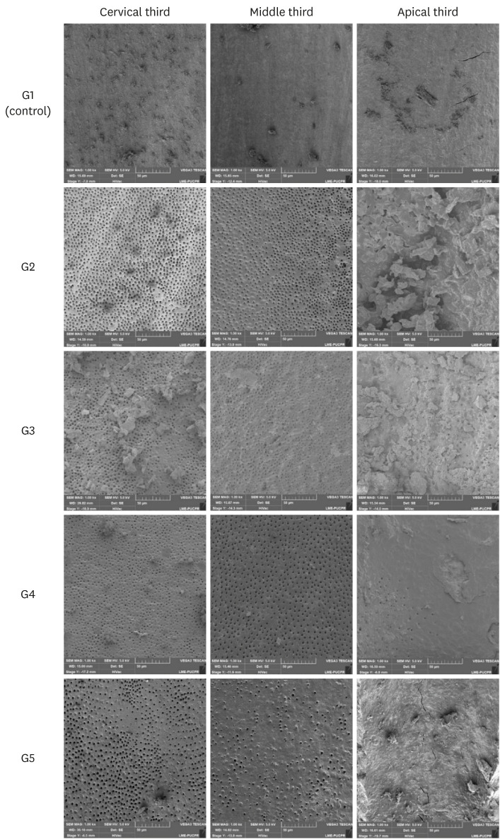
The aim of this study was to compare smear layer removal by conventional application (CA), passive ultrasonic irrigation (PUI), EasyClean (EC), and XP-Endo Finisher (XPF), using 17% ethylenediaminetetraacetic acid (EDTA) after chemomechanical preparation, as evaluated with scanning electron microscopy (SEM). Materials and MethodsForty-five single-rooted human mandibular premolars were selected for this study. After chemomechanical preparation, the teeth were randomly divided into 5 groups according to the protocol for smear layer removal, as follows: G1 (control): CA of distilled water; G2 (CA): CA of 17% EDTA; G3 (PUI): 17% EDTA activated by PUI; G4 (EC): 17% EDTA activated by EC; and G5 (XPF): 17% EDTA activated by XPF. SEM images (×1,000) were obtained from each root third and scored by 3 examiners. Data were evaluated using the Kruskal-Wallis and Dunn tests (p < 0.05). ResultsIn the apical third, there were no statistically significant differences among the groups (p > 0.05). In the cervical and middle thirds, the experimental groups performed better than the control group (p < 0.05); however, G2 presented better results than G3, G4, and G5 (p < 0.05), which showed no differences among one another (p > 0.05). ConclusionsNo irrigation method was able to completely remove the smear layer, especially in the apical third. Using CA for the chelating solution performed better than any form of activation.
-
Citations
Citations to this article as recorded by - Nanoscale Approaches to Oro-Dental Tissue Engineering: A Review of Strategies, Composites, and Translational Challenges
Pei Wang, Yingtong Ye, Keyi Mei, Biaoqi Chen, Ranjith Kankala, Fei Tong
International Journal of Nanomedicine.2026; Volume 21: 1. CrossRef - Comparative Evaluation of Smear Layer Removal Efficacy of Ethylenediaminetetraacetic Acid (EDTA) Gel Versus Solution With Sonic Activation: A Scanning Electron Microscopic Study
Gurkirat Singh, Rajinder Bansal, Manu Bansal, Jasvinder Kaur, Sangam Mittal, Nishant Sharma
Cureus.2026;[Epub] CrossRef - Smear layer removal comparing conventional irrigation, passive ultrasonic irrigation, EndoActivator System, and a new sonic device (Perfect Clean System) by scanning electron microscopy: An ex vivo study
Bruna Fernanda Alionço Gonçalves, Divya Reddy, Ricardo Machado, Paulo César Soares Júunior, Sérgio Aparecido Ignácio, Douglas Augusto Fernandes Couto, Karine Santos Frasquetti, Vânia Portela Ditzel Westphalen, Everdan Carneiro, Ulisses Xavier da Silva Net
PLOS ONE.2024; 19(12): e0314940. CrossRef - Impact of different agitation methods on smear layer cleaning of mesial canals with accentuated curvature
Abel Teves Cordova, Murilo Priori Alcalde, Michel Espinosa Klymus, Leonardo Rigoldi Bonjardim, Rodrigo Ricci Vivan, Marco Antonio Hungaro Duarte
Restorative Dentistry & Endodontics.2024;[Epub] CrossRef - Advances in hybridized nanoarchitectures for improved oro-dental health
Jun Guo, Pei Wang, Yuyao Li, Yifan Liu, Yingtong Ye, Yi Chen, Ranjith Kumar Kankala, Fei Tong
Journal of Nanobiotechnology.2024;[Epub] CrossRef - Cleaning and disinfection of the root canal system provided by four active supplementary irrigation methods
Alessandra Timponi Goes Cruz, Adriane Antoniw Klemz, Edvaldo Antônio Ribeiro Rosa, Fabiana Soares Grecca, Bianca Mattos, Lucila Piasecki, Ricardo Machado, Sérgio Aparecido Ignácio, Ulisses Xavier da Silva Neto
Scientific Reports.2024;[Epub] CrossRef - Scanning electron microscopic study of smear layer changes following ultrasonic endoactivator irrigation system during root canal treatment of primary teeth
Mohamed Ghaly, Aya Alsherif, Arafa Khatab
Tanta Dental Journal.2023; 20(2): 137. CrossRef - Influence of agitation methods of irrigants after methylene blue-mediated PDT on the bonding interface of a fiber post cementation system
Lucas David Galvani, Joatan Lucas de Sousa Gomes Costa, João Felipe Besegato, Joissi Ferrari Zaniboni, Wilfredo Gustavo Escalante-Otárola, Milton Carlos Kuga
Photodiagnosis and Photodynamic Therapy.2022; 37: 102708. CrossRef
-
3,632
View
-
39
Download
-
9
Web of Science
-
8
Crossref
-
Smear layer removal by different chemical solutions used with or without ultrasonic activation after post preparation
-
Daniel Poletto, Ana Claudia Poletto, Andressa Cavalaro, Ricardo Machado, Leopoldo Cosme-Silva, Cássia Cilene Dezan Garbelini, Márcio Grama Hoeppner
-
Restor Dent Endod 2017;42(4):324-331. Published online November 1, 2017
-
DOI: https://doi.org/10.5395/rde.2017.42.4.324
-
-
Abstract
PDFPubReaderePub
- Objectives
This study evaluated smear layer removal by different chemical solutions used with or without ultrasonic activation after post preparation. Materials and MethodsForty-five extracted uniradicular human mandibular premolars with single canals were treated endodontically. The cervical and middle thirds of the fillings were then removed, and the specimens were divided into 9 groups: G1, saline solution (NaCl); G2, 2.5% sodium hypochlorite (NaOCl); G3, 2% chlorhexidine (CHX); G4, 11.5% polyacrylic acid (PAA); G5, 17% ethylenediaminetetraacetic acid (EDTA). For the groups 6, 7, 8, and 9, the same solutions used in the groups 2, 3, 4, and 5 were used, respectively, but activated with ultrasonic activation. Afterwards, the roots were analyzed by a score considering the images obtained from a scanning electron microscope. ResultsEDTA achieved the best performance compared with the other solutions evaluated regardless of the irrigation method (p < 0.05). ConclusionsUltrasonic activation did not significantly influence smear layer removal.
-
Citations
Citations to this article as recorded by - Cerium Oxide Nanoparticle Loaded with Toluidine Blue as Cavity Disinfectant Activated via Light-Emitting Diode on the Shear Bond Strength and Resin Tag Length of Universal Adhesive: A Scanning Electron Microscope-EDX Study
Amer M. Alanazi, Syed Hussain Askary, Ibrahim Warsi, Aamir Afzal, Muhammad Omar Niaz, Ambrina Qureshi
Photobiomodulation, Photomedicine, and Laser Surgery.2026;[Epub] CrossRef - O papel do ultrassom no tratamento e retratamento de canais radiculares: Revisão de literatura
Carlos Roberto Souza Hipp, Joaquim Carlos Fest da Silveira, Luiz Felipe Gilson de Oliveira Rangel, Tatiana Federici de Souza Fest da Silveira, Carla Minozzo Mello, Rodrigo Simões de Oliveira
Research, Society and Development.2025; 14(8): e1314849323. CrossRef - Effect of sodium hypochlorite, ethylenediaminetetraacetic acid, and dual-rinse irrigation on dentin adhesion using an etch-and-rinse or self-etch approach
Matej Par, Tobias Steffen, Selinay Dogan, Noah Walser, Tobias T. Tauböck
Scientific Reports.2024;[Epub] CrossRef - Evaluation of Effect of Poloxamer on Smear Layer Removal Using Apical Negative Pressure: An In Vitro Scanning Electron Microscopy Study
Chandra Prabha, Chitharanjan Shetty, Aditya Shetty
Journal of International Oral Health.2024; 16(6): 498. CrossRef - Laboratory Assessment of Antibacterial Efficacy of Five Different Herbal-based Potential Endodontic Irrigants
Anjali A Oak, Kailash Attur, Kamal Bagda, Nitish Mathur, Lubna Mohammad, Nikhat M Attar
Advances in Human Biology.2023; 13(4): 350. CrossRef - Dental Surface Conditioning Techniques to Increase the Micromechanical Retention to Fiberglass Posts: A Literature Review
Paulina Leticia Moreno-Sánchez, Maricela Ramírez-Álvarez, Alfredo del Rosario Ayala-Ham, Erika de Lourdes Silva-Benítez, Miguel Ángel Casillas-Santana, Diana Leyva del Rio, León Francisco Espinosa-Cristóbal, Erik Lizárraga-Verdugo, Mariana Melisa Avendaño
Applied Sciences.2023; 13(14): 8083. CrossRef - Effect of irrigation protocols on smear layer removal, bond strength and nanoleakage of fiber posts using a self-adhesive resin cement
Rodrigo Stadler Alessi, Renata Terumi Jitumori, Bruna Fortes Bittencourt, Giovana Mongruel Gomes, João Carlos Gomes
Restorative Dentistry & Endodontics.2023;[Epub] CrossRef - Effects of using different root canal sealers and protocols for cleaning intraradicular dentin on the bond strength of a composite resin used to reinforce weakened roots
Luiz Pascoal Vansan, Ricardo Machado, Celso Bernardes de Souza, Ricardo Gariba, Antônio Miranda da Cruz, Cinara Muniz, Jardel FranciscoX Jardel Francisco Mazzi-Chaves, Lucas da Fonseca Roberti Garcia
Journal of Oral Research.2022; 11(6): 1. CrossRef - Influence of the use of chelating agents as final irrigant on the push‐out bond strength of epoxy resin‐based root canal sealers: A systematic review
Carla M. Augusto, Miguel A. Cunha Neto, Karem P. Pinto, Ana Flavia A. Barbosa, Emmanuel J. N. L. Silva, Ana Paula P. dos Santos, Luciana M. Sassone
Australian Endodontic Journal.2022; 48(2): 347. CrossRef - Adhesion and whitening efficacy of P11-4 self-assembling peptide and HAP suspension after using NaOCl as a pre-treatment agent
Niloofar Hojabri, Karl-Heinz Kunzelmann
BMC Oral Health.2022;[Epub] CrossRef - Influence of resin cements and root canal disinfection techniques on the adhesive bond strength of fibre reinforced composite post to radicular dentin
Zaid A. Al Jeaidi
Photodiagnosis and Photodynamic Therapy.2021; 33: 102108. CrossRef - The Antibacterial Efficacy and In Vivo Toxicity of Sodium Hypochlorite and Electrolyzed Oxidizing (EO) Water-Based Endodontic Irrigating Solutions
Sung-Chih Hsieh, Nai-Chia Teng, Chia Chun Chu, You-Tai Chu, Chung-He Chen, Liang-Yu Chang, Chieh-Yun Hsu, Ching-Shuan Huang, Grace Ying-Wen Hsiao, Jen-Chang Yang
Materials.2020; 13(2): 260. CrossRef
-
3,256
View
-
19
Download
-
12
Crossref
|