-
Resin infiltrant protects deproteinized dentin against erosive and abrasive wear
-
Ana Theresa Queiroz de Albuquerque, Bruna Oliveira Bezerra, Isabelly de Carvalho Leal, Maria Denise Rodrigues de Moraes, Mary Anne S. Melo, Vanara Florêncio Passos
-
Restor Dent Endod 2022;47(3):e29. Published online July 1, 2022
-
DOI: https://doi.org/10.5395/rde.2022.47.e29
-
-
 Abstract Abstract
 PDF PDF PubReader PubReader ePub ePub
- Objectives
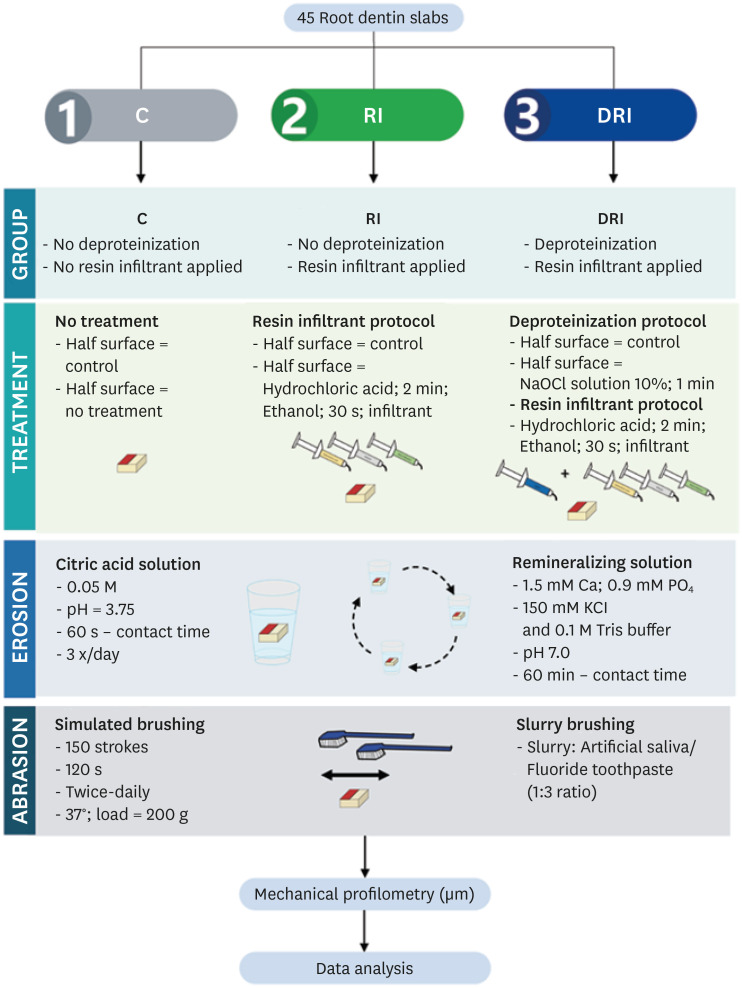
This study aimed to investigate the anti-erosive/abrasive effect of resin infiltration of previous deproteinized dentin. Materials and MethodsDentin slabs were randomly assigned to 3 groups (n = 15): Control (no deproteinization; no resin infiltrant applied), RI (no deproteinization; resin infiltrant applied), and DRI (deproteinization; resin infiltrant applied). After undergoing the assigned treatment, all slabs were subjected to an in vitro cycling model for 5 days. The specimens were immersed in citric acid (0.05 M, pH = 3.75; 60 seconds; 3 times/day) and brushed (150 strokes). Between the challenges, the specimens were exposed to a remineralizing solution (60 minutes). The morphological alterations were analyzed by mechanical profilometry (µm) and scanning electron microscopy (SEM). Data were submitted to one-way analysis of variance (ANOVA) and Tukey tests (p < 0.05). ResultsControl and RI groups presented mineral wear and did not significantly differ from each other (p = 0.063). DRI maintained a protective layer preserving the dentin (p < 0.001). After erosive/abrasive cycles, it was observed that in group RI, only 25% of the slabs partially evidenced the presence of the infiltrating, while, in the DRI group, 80% of the slabs presented the treated surface entirely covered by a resin-component layer protecting the dentin surface as observed in SEM images. ConclusionsThe removal of the organic content allows the resin infiltrant to efficiently protect the dentin surface against erosive/abrasive lesions.
-
Citations
Citations to this article as recorded by  - Acidic/abrasive challenges on simulated non-carious cervical lesions development and morphology
Giovanna C. Denucci, Ian Towle, Cecilia P. Turssi, George J. Eckert, Anderson T. Hara
Archives of Oral Biology.2025; 169: 106120. CrossRef - Physio‐Mechanic and Microscopic Analyses of Bioactive Glass‐Based Resin Infiltrants
Syed Zubairuddin Ahmed, Abdul Samad Khan, Wejdan Waleed Nasser, Methayel Abdulrahman Alrushaid, Zahrah Mohammed Alfaraj, Moayad Mohammed Aljeshi, Asma Tufail Shah, Budi Aslinie Md Sabri, Sultan Akhtar, Mohamed Ibrahim Abu Hassan
Microscopy Research and Technique.2025; 88(2): 595. CrossRef
-
1,841
View
-
39
Download
-
2
Web of Science
-
2
Crossref
-
Errors in light-emitting diodes positioning when curing bulk fill and incremental composites: impact on properties after aging
-
Abdulrahman A. Balhaddad, Isadora M. Garcia, Haifa Maktabi, Maria Salem Ibrahim, Qoot Alkhubaizi, Howard Strassler, Fabrício M. Collares, Mary Anne S. Melo
-
Restor Dent Endod 2021;46(4):e51. Published online September 24, 2021
-
DOI: https://doi.org/10.5395/rde.2021.46.e51
-
-
 Abstract Abstract
 PDF PDF PubReader PubReader ePub ePub
- Objectives
This study aimed to evaluate the effect of improper positioning single-peak and multi-peak lights on color change, microhardness of bottom and top, and surface topography of bulk fill and incremental composites after artificial aging for 1 year. Materials and MethodsBulk fill and incremental composites were cured using multi-peak and single-peak light-emitting diode (LED) following 4 clinical conditions: (1) optimal condition (no angulation or tip displacement), (2) tip-displacement (2 mm), (3) slight tip angulation (α = 20°) and (4) moderate tip angulation (α = 35°). After 1-year of water aging, the specimens were analyzed for color changes (ΔE), Vickers hardness, surface topography (Ra, Rt, and Rv), and scanning electron microscopy. ResultsFor samples cured by single-peak LED, the improper positioning significantly increases the color change compared to the optimal position regardless of the type of composite (p < 0.001). For multi-peak LED, the type of resin composite and the curing condition displayed a significant effect on ΔE (p < 0.001). For both LEDs, the Vickers hardness and bottom/top ratio of Vickers hardness were affected by the type of composite and the curing condition (p < 0.01). ConclusionsThe bulk fill composite presented greater resistance to wear, higher color stability, and better microhardness than the incremental composite when subjected to improper curing. The multi-peak LED improves curing under improper conditions compared to single-peak LED. Prevention of errors when curing composites requires the attention of all personnel involved in the patient's care once the clinical relevance of the appropriate polymerization reflects on reliable long-term outcomes.
-
Citations
Citations to this article as recorded by  - The demineralization resistance and mechanical assessments of different bioactive restorative materials for primary and permanent teeth: an in vitro study
Maria Salem Ibrahim, Fahad Rakad Aldhafeeri, Abdullah Sami Banaemah, Mana S. Alhaider, Yousif A. Al-Dulaijan, Abdulrahman A. Balhaddad
BDJ Open.2024;[Epub] CrossRef - Inorganic Compounds as Remineralizing Fillers in Dental Restorative Materials: Narrative Review
Leena Ibraheem Bin-Jardan, Dalal Ibrahim Almadani, Leen Saleh Almutairi, Hadi A. Almoabid, Mohammed A. Alessa, Khalid S. Almulhim, Rasha N. AlSheikh, Yousif A. Al-Dulaijan, Maria S. Ibrahim, Afnan O. Al-Zain, Abdulrahman A. Balhaddad
International Journal of Molecular Sciences.2023; 24(9): 8295. CrossRef
-
1,299
View
-
16
Download
-
2
Web of Science
-
2
Crossref
-
Inhibition of nicotine-induced Streptococcus mutans biofilm formation by salts solutions intended for mouthrinses
-
Abdulrahman A. Balhaddad, Mary Anne S. Melo, Richard L. Gregory
-
Restor Dent Endod 2019;44(1):e4. Published online January 16, 2019
-
DOI: https://doi.org/10.5395/rde.2019.44.e4
-
-
 Abstract Abstract
 PDF PDF PubReader PubReader ePub ePub
- Objectives
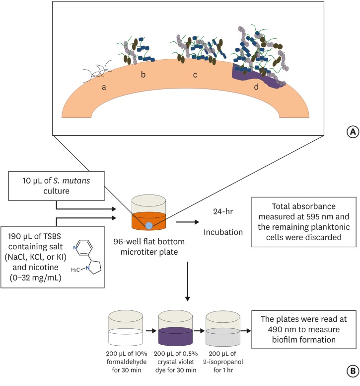
Biofilm formation is critical to dental caries initiation and development. The aim of this study was to investigate the effects of nicotine exposure on Streptococcus mutans (S. mutans) biofilm formation concomitantly with the inhibitory effects of sodium chloride (NaCl), potassium chloride (KCl) and potassium iodide (KI) salts. This study examined bacterial growth with varying concentrations of NaCl, KCl, and KI salts and nicotine levels consistent with primary levels of nicotine exposure. Materials and MethodsA preliminary screening experiment was performed to investigate the appropriate concentrations of NaCl, KCl, and KI to use with nicotine. With the data, a S. mutans biofilm growth assay was conducted using nicotine (0–32 mg/mL) in Tryptic Soy broth supplemented with 1% sucrose with and without 0.45 M of NaCl, 0.23 M of KCl, and 0.113 M of KI. The biofilm was stained with crystal violet dye and the absorbance measured to determine biofilm formation. ResultsThe presence of 0.45 M of NaCl, 0.23 M of KCl, and 0.113 M of KI significantly inhibited (p < 0.05) nicotine-induced S. mutans biofilm formation by 52%, 79.7%, and 64.1%, respectively. ConclusionsThe results provide additional evidence regarding the biofilm-enhancing effects of nicotine and demonstrate the inhibitory influence of these salts in reducing the nicotine-induced biofilm formation. A short-term exposure to these salts may inhibit S. mutans biofilm formation.
-
Citations
Citations to this article as recorded by  - The Inhibition of Streptococcus mutans Biofilms following Exposure to Different Chocolate Ingredients
Hadi A. Almoabid, Leen Saleh Almutairi, Abdul Samad Khan, Mohammed A. Aljaffary, Rasha AlSheikh, Khalid S. Almulhim, Abdulrahman A. Balhaddad
European Journal of Dentistry.2025;[Epub] CrossRef - The impact of Caralluma munbyana extracts on Streptococcus mutans biofilm formation
Turki Alshehri, Israa Alkhalifah, Areeb Alotaibi, Alaa F. Alsulaiman, Abdullah Al Madani, Basil Almutairi, Abdulrahman A. Balhaddad
Frontiers in Dental Medicine.2025;[Epub] CrossRef - Tobacco‐enhanced biofilm formation by Porphyromonas gingivalis and other oral microbes
Jinlian Tan, Gwyneth J. Lamont, David A. Scott
Molecular Oral Microbiology.2024; 39(5): 270. CrossRef - Nicotine is a potent extracellular polysaccharide inducer in Fusobacterium nucleatum biofilms
Adaias Oliveira Matos, Valentim Adelino Ricardo Barão, Richard Lee Gregory
Brazilian Journal of Oral Sciences.2023;[Epub] CrossRef - Effect of eucalyptus oil on Streptococcus mutans and Enterococcus faecalis growth
Abdulrahman A. Balhaddad, Rasha N. AlSheikh
BDJ Open.2023;[Epub] CrossRef - Microorganisms: crucial players of smokeless tobacco for several health attributes
Akanksha Vishwakarma, Digvijay Verma
Applied Microbiology and Biotechnology.2021; 105(16-17): 6123. CrossRef - Microbiology of the American Smokeless Tobacco
A. J. Rivera, R. E. Tyx
Applied Microbiology and Biotechnology.2021; 105(12): 4843. CrossRef - The Impact of Photosensitizer Selection on Bactericidal Efficacy Of PDT against Cariogenic Biofilms: A Systematic Review and Meta-Analysis
Maurício Ítalo Silva Teófilo, Teresa Maria Amorim Zaranza de Carvalho Russi, Paulo Goberlanio de Barros Silva, Abdulrahman A. Balhaddad, Mary Anne S. Melo, Juliana P.M.L. Rolim
Photodiagnosis and Photodynamic Therapy.2021; 33: 102046. CrossRef - Antibacterial Activities of Methanol and Aqueous Extracts of Salvadora persica against Streptococcus mutans Biofilms: An In Vitro Study
Abdulrahman A. Balhaddad, Lamia Mokeem, Mary Anne S. Melo, Richard L. Gregory
Dentistry Journal.2021; 9(12): 143. CrossRef - The burden of root caries: Updated perspectives and advances on management strategies
Mohammed S. AlQranei, Abdulrahman A. Balhaddad, Mary A.S. Melo
Gerodontology.2021; 38(2): 136. CrossRef - Emerging Contact-Killing Antibacterial Strategies for Developing Anti-Biofilm Dental Polymeric Restorative Materials
Heba Mitwalli, Rashed Alsahafi, Abdulrahman A. Balhaddad, Michael D. Weir, Hockin H. K. Xu, Mary Anne S. Melo
Bioengineering.2020; 7(3): 83. CrossRef - In-Vitro Model of Scardovia wiggsiae Biofilm Formation and Effect of Nicotine
Abdulrahman A. Balhaddad, Hadeel M. Ayoub, Richard L. Gregory
Brazilian Dental Journal.2020; 31(5): 471. CrossRef - Antibacterial efficacy and remineralization capacity of glycyrrhizic acid added casein phosphopeptide‐amorphous calcium phosphate
Feride Sahin, Fatih Oznurhan
Microscopy Research and Technique.2020; 83(7): 744. CrossRef - Concentration dependence of quaternary ammonium monomer on the design of high-performance bioactive composite for root caries restorations
Abdulrahman A. Balhaddad, Maria S. Ibrahim, Michael D. Weir, Hockin H.K. Xu, Mary Anne S. Melo
Dental Materials.2020; 36(8): e266. CrossRef
-
1,334
View
-
11
Download
-
14
Crossref
|


