-
A cone-beam computed tomography study of the prevalence and location of the second mesiobuccal root canal in maxillary molars
-
Seong-Ju Lee, Eun-Hye Lee, Se-Hee Park, Kyung-Mo Cho, Jin-Woo Kim
-
Restor Dent Endod 2020;45(4):e46. Published online September 3, 2020
-
DOI: https://doi.org/10.5395/rde.2020.45.e46
-
-
 Abstract Abstract
 PDF PDF PubReader PubReader ePub ePub
- Objectives
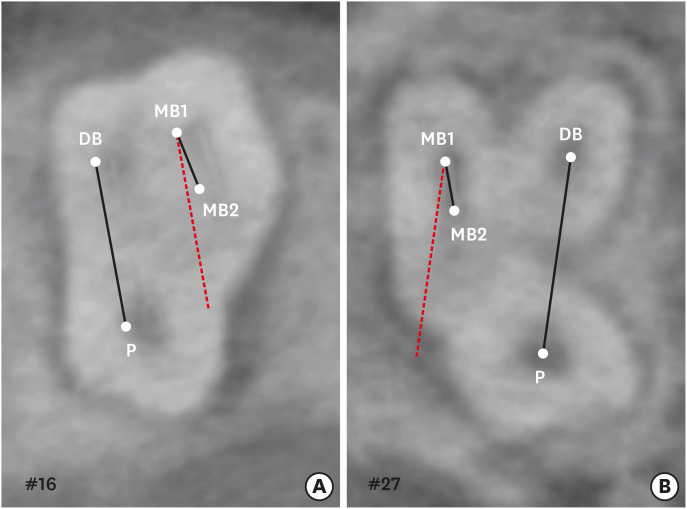
This study aimed to investigate the incidence and location of the second mesiobuccal root (MB2) canal in maxillary molars with the aid of various measuring points and lines using cone-beam computed tomography (CT). Materials and MethodsA total of 205 images of patients who underwent cone-beam CT examinations between 2011 and 2015 as part of their dental diagnosis and treatment were included. There were 76 images of the maxillary first molar and 135 images of the maxillary second molar. Canal orifices were detected at −1 mm from the top of the pulpal floor on cone-beam CT images. Image assessment was performed by 2 observers in reformatted image planes using software. Assessments included measurement of the distance between the MB1 and MB2 canals, and the angles between the lines connecting the MB1-MB2 and distobuccal (DB)-palatal (P) canals. The data were analyzed using the student's t-test. ResultsThe prevalence of the MB2 canal was 86.8% in the first molar and 28.9% in the second molar. The angle between the lines connecting the MB1-MB2 and DB-P canals was 2.3° ± 5.7° in the first molar and −3.95° ± 7.73° in the second molar. The distance between the MB1 and MB2 canals was 2.1 ± 0.44 mm in the first molar and 1.98 ± 0.42 mm in the second molar. ConclusionsThe angles between the lines connecting the MB1-MB2 and DB-P canals was almost parallel. These findings may aid in the prediction of the location of the MB2 canal orifice.
-
Citations
Citations to this article as recorded by  - Study on the Geometric Location Method of the Danger Zone in the Mesial Roots of Mandibular First Molars
Jinjie Yan, Yuanling Peng, Jing Yang, Jie Liu, Linxian Wang, Tingyuan Zhao, Jian Zhang, Kehua Que
Journal of Endodontics.2026; 52(3): 387. CrossRef - Comparative diagnostic accuracy of ChatGPT in second mesiobuccal canal detection
Mehmet Ali Altunkum, Sadullah Kaya
BMC Oral Health.2026;[Epub] CrossRef - Diagnostic Performance of Magnification and Ultrasonic Troughing in Detecting Second Mesiobuccal Canals: A Two-Factor Experimental Study
Mehmet Adiguzel, Furkan Ozeken
Cureus.2026;[Epub] CrossRef - Position of Second Mesiobuccal Canal Relative to Distobuccal and Palatal Canals of Maxillary Molars in an Iranian Population
Sina Mosadeghian, Azadeh Torkzadeh, Parisa Ranjbarian, Roya Asaadi
Journal of Research in Dental and Maxillofacial Sciences.2025; 10(1): 34. CrossRef - Machine Learning Models in the Detection of MB2 Canal Orifice in CBCT Images
Shishir Shetty, Meliz Yuvali, Ilker Ozsahin, Saad Al-Bayatti, Sangeetha Narasimhan, Mohammed Alsaegh, Hiba Al-Daghestani, Raghavendra Shetty, Renita Castelino, Leena R David, Dilber Uzun Ozsahin
International Dental Journal.2025; 75(3): 1640. CrossRef - EVALUATION OF THE PREVALENCE AND LOCATION OF SECOND MESIOBUCCAL CANALS IN 2100 UPPER FIRST AND SECOND MOLAR TEETH: A CONE BEAM COMPUTED TOMOGRAPHY STUDY
Bahar Kaplan, Özkan Adıgüzel, Ayşe Gül Öner Talmaç, Elif Meltem Aslan
İnönü Üniversitesi Sağlık Hizmetleri Meslek Yüksek Okulu Dergisi.2025; 13(3): 752. CrossRef - A novel method for the precise second mesiobuccal canal orifice location: A combined strategy for enhanced clinical practice
Yuhan Wang, Lingyun Li, Lu Zhang, Xiaoyan Wang
Journal of Dental Sciences.2025;[Epub] CrossRef - The Correlation between Intraorifice Distance and the Anatomical Characteristics of the Second Mesiobuccal Canal of Maxillary Molars: A CBCT Study
Isabella Perondi, Silvio Taschieri, Martino Baruffaldi, Roberto Fornara, Luca Francetti, Stefano Corbella, Deepa Gurunathan
International Journal of Dentistry.2024;[Epub] CrossRef - Endodontic management of type I maxillary first molar with two palatal roots using cone-beam computed tomography
Nuha Alghamdi
Dental Journal.2024; 57(1): 1. CrossRef - 3D geometric analysis of second mesiobuccal canal in permanent maxillary first molar tooth
Indrani Khadilkar, Divya Nangia, Amrita Chawla, Sidhartha Sharma, Vijay Kumar, Shalini Gupta, Ajay Logani
Australian Endodontic Journal.2023; 49(1): 140. CrossRef - Prevalence of mesiobuccal-2 canals in maxillary first and second molars among the Bruneian population—CBCT analysis
Hui Yi Onn, Malissa Siao Yun Abdullah Sikun, Hanif Abdul Rahman, Jagjit Singh Dhaliwal
BDJ Open.2022;[Epub] CrossRef - Location angle of second mesio-buccal canal in maxillary molars of an Indian population: an in vivo retrospective CBCT evaluation and proposal of a new classification
Kishor Vhorkate, Kulvinder Banga, Ajinkya M. Pawar, Shugufta Mir, Suraj Arora, Dian Agustin Wahjuningrum, Anuj Bhardwaj, Alexander Maniangat Luke
PeerJ.2022; 10: e14234. CrossRef - Maxillary molar root and canal morphology of Neolithic and modern Chinese
H.Y. Ren, K.Y. Kum, Y.S. Zhao, Y.J. Yoo, J.S. Jeong, Hiran Perinpanayagam, X.Y. Wang, G.J. Li, F. Wang, H. Fang, Y. Gu
Archives of Oral Biology.2021; 131: 105272. CrossRef
-
4,902
View
-
63
Download
-
13
Crossref
-
Comparison of vibration characteristics of file systems for root canal shaping according to file length
-
Seong-Jun Park, Se-Hee Park, Kyung-Mo Cho, Hyo-Jin Ji, Eun-Hye Lee, Jin-Woo Kim
-
Restor Dent Endod 2020;45(4):e51. Published online October 14, 2020
-
DOI: https://doi.org/10.5395/rde.2020.45.e51
-
-
Abstract
PDFPubReaderePub
- Objectives
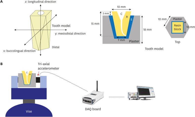
No studies have yet assessed vibration characteristics according to endodontic file length. Accordingly, the objective of the present study was to examine the vibration characteristics according to nickel-titanium file length and to compare these characteristics between different file systems. Materials and MethodsA total of 45 root canal models were divided into 3 experimental groups (n = 15 each) based on the file system used (ProTaper Gold [PTG], ProTaper Next, or WaveOne Gold [WOG]). Each experimental group was further divided into 3 subgroups according to file length (21, 25, or 31 mm). An electric motor (X-SMART PLUS) was used in the experiment. For each file system, vibrations generated when using a size 25 file were measured and used to calculate the average vibration acceleration. The differences in vibrations were analyzed using 1-way analysis of variance and the Scheffé post hoc test with a confidence interval of 95%. ResultsIn the PTG file system, significantly lower vibration acceleration was observed when using a 21-mm file than when using a 31-mm file. In the WOG file system, significantly stronger vibration acceleration was observed when using a 31-mm file than when using 21- or 25-mm files. Regardless of the file length, the WOG group exhibited significantly stronger vibration acceleration than the other 2 experimental groups. ConclusionsIn clinical practice, choosing a file with the shortest length possible could help reduce vibrations. Additionally, consideration should be given to vibrations that could be generated when using WOG files with reciprocating motion.
-
Citations
Citations to this article as recorded by - Comparison vibration characteristics of several wireless endodontic handpieces
Bo-Kyung Lee, Yoon Lee, Se-Hee Park, Kyung-Mo Cho, Jin-Woo Kim
Journal of Dental Rehabilitation and Applied Science.2022; 38(2): 81. CrossRef
-
1,950
View
-
5
Download
-
1
Crossref
-
Fiber-reinforced composite resin bridges: an alternative method to treat root-fractured teeth
-
Gun Heo, Eun-Hye Lee, Jin-Woo Kim, Kyung-Mo Cho, Se-Hee Park
-
Restor Dent Endod 2020;45(1):e8. Published online December 27, 2019
-
DOI: https://doi.org/10.5395/rde.2020.45.e8
-
-
Abstract
PDFPubReaderePub
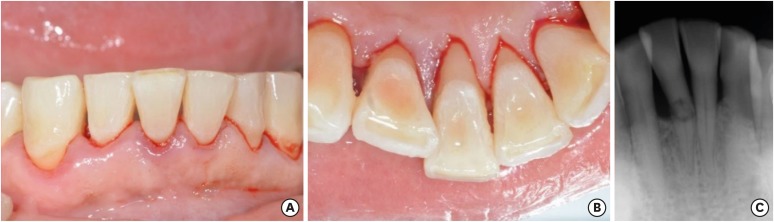
The replacement of missing teeth, especially in the anterior region, is an essential part of dental practice. Fiber-reinforced composite resin bridges are a conservative alternative to conventional fixed dental prostheses or implants. It is a minimally invasive, reversible technique that can be completed in a single visit. The two cases presented herein exemplify the treatment of root-fractured anterior teeth with a natural pontic immediately after extraction. -
Citations
Citations to this article as recorded by - Prosthodontic Aspects of Splinting the Mandibular Anterior Teeth by Fiber Reinforced Composites
Hrelja Miroslav, Laškarin Mirko, Čimić Samir, Kraljević Sonja, Dulčić Nikša, Badel Tomislav
Journal of Dental Problems and Solutions.2025; 12(1): 004. CrossRef - Current Evidence on the Fiber-reinforced Composite Bridges
Ramesh Chowdhary, Sunil Kumar Mishra
International Journal of Prosthodontics and Restorative Dentistry.2023; 12(4): 159. CrossRef - Bridging the Gap: A Case Report of Tooth Replacement using Resin-Bonded Fiber- Reinforced Composite Resin
Vineet Sharma, Sumit Bhansali, Sonal Priya Bhansali
Journal of Pierre Fauchard Academy (India Section).2023; : 66. CrossRef - Reconstruction of Natural Smile and Splinting with Natural Tooth Pontic Fiber‐Reinforced Composite Bridge
Maryam S. Tavangar, Fatemeh Aghaei, Massoumeh Nowrouzi, Andrea Scribante
Case Reports in Dentistry.2022;[Epub] CrossRef
-
2,767
View
-
23
Download
-
4
Crossref
-
Evaluation of radiopacity and discriminability of various fiber reinforced composite posts
-
Eun-Hye Lee, Hang-Moon Choi, Se-Hee Park, Jin-Woo Kim, Kyung-Mo Cho
-
J Korean Acad Conserv Dent 2010;35(3):188-197. Published online May 31, 2010
-
DOI: https://doi.org/10.5395/JKACD.2010.35.3.188
-
-
Abstract
PDFPubReaderePub
The purpose of this study was to compare radiopacity and radiographic discriminability of various FRC-Posts.
Six FRC-Posts were investigated ; 1) FRC Postec Plus (Ivoclar Vivadent AG, Schaan, Liechtenstein), 2) Snowlight (Carbotech, Lewis center, OH, USA), 3) Dentin Post (Komet Brasseler, Lamgo, Germany), 4) Rely-X Fiber Post (3M ESPE, St.paul, MN, USA), 5) D.T.-Light Post (BISCO, Schaumburg, IL,USA), 6) Luxapost (DMG, Hamburg, Germany)
The radiographs of each post with a reference 1 mm / 2 mm aluminum step-wedge was taken using digital sensor. The optical density were calculated by gray value of 10 × 10 pixel and compared in mm Al equivalent at five points.
Six maxillary incisors of similar radiopacity were used. Radiographs of posts in Mx. incisors of lingual side of dry mandible were taken.
We showed radiographs and asked the questionnaire to 3 radiologists, 3 endodontists, 3 general practitioners. The questionnaire was comprised of choices of the highest, lowest radiopaque individual post and the choices of best discriminable post at apical, coronal area.
The following results were obtained.
Each post system showed various radiopacity.
There was change of discriminability between each post and simulated specimens regardless of examiner.
Although each post showed various radiopacity, the difference of radiopacity did not affect on discriminability.
|