Articles
- Page Path
- HOME > Restor Dent Endod > Volume 27(3); 2002 > Article
- Original Article Leakage of SuperEBA in root-end cavities prepared with 3 new ultrasonic tips;KaVo Isthmus, KaVo T-shape and KiS tip
- Woo-Cheol Lee
-
2002;27(3):-276.
DOI: https://doi.org/10.5395/JKACD.2002.27.3.270
Published online: May 31, 2002
Department of conservative Dentistry, School of Dentistry, Seoul National University, Korea.
Copyright © 2002 Korean Academy of Conservative Dentistry
- 767 Views
- 1 Download
I. Introduction
Endodontic treatment is known to have varying rates of success by several studies from 89 to 95%7,13,14). If non-surgical retreatment of failed root canal treatment is unfeasible, surgical therapy is recommended. Periapical surgery includes resection of root apex, preparation of retrograde cavity, and improving the apical seal by filling the root end cavity. When retrograde fillings were placed, the cavity preparation was accomplished with burs. However, these fillings tend to be shallow, leaky and ineffective. Recently, ultrasonic tips are developed and introduced to prepare retrograde cavity cleaner, deeper and therefore provide enhanced retrograde seal6). Ultrasonic preparation also allows retrograde cavities parallel to the long axis of the root. Ilgenstein19) has developed a new set of KaVo SONICflex retro tips, claiming fast and efficient cutting with 3 to 4mm diamond preparation surface. The retrotips are available in two shapes, Isthmus and T-shape, each in two sizes and left and right configuration. Kim also designed KiS microsurgical ultrasonic tips with six different configurations which give better access than other ultrasonic instruments.
Several materials such as amalgam, Glass ionomer cement, and zinc oxide-eugenol cement have been evaluated for clinical use as retrograde filling materials. Oynick and Oynick10) reported use of SuperEBA to be suitable root-end filling material by demonstrating in vivo healing and biological compatibility. Dorn and Gartner5) also reported a 95% success rate utilizing SuperEBA as a root-end filling material. In a number of in vitro leakage studies1,7) demonstrated that SuperEBA has better sealing ability compared to other root-end filling materials. However, none of these studies compares leakage created by newly designed ultrasonic tips.
The purpose of this study was to compare the leak-age of SuperEBA placed into retrograde cavities prepared with 3 different ultrasonic tips; KaVo SONICflex retro Isthmus, T-shape and Obtura Spartan KiS tips.
II. Materials and Methods
56 single-rooted extracted human maxillary and madibular teeth were collected and stored in deionized water and thymol. No previous root canal treatment had been done on the teeth, and the specimens with large restorations, cervical defects, crack, and root fractures were excluded from this study. Adherent soft tissue, periodontal ligament and calculus were removed after soaking in physiological sterile saline. Following access preparation, the working length was determined by inserting a #10 K-file into the orifice of the canal and advancing apically until it was visible at the foramen then subtracting 1mm from this measurement. The root canals were prepared with Gates Glidden burs and Profile .06 taper rotary instruments using crown-down technique, rinsed with 2.5% sodium hypochlorite, and dried with paper points. The canals were then obturated with non-standardized gutta-percha using warm vertical technique with Obtura back fill and Grossman's sealer. Teeth were then stored in physiologic saline at 37℃ for 7 days. All root-end resection procedures were executed under the surgical microscope (Carl Zeiss Inc., Thornwood, NY). As shown in flow chart (Fig. 1), the obturated teeth were randomly divided into 3 groups; KaVo SONICflex retro Isthmus, Tshape (Kaltenbach & Voigt GmbH, Biberach, Germany) and KiS tip (ObturaSpartan, Fenton, MO) preparation group, 18 teeth each. Among these, two teeth from each group served as positive controls. After decoronated the obturated teeth, the apical 3mm of each root was resected under copious water spray with diamond burs in a high-speed handpiece perpendicular to the long axis of the tooth. Retrograde cavity was prepared in resected root-end to a depth of 3mm parallel to the long axis of the tooth with each ultrasonic tip (Fig. 3, 4 and 5). The retrograde cavities were judged to be completed when the gutta-percha was not seen on the wall and the cavity depth was at least 3mm measured with retrograde plugger. Cavity was then dried with air stream using Stropko irrigator (SybronEndo, Orange, CA). All retrograde cavities except six positive controls were filled with SuperEBA (Harry J. Bosworth, Skokie, IL) mixed according to manufacturer's instructions. All teeth were then stored at 37℃, 100% humidity for 72hrs. Three coats of nail varnish were applied to the external root surface except for the resected root surface before teeth were assembled in leakage model shown in Fig. 6. Two negative control teeth with no retrograde filling were covered with nail varnish on entire root surface. Teeth were suspended in an aqueous solution of 1% methylene blue (Sigma Chemical Co., St.Louis, MO) to a level 2mm from the root apex. After 2weeks, all specimens were removed from the vials, and the external surfaces of the roots were rinsed under tap water to remove excess dye. The teeth were then longitudinally grooved and split by prying with a chisel.
The amount of dye penetration of the retrograde fillings was measured using a stereomicroscope at ×6 magnification. The maximum depth of penetration of dye from the resected root-end was evaluated with the aid of a calibrated eyepiece graticule. These data were analyzed using ANOVA test to determine if there were statistically significant differences between the groups.
III. Results
Negative control demonstrated no dye leakage, while the positive controls showed dye penetration along the length of the retrograde fillings.
Linear leakage was 1.5±1.4mm in KaVo Isthmus group, 1.8±1.2mm in KaVo T-shape group, and 1.1±0.7mm in KiS tip group. Fig. 2 shows the amount of leakage for each experimental group. Fig. 7, 8, 9 are representative roots from each group with ultrasonic tip used.
The data revealed that the KiS tip group demonstrated less leakage than other groups, however, there were no statistically significant differences between the groups.
IV. Discussion
The results of this study demonstrated that the ultrasonic tips with which the retrograde cavity was prepared failed to influence the apical seal of SuperEBA root-end filling. However, it is noteworthy that there is difference in ease of use and ability of approaching surgical field among the ultrasonic tips. KaVo SONICflex retro Isthmus tip was designed to prepare isthmus for easier access even with complicated anatomical conditions. KaVo SONICflex retro T-shape claims that it gives better retention with Tshape retrograde cavity preparation. Fig. 10 shows both KaVo tips in high magnifications and its clinical application is shown as a diagram in Fig. 11 as well as radiographic view after retrograde filling in Fig. 12. Diamond coated KiS ultrasonic tips possess high cutting efficiency due to their increased shaft and provide better access to any anatomical situations with six tip design (Fig. 13). No.1 KiS tip was designed for anterior teeth with a tip angle of 80 degrees while No.2 KiS tip has greater diameter for larger anterior teeth. KiS No. 3 tip was fabricated with double angled 75 degree tip with good access to buccal roots of lower left or upper right molars. On the other hand, 110 degree No. 4 KiS tip gives you a better approach to the lingual roots of the lower left or upper right posterior teeth. No. 5 and No.6 KiS tips are the other pair for opposite side teeth such as lower right or upper left molars. KiS tips also provide better visibility by strategically placed irrigation port (Fig. 14). Clinical ability of each ultrasonic tip such as accessibility or visibility cannot be evaluated in in vitro study, however, it can be judged by clinicians who use ultrasonic tips in everyday practice.
Ultrasonic and sonic instruments became available commercially from early 1990s by several clinicians 3,6). These instruments have some advantages include; minimum bony reduction, less risk of perforation with parallel retrograde cavity, and time-saving technique2,4,12,16). Despite its frequent clinical use, not many clinical studies have been done to evaluate the performance in apical surgery. One of the clinical studies for evaluation of ultrasonic root-end preparation was done by Sumi et al.15). They reported that the success rate of 157 root-end resected teeth was 92.4%. Rubinstein and Kim11) investigated the success rate of SuperEBA retrograde filling and surgical microscope in preparing root end with EIE ultrasonic instruments. At the completion of their study after 14 months, a 96.8% of success rate was reported. Especially there was positive correlation between the healing time and the small osteotomy. von Arx and their colleagues17) executed clinical preliminary studies using new KaVo retrotips and what they found was the access with new instrument was found to be excellent in 80% of cases.
As we conclude from the data that there were no differences among the leakage level of retrograde fillings prepared with 3 ultrasonic tips, we inferred that their in vivo performance will be the same. However, there is no report regarding the clinical ability of KiS tips, therefore, our future research will be focused on determining the quality of the treatment modality for preparation of retrograde cavities using KiS ultrasonic tips in apical surgery.
- 1. Bondra DL, Hartwell GR, MacPherson MG, Portell FR. Leakage in vitro with IRM, high copper amalgam, and EBA cement as retrofilling materials. J Endod. 1989;15: 157-160.ArticlePubMed
- 2. Brent PD, et al. Evaluation of diamond-coated ultrasonic instruments for root-end preparation. J Endod. 1999;25: 672-675.ArticlePubMed
- 3. Carr GB. Advanced techniques and visual enhancement for endodontic surgery. Endod Rep. 1992;7: 6-9.
- 4. Chailertvanitkul P, et al. Polymicrobial coronal leakage of super-EBA root-end filling following two methods of root-end preparation. Int Endod J. 1998;31: 348-353.ArticlePubMed
- 5. Dorn SO, Gartner AH. Retrograde filling materials: a retrospective success-failure study of amalgam, EBA, and IRM. J Endod. 1990;16: 391-393.ArticlePubMed
- 6. Fong CD. A sonic instrument for retrograde preparation. J Endod. 1993;19: 374-375.ArticlePubMed
- 7. Ingle JI. Endodontics. 1976;2nd ed. Philadelphia: Lea and Febiger.
- 8. King KT, Anderson RW, Pashley DH, Pantera EA. Longitudinal evaluation of the seal of endodontic retrofillings. J Endod. 1990;16: 307-310.ArticlePubMed
- 9. O'Connor RP, et al. Leakage of amalgam and super EBA root-end fillings using two preparation techniques and surgical microscopy. J Endod. 1995;21: 74-78.ArticlePubMed
- 10. Oynick J, Oynick T. A study of a new material for retrograde fillings. J Endod. 1978;4: 203-206.ArticlePubMed
- 11. Rubinstein RA, Kim S. Short-term observation of the results of endodontic surgery with the use of a surgical operation microscope and super-EBA as root-end filling material. J Endod. 1999;25: 43-48.Article
- 12. Saunders WP, et al. Ultrasonic root-end preparation part 2. Microleakage of EBA root-end fillings. Int Endod J. 1994;27: 325-329.ArticlePubMed
- 13. Sjogren U, Hagglund B, Sundqvist G, Wing K. Factors affecting the long-term results of endodontic treatment. J Endod. 1990;16: 498-504.ArticlePubMed
- 14. Swartz DB, Skidmore AE, Griffin JA. Twenty years of endodontic success and failure. J Endod. 1983;9: 198-202.ArticlePubMed
- 15. Sumi Y, et al. Ultrasonic root-end preparation: clinical and radiographic evaluation of results. J Oral Maxillofac Surg. 1996;54: 590-593.ArticlePubMed
- 16. Vertucci FJ, Beatty RG. Apical leakage associated with retrofilling techniques: a dye study. J Endod. 1986;12: 331-336.ArticlePubMed
- 17. von Arx T, Kurt B, Ilgenstein B, Hardt N. Preliminary results and analysis of a new set of sonic instruments for root-end cavity preparation. Int Endod J. 1998;31: 32-38.ArticlePubMed
REFERENCES
Tables & Figures
REFERENCES
Citations
Citations to this article as recorded by 

Leakage of SuperEBA in root-end cavities prepared with 3 new ultrasonic tips;KaVo Isthmus, KaVo T-shape and KiS tip
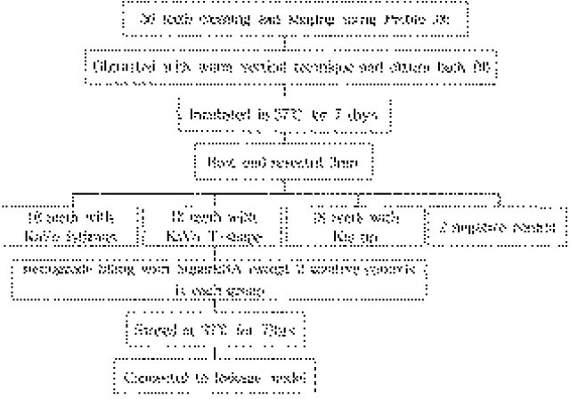
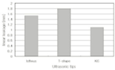
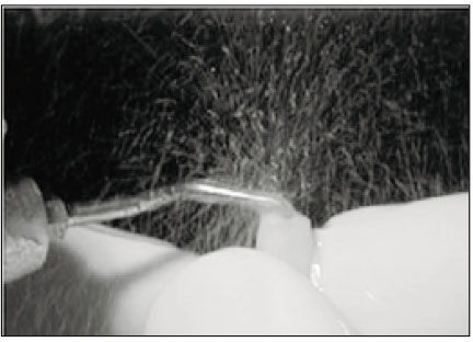
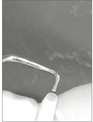
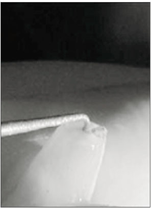
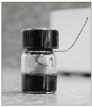
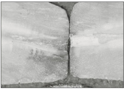
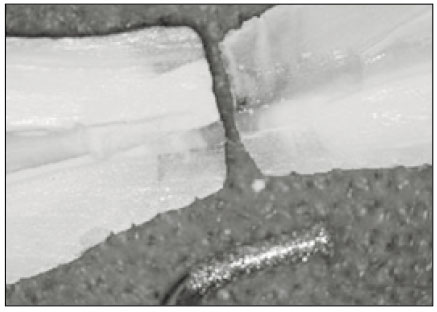
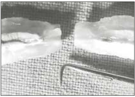
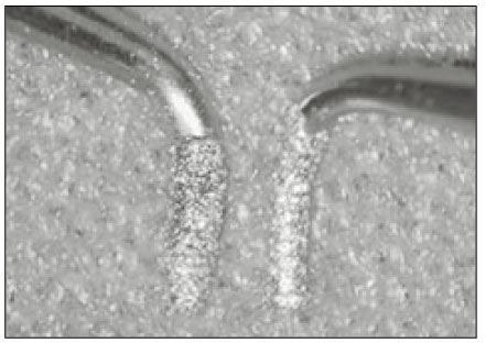
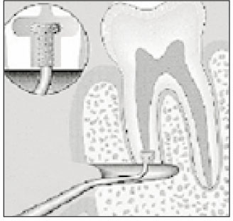
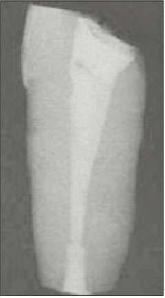
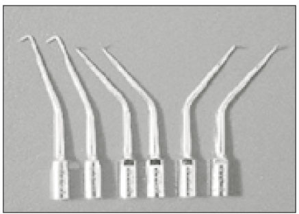
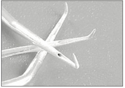
Fig. 1
Flow chart of the experimental design.
Fig. 2
Average linear leakage observed from the each experimental group.
Fig. 3
Retrograde cavity preparation with KaVo SONICflex retro Isthmus ultrasonic tip.
Fig. 4
Retrograde cavity preparation with KaVo SONICflex retro T-shape ultrasonic tip.
Fig. 5
Retrograde cavity preparation with ObturaSpartan KiS ultrasonic tip.
Fig. 6
Dye leakage model for this experiment.
Fig. 7
Linear leakage measured in the retrograde cavity prepared with KaVo Isthmus tip.
Fig. 8
Linear leakage measured in the retrograde cavity prepared with KaVo T-shape tip.
Fig. 9
Linear leakage measured in the retrograde cavity prepared with KiS tip.
Fig. 10
High magnification view of both KaVo ultrasonic tips.
Fig. 11
Shematic illustration of KaVo T-shape preparation.
Fig. 12
Radiographic view of SuperEBA filling after KaVo T-shape preparation.
Fig. 13
Six different configurations of KiS tips.
Fig. 14
Irrigation port placed at the front of the tip for better efficiency.
Fig. 1
Fig. 2
Fig. 3
Fig. 4
Fig. 5
Fig. 6
Fig. 7
Fig. 8
Fig. 9
Fig. 10
Fig. 11
Fig. 12
Fig. 13
Fig. 14
Leakage of SuperEBA in root-end cavities prepared with 3 new ultrasonic tips;KaVo Isthmus, KaVo T-shape and KiS tip

 KACD
KACD


 ePub Link
 Cite
Cite

