Warning: mkdir(): Permission denied in /home/virtual/lib/view_data.php on line 81

Warning: fopen(upload/ip_log/ip_log_2024-12.txt): failed to open stream: No such file or directory in /home/virtual/lib/view_data.php on line 83

Warning: fwrite() expects parameter 1 to be resource, boolean given in /home/virtual/lib/view_data.php on line 84
A confocal microscopic study on dentinal infiltration of one-bottle adhesive systems and self-etching priming system bonded to class V cavities
Skip Navigation
Skip to contents

Restor Dent Endod : Restorative Dentistry & Endodontics

OPEN ACCESS

Articles

Page Path
HOME > Restor Dent Endod > Volume 27(3); 2002 > Article
Original Article A confocal microscopic study on dentinal infiltration of one-bottle adhesive systems and self-etching priming system bonded to class V cavities
Hyung-Su Kim, Sung-Ho Park
Journal of Korean Academy of Conservative Dentistry 2002;27(3):257-269.
DOI: https://doi.org/10.5395/JKACD.2002.27.3.257
Published online: May 31, 2002

Department of Dentistry, The Graduate School of Dentistry, Yonsei University, Korea.

Copyright © 2002 Korean Academy of Conservative Dentistry

  • 18 Views
  • 0 Download
prev next
  • Objective
    The purpose of this study was to evaluate the resin infiltration into dentin of one-bottle adhesive systems and self-etching primer bonded to Class V cavities using confocal laser scanning microscope(CLSM).
  • Material and Methods
    Forty Class V cavities were prepared from freshly extracted caries-free human teeth. These teeth were divided into two groups based on the presence of cervical abrasion: Group I, cervical abrasion; Group II, wedge-shaped cavity preparation. Resin-dentin interfaces were produced with two one-bottle dentin bonding systems-ONE COAT BOND(OCB; Coltene®) and Syntac®Srint™(SS; VIVADENT)-, one self-etching priming system-CLEARFIL™ SE BOND(SB; KURARAY)- and one multi-step dentin bonding system-Scotchbond™Multi-Purpose(SBMP, 3M Dental Products)-as control according to manufacturers'instructions. Cavities were restored with Spectrum®(Dentsply). Specimens were immersed in saline for 24 hours and sectioned longitudinally with a low-speed diamond disc. The resin-dentin interfaces were microscopically observed using CLSM. The quality of resin-infiltrated dentin layers were evaluated by five dentists using 0-4 scale.
  • Results
    Confocal laser scanning microscopal investigations using primer labeled with rhodamine B showed that the penetration of the primer occurred along the cavity margins.
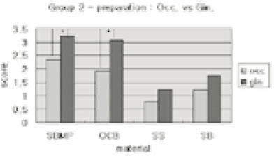
    Statistical analysis using one-way ANOVA followed by Duncan's Multiple Range test revealed that the primer penetration of the group 2(wedge-shaped cavity preparation) was more effective than group 1(cervical abrasion) and that of the gingival interfaces was more effective than the occlusal interfaces. In the one-bottle dentin bonding systems, the resin penetration score of OCB was compatible to SBMP, but those of SS and self-etching priming system, SB were lower than SBMP.
  • 1. Pashley DH, Carvalho RM. Dentine permeability and dentine adhesion. J Dent. 1997;25: 355-372.ArticlePubMed
  • 2. Ferrari M, Mannocci F, Cagidiaco MC, Kugel G. Short-term assessment of leakage of Class V composite restorations placed in vivo. Clin Oral Investig. 1997;1(2):61-64.ArticlePubMedPDF
  • 3. Gwinett AJ, Jendersen MD. Micromorphological features of cervical erosion after acid conditioning and its relation with composite resin. J Dent Res. 1978;57: 543-549.ArticlePubMedPDF
  • 4. Mixon JM, Spencer P, Moore DL, Chappell RP, Adams S. Surface morphology and chemical characterization of abrasion/erosion lesions. Am J Dent. 1995;8: 5-9.PubMed
  • 5. Heymann HO, Bayne SC. Current concepts in dentin bonding; focusing on dentinal adhesion factors. J Am Dent Assoc. 1993;124: 27-36.Article
  • 6. Schupbach P, Krejci I, Lutz F. Dentin bonding: effect of tubule orientation on hybrid layer formation. Eur J Oral Sci. 1997;105: 344-352.ArticlePubMed
  • 7. Van Meerbeek B, Braem M, Lambrechts P, Vanherle G. Morphological characterization of the interface between resin and sclerotic dentine. J Dent. 1994;22: 141-146.ArticlePubMed
  • 8. Tay FR, Gwinnett AJ, Wei SH. Micromorphological spectrum from overdrying to overwetting acid conditioned dentin in water-free, acetone-based, single-bottle primer/adhesives. Dent Mater. 1996;236-244.ArticlePubMed
  • 9. Watson TF, De Wilmot DM. A confocal microscopic evaluation of the interface between Syntac adhesive and tooth tissue. J Dent. 1992;20: 302-310.ArticlePubMed
  • 10. Duke ES, Lindemuth J. Variability of clinical dentin sustrates. Am J Dent. 1991;4: 241-246.PubMed
  • 11. Chappell RP, Cobb CM, Spencer P, Eick JD. Dentinal tubule anastomosis: a potential factor in adhesive bonding? J Prosthet Dent. 1994;72(2):183-188.ArticlePubMed
  • 12. Ferrari M, Cagidiaco CM, Mason PN. Morphologic aspects of the resin-dentin interdiffusion zone with five different dentin adhesive systems tested in vivo. J Prosthet Dent. 1994 4;71(4):404-408.ArticlePubMed
  • 13. Duke ES. Clinical studies of adhesive systems. Oper Dent. 1992;Suppl 5. 103-110.
  • 14. Yoshiyama M, Carvalho R, Sano H, Horner JA, Brewer PD, Pashley DH. Regional bond strengths of resins to human root dentine. J Dent. 1996;435-442.ArticlePubMed
  • 15. Hannig M, Reinhardt KJ, Bott B. Self-etching primer vs phosphoric acid:An alternative concept for composite-to-enamel bonding. Oper Dent. 1999;172-180.PubMed
  • 16. Hannig M, Reinhardt KJ, Bott B. Self-etching primer vs phosphoric acid: An alternative concept for composite to enamel bonding. Oper Dent. 1999;24: 172-180.PubMed
  • 17. Yoshiyama M, Sano H, Ebisu S, Tagami J, Ciucchi B, Carvalho RM, Johnson MH, Pashley DH. Regional strengths of bonding agents to cervical sclerotic root dentin. J Dent Res. 1996;75: 1404-1413.ArticlePubMedPDF
  • 18. Watson TF. Application of confocal scanning optical microscopy to dentistry. Br Dent J. 1991;9: 287-291.
  • 19. Minsky M. Microscopy apparatus. 1957 Nov 7 United States Patent Office Filed.
  • 20. Yoshiyama M, Carvalho R, Sano H, Horner JA, Brewer PD, Pashley DH. Interfacial morphology and strength of bonds made to superficial versus deep dentin. Am J Dent. 1995;297-302.
  • 21. Gwinnett AJ. Quantitative contribution of resin infiltration/hybridization to dentin bonding. Am J Dent. 1993;6: 7-9.PubMed
  • 22. Titley K, Chernecky R, Chan A, Smith D. The compositon and ultrastructure of resin tags in etched dentin. Am J Dent. 1995;8: 224-230.PubMed
  • 23. Pashley DH, et al. Permeability of dentin to adhesive agents. Quintessence Int. 1993;24: 618-631.PubMed
  • 24. Prati C, Chersoni S, Moniorgi R, Pashley DH. Resin-infiltrated dentin layer formation of new bonding systems. Oper Dent. 1998;23: 185-194.PubMed
Fig. 1
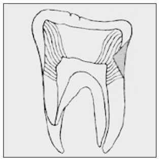
Diagram of longitudinally sectioned tooth showing cavity shapes (shadowed) and schematic orientation of dentinal tubules to the class V cavity surfaces.
jkacd-27-257-g001.jpg
Fig. 2
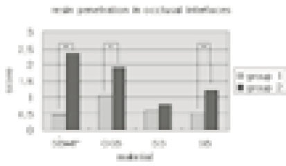
Resin penetration scores in occlusal interfaces of class V restorations. Asterisk(*) means statistically significant differences(p<0.05)
jkacd-27-257-g002.jpg
Fig. 3
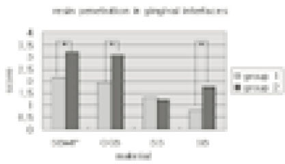
Resin penetration scores in gingival interfaces of class V restorations. Asterisk(*) means statistically significant differences(p<0.05)
jkacd-27-257-g003.jpg
Fig. 4
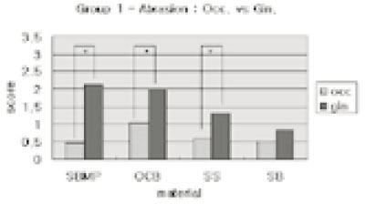
Comparison of resin penetrations of occlusal and gingival interfaces in group 1(cervical abrasion). Asterisk(*) means statistically significant differences (p<0.05)
jkacd-27-257-g004.jpg
Fig. 5
Comparison of resin penetrations of occlusal and gingival interfaces in group 2(V-shaped cavity preparaion). Asterisk(*) means statistically significant differences(p<0.05)
jkacd-27-257-g005.jpg
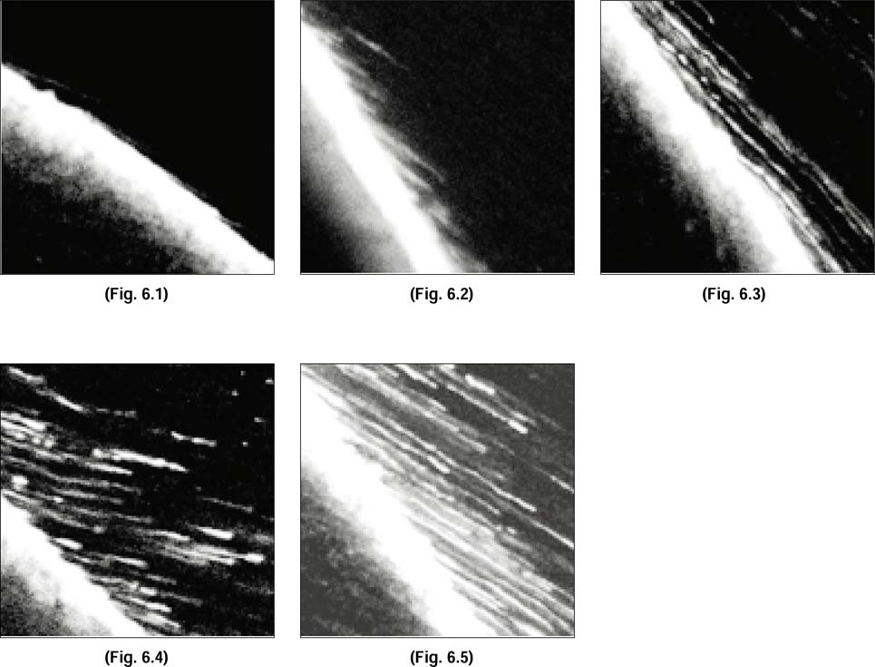
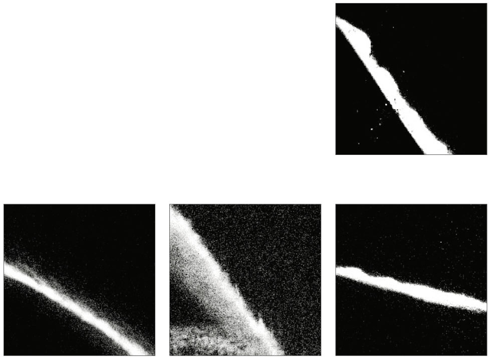
Fig. 6.1-6.5
Standard confocal laser scanning microscopic(CLSM) images at resin-dentin interfaces in occlusal interfaces of Class V cavity used for scoring the resin tag penetrations. Original magnification ×200
6.1. Score 0., 6.2. Score 1., 6.3. Score 2.,
6.4. Score 3., 6.5., Score 4.
jkacd-27-257-g006.jpg
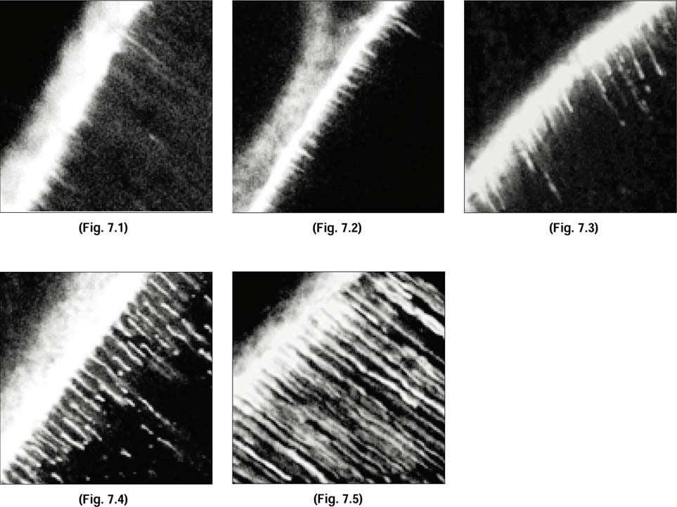
Fig. 7.1-7.5
Standard confocal laser scanning microscopic(CLSM) images at resin-dentin interfaces in gingival interfaces of Class V cavity used for scoring the resin tag penetrations. Original magnification ×200
7.1. Score 0., 7.2. Score 1., 7.3. Score 2.,
7.4. Score 3., 7.5., Score 4.
jkacd-27-257-g007.jpg
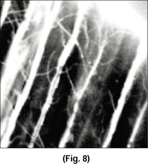
Fig. 8
Confocal laser scanning microscopic(CLSM) images at resin-dentin interfaces in gingival interfaces of Class V cavity with Scotchbond multi purpose. magnification × 800 23
jkacd-27-257-g008.jpg
Fig. 9.1-9.4
Confocal laser scanning microscopic(CLSM) images at resin-dentin interfaces in occlusal interfaces of cervical abrasion. Original magnification ×200
jkacd-27-257-g009.jpg
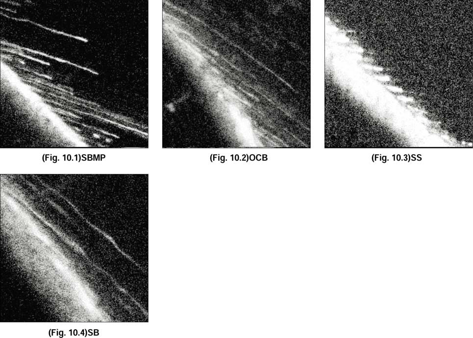
Fig. 10.1-10.4
Confocal laser scanning microscopic(CLSM) images at resin-dentin interfaces in occlusal interfaces of Class V cavity. Original magnification ×200
jkacd-27-257-g010.jpg
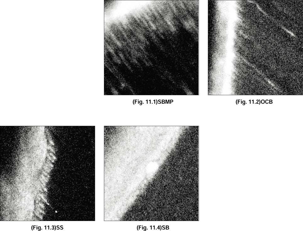
Fig. 11.1-11.4
Confocal laser scanning microscopic(CLSM) images at resin-dentin interfaces in gingival interfaces of cervical abrasion. Original magnification ×200
jkacd-27-257-g011.jpg
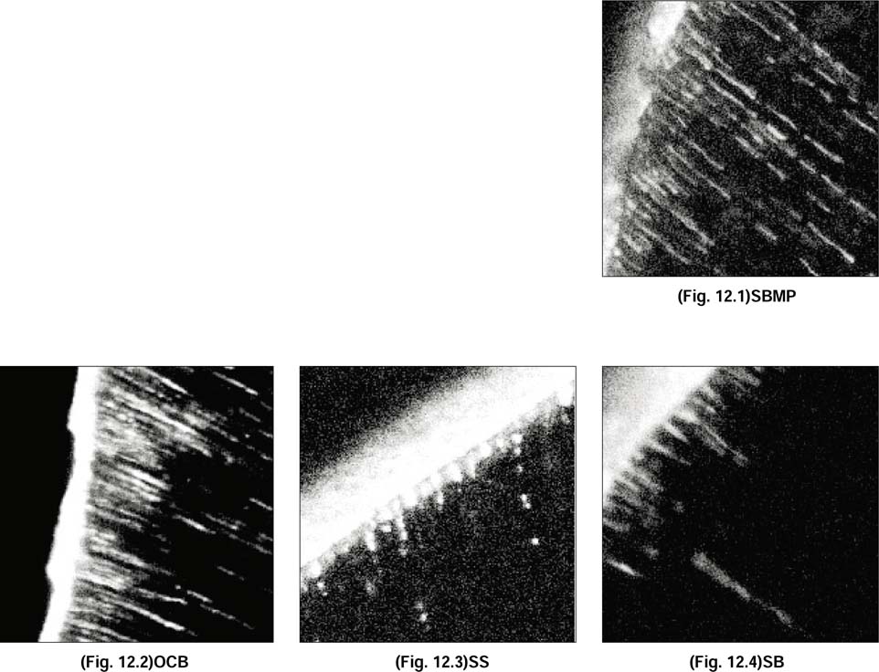
Fig. 12.1-12.4
Confocal laser scanning microscopic(CLSM) images at resin-dentin interfaces in gingival interfaces of Class V cavity. Original magnification ×200
jkacd-27-257-g012.jpg
Table 1
Names, numbers and characteristics of each groups of experiment
jkacd-27-257-i001.jpg
Table 2
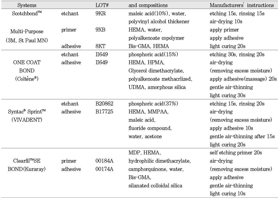
Chemical components and instructions for use of the dentin bonding systems used in this study
jkacd-27-257-i002.jpg
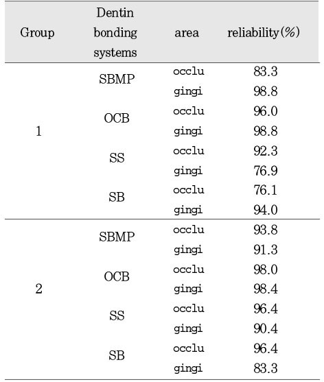
Table 3
Inter-observer correlations of each groups, materials, and parameters

occlu: resin penetrations in occlusal interfaces.

gingi: resin penetrations in gingival interfaces.

jkacd-27-257-i003.jpg
Table 4
Resin penetration scores(Mean±SD) at the occlusal interfaces

Comparison among the groups: one-way ANOVA, p<0.05

jkacd-27-257-i004.jpg
Table 5
Resin penetration scores(Mean±SD) at the gingival interfaces
jkacd-27-257-i005.jpg

Tables & Figures

REFERENCES

    Citations

    Citations to this article as recorded by  

      • ePub LinkePub Link
      • Cite
        CITE
        export Copy Download
        Close
        Download Citation
        Download a citation file in RIS format that can be imported by all major citation management software, including EndNote, ProCite, RefWorks, and Reference Manager.

        Format:
        • RIS — For EndNote, ProCite, RefWorks, and most other reference management software
        • BibTeX — For JabRef, BibDesk, and other BibTeX-specific software
        Include:
        • Citation for the content below
        A confocal microscopic study on dentinal infiltration of one-bottle adhesive systems and self-etching priming system bonded to class V cavities
        J Korean Acad Conserv Dent. 2002;27(3):257-269.   Published online May 31, 2002
        Close
      • XML DownloadXML Download
      Figure
      • 0
      • 1
      • 2
      • 3
      • 4
      • 5
      • 6
      • 7
      • 8
      • 9
      • 10
      • 11
      A confocal microscopic study on dentinal infiltration of one-bottle adhesive systems and self-etching priming system bonded to class V cavities
      Image Image Image Image Image Image Image Image Image Image Image Image
      Fig. 1 Diagram of longitudinally sectioned tooth showing cavity shapes (shadowed) and schematic orientation of dentinal tubules to the class V cavity surfaces.
      Fig. 2 Resin penetration scores in occlusal interfaces of class V restorations. Asterisk(*) means statistically significant differences(p<0.05)
      Fig. 3 Resin penetration scores in gingival interfaces of class V restorations. Asterisk(*) means statistically significant differences(p<0.05)
      Fig. 4 Comparison of resin penetrations of occlusal and gingival interfaces in group 1(cervical abrasion). Asterisk(*) means statistically significant differences (p<0.05)
      Fig. 5 Comparison of resin penetrations of occlusal and gingival interfaces in group 2(V-shaped cavity preparaion). Asterisk(*) means statistically significant differences(p<0.05)
      Fig. 6.1-6.5 Standard confocal laser scanning microscopic(CLSM) images at resin-dentin interfaces in occlusal interfaces of Class V cavity used for scoring the resin tag penetrations. Original magnification ×200 6.1. Score 0., 6.2. Score 1., 6.3. Score 2., 6.4. Score 3., 6.5., Score 4.
      Fig. 7.1-7.5 Standard confocal laser scanning microscopic(CLSM) images at resin-dentin interfaces in gingival interfaces of Class V cavity used for scoring the resin tag penetrations. Original magnification ×200 7.1. Score 0., 7.2. Score 1., 7.3. Score 2., 7.4. Score 3., 7.5., Score 4.
      Fig. 8 Confocal laser scanning microscopic(CLSM) images at resin-dentin interfaces in gingival interfaces of Class V cavity with Scotchbond multi purpose. magnification × 800 23
      Fig. 9.1-9.4 Confocal laser scanning microscopic(CLSM) images at resin-dentin interfaces in occlusal interfaces of cervical abrasion. Original magnification ×200
      Fig. 10.1-10.4 Confocal laser scanning microscopic(CLSM) images at resin-dentin interfaces in occlusal interfaces of Class V cavity. Original magnification ×200
      Fig. 11.1-11.4 Confocal laser scanning microscopic(CLSM) images at resin-dentin interfaces in gingival interfaces of cervical abrasion. Original magnification ×200
      Fig. 12.1-12.4 Confocal laser scanning microscopic(CLSM) images at resin-dentin interfaces in gingival interfaces of Class V cavity. Original magnification ×200
      A confocal microscopic study on dentinal infiltration of one-bottle adhesive systems and self-etching priming system bonded to class V cavities

      Names, numbers and characteristics of each groups of experiment

      Chemical components and instructions for use of the dentin bonding systems used in this study

      Inter-observer correlations of each groups, materials, and parameters

      occlu: resin penetrations in occlusal interfaces.

      gingi: resin penetrations in gingival interfaces.

      Resin penetration scores(Mean±SD) at the occlusal interfaces

      Comparison among the groups: one-way ANOVA, p<0.05

      Resin penetration scores(Mean±SD) at the gingival interfaces

      Table 1 Names, numbers and characteristics of each groups of experiment

      Table 2 Chemical components and instructions for use of the dentin bonding systems used in this study

      Table 3 Inter-observer correlations of each groups, materials, and parameters

      occlu: resin penetrations in occlusal interfaces.

      gingi: resin penetrations in gingival interfaces.

      Table 4 Resin penetration scores(Mean±SD) at the occlusal interfaces

      Comparison among the groups: one-way ANOVA, p<0.05

      Table 5 Resin penetration scores(Mean±SD) at the gingival interfaces


      Restor Dent Endod : Restorative Dentistry & Endodontics
      Close layer
      TOP