
Search
- Page Path
- HOME > Search
Research Articles
- Effects of dentin surface preparations on bonding of self-etching adhesives under simulated pulpal pressure
- Chantima Siriporananon, Pisol Senawongse, Vanthana Sattabanasuk, Natchalee Srimaneekarn, Hidehiko Sano, Pipop Saikaew
- Restor Dent Endod 2022;47(1):e4. Published online December 28, 2021
- DOI: https://doi.org/10.5395/rde.2022.47.e4
-
 Abstract
 PDF
PDF PubReader
 ePub
ePub Objectives This study evaluated the effects of different smear layer preparations on the dentin permeability and microtensile bond strength (µTBS) of 2 self-etching adhesives (Clearfil SE Bond [CSE] and Clearfil Tri-S Bond Universal [CTS]) under dynamic pulpal pressure.
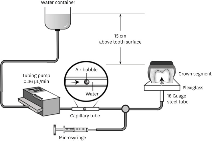
Materials and Methods Human third molars were cut into crown segments. The dentin surfaces were prepared using 4 armamentaria: 600-grit SiC paper, coarse diamond burs, superfine diamond burs, and carbide burs. The pulp chamber of each crown segment was connected to a dynamic intra-pulpal pressure simulation apparatus, and the permeability test was done under a pressure of 15 cmH2O. The relative permeability (%P) was evaluated on the smear layer-covered and bonded dentin surfaces. The teeth were bonded to either of the adhesives under pulpal pressure simulation, and cut into sticks after 24 hours water storage for the µTBS test. The resin-dentin interface and nanoleakage observations were performed using a scanning electron microscope. Statistical comparisons were done using analysis of variance and
post hoc tests.Results Only the method of surface preparation had a significant effect on permeability (
p < 0.05). The smear layers created by the carbide and superfine diamond burs yielded the lowest permeability. CSE demonstrated a higher µTBS, with these values in the superfine diamond and carbide bur groups being the highest. Microscopic evaluation of the resin-dentin interface revealed nanoleakage in the coarse diamond bur and SiC paper groups for both adhesives.Conclusions Superfine diamond and carbide burs can be recommended for dentin preparation with the use of 2-step CSE.
-
Citations
Citations to this article as recorded by
- Effect of smear layer pretreatment with EDTA and sodium hypochlorite on the dentin bond durability of universal adhesives
Thanawat Ruaydee, Chantida Pawaputanon Na Mahasarakham, Vanthana Sattabanasuk, Pipop Saikaew
Frontiers in Dental Medicine.2026;[Epub] CrossRef - Determination of marginal permeability of restorations in the cervical region using a universal adhesive system: a randomized controlled open-label laboratory study
Svetlana N. Razumova, Anzhela S. Brago, Oxana R. Ruda, Artur G. Talandis, Lamara M. Khaskhanova, Ruzanna M. Bragunova, Bohdan O. Pecherskyi
Russian Journal of Dentistry.2026; 30(2): 113. CrossRef - Catechol–Phosphonate–Augmented Universal Adhesive for Hydrolysis-Resistant Dentin Bonds: A µTBS and Spectroscopic Study
Rabeia J. Khalil, Suha K. Ibrahim, Athraa H. Madhat, Ali H. Tawfieq
European Journal of Dentistry.2026;[Epub] CrossRef - The effect of different adhesive strategies and diamond burs on dentin bond strength of universal resin cements
Chavakorn Atsavathavornset, Pipop Saikaew, Choltacha Harnirattisai, Hidehiko Sano
Clinical Oral Investigations.2025;[Epub] CrossRef - Universal adhesive systems in dentistry: A narrative review
Svetlana N. Razumova, Anzhela S. Brago, Oxana R. Ruda, Zoya A. Guryeva, Elvira V. Adzhieva
Russian Journal of Dentistry.2024; 28(5): 512. CrossRef - Delayed light activation of resin composite affects the bond strength of adhesives under dynamic simulated pulpal pressure
Nattaporn Sukprasert, Choltacha Harnirattisai, Pisol Senawongse, Hidehiko Sano, Pipop Saikaew
Clinical Oral Investigations.2022; 26(11): 6743. CrossRef
- Effect of smear layer pretreatment with EDTA and sodium hypochlorite on the dentin bond durability of universal adhesives
- 4,353 View
- 65 Download
- 4 Web of Science
- 6 Crossref

-
In vitro apical pressure created by 2 irrigation needles and a multisonic system in mandibular molars - Ronald Ordinola-Zapata, Joseph T. Crepps, Ana Arias, Fei Lin
- Restor Dent Endod 2021;46(1):e14. Published online February 8, 2021
- DOI: https://doi.org/10.5395/rde.2021.46.e14
-
Abstract
PDFPubReaderePub
Objectives The aim of this study was to evaluate the apical pressure generated by 2 endodontic irrigation needles and the GentleWave system in mandibular molars.
Materials and Methods The mesial and distal root canals of 12 mandibular molars were irrigated with a 30-gauge close-end needle or with a 30-gauge open-end needle. Procedures were performed in the mesial and distal canals. The GentleWave procedure and irrigation at 1 mm from the apex in the distal roots using an open-end needle were used, respectively, as negative and positive controls. The apical pressure was measured using a data acquisition pressure setup. Apical pressure exerted by the different needles in the 2 different canal types was statistically compared using 2-way analysis of variance.
Results Significant differences were found in the apical pressure for both needles and the canal type. The lowest values were obtained with close-end needles and in mesial canals. Negative apical pressure values were obtained using GentleWave.
Conclusions The needle and the canal type influenced the apical pressure. The GentleWave procedure produced negative apical pressure.
-
Citations
Citations to this article as recorded by- GentleWave versus Established Irrigation Techniques: Current Evidence from a Scoping Review
Mohmed Isaqali Karobari, Abdul Habeeb Adil, Niher Tabassum Snigdha, Emmanuel João Nogueira Leal da Silva
Journal of Endodontics.2026; 52(5): 713. CrossRef - Influence of kinematic motion and instrumentation strategy on apical debris extrusion during root canal preparation: An in vitro study
Amira Alghazaly, Jumanah Aljohani, Khadijah Mohabat, Rafah Ghous
Journal of Conservative Dentistry and Endodontics.2026; 29(7): 741. CrossRef - Use of the gentlewave system in endodonticsUse of the gentlewave system in endodontics
Daiana Jacobi Lazzarotto, Mayara Colpo Prado, Lara Dotto, Rafael Sarkis-Onofre
Brazilian Journal of Oral Sciences.2025; 24: e254250. CrossRef - Bibliometric analysis of the GentleWave system: trends, collaborations, and research gaps
Raimundo Sales de Oliveira Neto, Thais de Moraes Souza, João Vitor Oliveira de Amorim, Thaine Oliveira Lima, Guilherme Ferreira da Silva, Rodrigo Ricci Vivan, Murilo Priori Alcalde, Marco Antonio Hungaro Duarte
Restorative Dentistry & Endodontics.2025; 50(2): e17. CrossRef - The effect of ultrasonic and multisonic irrigation on root canal microbial communities: An ex vivo study
Ki Hong Park, Ronald Ordinola‐Zapata, W. Craig Noblett, Bruno P. Lima, Christopher Staley
International Endodontic Journal.2024; 57(7): 895. CrossRef - Efficacy of the GentleWave System in the removal of biofilm from the mesial roots of mandibular molars before and after minimal instrumentation: An ex vivo study
Kwang Ho Kim, Céline Lévesque, Gevik Malkhassian, Bettina Basrani
International Endodontic Journal.2024; 57(7): 922. CrossRef - A critical analysis of research methods and experimental models to study irrigants and irrigation systems
Christos Boutsioukis, Maria Teresa Arias‐Moliz, Luis E. Chávez de Paz
International Endodontic Journal.2022; 55(S2): 295. CrossRef - Outcomes of the GentleWave system on root canal treatment: a narrative review
Hernán Coaguila-Llerena, Eduarda Gaeta, Gisele Faria
Restorative Dentistry & Endodontics.2022;[Epub] CrossRef
- GentleWave versus Established Irrigation Techniques: Current Evidence from a Scoping Review
- 3,232 View
- 39 Download
- 7 Web of Science
- 8 Crossref
Case Report
- An embouchure aid for saxophone player
- Ho-Jin Moon
- Restor Dent Endod 2012;37(1):54-60. Published online March 2, 2012
- DOI: https://doi.org/10.5395/rde.2012.37.1.54
-
Abstract
PDFPubReaderePub
This study aims to introduce the method that can relieve vibrating forces to oral environment by making an embouchure aid. Thin plastic crown forms were fabricated to prevent tooth abrasion and irritation to lip mucosa for the saxophone player. After application to the player, the most comfort form was chosen and delivered to 3 professional saxophone players. After 5 mon, the players responded to the survey. This embouchure aid did not disturb playing and gave comfort to lower lip. In general, the players preferred thin soft type and thought it caused little effect on sound. Far too little attention has been paid to the problems encountered by single-reed wind instrumentalist who suffer from tooth abrasion and irritation to lip mucosa. The embouchure aid not only prevent tooth damage but also diminish the discomfort of tight embouchure.
- 2,335 View
- 10 Download
Basic Research
- The evaluation of periodontal ligament cells of rat teeth after low-temperature preservation under high pressure
- Jin-Ho Chung, Jin Kim, Seong-Ho Choi, Eui-Seong Kim, Jiyong Park, Seung-Jong Lee
- J Korean Acad Conserv Dent 2010;35(4):285-294. Published online July 31, 2010
- DOI: https://doi.org/10.5395/JKACD.2010.35.4.285
-
Abstract
PDFPubReaderePub
The purpose of this study was to evaluate the viability of periodontal ligament cells of rat teeth after low-temperature preservation under high pressure by means of MTT assay, WST-1 assay. 12 teeth of Sprague-Dawley white female rats of 4 week-old were used for each group.
Both side of the first and second maxillary molars were extracted as atraumatically as possible under tiletamine anesthesia. The experimental groups were group 1 (Immediate extraction), group 2 (Slow freezing under pressure of 3 MPa), group 3 (Slow freezing under pressure of 2 MPa), group 4 (Slow freezing under no additional pressure), group 5 (Rapid freezing in liquid nitrogen under pressure of 2 MPa), group 6 (Rapid freezing in liquid nitrogen under no additional pressure), group 7 (low-temperature preservation at 0℃ under pressure of 2 MPa), group 8 (low-temperature preservation at 0℃ under no additional pressure), group 9 (low-temperature preservation at -5℃ under pressure of 90 MPa). F-medium and 10% DMSO were used as preservation medium and cryo-protectant. For cryo-preservation groups, thawing was performed in 37℃ water bath, then MTT assay, WST-1 assay were processed. One way ANOVA and Tukey HSD method were performed at the 95% level of confidence. The values of optical density obtained by MTT assay and WST-1 were divided by the values of eosin staining for tissue volume standardization.
In both MTT and WST-1 assay, group 7 (0℃/2 MPa) showed higher viability of periodontal ligament cells than other group (2-6, 8) and this was statistically significant (p < 0.05), but showed lower viability than group 1, immediate extraction group (no statistical significance).
By the results of this study, low-temperature preservation at 0℃ under pressure of 2 MPa suggest the possibility for long term preservation of teeth.
-
Citations
Citations to this article as recorded by- Evaluation of the Viability of Rat Periodontal Ligament Cells after Storing at 0℃/2 MPa Condition up to One Week: In Vivo MTT Method
Sun Mi Jang, Sin-Yeon Cho, Eui-Seong Kim, Il-Young Jung, Seung Jong Lee
Journal of Korean Dental Science.2016; 9(1): 1. CrossRef
- Evaluation of the Viability of Rat Periodontal Ligament Cells after Storing at 0℃/2 MPa Condition up to One Week: In Vivo MTT Method
- 1,833 View
- 2 Download
- 1 Crossref
Original Articles
- Comparison of viability of oral epithelial cells stored by different freezing methods
- Do-Young Baek, Seung-Jong Lee, Han-Sung Jung, EuiSeong Kim
- J Korean Acad Conserv Dent 2009;34(6):491-499. Published online November 30, 2009
- DOI: https://doi.org/10.5395/JKACD.2009.34.6.491
-
Abstract
PDFPubReaderePub
This study examined the influence of the storage methods on the viability of oral epithelial cells using conventional cell freezing storage, slow freezing preservation, rapid freezing preservation, and slow freezing preservation with a pressure of 2 Mpa or 3 Mpa. The cell viability was evaluated by cell counting, WST-1 and the clonogenic capacity after 6 days of freezing storage. After 6 days, the frozen cells were thawed rapidly, and the cell counting, WST-1, and clonogenic capacity values were measured and compared.
The results from cell counting demonstrated that conventional cryopreservation, slow freezing under a 2 Mpa pressure and slow freezing under a 3 Mpa pressure showed significantly higher values than slow freezing preservation and rapid freezing preservation (p<0.05).
The results from the optical density by WST-1 demonstrated that slow freezing under a 2 Mpa pressure showed significantly higher values than slow freezing preservation and rapid freezing preservation (p<0.05).
The clonogenic capacity demonstrated that slow freezing under a 2 Mpa pressure showed significantly higher values than slow freezing preservation and rapid freezing preservation (p<0.05).
-
Citations
Citations to this article as recorded by- Evaluation of the Viability of Rat Periodontal Ligament Cells after Storing at 0℃/2 MPa Condition up to One Week: In Vivo MTT Method
Sun Mi Jang, Sin-Yeon Cho, Eui-Seong Kim, Il-Young Jung, Seung Jong Lee
Journal of Korean Dental Science.2016; 9(1): 1. CrossRef - The evaluation of periodontal ligament cells of rat teeth after low-temperature preservation under high pressure
Jin-Ho Chung, Jin Kim, Seong-Ho Choi, Eui-Seong Kim, Jiyong Park, Seung-Jong Lee
Journal of Korean Academy of Conservative Dentistry.2010; 35(4): 285. CrossRef
- Evaluation of the Viability of Rat Periodontal Ligament Cells after Storing at 0℃/2 MPa Condition up to One Week: In Vivo MTT Method
- 2,109 View
- 2 Download
- 2 Crossref
-
EVALUATION OF
ENTEROCOCCUS FAECALIS REMOVAL EFFICACY OF THE ENDOVAC® AND ENDOACTIVATOR® INTRACANAL IRRIGATION METHODS - Seung-Gon Song, Kyung-Mo Cho, Jin-Woo Kim
- J Korean Acad Conserv Dent 2009;34(5):390-396. Published online January 14, 2009
- DOI: https://doi.org/10.5395/JKACD.2009.34.5.390
-
Abstract
PDFPubReaderePub
Abstract The aim of this study was to evaluate endodontic irrigation methods with EndoVac® and EndoActivator® in the elimination of
Enterococcus faecalis from the root canals. Extracted 70 human single-rooted teeth were used. The canals were instrumented by a crown-down technique with .04 taper ProFile to ISO size 40. After the teeth were autoclaved, the canals were inoculated withE. faecalis and incubated for 48 h. The teeth were randomly divided into three experimental groups of 20 teeth each according to canal irrigation methods and two control groups as follows: group 1 - EndoVac®; group 2 - EndoActivator®; group 3 - Conventional needle irrigation method. After canal irrigation using 2.5% NaOCl, first samples (S1) were taken using sterile paper point. And the canals were filled with sterile brain heart infusion (BHI) broth and incubated for 24 h, then second samples (S2) were taken. The samples were cultured on BHI agar plate to determine the numbers of colony forming units (CFU). In first sampling (S1), only one canal of conventional method among the all experimental groups was positive cultured. In second sampling (S2), EndoVac® group showed the least positive culture numbers ofE. faecalis . There was statistically significant difference between the EndoVac® and conventional needle irrigation methods in the mean value of Log CFU. According to the results of this study, EndoVac® showed better efficacy than conventional needle irrigation method in the elimination ofE. faecalis from the root canal.
- 1,328 View
- 9 Download
- THE EFFICACY OF PROGRAMMED CRYO-PRESERVATION UNDER PRESSURE IN RAT PERIODONTAL LIGAMENT CELLS
- Young-Eun Lee, Eui-Seong Kim, Jin Kim, Seung-Hoon Han, Seung-Jong Lee
- J Korean Acad Conserv Dent 2009;34(4):356-363. Published online January 14, 2009
- DOI: https://doi.org/10.5395/JKACD.2009.34.4.356
-
Abstract
PDFPubReaderePub
Abstract The purpose of this study was to evaluate the viability of periodontal ligament cells in rat teeth using slow cryo-preservation method under pressure by means of MTT assay and WST-1 assay. Eighteen teeth of Sprague-Dawley white female rats of 4 week-old were used for each group.
Both sides of the first and second maxillary molars were extracted as atraumatically as possible under Tiletamine anesthesia. The experimental groups were group 1 (Immediate control), group 2 (Cold preservation at 4°C for 1 week), group 3 (Slow freezing), group 4 (Slow freezing under pressure of 3 MPa). F-medium and 10% DMSO were used as preservation medium and cryo-protectant. For cryo-preservation groups, thawing was performed in 37°C water bath, then MTT assay and WST-1 assay were processed. One way ANOVA and Tukey method were performed at the 95% level of confidence. The values of optical density obtained by MTT assay and WST-1 were divided by the values of eosin staining for tissue volume standardization.
In both MTT and WST-1 assay, group 4 showed significantly higher viability of periodontal ligament cells than group 2 and 3 (p < 0.05), but showed lower viability than immediate control group.
By the results of this study, slow cryo-preservation method under pressure suggests the possibility for long term cryo-preservation of the teeth.
-
Citations
Citations to this article as recorded by- Effects of Slow Programmable Cryopreservation on Preserving Viability of the Cultured Periodontal Ligament Cells from Human Impacted Third Molar
Jin-Woo Kim, Tae-Yi Kim, Ye-mi Kim, Eun-Kyoung Pang, Sun-Jong Kim
Journal of Korean Dental Science.2015; 8(2): 57. CrossRef - The evaluation of periodontal ligament cells of rat teeth after low-temperature preservation under high pressure
Jin-Ho Chung, Jin Kim, Seong-Ho Choi, Eui-Seong Kim, Jiyong Park, Seung-Jong Lee
Journal of Korean Academy of Conservative Dentistry.2010; 35(4): 285. CrossRef - Comparison of viability of oral epithelial cells stored by different freezing methods
Do-Young Baek, Seung-Jong Lee, Han-Sung Jung, EuiSeong Kim
Journal of Korean Academy of Conservative Dentistry.2009; 34(6): 491. CrossRef
- Effects of Slow Programmable Cryopreservation on Preserving Viability of the Cultured Periodontal Ligament Cells from Human Impacted Third Molar
- 1,818 View
- 1 Download
- 3 Crossref
- Micro-shear bond strength to dentin under simulated pulpal pressure
- Yun-Jung Song, Sung-Ho Park
- J Korean Acad Conserv Dent 2004;29(4):339-345. Published online July 31, 2004
- DOI: https://doi.org/10.5395/JKACD.2004.29.4.339
-
Abstract
PDFPubReaderePub
The aim of this study was to measure and compare the micro shear bond strengths of the following dentin bonding systems to the dentin surfaces under simulated pulpal pressure; All Bond 2®, Second®, AdheSE®, Adper Prompt L-Pop®. The occlusal surfaces of 180 extracted human molars were prepared so the dentin bonding surfaces could be exposed. The teeth were randomly assigned to 3 equal groups of 60 each and subdivided. The dentin surfaces were treated with the above mentioned bonding system and resin composite cylinders were built up under a simulated pulpal pressure when saline (Group II) or diluted bovine serum (Group III) was used as the pulpal fluid. As a control, the same procedures were performed in the dried dentin surfaces (Group I). After one day of storage in water, the micro shear bond strengths were measured using an EZ tester. Group II and III showed significantly lower shear bond strength than Group I statistically (p < 0.05). SEbond® and AdheSE® showed no difference among the different dentin condition. In the Adper Prompt L-Pop®, a simulated pulpal pressure were applied to the specimens using diluted bovine serum, which showed a higher strength than the specimens in which saline was used (p < 0.05).
- 1,171 View
- 2 Download
 First
First Prev
Prev

