Search
- Page Path
- HOME > Search
-
A new minimally invasive guided endodontic microsurgery by cone beam computed tomography and 3-dimensional printing technology

- Jong-Eun Kim, June-Sung Shim, Yooseok Shin
- Restor Dent Endod 2019;44(3):e29. Published online July 25, 2019
- DOI: https://doi.org/10.5395/rde.2019.44.e29

-
 Abstract
 PDF
PDF Supplementary Material
 PubReader
 ePub
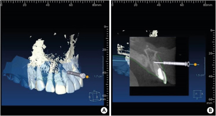
ePub Endodontic microsurgery is defined as the treatment performed on the root apices of an infected tooth, which was unresolved with conventional root canal therapy. Recently, the advanced technology in 3-dimensional model reconstruction based on computed tomography such as cone beam computed tomography has opened a new avenue in application of personalized, accurate diagnosis and has been increasingly used in the field of dentistry. Nevertheless, direct intra-oral localization of root apex based on the 3-dimensional information is extremely difficult and significant amount of bone removal is inevitable when freehand surgical procedure was employed. Moreover, gingival flap and alveolar bone fenestration are usually required, which leads to prolonged time of surgery, thereby increasing the chance of trauma as well as the risk of infection. The purpose of this case report is to present endodontic microsurgery using the guide template that can accurately target the position of apex for the treatment of an anterior tooth with calcified canal which was untreatable with conventional root canal therapy and unable to track the position of the apex due to the absence of fistula.
- 23 View
- 0 Download

- Accidental injury of the inferior alveolar nerve due to the extrusion of calcium hydroxide in endodontic treatment: a case report
- Yooseok Shin, Byoung-Duck Roh, Yemi Kim, Taehyeon Kim, Hyungjun Kim
- Restor Dent Endod 2016;41(1):63-67. Published online January 6, 2016
- DOI: https://doi.org/10.5395/rde.2016.41.1.63

-
 Abstract
 PDF
PDF PubReader
 ePub
ePub During clinical endodontic treatment, we often find radiopaque filling material beyond the root apex. Accidental extrusion of calcium hydroxide could cause the injury of inferior alveolar nerve, such as paresthesia or continuous inflammatory response. This case report presents the extrusion of calcium hydroxide and treatment procedures including surgical intervention. A 48 yr old female patient experienced Calcipex II extrusion in to the inferior alveolar canal on left mandibular area during endodontic treatment. After completion of endodontic treatment on left mandibular first molar, surgical intervention was planned under general anesthesia. After cortical bone osteotomy and debridement, neuroma resection and neurorrhaphy was performed, and prognosis was observed. But no improvement in sensory nerve was seen following surgical intervention after 20 mon. A clinician should be aware of extrusion of intracanal medicaments and the possibility of damage on inferior alveolar canal. Injectable type of calcium hydroxide should be applied with care for preventing nerve injury. The alternative delivery method such as lentulo spiral was suggested on the posterior mandibular molar.
- 21 View
- 1 Download

- A study on the compatibility between one-bottle dentin adhesives and composite resins using micro-shear bond strength
- Minju Song, Yooseok Shin, Jeong-Won Park, Byoung-Duck Roh
- Restor Dent Endod 2015;40(1):30-36. Published online September 26, 2014
- DOI: https://doi.org/10.5395/rde.2015.40.1.30
-
 Abstract
 PDF
PDF PubReader
 ePub
ePub Objectives This study was performed to determine whether the combined use of one-bottle self-etch adhesives and composite resins from same manufacturers have better bond strengths than combinations of adhesive and resins from different manufacturers.
Materials and Methods 25 experimental micro-shear bond test groups were made from combinations of five dentin adhesives and five composite resins with extracted human molars stored in saline for 24 hr. Testing was performed using the wire-loop method and a universal testing machine. Bond strength data was statistically analyzed using two way analysis of variance (ANOVA) and Tukey's
post hoc test.Results Two way ANOVA revealed significant differences for the factors of dentin adhesives and composite resins, and significant interaction effect (
p < 0.001). All combinations with Xeno V (Dentsply De Trey) and Clearfil S3 Bond (Kuraray Dental) adhesives showed no significant differences in micro-shear bond strength, but other adhesives showed significant differences depending on the composite resin (p < 0.05). Contrary to the other adhesives, Xeno V and BondForce (Tokuyama Dental) had higher bond strengths with the same manufacturer's composite resin than other manufacturer's composite resin.Conclusions Not all combinations of adhesive and composite resin by same manufacturers failed to show significantly higher bond strengths than mixed manufacturer combinations.
- 32 View
- 0 Download

- Evaluation of canal preparation for apical sealing with various Ni-Ti rotary instruments
- Yooseok Shin, Su-Jung Shin, Minju Song, Euiseong Kim
- J Korean Acad Conserv Dent 2011;36(4):300-305. Published online July 31, 2011
- DOI: https://doi.org/10.5395/JKACD.2011.36.4.300
-
 Abstract
 PDF
PDF PubReader
 ePub
ePub Objectives The aim of this study was to evaluate the various NiTi rotary instruments regarding their ability to provide a circular apical preparation.
Materials and Methods 50 single canal roots were selected, cut at the cementodentinal junction and the coronal 1/3 of the canals was flared using Gates Glidden burs. Samples were randomly divided into 5 experimental groups of 10 each. In group I, GT files, Profile 04 and Quantec #9 and #10 files were used. In Group II Lightspeed was used instead of Quantec. In Group III, Orifice shaper, Profile .06 series and Lightspeed were used. In Group IV, Quantec #9 and #10 files were used instead of Lightspeed. In Group V, the GT file and the Profile .04 series were used to prepare the entire canal length. All tooth samples were cut at 1 mm, 3 mm and 5 mm from the apex and were examined under the microscope.
Results Groups II and III (Lightspeed) showed a more circular preparation in the apical 1mm samples than the groups that used Quantec (Group I & IV) or GT files and Profile .04 series.(Group V)(
p < 0.05) There was no significant difference statistically among the apical 3, 5 mm samples. In 5 mm samples, most of the samples showed complete circularity and none of them showed irregular shape.Conclusions Lightspeed showed circular preparation at apical 1 mm more frequently than other instruments used in this study. However only 35% of samples showed circularity even in the Lightspeed Group which were enlarged 3 ISO size from the initial apical binding file (IAF) size. So it must be considered that enlarging 3 ISO size isn't enough to make round preparation.
- 19 View
- 0 Download


 KACD
KACD
 First
First Prev
Prev


