Search
- Page Path
- HOME > Search
- Root canal volume change and transportation by Vortex Blue, ProTaper Next, and ProTaper Universal in curved root canals
- Hyun-Jin Park, Min-Seock Seo, Young-Mi Moon
- Restor Dent Endod 2018;43(1):e3. Published online December 24, 2017
- DOI: https://doi.org/10.5395/rde.2018.43.e3

-
 Abstract
 PDF
PDF PubReader
 ePub
ePub Objectives The aim of this study was to compare root canal volume change and canal transportation by Vortex Blue (VB; Dentsply Tulsa Dental Specialties), ProTaper Next (PTN; Dentsply Maillefer), and ProTaper Universal (PTU; Dentsply Maillefer) nickel-titanium rotary files in curved root canals.
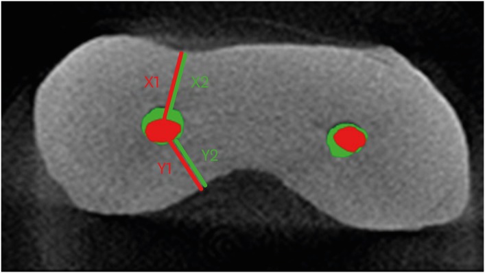
Materials and Methods Thirty canals with 20°–45° of curvature from extracted human molars were used. Root canal instrumentation was performed with VB, PTN, and PTU files up to #30.06, X3, and F3, respectively. Changes in root canal volume before and after the instrumentation, and the amount and direction of canal transportation at 1, 3, and 5 mm from the root apex were measured by using micro-computed tomography. Data of canal volume change were statistically analyzed using one-way analysis of variance and Tukey test, while data of amount and direction of transportation were analyzed using Kruskal-Wallis and Mann-Whitney
U test.Results There were no significant differences among 3 groups in terms of canal volume change (
p > 0.05). For the amount of transportation, PTN showed significantly less transportation than PTU at 3 mm level (p = 0.005). VB files showed no significant difference in canal transportation at all 3 levels with either PTN or PTU files. Also, VB files showed unique inward transportation tendency in the apical area.Conclusions Other than PTN produced less amount of transportation than PTU at 3 mm level, all 3 file systems showed similar level of canal volume change and transportation, and VB file system could prepare the curved canals without significant shaping errors.
-
Citations
Citations to this article as recorded by- The effect of nickel-titanium rotary systems on the biomechanical behaviour of mandibular first molars with curved and straight mesial roots: a finite element analysis study
Yaprak Cesur, Sevinc Askerbeyli Örs, Ahmet Serper, Mert Ocak
BMC Oral Health.2025;[Epub] CrossRef - Micro-Computed Tomographic Evaluation of the Shaping Ability of Vortex Blue and TruNatomyTM Ni-Ti Rotary Systems
Batool Alghamdi, Mey Al-Habib, Mona Alsulaiman, Lina Bahanan, Ali Alrahlah, Leonel S. J. Bautista, Sarah Bukhari, Mohammed Howait, Loai Alsofi
Crystals.2024; 14(11): 980. CrossRef - Evaluation of the Centering Ability and Canal Transportation of Rotary File Systems in Different Kinematics Using CBCT
Nupur R Vasava, Shreya H Modi, Chintan Joshi, Mona C Somani, Sweety J Thumar, Aashray A Patel, Anisha D Parmar, Kruti M Jadawala
World Journal of Dentistry.2024; 14(11): 983. CrossRef - Comparative evaluation of nickel titanium rotary instruments on canal transportation and centering ability in curved canals by using cone beam computed tomography: An in vitro study
Krishnaveni Krishnaveni, Nikitha Kalla, Nagalakshmi Reddy, Sharvanan Udayar
Journal of Dental Specialities.2023; 11(2): 105. CrossRef - Comparative Evaluation of Root Canal Centering Ability of Two Heat-treated Single-shaping NiTi Rotary Instruments in Simulated Curved Canals: An In Vitro Study
Preethi Varadan, Chakravarthy Arumugam, Athira Shaji, R R Mathan
World Journal of Dentistry.2023; 14(6): 535. CrossRef - A Comparison of Canal Width Changes in Simulated Curved Canals prepared with Profile and Protaper Rotary Systems
Aisha Faisal, Huma Farid, Robia Ghafoor
Pakistan Journal of Health Sciences.2022; : 55. CrossRef - Evaluation of the Respect of the Root Canal Trajectory by Rotary Niti Instruments (Protaper®Universal): Retrospective Radiographic Study
Salma El Abbassi, Sanaa Chala, Majid Sakout, Faïza Abdallaoui
Integrative Journal of Medical Sciences.2022;[Epub] CrossRef
- The effect of nickel-titanium rotary systems on the biomechanical behaviour of mandibular first molars with curved and straight mesial roots: a finite element analysis study
- 1,647 View
- 11 Download
- 7 Crossref

- In-depth morphological study of mesiobuccal root canal systems in maxillary first molars: review
- Seok-Woo Chang, Jong-Ki Lee, Yoon Lee, Kee-Yeon Kum
- Restor Dent Endod 2013;38(1):2-10. Published online February 26, 2013
- DOI: https://doi.org/10.5395/rde.2013.38.1.2
-
 Abstract
 PDF
PDF PubReader
 ePub
ePub A common failure in endodontic treatment of the permanent maxillary first molars is likely to be caused by an inability to locate, clean, and obturate the second mesiobuccal (MB) canals. Because of the importance of knowledge on these additional canals, there have been numerous studies which investigated the maxillary first molar MB root canal morphology using
in vivo and laboratory methods. In this article, the protocols, advantages and disadvantages of various methodologies for in-depth study of maxillary first molar MB root canal morphology were discussed. Furthermore, newly identified configuration types for the establishment of new classification system were suggested based on two image reformatting techniques of micro-computed tomography, which can be useful as a further 'Gold Standard' method for in-depth morphological study of complex root canal systems.-
Citations
Citations to this article as recorded by- An epidemiological study of extracted mandibular premolars from adolescent patients in Damascus using two classification system analyzed with CBCT and digital periapical radiographs
Yasser Alsayed Tolibah, Mohammed N. Al-Shiekh, Mohammad Tamer Abbara, Marwan Alhaji, Osama Aljabban, Nada Bshara
BMC Oral Health.2025;[Epub] CrossRef - Cone beam computed tomography analysis of the root and canal morphology of the maxillary second molars in a Hail province of the Saudi population
Ahmed A. Madfa, Moazzy I. Almansour, Saad M. Al-Zubaidi, Albandari H. Alghurayes, Safanah D. AlDAkhayel, Fatemah I. Alzoori, Taif F. Alshammari, Abrar M. Aldakhil
Heliyon.2023; 9(9): e19477. CrossRef - Signs of a missed root canal
M. Yu. Pokrovsky, O. A. Aleshina, T. P. Goryacheva, A. M. Pokrovskiy
Endodontics Today.2023; 21(3): 205. CrossRef - Root Canal Morphology of Maxillary First and Second Molars in a Qatari Population: A Cone-Beam Computed Tomography Study
Maryam Mohammed Al-Obaid, Fatima Abdullah Al-Sheeb
European Dental Research and Biomaterials Journal.2021; 2(01): 34. CrossRef - A Study Comparing the Characteristics of Zinc Oxide Eugenol-Based and Mineral Trioxide Aggregate-Based Root Canal Sealers
Seok-Eun Lee, Ja-Won Cho, Hyun-Jun Yoo, Myung-Gu Lee, Yeol-Mae Jeon, Da-Hui Kim, Hye-Won Park
International Journal of Clinical Preventive Dentistry.2021; 17(3): 117. CrossRef - Root Canal Configuration of Burmese (Myanmar) Maxillary First Molar: A Micro-Computed Tomography Study
M. M. Kyaw Moe, H. J. Jo, J. H. Ha, S. K. Kim, Antonino Lo Giudice
International Journal of Dentistry.2021; 2021: 1. CrossRef - Three-Dimensional Analysis of Root Anatomy and Root Canal Curvature in Mandibular Incisors Using Micro-Computed Tomography with Novel Software
JongKi Lee, Shin-Hoon Lee, Jong-Rak Hong, Kee-Yeon Kum, Soram Oh, Adel Saeed Al-Ghamdi, Fawzi Ali Al-Ghamdi, Ayman Omar Mandorah, Ji-Hyun Jang, Seok Woo Chang
Applied Sciences.2020; 10(12): 4385. CrossRef - An investigation into dose optimisation for imaging root canal anatomy using cone beam CT
Margarete B McGuigan, Christie Theodorakou, Henry F Duncan, Jonathan Davies, Anita Sengupta, Keith Horner
Dentomaxillofacial Radiology.2020; 49(7): 20200072. CrossRef - Analysis of Root Canal Anatomy and Variation in Morphology of Maxillary First Molar Using Various Methods: An In Vitro Study
Youssef A Algarni
World Journal of Dentistry.2019; 10(4): 291. CrossRef - Root Canal Morphology of Mandibular Primary Molars: A Micro-CT Study
Meryem ZİYA, Burcu Nihan YÜKSEL, Şaziye SARI
Cumhuriyet Dental Journal.2019; 22(4): 382. CrossRef - Comparison of the implementation of extra root canal treatment before and after fee schedule change in the Taiwan National Health Insurance System
Nien-Chieh Lee, Yen-Hsiang Chang, Hui-Tzu Tu, Chang-Fu Kuo, Kuang-Hui Yu, Lai-Chu See
Journal of Dental Sciences.2018; 13(2): 145. CrossRef - Influence of environment on testing of hydraulic sealers
Mira Kebudi Benezra, Pierre Schembri Wismayer, Josette Camilleri
Scientific Reports.2017;[Epub] CrossRef - CBCT uses in clinical endodontics: the effect of CBCT on the ability to locate MB2 canals in maxillary molars
J. Parker, A. Mol, E. M. Rivera, P. Tawil
International Endodontic Journal.2017; 50(12): 1109. CrossRef - Comparison of Alternative Image Reformatting Techniques in Micro–Computed Tomography and Tooth Clearing for Detailed Canal Morphology
Ki-Wook Lee, Yeun Kim, Hiran Perinpanayagam, Jong-Ki Lee, Yeon-Jee Yoo, Sang-Min Lim, Seok Woo Chang, Byung-Hyun Ha, Qiang Zhu, Kee-Yeon Kum
Journal of Endodontics.2014; 40(3): 417. CrossRef - In Vitro Biocompatibility, Inflammatory Response, and Osteogenic Potential of 4 Root Canal Sealers: Sealapex, Sankin Apatite Root Sealer, MTA Fillapex, and iRoot SP Root Canal Sealer
Seok-Woo Chang, So-Youn Lee, Soo-Kyung Kang, Kee-Yeon Kum, Eun-Cheol Kim
Journal of Endodontics.2014; 40(10): 1642. CrossRef - Análise do preparo de canais radiculares utilizando-se a diafanização
Georje de Martin, Rogério Albuquerque Azeredo
Revista de Odontologia da UNESP.2014; 43(2): 111. CrossRef
- An epidemiological study of extracted mandibular premolars from adolescent patients in Damascus using two classification system analyzed with CBCT and digital periapical radiographs
- 1,968 View
- 14 Download
- 16 Crossref

- The effect of C-factor and volume on microleakage of composite resin restorations with enamel margins
- Bong-Joo Koo, Dong-Hoon Shin
- J Korean Acad Conserv Dent 2006;31(6):452-459. Published online November 30, 2006
- DOI: https://doi.org/10.5395/JKACD.2006.31.6.452
-
 Abstract
 PDF
PDF PubReader
 ePub
ePub Competition will usually develop between the opposing walls as the restorative resin shrinks during polymerization. Magnitude of this phenomenon may be depended upon cavity configuration and volume.
The purpose of this sturdy was to evaluate the effect of cavity configuration and volume on microleakage of composite resin restoration that has margins on the enamel site only.
The labial enamel of forty bovine teeth was ground using a model trimmer to expose a flat enamel surface. Four groups with cylindrical cavities were defined, according to volume and configuration factor (Depth × Diameter / C-factor) - Group I: 1.5 mm × 2.0 mm / 4.0, Group II: 1.5 mm × 6.0 mm / 2.0, Group III: 2.0 mm × 1.72 mm / 5.62, Group IV: 2.0 mm × 5.23 mm / 2.54.
After treating with fifth-generation one-bottle adhesive - BC Plus™ (Vericom, AnYang, Korea), cavities were bulk filled with microhybrid composite resin - Denfill™ (Vericom). Teeth were stored in distilled water for one day at room temperature and were finished and polished with Sof-Lex system. Specimens were thermocycled 500 times between 5℃ and 55℃ for 30 second at each temperature.
Teeth were isolated with two layers of nail varnish except the restoration surface and 1 mm surrounding margins. Electrical conductivity (µA) was recorded in distilled water by electrochemical method. Microleakage scores were compared and analyzed using two-way ANOVA at 95% level.
The results were as follows:
1. Small cavity volume showed lower microleakage score than large one, however, there was no statistically significant difference.
2. There was no relationship between cavity configuration and microleakage.
Factors of cavity configuration and volume did not affect on microleakage of resin restorations with enamel margins only.
-
Citations
Citations to this article as recorded by- Influence of rebonding procedures on microleakage of composite resin restorations
Mi-Ae Lee, Duck-Kyu Seo, Ho-Hyun Son, Byeong-Hoon Cho
Journal of Korean Academy of Conservative Dentistry.2010; 35(3): 164. CrossRef - Microleakage of the experimental composite resin with three component photoinitiator systems
Ji-Hoon Kim, Dong-Hoon Shin
Journal of Korean Academy of Conservative Dentistry.2009; 34(4): 333. CrossRef - A survey on the use of composite resin in Class II restoration in Korea
Dong-Ho Shin, Se-Eun Park, In-Seok Yang, Juhea Chang, In-Bog Lee, Byeong-Hoon Cho, Ho-Hyun Son
Journal of Korean Academy of Conservative Dentistry.2009; 34(2): 87. CrossRef - Difference in bond strength according to filling techniques and cavity walls in box-type occlusal composite resin restoration
Eun-Joo Ko, Dong-Hoon Shin
Journal of Korean Academy of Conservative Dentistry.2009; 34(4): 350. CrossRef
- Influence of rebonding procedures on microleakage of composite resin restorations
- 1,226 View
- 4 Download
- 4 Crossref

- Effect of cavity shape, bond quality and volume on dentin bond strength
- Hyo-Jin Lee, Jong-Soon Kim, Shin-Jae Lee, Bum-Soon Lim, Seung-Ho Baek, Byeong-Hoon Cho
- J Korean Acad Conserv Dent 2005;30(6):450-460. Published online November 30, 2005
- DOI: https://doi.org/10.5395/JKACD.2005.30.6.450
-
 Abstract
 PDF
PDF PubReader
 ePub
ePub The aim of this study was to evaluate the effect of cavity shape, bond quality of bonding agent and volume of resin composite on shrinkage stress developed at the cavity floor. This was done by measuring the shear bond strength with respect to iris materials (cavity shape; adhesive-coated dentin as a high C-factor and Teflon-coated metal as a low C-factor), bonding agents (bond quality; Scotchbond™ Multi-purpose and Xeno®III) and iris hole diameters (volume; 1 mm or 3 mm in diameter × 1.5 mm in thickness). Ninety-six molars were randomly divided into 8 groups (2 × 2 × 2 experimental setup). In order to simulate a Class I cavity, shear bond strength was measured on the flat occlusal dentin surface with irises. The iris hole was filled with Z250 restorative resin composite in a bulk-filling manner. The data was analyzed using three-way ANOVA and the Tukey test. Fracture mode analysis was also done. When the cavity had high C-factor, good bond quality and large volume, the bond strength decreased significantly. The volume of resin composite restricted within the well-bonded cavity walls is also be suggested to be included in the concept of C-factor, as well as the cavity shape and bond quality. Since the bond quality and volume can exaggerate the effect of cavity shape on the shrinkage stress developed at the resin-dentin bond, resin composites must be filled in a method, which minimizes the volume that can increase the C-factor.
- 1,083 View
- 0 Download

- Effect of light intensity on the polymerization rate of composite resin using real-time measurement of volumetric change
- Sung-Ho La, In-Bog Lee, Chang-Keun Kim, Byeong-Hoon Cho, Kwang-Won Lee, Ho-Hyun Son
- J Korean Acad Conserv Dent 2002;27(2):135-141. Published online March 31, 2002
- DOI: https://doi.org/10.5395/JKACD.2002.27.2.135
-
 Abstract
 PDF
PDF PubReader
 ePub
ePub Objectives The aim of this study is to evaluate the effect of light intensity variation on the polymerization rate of composite resin using IB system (the experimental equipment designed by Dr. IB Lee) by which real-time volumetric change of composite can be measured.
Methods Three commercial composite resins [Z100(Z1), AeliteFil(AF), SureFil(SF)] were photopolymerized with Variable Intensity Polymerizer unit (Bisco, U.S.A.) under the variable light intensity (75/150/225/300/375/450mW2) during 20 sec. Polymerization shrinkage of samples was detected continuously by IB system during 110 sec and the rate of polymerization shrinkage was obtained by its shrinkage data. Peak time(P.T.) showing the maximum rate of polymerization shrinkage was used to compare the polymerization rate.
Results Peak time decreased with increasing light intensity(p<0.05). Maximum rate of polymerization shrinkage increased with increasing light intensity(p<0.05). Statistical analysis revealed a significant positive correlation between peak time and inverse square root of the light intensity (AF:R=0.965, Z1:R=0.974, SF:R=0.927). Statistical analysis revealed a significant negative correlation between the maximum rate of polymerization shrinkage and peak time(AF:R=-0.933, Z1:R=-0.892, SF:R=-0.883), and a significant positive correlation between the maximum rate of polymerization shrinkage and square root of the light intensity (AF:R=0.988, Z1:R=0.974, SF:R=0.946).
Discussion and Conclusions The polymerization rate of composite resins used in this study was proportional to the square root of light intensity. Maximum rate of polymerization shrinkage as well as peak time can be used to compare the polymerization rate. Real-time volume method using IB system can be a simple, alternative method to obtain the polymerization rate of composite resins.
-
Citations
Citations to this article as recorded by- Effect of instrument compliance on the polymerization shrinkage stress measurements of dental resin composites
Deog-Gyu Seo, Sun-Hong Min, In-Bog Lee
Journal of Korean Academy of Conservative Dentistry.2009; 34(2): 145. CrossRef
- Effect of instrument compliance on the polymerization shrinkage stress measurements of dental resin composites
- 1,065 View
- 3 Download
- 1 Crossref


 KACD
KACD

 First
First Prev
Prev


