Search
- Page Path
- HOME > Search
- Effect of surface sealant on the color stability and whiteness index of single-shade resin composites after staining and bleaching
- Muhammet Fidan, Özhan Yağcı
- Restor Dent Endod 2024;49(3):e30. Published online July 11, 2024
- DOI: https://doi.org/10.5395/rde.2024.49.e30

-
 Abstract
 PDF
PDF PubReader
 ePub
ePub Objectives The aim of the current study was to evaluate the effect of polishing systems and surface sealant on the color stability and whiteness index of single-shade resin composites after staining and bleaching.
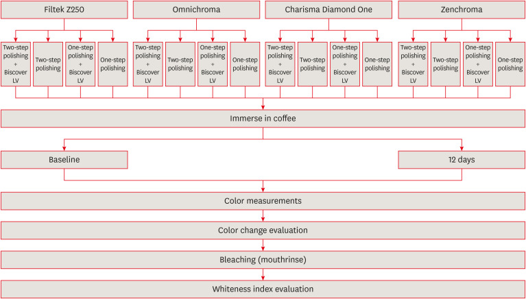
Materials and Methods Three single-shade (Omnichroma, Charisma Diamond One, Zenchroma) and one multi-shade (Filtek Z250) materials were tested. From each resin composite, 40 specimens were prepared. The specimens were divided into 4 subgroups (
n = 10) according to the surface treatments: 1-step polishing, 1-step + Biscover LV, 2-step polishing, and 2-step polishing + Biscover LV. Color differences (ΔE00) were calculated after being immersed in the coffee solution for 12 days. After the staining, the specimens were immersed in a whitening mouthrinse (Crest-3D White) for 12 hours. Whiteness index differences (∆WID = WID after staining − WID after bleaching) values were recorded. The generalized linear model was used for analysis (p < 0.05).Results The lowest and highest ΔE00 values were found for Zenchroma and Charisma Diamond One respectively. Sealed groups indicated higher ΔE00 values than nonsealed groups with significant differences (
p = 0.008). The lowest and highest ΔWID values were found for Zenchroma and Charisma Diamond One respectively. Sealed groups indicated lower ΔWID values than nonsealed groups with significant differences (p = 0.022).Conclusions The use of surface sealant increased the discoloration and showed less whiteness change in resin materials. When the 1-step was compared with the 2-step polishing, the effects on the color stability and whiteness index values of the resin materials were similar.
-
Citations
Citations to this article as recorded by- Evaluating the effects of bleaching on color stability and surface roughness in single-shade and multi-shade resin composites
Hatice Tepe, Özge Çeliksöz, Zeynep Biçer, Batucan Yaman
Anatolian Current Medical Journal.2024; 6(6): 372. CrossRef
- Evaluating the effects of bleaching on color stability and surface roughness in single-shade and multi-shade resin composites
- 2,700 View
- 83 Download
- 1 Crossref

- Color discrepancy of single-shade composites at different distances from the interface measured using cell phone images
- Márcia Luciana Carregosa Santana, Gabriella de Jesus Santos Livi, André Luis Faria-e-Silva
- Restor Dent Endod 2024;49(1):e7. Published online January 24, 2024
- DOI: https://doi.org/10.5395/rde.2024.49.e7

-
 Abstract
 PDF
PDF PubReader
 ePub
ePub Objectives This study aimed to evaluate the impact of substrate color and interface distance on the color adjustment of 2 single-shade composites, Vittra APS Unique and Charisma Diamond One.
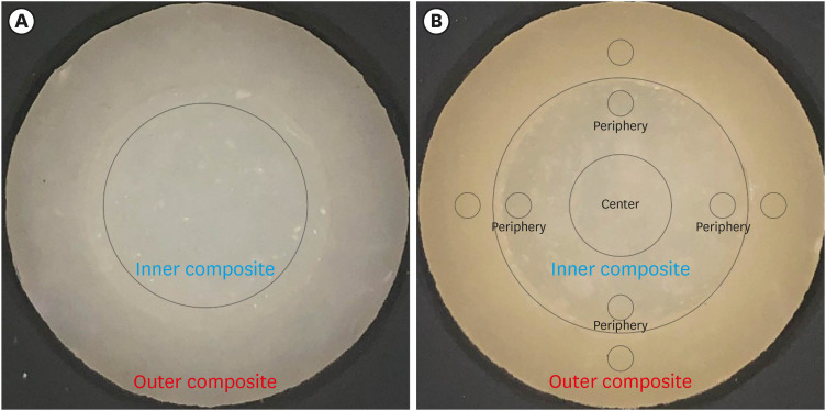
Materials and Methods Dual disc-shaped specimens were created using Vittra APS Unique or Charisma Diamond One as the center composite, surrounded by shaded composites (A1 or A3). Color measurements were taken with a spectrophotometer against a gray background, recording the color coordinates in the CIELAB color space. Illumination with a light-correcting device and image acquisition using a polarizing filter-equipped cell phone were performed on specimens over the same background. Image processing software was used to measure the color coordinates in the center and periphery of the inner composite and in the outer composite. The color data were then converted to CIELAB coordinates and adjusted using data from the spectrophotometer. Color differences (ΔE00) between the center/periphery of single-shade and outer composites were calculated, along with color changes in single-shade composites caused by different outer composites. Color differences for the inner composites surrounded by A1 and A3 were also calculated. Data were analyzed using repeated-measures analysis of variance (α = 0.05).
Results The results showed that color discrepancies were lowest near the interface and when the outer composite was whiter (A1). Additionally, Charisma Diamond One exhibited better color adjustment ability than Vittra APS Unique.
Conclusions Color discrepancies between the investigated single-shade composites diminished towards the interface with the surrounding composite, particularly when the latter exhibited a lighter shade.
-
Citations
Citations to this article as recorded by- Evaluation of color stability in single-shade composite resins using spectrophotometer and cross-polarized mobile photography
Hatice Tepe, Ozge Celiksoz, Batu Can Yaman
BMC Oral Health.2025;[Epub] CrossRef - Comparative Evaluation of the Staining Resistance of Two Single-Shade Composites in Coffee and Chlorhexidine: A Spectrophotometric Analysis
Unmesh Khanvilkar, Shrinath D Kulkarni, Siddhesh Bandekar, Ved M Talathi, Oshin Baghel, Priyanka Razdan, Seema Gupta
Cureus.2025;[Epub] CrossRef - Clinical Implications of Color Adjustment in Single-Shade Resins Post-Dental Bleaching: A Systematic Review
Samille Biasi Miranda, Caroline de Farias Charamba Leal, Rodrigo Barros Esteves Lins, Marcos Antonio Japiassu Resende Montes
Journal of Clinical Medicine.2025; 14(9): 3194. CrossRef - Accuracy and Reliability of Smartphone Versus Mirrorless Camera Images-Assisted Digital Shade Guides: An In Vitro Study
Soo Teng Chew, Suet Yeo Soo, Mohd Zulkifli Kassim, Khai Yin Lim, In Meei Tew
Applied Sciences.2025; 15(14): 8070. CrossRef
- Evaluation of color stability in single-shade composite resins using spectrophotometer and cross-polarized mobile photography
- 2,133 View
- 76 Download
- 3 Web of Science
- 4 Crossref

- Comparison of instrumental methods for color change assessment of Giomer resins
- Luiza de Almeida Queiroz Ferreira, Rogéli Tibúrcio Ribeiro da Cunha Peixoto, Cláudia Silami de Magalhães, Tassiana Melo Sá, Monica Yamauti, Francisca Daniele Moreira Jardilino
- Restor Dent Endod 2022;47(1):e8. Published online February 3, 2022
- DOI: https://doi.org/10.5395/rde.2022.47.e8

-
 Abstract
 PDF
PDF PubReader
 ePub
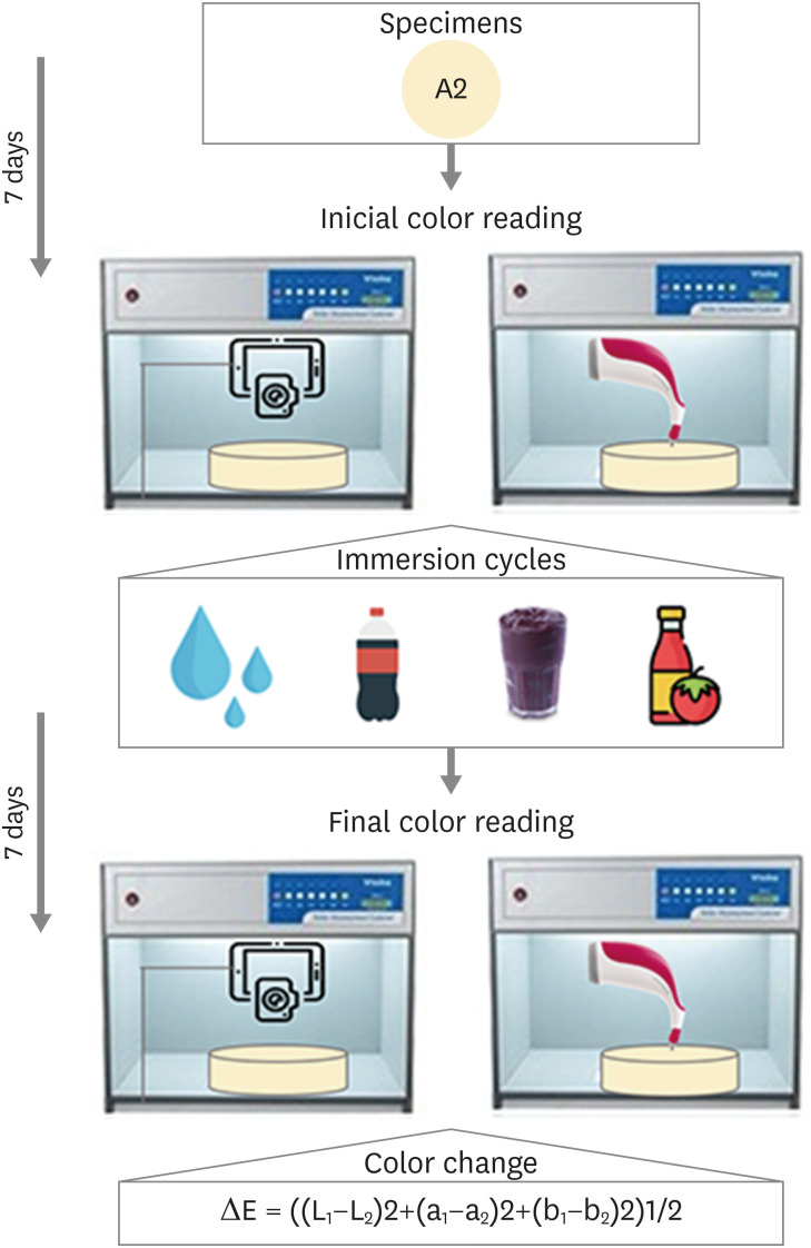
ePub Objectives The aim of this study was to compare the color change of the Giomer resin composite (Beautifil-Bulk) by using photographs obtained with a smartphone (iPhone 6S) associated with Adobe Photoshop software (digital method), with the spectrophotometric method (Vita Easyshade) after immersion in different pigment solutions.
Materials and Methods Twenty resin composite samples with a diameter of 15.0 mm and thickness of 1.0 mm were confectioned in A2 color (
n = 5). Photographs and initial color readings were performed with a smartphone and spectrophotometer, respectively. Then, samples were randomly divided and subjected to cycles of immersion in distilled water (control), açai, Coke, and tomato sauce, 3 times a day, 20 minutes for 7 days. Later, new photographs and color readings were taken.Results The analysis (2-way analysis of variance, Holm-Sidak,
p < 0.05) demonstrated no statistical difference (p < 0.005) between the methods in all groups. Similar color changes were observed for all pigment solutions when using the spectrophotometric method. For the digital method, all color changes were clinically unacceptable, with distilled water and tomato sauce similar to each other and with statistical differences (p < 0.005) for Coke and açai.Conclusions Only the tomato sauce produced a color change above the acceptability threshold using both methods of color assessment. The spectrophotometric and digital methods produce different patterns of color change. According to our results, the spectrophotometric method is more recommended in color change assessment.
-
Citations
Citations to this article as recorded by- The effects of mechanical and chemical degradation on the surface roughness, gloss, and color stability of bulk-fill resin composites
Merve Nezir, Hanife Altınışık, Esra Özyurt, Naz Bayar, Mediha Büyükgöze Dindar
BMC Oral Health.2025;[Epub] CrossRef - Are Sculptable Bulk‐Fill Composites Susceptible to Color Change: A Systematic Review
Jamieson Wong, Constance Yeo, Michelle The, Filip Taneski, Uros Josic, Lorenzo Breschi, Vesna Miletic
Journal of Esthetic and Restorative Dentistry.2025;[Epub] CrossRef - Color Image Expression through CIE L*a*b* System in Foods
Hyun-Woong Choi, Seong-Eun Park, Hong-Seok Son
Journal of the Korean Society of Food Science and Nutrition.2023; 52(2): 223. CrossRef
- The effects of mechanical and chemical degradation on the surface roughness, gloss, and color stability of bulk-fill resin composites
- 2,472 View
- 36 Download
- 3 Web of Science
- 3 Crossref

- Color assessment of resin composite by using cellphone images compared with a spectrophotometer
- Rafaella Mariana Fontes de Bragança, Rafael Ratto Moraes, André Luis Faria-e-Silva
- Restor Dent Endod 2021;46(2):e23. Published online April 5, 2021
- DOI: https://doi.org/10.5395/rde.2021.46.e23

-
 Abstract
 PDF
PDF PubReader
 ePub
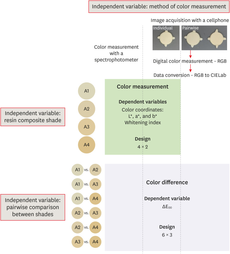
ePub Objectives This study assessed the reliability of digital color measurements using images of resin composite specimens captured with a cellphone.
Materials and Methods The reference color of cylindrical specimens built-up with the use of resin composite (shades A1, A2, A3, and A4) was measured with a portable spectrophotometer (CIELab). Images of the specimens were obtained individually or pairwise (compared shades in the same photograph) under standardized parameters. The color of the specimens was measured in the images using RGB system and converted to CIELab system using image processing software. Whiteness index (WID) and color differences (ΔE00) were calculated for each color measurement method. For the cellphone, the ΔE00 was calculated between the pairs of shades in separate images and in the same image. Data were analyzed using 2-way repeated-measures analysis of variance (α = 0.05). Linear regression models were used to predict the reference ΔE00 values of those calculated using color measured in the images.
Results Images captured with the cellphone resulted in different WID values from the spectrophotometer only for shades A3 and A4. No difference to the reference ΔE00 was observed when individual images were used. In general, a similar ranking of ΔE00 among resin composite shades was observed for all methods. Stronger correlation coefficients with the reference ΔE00 were observed using individual than pairwise images.
Conclusions This study showed that the use of cellphone images to measure the color difference seems to be a feasible alternative providing outcomes similar to those obtained with the spectrophotometer.
-
Citations
Citations to this article as recorded by- Evaluation of color stability in single-shade composite resins using spectrophotometer and cross-polarized mobile photography
Hatice Tepe, Ozge Celiksoz, Batu Can Yaman
BMC Oral Health.2025;[Epub] CrossRef - Color discrepancy of single-shade composites at different distances from the interface measured using cell phone images
Márcia Luciana Carregosa Santana, Gabriella de Jesus Santos Livi, André Luis Faria-e-Silva
Restorative Dentistry & Endodontics.2024;[Epub] CrossRef - How the Translucency and Color Stability of Single-Shade Universal Resin Composites Are Affected by Coffee?
Büşra Özdemir, Betül Kübra Kurucu Karadeniz, Seyit Bilal Özdemir, Ömer Akbulut
Current Research in Dental Sciences.2024; 34(4): 270. CrossRef - Color Image Expression through CIE L*a*b* System in Foods
Hyun-Woong Choi, Seong-Eun Park, Hong-Seok Son
Journal of the Korean Society of Food Science and Nutrition.2023; 52(2): 223. CrossRef - Comparative Evaluation of VITA Shade Guide and Various Composite Shades Using Spectrophotometer, Digital Single-lens Reflex, and Cellphone: An In Vitro Study
Aman Verma, Sonali Taneja, Surabhi Ghosh
World Journal of Dentistry.2023; 14(9): 803. CrossRef - Comparison of instrumental methods for color change assessment of Giomer resins
Luiza de Almeida Queiroz Ferreira, Rogéli Tibúrcio Ribeiro da Cunha Peixoto, Cláudia Silami de Magalhães, Tassiana Melo Sá, Monica Yamauti, Francisca Daniele Moreira Jardilino
Restorative Dentistry & Endodontics.2022;[Epub] CrossRef
- Evaluation of color stability in single-shade composite resins using spectrophotometer and cross-polarized mobile photography
- 2,366 View
- 24 Download
- 2 Web of Science
- 6 Crossref


 KACD
KACD

 First
First Prev
Prev


