Search
- Page Path
- HOME > Search
- Effect of smear layer deproteinization on bonding of self-etch adhesives to dentin: a systematic review and meta-analysis
- Khaldoan H. Alshaikh, Hamdi H. H. Hamama, Salah H. Mahmoud
- Restor Dent Endod 2018;43(2):e14. Published online March 6, 2018
- DOI: https://doi.org/10.5395/rde.2018.43.e14

-
 Abstract
 PDF
PDF PubReader
 ePub
ePub Objectives The aim of this systematic review was to critically analyze previously published studies of the effects of dentin surface pretreatment with deproteinizing agents on the bonding of self-etch (SE) adhesives to dentin. Additionally, a meta-analysis was conducted to quantify the effects of the above-mentioned surface pretreatment methods on the bonding of SE adhesives to dentin.
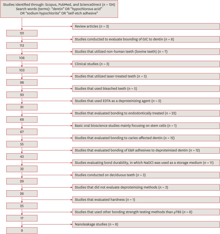
Materials and Methods An electronic search was performed using the following databases: Scopus, PubMed and ScienceDirect. The online search was performed using the following keywords: ‘dentin’ or ‘hypochlorous acid’ or ‘sodium hypochlorite’ and ‘self-etch adhesive.’ The following categories were excluded during the assessment process: non-English articles, randomized clinical trials, case reports, animal studies, and review articles. The reviewed studies were subjected to meta-analysis to quantify the effect of the application time and concentration of sodium hypochlorite (NaOCl) and hypochlorous acid (HOCl) deproteinizing agents on bonding to dentin.
Results Only 9 laboratory studies fit the inclusion criteria of this systematic review. The results of the meta-analysis revealed that the pooled average microtensile bond strength values to dentin pre-treated with deproteinizing agents (15.71 MPa) was significantly lower than those of the non-treated control group (20.94 MPa).
Conclusions In light of the currently available scientific evidence, dentin surface pretreatment with deproteinizing agents does not enhance the bonding of SE adhesives to dentin. The HOCl deproteinizing agent exhibited minimal adverse effects on bonding to dentin in comparison with NaOCl solutions.
-
Citations
Citations to this article as recorded by-
Evaluating the remnants of Al
2
O
3
particles on different dentine substrate after sandblasting and various cleaning protocols
Faeze Hamze, Khotan Aflatoonian, Mahshid Mohammadibassir, Mohammad-Bagher Rezvani
Journal of Adhesion Science and Technology.2025; 39(6): 869. CrossRef - Preservation Strategies for Interfacial Integrity in Restorative Dentistry: A Non-Comprehensive Literature Review
Carmem S. Pfeifer, Fernanda S. Lucena, Fernanda M. Tsuzuki
Journal of Functional Biomaterials.2025; 16(2): 42. CrossRef - Outcome of Er, Cr:YSGG laser and antioxidant pretreatments on bonding quality to caries-induced dentin
Lamiaa M. Moharam, Haidy N. Salem, Ahmed Abdou, Rasha H. Afifi
BMC Oral Health.2025;[Epub] CrossRef - A comparison of different cleaning approaches for blood contamination after curing universal adhesives on the dentine surface
Ting Liu, Haifeng Xie, Chen Chen
Dental Materials.2024; 40(11): 1786. CrossRef - Effect of fiber-reinforced direct restorative materials on the fracture resistance of endodontically treated mandibular molars restored with a conservative endodontic cavity design
Merve Nezir, Beyza Arslandaş Dinçtürk, Ceyda Sarı, Cemile Kedici Alp, Hanife Altınışık
Clinical Oral Investigations.2024;[Epub] CrossRef - Effect of the use of bromelain associated with bioactive glass-ceramic on dentin/adhesive interface
Rocio Geng Vivanco, Ana Beatriz Silva Sousa, Viviane de de Cássia Oliveira, Mário Alexandre Coelho Sinhoreti, Fernanda de Carvalho Panzeri Pires-de-Souza
Clinical Oral Investigations.2024;[Epub] CrossRef - Experimental and Chitosan-Infused Adhesive with Dentin Pretreated with Femtosecond Laser, Methylene Blue-Activated Low-Level Laser, and Phosphoric Acid
Fahad Alkhudhairy
Photobiomodulation, Photomedicine, and Laser Surgery.2024; 42(10): 634. CrossRef - Evaluation of Effective Bond Strength of Composite Resin to Etched Dentin after Dentin Pretreatment: An In-vitro Study
Muhammed Bilal, Shiraz Pasha, Arathi S. Nair
Journal of the Scientific Society.2024; 51(4): 545. CrossRef - Comparison of Different Dentin Deproteinizing Agents on Bond Strength and Microleakage of Universal Adhesive to Dentin
Fatih Bedir, Gül Yıldız Telatar
Journal of Advanced Oral Research.2023; 14(1): 44. CrossRef - Addition of metal chlorides to a HOCl conditioner can enhance bond strength to smear layer deproteinized dentin
Kittisak Sanon, Antonin Tichy, Takashi Hatayama, Ornnicha Thanatvarakorn, Taweesak Prasansuttiporn, Takahiro Wada, Yasushi Shimada, Keiichi Hosaka, Masatoshi Nakajima
Dental Materials.2022; 38(8): 1235. CrossRef - Internal and Marginal Adaptation of Adhesive Resin Cements Used for Luting Inlay Restorations: An In Vitro Micro-CT Study
Linah M. Ashy, Hanadi Marghalani
Materials.2022; 15(17): 6161. CrossRef - Collagen-depletion strategies in dentin as alternatives to the hybrid layer concept and their effect on bond strength: a systematic review
António H. S. Delgado, Madalena Belmar Da Costa, Mário Cruz Polido, Ana Mano Azul, Salvatore Sauro
Scientific Reports.2022;[Epub] CrossRef - NaOCl Application after Acid Etching and Retention of Cervical Restorations: A 3-Year Randomized Clinical Trial
M Favetti, T Schroeder, AF Montagner, RR Moraes, T Pereira-Cenci, MS Cenci
Operative Dentistry.2022; 47(3): 268. CrossRef - Resin infiltrant protects deproteinized dentin against erosive and abrasive wear
Ana Theresa Queiroz de Albuquerque, Bruna Oliveira Bezerra, Isabelly de Carvalho Leal, Maria Denise Rodrigues de Moraes, Mary Anne S. Melo, Vanara Florêncio Passos
Restorative Dentistry & Endodontics.2022;[Epub] CrossRef - Bis[2-(Methacryloyloxy) Ethyl] Phosphate as a Primer for Enamel and Dentine
R. Alkattan, G. Koller, S. Banerji, S. Deb
Journal of Dental Research.2021; 100(10): 1081. CrossRef - Influence of Dentine Pre-Treatment by Sandblasting with Aluminum Oxide in Adhesive Restorations. An In Vitro Study
Bruna Sinjari, Manlio Santilli, Gianmaria D’Addazio, Imena Rexhepi, Alessia Gigante, Sergio Caputi, Tonino Traini
Materials.2020; 13(13): 3026. CrossRef - A novel prime-&-rinse mode using MDP and MMPs inhibitors improves the dentin bond durability of self-etch adhesive
Jingqiu Xu, Mingxing Li, Wenting Wang, Zhifang Wu, Chaoyang Wang, Xiaoting Jin, Ling Zhang, Wenxiang Jiang, Baiping Fu
Journal of the Mechanical Behavior of Biomedical Materials.2020; 104: 103698. CrossRef - The effects of deproteinization and primer treatment on microtensile bond strength of self-adhesive resin cement to dentin
In-Hye Bae, Sung-Ae Son, Jeong-Kil Park
Korean Journal of Dental Materials.2019; 46(2): 99. CrossRef - Effect of Papain and Bromelain Enzymes on Shear Bond Strength of Composite to Superficial Dentin in Different Adhesive Systems
Farahnaz Sharafeddin, Mina Safari
The Journal of Contemporary Dental Practice.2019; 20(9): 1077. CrossRef
-
Evaluating the remnants of Al
2
O
3
particles on different dentine substrate after sandblasting and various cleaning protocols
- 327 View
- 4 Download
- 19 Crossref

- Enamel adhesion of light- and chemical-cured composites coupled by two step self-etch adhesives
- Sae-Hee Han, Eun-Soung Kim, Young-Gon Cho
- J Korean Acad Conserv Dent 2007;32(3):169-179. Published online May 31, 2007
- DOI: https://doi.org/10.5395/JKACD.2007.32.3.169
-
 Abstract
 PDF
PDF PubReader
 ePub
ePub This study was to compare the microshear bond strength (µSBS) of light- and chemically cured composites to enamel coupled with four 2-step self-etch adhesives and also to evaluate the incompatibility between 2-step self-etch adhesives and chemically cured composite resin.
Crown segments of extracted human molars were cut mesiodistally, and a 1 mm thickness of specimen was made. They were assigned to four groups by adhesives used: SE group (Clearfil SE Bond), AdheSE group (AdheSE), Tyrian group (Tyrian SPE/One-Step Plus), and Contax group (Contax). Each adhesive was applied to a cut enamel surface as per the manufacturer's instruction. Light-cured (Filtek Z250) or chemically cured composite (Luxacore Smartmix Dual) was bonded to the enamel of each specimen using a Tygon tube. After storage in distilled water for 24 hours, the bonded specimens were subjected to µSBS testing with a crosshead speed of 1 mm/minute. The mean µSBS (n=20 for each group) was statistically compared using two-way ANOVA, Tukey HSD, and t test at 95% level. Also the interface of enamel and composite was evaluated under FE-SEM.
The results of this study were as follows;
1. The µSBS of the SE Bond group to the enamel was significantly higher than that of the AdheSE group, the Tyrian group, and the Contax group in both the light-cured and the chemically cured composite resin (p < 0.05).
2. There was not a significant difference among the AdheSE group, the Tyrian group, and the Contax group in both the light-cured and the chemically cured composite resin.
3. The µSBS of the light-cured composite resin was significantly higher than that of the chemically cured composite resin when same adhesive was applied to the enamel (p < 0.05).
4. The interface of enamel and all 2-step self-etch adhesives showed close adaptation, and so the incompatibility of the chemically cured composite resin did not show.
-
Citations
Citations to this article as recorded by- Effect of pre-heating on some physical properties of composite resin
Myoung Uk Jin, Sung Kyo Kim
Journal of Korean Academy of Conservative Dentistry.2009; 34(1): 30. CrossRef
- Effect of pre-heating on some physical properties of composite resin
- 218 View
- 2 Download
- 1 Crossref

- Microshear bond strength of adhesives according to the direction of enamel rods
- Young-Gon Cho, Jong-Jin Kim
- J Korean Acad Conserv Dent 2005;30(4):344-351. Published online July 30, 2005
- DOI: https://doi.org/10.5395/JKACD.2005.30.4.344
-
 Abstract
 PDF
PDF PubReader
 ePub
ePub This study compared the microshear bond strength (µSBS) to end and side of enamel rod bonded by four adhesives including two total etch adhesives and two self-etch adhesives.
Crown segments of extracted human molars were cut mesiodistally. The outer buccal or lingual surface was used as specimens cutting the ends of enamel rods, and inner slabs used as specimens cutting the sides of enamel rods.
They were assigned to four groups by used adhesives: Group 1 (All-Bond 2), Group 2 (Single Bond), Group 3 (Tyrian SPE/One-Step Plus), Group 4 (Adper Prompt L-Pop). After each adhesive was applied to enamel surface, three composite cylinders were adhered to it of each specimen using Tygon tube. After storage in distilled water for 24 hours, the bonded specimens were subjected to µSBS testing with a crosshead speed of 1 mm/minute. The results of this study were as follows;
1. The µSBS of Group 2 (16.50 ± 2.31 MPa) and Group 4 (15.83 ± 2.33 MPa) to the end of enamel prism was significantly higher than that of Group 1 (11.93 ± 2.25 MPa) and Group 3 (11.97 ± 2.05 MPa) (p < 0.05).
2. The µSBS of Group 2 (13.43 ± 2.93 MPa) to the side of enamel prism was significantly higher than that of Group 1 (8.64 ± 1.53 MPa), Group 3 (9.69 ± 1.80 MPa), and Group 4 (10.56 ± 1.75 MPa) (p < 0.05).
3. The mean µSBS to the end of enamel rod was significantly higher than that to the side of enamel rod in all group (p < 0.05).
-
Citations
Citations to this article as recorded by- Enamel adhesion of light- and chemical-cured composites coupled by two step self-etch adhesives
Sae-Hee Han, Eun-Soung Kim, Young-Gon Cho
Journal of Korean Academy of Conservative Dentistry.2007; 32(3): 169. CrossRef
- Enamel adhesion of light- and chemical-cured composites coupled by two step self-etch adhesives
- 246 View
- 1 Download
- 1 Crossref


 KACD
KACD

 First
First Prev
Prev


