Search
- Page Path
- HOME > Search
- Effect of irrigants on the color stability, solubility, and surface characteristics of calcium-silicate based cements
- Selen Küçükkaya Eren, Sevinc Askerbeyli Örs, Hacer Aksel, Şenay Canay, Duygu Karasan
- Restor Dent Endod 2022;47(1):e10. Published online February 10, 2022
- DOI: https://doi.org/10.5395/rde.2022.47.e10

-
 Abstract
 PDF
PDF PubReader
 ePub
ePub Objectives This study aimed to investigate the color stability, solubility, and surface characteristics of 3 calcium silicate-based cements (CSCs) after immersion in different solutions.
Materials and Methods ProRoot white mineral trioxide aggregate (MTA), Biodentine, and Endosequence Root Repair Material (ERRM) were placed in cylindrical molds and stored at 37°C for 24 hours. Each specimen was immersed in distilled water, 5% sodium hypochlorite (NaOCl), 2% chlorhexidine, or 0.1% octenidine hydrochloride (OCT) for 24 hours. Color changes were measured with a spectrophotometer. Solubility was determined using an analytical balance with 10−5 g accuracy. The surface characteristics were analyzed using scanning electron microscopy and energy-dispersive spectroscopy. Data were analyzed using 2-way analysis of variance, the Tukey test, and the paired
t -test.Results MTA exhibited significant discoloration in contact with NaOCl (
p < 0.05). White precipitation occurred on the surfaces of Biodentine and ERRM after contact with the solutions, and none of the materials presented dark brown discoloration. All materials showed significant solubility after immersion in the solutions (p < 0.05), irrespective of the solution type (p > 0.05). The surface topography and elemental composition of the samples showed different patterns of crystal formation and precipitation depending on the solution type.Conclusions All materials presented some amount of solubility and showed crystal precipitation after contact with the solutions. Biodentine and ERRM are suitable alternatives to ProRoot MTA as they do not exhibit discoloration. The use of OCT can be considered safe for CSCs.
-
Citations
Citations to this article as recorded by- Chemical and in vivo analyses of calcium silicate‐based materials in bone and connective tissues
Ana Cristina Padilha Janini, Lauter Eston Pelepenko, Brenda Fornazaro Moraes, Victor Augusto Benedicto dos Santos, Matheus Barros‐Costa, Isabela Alvarenga Maciel dos Santos, Fábio Roberto de Souza Batista, Juliana de Aguiar Silveira Meira, Mariza Akemi Ma
International Endodontic Journal.2025; 58(3): 484. CrossRef - Topic: Perspectives on Success and Failure of Endodontic Treatments
Ilma Robo, Manola Kelmendi, Eva Habazaj, Kleves Elezi, Rialda Xhizdari, Nevila Alliu
SN Comprehensive Clinical Medicine.2025;[Epub] CrossRef - Bismuth release from endodontic materials: Proposed mechanisms for systemic circulation and organ accumulation
Lauter Eston Pelepenko, Benjamin Hewitt, Rodrigo Bueno de Oliveira, Brenda Fornazaro Moraes, Débora C. Coraça-Huber, Ana Cristina Padilha Janini, Marina Angélica Marciano
Journal of Hazardous Materials.2025; 494: 138580. CrossRef - Effect of Vital Pulp Therapy Biomaterials on Tooth Discolouration: A Review of the Literature
Maedeh Gilvari Sarshari, Kiana Shakeri, Ardavan Parhizkar, Naresh Kasoju
International Journal of Biomaterials.2025;[Epub] CrossRef - Data about application of chlorhexidine as a periodontal irrigant –
Systematic Review.
Ilma Robo, Manola Kelmendi , Eva Habazaj , Kristi Sulanjaku , Nevila Alliu
Acta Stomatologica Marisiensis Journal.2025; 8(1): 6. CrossRef - Biocompatibility of Hydraulic Calcium Silicate-Based Cement MTA FlowTM on Human Dental Pulp Stem Cells In Vitro
Paulius Tušas, Josette Camilleri, Milda Alksnė, Egidijus Šimoliūnas, Saulius Drukteinis, Eglė Marija Urbonė, Virginija Bukelskienė, Vygandas Rutkūnas, Vytautė Pečiulienė
Journal of Functional Biomaterials.2025; 16(7): 252. CrossRef - Evaluation of the Effects of Different Irrigation Solutions on MTA and Dentin Microhardness
Gokay Buyukcolpan, İdil Özden, Hesna Sazak Öveçoğlu
Clinical and Experimental Health Sciences.2025; 15(3): 524. CrossRef - Comparative Evaluation of the Intratubular Penetration Ability of Two Retrograde Obturation Techniques in Micro-Endodontic Surgical Procedure: An In Vitro Study with Confocal Laser Scanning Microscopy
Alberto Casino Alegre, Michell Ramírez López, Manuel Monterde Hernández, Susana Aranda Verdú, Jorge Rubio Climent, Antonio Pallarés Sabater
Dentistry Journal.2025; 13(11): 509. CrossRef - Time-dependent Tooth Color Changes Following Conventional, Silver-based, and Photodynamic Root Canal Irrigants: An In Vitro Study
Laila Mohamed Mohamed Kenawi, Mohamed Fattouh, Khaled Abid Althaqafi, Abla Arafa
The Open Dentistry Journal.2025;[Epub] CrossRef - The outcome of combined use of iRoot BP Plus and iRoot SP for root-end filling in endodontic microsurgery: a randomized controlled trial
Xu Dong, Qin Su, Wen Li, Jinbo Yang, Dongzhe Song, Jing Yang, Xin Xu
Clinical Oral Investigations.2024;[Epub] CrossRef - How Does Ethylenediaminetetraacetic Acid Irrigation Affect Biodentine? A Multimethod Ex Vivo Study
Katarzyna Dąbrowska, Aleksandra Palatyńska-Ulatowska, Leszek Klimek
Materials.2024; 17(6): 1230. CrossRef - Color stability and solubility of Biodentine and NeoPutty in contact with different irrigation solutions
Sıla Nur Usta, Cangül Keskin
Restorative Dentistry & Endodontics.2024;[Epub] CrossRef - The impact of various irrigation solutions on the color stabilities of five calcium silicate cement: an in-vitro study
Aslı Soğukpınar Onsuren, Onur Kesici, Elif Uğurbekler Hündü
Selcuk Dental Journal.2024; 11(3): 313. CrossRef - Bioceramics in Endodontics: Updates and Future Perspectives
Xu Dong, Xin Xu
Bioengineering.2023; 10(3): 354. CrossRef - Effect of calcium silicate-based endodontic sealers on tooth color: A 3-year in vitro experimental study
Carmen Llena, Ana Herrero, Sandra Lloret, Martha Barraza, Jose Luis Sanz
Heliyon.2023; 9(2): e13237. CrossRef - Evaluation of the Shear Bond Strength of Four Bioceramic Materials with Different Restorative Materials and Timings
Abeer S. Alqahtani, Ayman M. Sulimany, Abdullah S. Alayad, Abdulaziz S. Alqahtani, Omar A. Bawazir
Materials.2022; 15(13): 4668. CrossRef
- Chemical and in vivo analyses of calcium silicate‐based materials in bone and connective tissues
- 1,975 View
- 32 Download
- 12 Web of Science
- 16 Crossref

-
Influence of reciprocating and rotary instrumentation on microbial reduction: a systematic review and meta-analysis of
in vitro studies - Selen Küçükkaya Eren, Emel Uzunoğlu-Özyürek, Sevilay Karahan
- Restor Dent Endod 2021;46(2):e19. Published online March 10, 2021
- DOI: https://doi.org/10.5395/rde.2021.46.e19

-
 Abstract
 PDF
PDF PubReader
 ePub
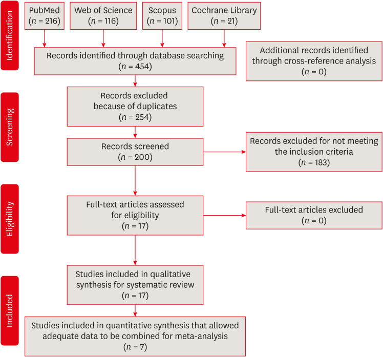
ePub Objectives The purpose of this study was to conduct a systematic review and meta-analysis of
in vitro studies regarding the effectiveness of reciprocating and rotary instrumentation on microbial reduction in root canals.Materials and Methods PubMed, Scopus, Web of Science, the Cochrane Library, and the gray literature were searched through December 2019. Studies comparing the influence of reciprocating and rotary instrumentation on the removal of microorganisms from root canals that quantified the antimicrobial effect were included. Data extraction was completed using a systematic form for data collection. The risk of bias of the studies was evaluated. Standardized mean differences (SMDs) and confidence intervals (CIs) were calculated using a random effects meta-analysis.
Results Seventeen
in vitro studies were included in this systematic review, of which 7 provided adequate data for inclusion in the meta-analysis. Both reciprocating and rotary systems were similarly effective in reducing the microbial load in infected root canals (SMD [95% CI], 0.0481 [−0.271, 0.367]). Three studies showed a low risk of bias, whereas most of the studies (82%) presented a medium risk.Conclusions Although both techniques decrease the microbial content (with reductions of 23.32%–88.47% and 23.33%–89.86% for reciprocating and rotary instrumentation, respectively)
, they are not able to provide complete disinfection of root canals.-
Citations
Citations to this article as recorded by- Mapping risk of bias criteria in systematic reviews of in vitro endodontic studies: an umbrella review
Rafaella Rodrigues da Gama, Lucas Peixoto de Araújo, Evandro Piva, Leandro Perello Duro, Adriana Fernandes da Silva, Wellington Luiz de Oliveira da Rosa
Evidence-Based Dentistry.2025; 26(4): 179. CrossRef - Fifteen years of engine‐driven nickel–titanium reciprocating instruments, what do we know so far? An umbrella review
Felipe Immich, Lucas Peixoto de Araújo, Rafaella Rodrigues da Gama, Wellington Luiz de Oliveira da Rosa, Evandro Piva, Giampiero Rossi‐Fedele
Australian Endodontic Journal.2024; 50(2): 409. CrossRef - Does minimally invasive canal preparation provide higher fracture resistance of endodontically treated teeth? A systematic review ofin vitrostudies
Sıla Nur Usta, Emmanuel João Nogueira Leal Silva, Seda Falakaloğlu, Mustafa Gündoğar
Restorative Dentistry & Endodontics.2023;[Epub] CrossRef - The Effect of Combined Ultrasonic Tip and Mechanized Instrumentation on the Reduction of the Percentage of Non-Instrumented Surfaces in Oval/Flat Root Canals: A Systematic Review and Meta-Analysis
Marcella Dewes Cassal, Pedro Cardoso Soares, Marcelo dos Santos
Cureus.2023;[Epub] CrossRef - Impact of Different Access Cavity Designs and Ni–Ti Files on the Elimination of Enterococcus faecalis from the Root Canal System: An In Vitro Study
Gizem Andac, Atakan Kalender, Buket Baddal, Fatma Basmaci
Applied Sciences.2022; 12(4): 2049. CrossRef - Shaping Properties and Outcomes of Nickel-Titanium Reciprocation Systems in Primary Teeth: A Systematic Review and Meta-Analysis of In Vitro Studies
SelvaKumar Haridoss, Bhavyaa R, Kavitha Swaminathan, Aruna P
Cureus.2022;[Epub] CrossRef - Influence of Root Canal Sealers and Obturation Techniques on Vertical Root Fracture Resistance. An In Vitro Experiment
Mazen F. Alkahtany, Khalid H. Almadi, Fahad A. Alahmad, Abdullah M. Alshehri, Abdulrahman A. AlSwayyed, Omar M. AlZahran, Ali AlHadan, Abdulaziz S. Almustafa, Fahim Vohra, Tariq Abduljabbar
Applied Sciences.2021; 11(17): 8022. CrossRef
- Mapping risk of bias criteria in systematic reviews of in vitro endodontic studies: an umbrella review
- 2,171 View
- 31 Download
- 10 Web of Science
- 7 Crossref

- Critical evaluation of fracture strength testing for endodontically treated teeth: a finite element analysis study
- Emel Uzunoglu-Özyürek, Selen Küçükkaya Eren, Oğuz Eraslan, Sema Belli
- Restor Dent Endod 2019;44(2):e15. Published online April 18, 2019
- DOI: https://doi.org/10.5395/rde.2019.44.e15

-
 Abstract
 PDF
PDF PubReader
 ePub
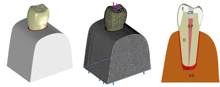
ePub Objectives The aim of this study was to investigate whether the diameter and direction of the plunger and simulation of the periodontal ligament (PDL) affected the stress distribution in endodontically treated premolars.
Methods A fracture strength test was simulated via finite element analysis. A base model was set up, and the following parameters were modified: plunger diameter (3 mm vs. 6 mm), plunger direction (vertical vs. 135° angular to the central fossa), and PDL simulation. The analysis was conducted using the CosmosWorks structural analysis program, and the results are presented in terms of von Mises stresses.
Results The smaller plunger increased the stresses at the contact area of the crown, but the plunger diameter had no effect on the stress distribution within the root. An angular plunger direction increased stresses within the root, as well as at the buccal cusp of the crown, compared with the vertical direction. Simulation of the PDL caused higher stress accumulation, especially in the cervical region of the root.
Conclusions The plunger diameter had no effect on the stress distribution in the roots, whereas the plunger direction and PDL simulation did affect the stress distribution. More stringent standards can be established by taking such parameters into account when performing fracture testing in future studies.
-
Citations
Citations to this article as recorded by- Access cavity in endodontics: Balancing precision, preservation, and clinical needs
Dina Abdellatif, Ismail Davut Capar, De Fontaine Sarah, Alfredo Iandolo, Christophe Meyer, Davide Mancino
Journal of Conservative Dentistry and Endodontics.2025; 28(6): 573. CrossRef - Assessment of Stress Distribution with 3 Taper Design Preparation of Root Canal Using Finite Element Analysis
Tejasree Rathod, G. Durgabhavani, Pudu Tirupathi, Nusrath Parveen, Yelloji Paramesh, Prabhakar Dharavattu
Journal of Pharmacy and Bioallied Sciences.2024; 16(Suppl 1): S112. CrossRef - The impact of the filling technique with two sealers in bulk or associated with gutta-percha on the fatigue behavior and failure patterns of endodontically treated teeth
Isabella Marian Lena, Luiza Colpo Chiaratti, Rafaela Oliveira Pilecco, Renan Vaz Machry, João Paulo Mendes Tribst, Cornelis Johannes Kleverlaan, Gabriel Kalil Rocha Pereira, Renata Dornelles Morgental
PeerJ.2024; 12: e18221. CrossRef - Stronger than Ever: Multifilament Fiberglass Posts Boost Maxillary Premolar Fracture Resistance
Naji Kharouf, Eugenio Pedullà, Gianluca Plotino, Hamdi Jmal, Mohammed-El-Habib Alloui, Philippine Simonis, Patrice Laquerriere, Valentina Macaluso, Dina Abdellatif, Raphaël Richert, Youssef Haikel, Davide Mancino
Journal of Clinical Medicine.2023; 12(8): 2975. CrossRef - Neural network approach to evaluate the physical properties of dentin
Mohammad Ali Saghiri, Ali Mohammad Saghiri, Elham Samadi, Devyani Nath, Julia Vakhnovetsky, Steven M. Morgano
Odontology.2023; 111(1): 68. CrossRef - Modelling and evaluating periodontal ligament mechanical behaviour and properties: A scoping review of current approaches and limitations
Enaiyat Ghani Ovy, Dan L. Romanyk, Carlos Flores Mir, Lindsey Westover
Orthodontics & Craniofacial Research.2022; 25(2): 199. CrossRef - FEAr no more! Finite element analysis in orthodontics
Shilpa Chawla, Shailesh Deshmukh
Journal of the International Clinical Dental Research Organization.2022; 14(1): 6. CrossRef - Influence of Methodological Variables on Fracture Strength Tests Results of Premolars with Different Number of Residual Walls. A Systematic Review with Meta-Analysis
Carlo Gaeta, Crystal Marruganti, Emanuele Mignosa, Giovanni Franciosi, Edoardo Ferrari, Simone Grandini
Dentistry Journal.2021; 9(12): 146. CrossRef
- Access cavity in endodontics: Balancing precision, preservation, and clinical needs
- 2,246 View
- 36 Download
- 8 Crossref

- Mineral content analysis of root canal dentin using laser-induced breakdown spectroscopy
- Selen Küçükkaya Eren, Emel Uzunoğlu, Banu Sezer, Zeliha Yılmaz, İsmail Hakkı Boyacı
- Restor Dent Endod 2018;43(1):e11. Published online February 4, 2018
- DOI: https://doi.org/10.5395/rde.2018.43.e11

-
 Abstract
 PDF
PDF PubReader
 ePub
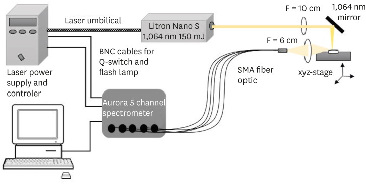
ePub Objectives This study aimed to introduce the use of laser-induced breakdown spectroscopy (LIBS) for evaluation of the mineral content of root canal dentin, and to assess whether a correlation exists between LIBS and scanning electron microscopy/energy dispersive spectroscopy (SEM/EDS) methods by comparing the effects of irrigation solutions on the mineral content change of root canal dentin.
Materials and Methods Forty teeth with a single root canal were decoronated and longitudinally sectioned to expose the canals. The root halves were divided into 4 groups (
n = 10) according to the solution applied: group NaOCl, 5.25% sodium hypochlorite (NaOCl) for 1 hour; group EDTA, 17% ethylenediaminetetraacetic acid (EDTA) for 2 minutes; group NaOCl+EDTA, 5.25% NaOCl for 1 hour and 17% EDTA for 2 minutes; a control group. Each root half belonging to the same root was evaluated for mineral content with either LIBS or SEM/EDS methods. The data were analyzed statistically.Results In groups NaOCl and NaOCl+EDTA, the calcium (Ca)/phosphorus (P) ratio decreased while the sodium (Na) level increased compared with the other groups (
p < 0.05). The magnesium (Mg) level changes were not significant among the groups. A significant positive correlation was found between the results of LIBS and SEM/EDS analyses (r = 0.84,p < 0.001).Conclusions Treatment with NaOCl for 1 hour altered the mineral content of dentin, while EDTA application for 2 minutes had no effect on the elemental composition. The LIBS method proved to be reliable while providing data for the elemental composition of root canal dentin.
-
Citations
Citations to this article as recorded by- In vitro evaluation of antimicrobial photodynamic therapy with photosensitizers and calcium hydroxide on bond strength, chemical composition, and sealing of glass-fiber posts to root dentin
Thalya Fernanda Horsth Maltarollo, Paulo Henrique dos Santos, Henrique Augusto Banci, Mariana de Oliveira Bachega, Beatriz Melare de Oliveira, Marco Hungaro Antonio Duarte, Índia Olinta de Azevedo Queiroz, Rodrigo Rodrigues Amaral, Luciano Angelo Tavares
Lasers in Medical Science.2025;[Epub] CrossRef - Effect of Using 5% Apple Vinegar Irrigation Solution Adjunct to Diode Laser on Smear Layer Removal and Calcium/Phosphorus Ion Ratio during Root Canal Treatment
Tarek AA Salam, Haythem SA Kader, Elsayed E Abdallah
CODS - Journal of Dentistry.2024; 15(1): 3. CrossRef - Evaluation of chemical composition of root canal dentin between two age groups using different irrigating solutions: An in vitro sem-eds study
Naresh Kumar K, Abhijith Kallu, Surender L.R, Sravani Nirmala, Narender Reddy
International Dental Journal of Student's Research.2024; 12(1): 18. CrossRef - Minimally invasive management of vital teeth requiring root canal therapy
E. Karatas, M. Hadis, W. M. Palin, M. R. Milward, S. A. Kuehne, J. Camilleri
Scientific Reports.2023;[Epub] CrossRef - The Effects of a Novel Nanohydroxyapatite Gel and Er: YAG Laser Treatment on Dentin Hypersensitivity
Demet Sahin, Ceren Deger, Burcu Oglakci, Metehan Demirkol, Bedri Onur Kucukyildirim, Mehtikar Gursel, Evrim Eliguzeloglu Dalkilic
Materials.2023; 16(19): 6522. CrossRef - Chitosan Homogenizing Coffee Ring Effect for Soil Available Potassium Determination Using Laser-Induced Breakdown Spectroscopy
Xiaolong Li, Rongqin Chen, Zhengkai You, Tiantian Pan, Rui Yang, Jing Huang, Hui Fang, Wenwen Kong, Jiyu Peng, Fei Liu
Chemosensors.2022; 10(9): 374. CrossRef - Quantitative analysis of cadmium in rice roots based on LIBS and chemometrics methods
Wei Wang, Wenwen Kong, Tingting Shen, Zun Man, Wenjing Zhu, Yong He, Fei Liu
Environmental Sciences Europe.2021;[Epub] CrossRef
- In vitro evaluation of antimicrobial photodynamic therapy with photosensitizers and calcium hydroxide on bond strength, chemical composition, and sealing of glass-fiber posts to root dentin
- 1,627 View
- 10 Download
- 7 Crossref

- Evaluation of the effects of two novel irrigants on intraradicular dentine erosion, debris and smear layer removal
- Melahat Görduysus, Selen Küçükkaya, Nursel Pekel Bayramgil, Mehmet Ömer Görduysus
- Restor Dent Endod 2015;40(3):216-222. Published online July 3, 2015
- DOI: https://doi.org/10.5395/rde.2015.40.3.216
-
 Abstract
 PDF
PDF PubReader
 ePub
ePub Objectives To evaluate the effects of copolymer of acrylic acid and maleic acid (Poly[AA-
co -MA]) and calcium hypochlorite (Ca(OCl)2) on root canal dentin using scanning electron microscope (SEM).Materials and Methods Twenty-four single-rooted teeth were instrumented and the apical and coronal thirds of each root were removed, leaving the 5 mm middle thirds, which were then separated into two pieces longitudinally. The specimens were randomly divided into six groups and subjected to each irrigant for 5 min as follows: G1, Ca(OCl)2; G2, Poly(AA-
co -MA); G3, Ca(OCl)2 + Poly(AA-co -MA); G4, sodium hypochlorite (NaOCl); G5, ethylenediaminetetraacetic acid (EDTA); G6, NaOCl+EDTA. The specimens were prepared for SEM evaluation. Smear layer, debris and erosion scores were recorded by two blinded examiners. One image from G3 was analyzed with energy dispersive spectroscopy (EDS) on suspicion of precipitate formation. Data were analyzed using the Kruskal-Wallis and Dunn tests.Results G1 and G4 showed the presence of debris and smear layer and they were statistically different from G2, G3, G5 and G6 where debris and smear layer were totally removed (
p < 0.05). In G1 and G4, erosion evaluation could not be done because of debris and smear layer. G2, G3 and G5 showed no erosion, and there was no significant difference between them. G6 showed severe erosion and was statistically different from G2, G3 and G5 (p < 0.05). EDS microanalysis showed the presence of Na, P, and Ca elements on the surface.Conclusions Poly(AA-
co -MA) is effective in removing the smear layer and debris without causing erosion either alone or with Ca(OCl)2.-
Citations
Citations to this article as recorded by- Effect of Heat and Irrigation Agitation on the Smear Layer Removal Ability of Calcium Hypochlorite: A Scanning Electron Microscope Study
Damla Erkal, Kürşat Er
Microscopy Research and Technique.2026; 89(1): 65. CrossRef - Effect of sodium and calcium hypochlorite with or without surfactant on the adhesion of epoxy resin-based endodontic sealer
Guilherme Pauletto, Natália Franco Brum, Israel Bangel Carlotto, Lucas Saldanha da Rosa, Carlos Alexandre Souza Bier
Brazilian Journal of Oral Sciences.2025; 24: e254080. CrossRef - Bonding and Cleaning Effects of Irrigation Protocols Using Calcium Hypochlorite on the Post-space Radicular Dentin
JF Besegato, GR Bravo, JF Zaniboni, LG Belizário, ENM de Almeida, MB Gelio, WG Escalante-Otárola, MC Kuga
Operative Dentistry.2024; 49(6): E1. CrossRef - Effect of sodium hypochlorite and calcium hypochlorite on the apical sealing ability of endodontic sealers
Israel Bangel Carlotto, Natália Franco Brum, Guilherme Pauletto, Lucas Saldanha da Rosa, Carlos Alexandre Souza Bier
Brazilian Journal of Oral Sciences.2024; 23: e242700. CrossRef - Comparative evaluation of antifungal activity of Sodium Hypochlorite, Calcium Hypochlorite and modified Salt Solution associated with passive ultrasonic irrigation against Candida albicans - An In-Vitro study
Helen Thomas, D. N. Nirupama, Mohan Thomas Nainan, D. N. Naveen, C. Y. Ranjini, R. Vijay
Journal of Conservative Dentistry and Endodontics.2024; 27(2): 159. CrossRef - Effect of calcium hypochlorite as an irrigant alternative in the removal of methylene blue after photodynamic therapy under the post-space adhesive interface
João Felipe Besegato, Joatan Lucas de Sousa Gomes Costa, Joissi Ferrari Zaniboni, Giovanna Righetti Bravo, Jéssika Mayhara Pereira Morais, Wilfredo Gustavo Escalante-Otárola, Milton Carlos Kuga
Laser Physics.2023; 33(5): 055601. CrossRef - Effectiveness of Different Irrigation Techniques on Post Space Smear Layer Removal: SEM Evaluation
Alfredo Iandolo, Massimo Pisano, Dina Abdellatif, Alessandra Amato, Francesco Giordano, Alessio Buonavoglia, Giuseppe Sangiovanni, Mario Caggiano
Prosthesis.2023; 5(2): 539. CrossRef - The dynamic interplay of dietary acid pH and concentration during early-stage human enamel and dentine erosion
J. Pattem, J. Field, P. J. Waterhouse, M. J. German
Frontiers in Dental Medicine.2022;[Epub] CrossRef - Effect of EDTA, sodium, and calcium hypochlorite on the inorganic component of root canal dentin: A SEM analysis
Luana Roleto Cardoso, Flávia Emi Razera Baldasso, Débora Delai, Francisco Montagner, Patrícia Maria Poli Kopper
Microscopy Research and Technique.2019; 82(2): 128. CrossRef - Comparative Analysis of Manual, Rotary and Reciprocal Systems on Primary Teeth Root Canals: An In Vitro Scanning Electron Microscopy Study
İpek ARSLAN, Sema AYDINOĞLU, Ozgul BAYGIN, Tamer TÜZÜNER, Murat ŞİRİN
Cumhuriyet Dental Journal.2019; 22(3): 299. CrossRef - Regenerating a monoblock to obturate root canalsvia a mineralising strategy
Le Zhang, Quan-Li Li, Ying Cao, Yun Wang
Scientific Reports.2018;[Epub] CrossRef - Antibacterial Efficacy of Calcium Hypochlorite with Vibringe Sonic Irrigation System onEnterococcus faecalis: An In Vitro Study
Aysin Dumani, Hatice Korkmaz Guvenmez, Sehnaz Yilmaz, Oguz Yoldas, Zeliha Gonca Bek Kurklu
BioMed Research International.2016; 2016: 1. CrossRef
- Effect of Heat and Irrigation Agitation on the Smear Layer Removal Ability of Calcium Hypochlorite: A Scanning Electron Microscope Study
- 1,599 View
- 6 Download
- 12 Crossref


 KACD
KACD

 First
First Prev
Prev


