Search
- Page Path
- HOME > Search
-
Dentinal defects induced by 6 different endodontic files when used for oval root canals: an
in vitro comparative study - Ajinkya M Pawar, Bhagyashree Thakur, Anda Kfir, Hyeon-Cheol Kim
- Restor Dent Endod 2019;44(3):e31. Published online July 29, 2019
- DOI: https://doi.org/10.5395/rde.2019.44.e31

-
 Abstract
 PDF
PDF PubReader
 ePub
ePub Objectives To compare the formation of dentinal defects using stainless-steel hand K-files (HFs), rotary files, reciprocating files, and Self-Adjusting File (SAF), when used for oval root canals.
Materials and Methods One hundred and forty extracted human mandibular premolar with single root and oval canal were selected for this study. Oval canals were confirmed by exposing to mesio-distal and bucco-lingual radiographs. Teeth with open apices or anatomic irregularities were excluded. All selected teeth were de-coronated perpendicular to the long axis of the tooth, leaving roots segments approximately of 16 mm in length. Twenty teeth were left unprepared (control), and the remaining 120 teeth were divided into 6 groups (
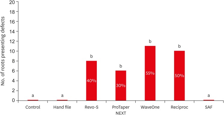
n = 20) and instrumented using HF (size 40/0.02), Revo-S (RS; size 40/0.06), ProTaper NEXT (PTN; size 40/0.06), WaveOne (WO; size 40/0.09), RECIPROC (RC; size 40/0.06), and the SAF (2 mm). Roots were then sectioned 3, 6, and 9 mm from the apex, and observed under stereomicroscope, for presence of dentinal defects. “No defect” was defined as root dentin that presented with no visible microcracks or fractures. “Defect” was defined by microcracks or fractures in the root dentin.Results The control, HF, and SAF did not exhibit any dentinal defects. In roots instrumented by RS, PTN, WO, and RC files exhibited microcracks (incomplete or complete) in 40%, 30%, 55%, and 50%, respectively.
Conclusions The motor-driven root canal instrumentation with rotary and reciprocating files may create microcracks in radicular dentine, whereas the stainless-steel hand file instrumentation, and the SAF produce minimal or less cracks.
-
Citations
Citations to this article as recorded by- Evaluation of dentinal crack formation during post space preparation using different fiber post systems with micro-computed tomography
Ayşe Nur Kuşuçar, Damla Kırıcı
BMC Oral Health.2025;[Epub] CrossRef - Computational Insights into Root Canal Treatment: A Survey of Selected Methods in Imaging, Segmentation, Morphological Analysis, and Clinical Management
Jianning Li, Kerstin Bitter, Anh Duc Nguyen, Hagay Shemesh, Paul Zaslansky, Stefan Zachow
Dentistry Journal.2025; 13(12): 579. CrossRef - Comparative evaluation of incidence of dentinal defects after root canal preparation using three different endodontic retreatment systems – An in vitro study
S. Aarthi, J. S. Sivakumar, A. Andamuthu Sivakumar, J. Saravanapriyan Soundappan, M. Chittrarasu, G. Jayanthi
Journal of Conservative Dentistry and Endodontics.2024; 27(3): 262. CrossRef - Evaluation of Dentin Cracks by Stereomicroscope after Preparation of Mesiobuccal Canal of Maxillary First Molars Using Edge Taper Platinum and ProTaper Gold Rotary Files: A Laboratory Study
Narjes Hoshyari, Seyedali Seyedmajidi, Anahita Lotfizadeh, Eghlima Malakan, Abolfazl Hosseinnataj, Azam Haddadi Kohsar
Avicenna Journal of Dental Research.2023; 15(4): 167. CrossRef - A comparative evaluation of dentinal defects after root canal preparation with different rotary and reciprocal systems
Ece Yakın, Berna Aslan, Emine Odabaşı Tezer
Northwestern Medical Journal.2023; 3(3): 147. CrossRef - Comparison of Dentinal Defects Induced by Rotary, Reciprocating, and Hand Files in Oval Shaped Root Canal - An In-Vitro Study
Harakh Chand Branawal, Neelam Mittal, Prachi Rani, Aiyman Ayubi, Silviya Samad
Indian Journal of Dental Research.2023; 34(4): 433. CrossRef - Comparative evaluation of incidence of dentinal defects after root canal preparation using hand, rotary, and reciprocating files: An ex vivo study
Debanjan Das, Sudipto Barai, Rohit Kumar, Sourav Bhattacharyya, AsimB Maity, Pushpa Shankarappa
Journal of International Oral Health.2022; 14(1): 78. CrossRef - Effect of XP‐endo Shaper versus conventional rotary files on postoperative pain and bacterial reduction in oval canals with necrotic pulps: a randomized clinical study
R. S. Emara, S. I. Gawdat, H. M. M. El‐Far
International Endodontic Journal.2021; 54(7): 1026. CrossRef - Comparative Evaluation of Dentinal Microcrack Formation by Single Reciprocating File Systems: An In Vitro Study
Baby James, A Devadathan, Manuja Nair, Ashitha T Kulangara, Jose Jacob
Conservative Dentistry and Endodontic Journal.2020; 5(1): 1. CrossRef - The potential effect of instrumentation with different nickel titanium rotary systems on dentinal crack formation—An in vitro study
Márk Fráter, András Jakab, Gábor Braunitzer, Zsolt Tóth, Katalin Nagy, Andrej M. Kielbassa
PLOS ONE.2020; 15(9): e0238790. CrossRef
- Evaluation of dentinal crack formation during post space preparation using different fiber post systems with micro-computed tomography
- 1,756 View
- 23 Download
- 10 Crossref

- Clinical evaluation of a new extraction method for intentional replantation
- Yong-Hoon Choi, Ji-Hyun Bae
- J Korean Acad Conserv Dent 2011;36(3):211-218. Published online May 31, 2011
- DOI: https://doi.org/10.5395/JKACD.2011.36.3.211
-
 Abstract
 PDF
PDF PubReader
 ePub
ePub Purpose Intentional replantation (IR) is a suitable treatment option when nonsurgical retreatment and periradicular surgery are unfeasible. For successful IR, fracture-free safe extraction is crucial step. Recently, a new extraction method of atraumatic safe extraction (ASE) for IR has been introduced.
Patients and Methods Ninety-six patients with the following conditions who underwent IR at the Department of Conservative Dentistry, Seoul National University Bundang Hospital, in 2010 were enrolled in this study: failed nonsurgical retreatment and periradicular surgery not recommended because of anatomical limitations or when rejected by the patient. Preoperative orthodontic extrusive force was applied for 2-3 weeks to increase mobility and periodontal ligament volume. A Physics Forceps was used for extraction and the success rate of ASE was assessed.
Results Ninety-six premolars and molars were treated by IR. The complete success rate (no crown and root fracture) was 93% (
n = 89); the limited success rates because of partial root tip fracture and partial osteotomy were 2% (n = 2) and 5% (n = 5), respectively. The clinical and overall success rates of ASE were 95% and 100%, respectively; no failure was observed.Conclusions ASE can be regarded as a reproducible, predictable method of extraction for IR.
-
Citations
Citations to this article as recorded by- Bone Loss and Soft Tissue Loss Following Orthodontic Extraction Using Conventional Forceps versus Physics Forceps: A Prospective Split Mouth Study
D. Alden Schnyder Jason, S. Gidean Arularasan, Murugesan Krishnan, M. P. Santhosh Kumar, Saravanan Lakshmanan
Journal of Maxillofacial and Oral Surgery.2025; 24(1): 301. CrossRef - Survival outcomes of third molar autotransplantation according to impaction severity: a retrospective cohort study
Kang-Hee Lee, Yong-Suk Choi, Pil-Young Yun, Ji-Young Yoon, Jeong-Kui Ku
Journal of the Korean Association of Oral and Maxillofacial Surgeons.2025; 51(4): 198. CrossRef - Minimally Invasive Extraction System Benex—Clinical Evaluation and Comparison
Lyubomir Chenchev, Vasilena Ivanova, Krikor Giragosyan, Tasho Gavrailov, Ivan Chenchev
Dentistry Journal.2024; 12(8): 234. CrossRef - Minimally invasive extractions with physics forceps – clinical evaluation and comparison
Lyubomir I. Chenchev, Vasilena V. Ivanova, Ivan L. Chenchev, Hristo I. Daskalov
Folia Medica.2024; 66(2): 235. CrossRef - Orthodontic Extrusion vs. Surgical Extrusion to Rehabilitate Severely Damaged Teeth: A Literature Review
Martina Cordaro, Edoardo Staderini, Ferruccio Torsello, Nicola Maria Grande, Matteo Turchi, Massimo Cordaro
International Journal of Environmental Research and Public Health.2021; 18(18): 9530. CrossRef - Comparison of the efficiency of arm force versus arm force plus wrist movement in closed method extractions an observational study
Prashanth Sundaram, Saravanan Kandasamy, Reena Rachel John, K. C. Keerthana Sri
National Journal of Maxillofacial Surgery.2021; 12(2): 250. CrossRef - Surgical extrusion of a maxillary premolar after orthodontic extrusion: a retrospective study
Yong-Hoon Choi, Hyo-Jung Lee
Journal of the Korean Association of Oral and Maxillofacial Surgeons.2019; 45(5): 254. CrossRef - A Cone-beam Computed Tomographic Study of Apical Surgery–related Morphological Characteristics of the Distolingual Root in 3-rooted Mandibular First Molars in a Chinese Population
Xiao Zhang, Ning Xu, Hanguo Wang, Qing Yu
Journal of Endodontics.2017; 43(12): 2020. CrossRef - Influence of Apical Root Resection on the Biomechanical Response of a Single-rooted Tooth—Part 2: Apical Root Resection Combined with Periodontal Bone Loss
Youngjune Jang, Hyoung-Taek Hong, Heoung-Jae Chun, Byoung-Duck Roh
Journal of Endodontics.2015; 41(3): 412. CrossRef - Comparison Between Physics and Conventional Forceps in Simple Dental Extraction
Mohamed H. El-Kenawy, Wael Mohamed Said Ahmed
Journal of Maxillofacial and Oral Surgery.2015; 14(4): 949. CrossRef - Clinical outcome of intentional replantation with preoperative orthodontic extrusion: a retrospective study
Y. H. Choi, J. H. Bae, Y. K. Kim, H. Y. Kim, S. K. Kim, B. H. Cho
International Endodontic Journal.2014; 47(12): 1168. CrossRef - Sealing Ability of Three Different Materials Used as Retrograde Filling
Ji-Hoon Park, Seung-Bok Kang, Yong-Hoon Choi, Ji-Hyun Bae
Journal of Korean Dental Science.2012; 5(2): 60. CrossRef - Cone-Beam Computed Tomography Study of Incidence of Distolingual Root and Distance from Distolingual Canal to Buccal Cortical Bone of Mandibular First Molars in a Korean Population
Sin-Young Kim, Sung-Eun Yang
Journal of Endodontics.2012; 38(3): 301. CrossRef
- Bone Loss and Soft Tissue Loss Following Orthodontic Extraction Using Conventional Forceps versus Physics Forceps: A Prospective Split Mouth Study
- 1,652 View
- 9 Download
- 13 Crossref

- A clinical evaluation of safety of an office bleaching gel containing 30% hydrogen peroxide
- Sin-Young Kim, Je-Uk Park, Chang-Hyen Kim, Sung-Eun Yang
- J Korean Acad Conserv Dent 2010;35(3):198-210. Published online May 31, 2010
- DOI: https://doi.org/10.5395/JKACD.2010.35.3.198
-
 Abstract
 PDF
PDF PubReader
 ePub
ePub This study evaluated the safety of an office bleaching gel (RemeWhite, Remedent Inc., Deurle, Belgium) containing 30% hydrogen peroxide. 37 volunteers were recieved office bleaching with the RemeWhite for 3 times at one visit, total 2 visits. As control group, the same gel in which hydrogen peroxide was not included was applied to 34 volunteers with the same protocol.
There was no difference between experimental group and control group using electric pulp test. In the result of gingival inflammation index and tooth sensitivity test, there was mild pain response in experimental group but it disappeared as time went by. Therefore, safety of the office bleaching gel containing 30% hydrogen peroxide was confirmed.
-
Citations
Citations to this article as recorded by- Clinical assessment of whitening efficacy and safety of in-office tooth whitening system containing 15% hydrogen peroxide with or without light activation
Young-Suk Noh, Young-Jee Rho, Yeon-Jee Yoo, Hyang-Ok Lee, Sang-Min Lim, Hyun-Jeong Kweon, Yeun Kim, Seong-Yeon Park, Hee-Young Yoon, Jung-Hyun Lee, Chan-Hee Lee, So-Ram Oh, Kee-Yeon Kum
Journal of Korean Academy of Conservative Dentistry.2011; 36(4): 306. CrossRef
- Clinical assessment of whitening efficacy and safety of in-office tooth whitening system containing 15% hydrogen peroxide with or without light activation
- 931 View
- 2 Download
- 1 Crossref

- Clinical study of shade improvement and safety of polymer-based pen type BlancTic Forte whitening agent containing 8.3% Carbamide peroxide
- Jin-Kyung Lee, Sun-Hong Min, Sung-Tae Hong, So-Ram Oh, Shin-Hye Chung, Young-Hye Hwang, Sung-Yeop You, Kwang-Shik Bae, Seung-Ho Baek, Woo-Cheol Lee, Won-Jun Son, Kee-Yeon Kum
- J Korean Acad Conserv Dent 2009;34(2):154-161. Published online March 31, 2009
- DOI: https://doi.org/10.5395/JKACD.2009.34.2.154
-
 Abstract
 PDF
PDF PubReader
 ePub
ePub This clinical study evaluated the whitening effect and safety of polymer based-pen type BlancTis Forte (NIBEC) containing 8.3% carbamide peroxide. Twenty volunteers used the BlancTis Forte whitening agent for 2 hours twice a day for 4 weeks. As a control, Whitening Effect Pen (LG) containing 3% hydrogen peroxide was used by 20 volunteers using the same protocol. The change in shade (ΔE*, color difference) was measured using Shadepilot™ (DeguDent) before, during, and after bleaching (2 weeks, 4 weeks, and post-bleaching 4 weeks). A clinical examination for any side effects (tooth hypersensitivity or soft tissue complications) was also performed at each check-up. The following results were obtained.
1. Both the experimental and control groups displayed a noticeable change in shade (ΔE) of over 2. No significant differences were found between the two groups (p > 0.05), implying that the two agents have a similar whitening effect.
2. The whitening effect was mainly due to changes in a and b values rather than in L value (brightness). The experimental group showed a significantly higher change in b value, thus yellow shade, than the control (p < 0.05).
3. None of the participants complained of tooth hypersensitivity or soft tissue complications, confirming the safety of both whitening agents.
-
Citations
Citations to this article as recorded by- Surface Damage and Bleaching Effect according to the Application Type of Home Tooth Bleaching Applicants
Na-Yeoun Tak, Do-Seon Lim, Hee-Jung Lim, Im-Hee Jung
Journal of Dental Hygiene Science.2020; 20(4): 252. CrossRef - Efficacy of a self - applied paint - on whitening gel combined with wrap
Soo-Yeon Kim, Jae-Hyun Ahn, Ji-Young Kim, Jin-Woo Kim, Se-Hee Park, Kyung-Mo Cho
Journal of Dental Rehabilitation and Applied Science.2018; 34(3): 175. CrossRef
- Surface Damage and Bleaching Effect according to the Application Type of Home Tooth Bleaching Applicants
- 1,225 View
- 2 Download
- 2 Crossref

- A clinical evaluation of a bleaching strip containing 2.9% hydrogen peroxide
- Eun-Sook Park, So-Rae Seong, Seong-Tae Hong, Ji-Eun Kim, So-Young Lee, Soo-Youn Hwang, Shin-Jae Lee, Bo-Hyoung Jin, Ho-Hyun Son, Byeong-Hoon Cho
- J Korean Acad Conserv Dent 2006;31(4):269-281. Published online July 31, 2006
- DOI: https://doi.org/10.5395/JKACD.2006.31.4.269
-
 Abstract
 PDF
PDF PubReader
 ePub
ePub This study evaluated the effectiveness and safety of an experimental bleaching strip (Medison dental whitening strip, Samsung medical Co., Anyang, Korea) containing 2.9% hydrogen peroxide. Twenty-three volunteers used the bleaching strips for one and a half hour daily for 2 weeks. As control group, the same strips in which hydrogen peroxide was not included were used by 24 volunteers with the same protocol. The shade change (ΔE*, color difference) of twelve anterior teeth was measured using Shade Vision (X-Rite Inc., S.W. Grandville, MI, USA), Chroma Meter (Minolta Co., Ltd. Osaka, Japan) and Vitapan classical shade guide (Vita Zahnfabrik, Germany). The shade change of overall teeth in the experimental group was significantly greater than that in the control group (p < 0.05) and was easily perceivable. The change resulted from the increase of lightness (CIE L* value) and the decrease of redness (CIE a* value) and yellowness (CIE b* value). The shade change of individual tooth was greatest in canine, and smallest in central incisor. The safety of the bleaching strip was also confirmed.
-
Citations
Citations to this article as recorded by- Effect of at-home agents and concentrations on bleaching efficacy: A systematic review and network meta-analysis
Renata Maria Oleniki Terra, Michael Willian Favoreto, Tom Morris, Alessandro D. Loguercio, Alessandra Reis
Journal of Dentistry.2025; 160: 105857. CrossRef - Effects of Citrus limon Extract on Oxidative Stress-Induced Nitric Oxide Generation and Bovine Teeth Bleaching
Soon-Jeong Jeong
Journal of Dental Hygiene Science.2021; 21(2): 96. CrossRef - Efficacy of a self - applied paint - on whitening gel combined with wrap
Soo-Yeon Kim, Jae-Hyun Ahn, Ji-Young Kim, Jin-Woo Kim, Se-Hee Park, Kyung-Mo Cho
Journal of Dental Rehabilitation and Applied Science.2018; 34(3): 175. CrossRef - Effects of a whitening strip combined with a desensitizing primer on tooth color
Hae-Eun Shin, Sang-Uk Im, Eun-Kyung Kim, Jong-Hun Kim, Jae-Hyun Ahn, Youn-Hee Choi, Keun-Bae Song
Journal of Korean Academy of Oral Health.2016; 40(1): 31. CrossRef - A clinical evaluation of efficacy of an office bleaching gel containing 30% hydrogen peroxide
Sin-Young Kim, Je-Uk Park, Chang-Hyen Kim, Sung-Eun Yang
Journal of Korean Academy of Conservative Dentistry.2010; 35(1): 40. CrossRef - The evaluation of clinical efficacy and longevity of home bleaching without combined application of In-office bleaching
Byunk-Gyu Shin, Sung-Eun Yang
Journal of Korean Academy of Conservative Dentistry.2010; 35(5): 387. CrossRef - Effect of the bleaching light on whitening efficacy
Jong-Hyun Park, Hye-Jin Shin, Deok-Young Park, Se-Hee Park, Jin-Woo Kim, Kyung-Mo Cho
Journal of Korean Academy of Conservative Dentistry.2009; 34(2): 95. CrossRef - Clinical study of shade improvement and safety of polymer-based pen type BlancTic Forte whitening agent containing 8.3% Carbamide peroxide
Jin-Kyung Lee, Sun-Hong Min, Sung-Tae Hong, So-Ram Oh, Shin-Hye Chung, Young-Hye Hwang, Sung-Yeop You, Kwang-Shik Bae, Seung-Ho Baek, Woo-Cheol Lee, Won-Jun Son, Kee-Yeon Kum
Journal of Korean Academy of Conservative Dentistry.2009; 34(2): 154. CrossRef
- Effect of at-home agents and concentrations on bleaching efficacy: A systematic review and network meta-analysis
- 1,438 View
- 3 Download
- 8 Crossref


 KACD
KACD

 First
First Prev
Prev


