Search
- Page Path
- HOME > Search
- Cryopreservation of mesenchymal stem cells derived from dental pulp: a systematic review
- Sabrina Moreira Paes, Yasmine Mendes Pupo, Bruno Cavalini Cavenago, Thiago Fonseca-Silva, Carolina Carvalho de Oliveira Santos
- Restor Dent Endod 2021;46(2):e26. Published online April 29, 2021
- DOI: https://doi.org/10.5395/rde.2021.46.e26

-
 Abstract
 PDF
PDF PubReader
 ePub
ePub Objectives The aim of the present systematic review was to investigate the cryopreservation process of dental pulp mesenchymal stromal cells and whether cryopreservation is effective in promoting cell viability and recovery.
Materials and Methods This systematic review was developed in accordance with the Preferred Reporting Items for Systematic Reviews and Meta-Analyses (PRISMA) statement and the research question was determined using the population, exposure, comparison, and outcomes strategy. Electronic searches were conducted in the PubMed, Cochrane Library, Science Direct, LILACS, and SciELO databases and in the gray literature (dissertations and thesis databases and Google Scholar) for relevant articles published up to March 2019. Clinical trial studies performed with dental pulp of human permanent or primary teeth, containing concrete information regarding the cryopreservation stages, and with cryopreservation performed for a period of at least 1 week were included in this study.
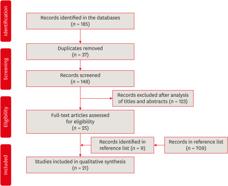
Results The search strategy resulted in the retrieval of 185 publications. After the application of the eligibility criteria, 21 articles were selected for a qualitative analysis.
Conclusions The cryopreservation process must be carried out in 6 stages: tooth disinfection, pulp extraction, cell isolation, cell proliferation, cryopreservation, and thawing. In addition, it can be inferred that the use of dimethyl sulfoxide, programmable freezing, and storage in liquid nitrogen are associated with a high rate of cell viability after thawing and a high rate of cell proliferation in both primary and permanent teeth.
-
Citations
Citations to this article as recorded by- Comparative study on biological characteristics of dental mesenchymal stem cells isolated from gingiva, periodontal ligament, and dental follicle and their derived conditioned medium
Xianyi He, Yichen Gao, Haiyin Wan, Xia Wang, Jie Shen, Yun He, Junliang Chen
Annals of Anatomy - Anatomischer Anzeiger.2026; 263: 152751. CrossRef - Effect of the pulp harvesting method on the viability of human dental pulp stem cells and their odontogenic differentiation potential
Justine De Visscher, Lore Vermeir, Natasja Van den Vreken, Liesbeth Temmerman, Noëmi De Roo, Jolanda van Hengel, Guy De Pauw
Cell and Tissue Banking.2025;[Epub] CrossRef - The Antimicrobial Effect of the Incorporation of Inorganic Substances into Heat-Cured Denture Base Resins—A Systematic Review
Mariana Lima, Helena Salgado, André Correia, Patrícia Fonseca
Prosthesis.2024; 6(5): 1189. CrossRef - Sphingosine-1-phosphate Treatment Improves Cryopreservation Efficiency in Human Mesenchymal Stem Cells
Seong-Ju Oh, Chan-Hee Jo, Tae-Seok Kim, Chae-Yeon Hong, Sung-Lim Lee, Young-Hoon Kang, Gyu-Jin Rho
Life.2023; 13(6): 1286. CrossRef - Time- and Concentration-Dependent Effects of the Stem Cells Derived from Human Exfoliated Deciduous Teeth on Osteosarcoma Cells
Razieh Alipour, Batool Hashemibeni, Vajihe Asgari, Hamid Bahramian
Advanced Biomedical Research.2023;[Epub] CrossRef
- Comparative study on biological characteristics of dental mesenchymal stem cells isolated from gingiva, periodontal ligament, and dental follicle and their derived conditioned medium
- 1,891 View
- 25 Download
- 4 Web of Science
- 5 Crossref

- The evaluation of periodontal ligament cells of rat teeth after low-temperature preservation under high pressure
- Jin-Ho Chung, Jin Kim, Seong-Ho Choi, Eui-Seong Kim, Jiyong Park, Seung-Jong Lee
- J Korean Acad Conserv Dent 2010;35(4):285-294. Published online July 31, 2010
- DOI: https://doi.org/10.5395/JKACD.2010.35.4.285
-
 Abstract
 PDF
PDF PubReader
 ePub
ePub The purpose of this study was to evaluate the viability of periodontal ligament cells of rat teeth after low-temperature preservation under high pressure by means of MTT assay, WST-1 assay. 12 teeth of Sprague-Dawley white female rats of 4 week-old were used for each group.
Both side of the first and second maxillary molars were extracted as atraumatically as possible under tiletamine anesthesia. The experimental groups were group 1 (Immediate extraction), group 2 (Slow freezing under pressure of 3 MPa), group 3 (Slow freezing under pressure of 2 MPa), group 4 (Slow freezing under no additional pressure), group 5 (Rapid freezing in liquid nitrogen under pressure of 2 MPa), group 6 (Rapid freezing in liquid nitrogen under no additional pressure), group 7 (low-temperature preservation at 0℃ under pressure of 2 MPa), group 8 (low-temperature preservation at 0℃ under no additional pressure), group 9 (low-temperature preservation at -5℃ under pressure of 90 MPa). F-medium and 10% DMSO were used as preservation medium and cryo-protectant. For cryo-preservation groups, thawing was performed in 37℃ water bath, then MTT assay, WST-1 assay were processed. One way ANOVA and Tukey HSD method were performed at the 95% level of confidence. The values of optical density obtained by MTT assay and WST-1 were divided by the values of eosin staining for tissue volume standardization.
In both MTT and WST-1 assay, group 7 (0℃/2 MPa) showed higher viability of periodontal ligament cells than other group (2-6, 8) and this was statistically significant (p < 0.05), but showed lower viability than group 1, immediate extraction group (no statistical significance).
By the results of this study, low-temperature preservation at 0℃ under pressure of 2 MPa suggest the possibility for long term preservation of teeth.
-
Citations
Citations to this article as recorded by- Evaluation of the Viability of Rat Periodontal Ligament Cells after Storing at 0℃/2 MPa Condition up to One Week: In Vivo MTT Method
Sun Mi Jang, Sin-Yeon Cho, Eui-Seong Kim, Il-Young Jung, Seung Jong Lee
Journal of Korean Dental Science.2016; 9(1): 1. CrossRef
- Evaluation of the Viability of Rat Periodontal Ligament Cells after Storing at 0℃/2 MPa Condition up to One Week: In Vivo MTT Method
- 1,078 View
- 1 Download
- 1 Crossref

- THE EFFICACY OF PROGRAMMED CRYO-PRESERVATION UNDER PRESSURE IN RAT PERIODONTAL LIGAMENT CELLS
- Young-Eun Lee, Eui-Seong Kim, Jin Kim, Seung-Hoon Han, Seung-Jong Lee
- J Korean Acad Conserv Dent 2009;34(4):356-363. Published online January 14, 2009
- DOI: https://doi.org/10.5395/JKACD.2009.34.4.356
-
 Abstract
 PDF
PDF PubReader
 ePub
ePub Abstract The purpose of this study was to evaluate the viability of periodontal ligament cells in rat teeth using slow cryo-preservation method under pressure by means of MTT assay and WST-1 assay. Eighteen teeth of Sprague-Dawley white female rats of 4 week-old were used for each group.
Both sides of the first and second maxillary molars were extracted as atraumatically as possible under Tiletamine anesthesia. The experimental groups were group 1 (Immediate control), group 2 (Cold preservation at 4°C for 1 week), group 3 (Slow freezing), group 4 (Slow freezing under pressure of 3 MPa). F-medium and 10% DMSO were used as preservation medium and cryo-protectant. For cryo-preservation groups, thawing was performed in 37°C water bath, then MTT assay and WST-1 assay were processed. One way ANOVA and Tukey method were performed at the 95% level of confidence. The values of optical density obtained by MTT assay and WST-1 were divided by the values of eosin staining for tissue volume standardization.
In both MTT and WST-1 assay, group 4 showed significantly higher viability of periodontal ligament cells than group 2 and 3 (p < 0.05), but showed lower viability than immediate control group.
By the results of this study, slow cryo-preservation method under pressure suggests the possibility for long term cryo-preservation of the teeth.
-
Citations
Citations to this article as recorded by- Effects of Slow Programmable Cryopreservation on Preserving Viability of the Cultured Periodontal Ligament Cells from Human Impacted Third Molar
Jin-Woo Kim, Tae-Yi Kim, Ye-mi Kim, Eun-Kyoung Pang, Sun-Jong Kim
Journal of Korean Dental Science.2015; 8(2): 57. CrossRef - The evaluation of periodontal ligament cells of rat teeth after low-temperature preservation under high pressure
Jin-Ho Chung, Jin Kim, Seong-Ho Choi, Eui-Seong Kim, Jiyong Park, Seung-Jong Lee
Journal of Korean Academy of Conservative Dentistry.2010; 35(4): 285. CrossRef - Comparison of viability of oral epithelial cells stored by different freezing methods
Do-Young Baek, Seung-Jong Lee, Han-Sung Jung, EuiSeong Kim
Journal of Korean Academy of Conservative Dentistry.2009; 34(6): 491. CrossRef
- Effects of Slow Programmable Cryopreservation on Preserving Viability of the Cultured Periodontal Ligament Cells from Human Impacted Third Molar
- 1,045 View
- 1 Download
- 3 Crossref

- Evaluation of the viability of periodontal ligament cell in rat teeth using slow cryopreservation method with magnetic field
- Hyun-Jung Ahn, Eui-Seong Kim, Jin Kim, Duck-Won Kim, Ki-Yeol Kim, Chan-Young Lee, Seung-Jong Lee
- J Korean Acad Conserv Dent 2008;33(4):332-340. Published online July 31, 2008
- DOI: https://doi.org/10.5395/JKACD.2008.33.4.332
-
 Abstract
 PDF
PDF PubReader
 ePub
ePub The purpose of this study was to evaluate the viability of periodontal ligament cell in rat teeth using slow cryopreservation method with magnetic field through MTT assay and TUNEL test. For each group, 12 teeth of 4 weeks old white female Sprague-Dawley rat were used for MTT assay, and 6 teeth in TUNEL test. The Maxillary left and right, first and second molars were extracted as atraumatically as possible under tiletamine anesthesia. The experimental groups were group1 (immediately extraction), group 2 (cold preservation at 4℃ for 1 week), group 3 (rapid cryopreservation in liquid nitrogen), group 4 (slow cryopreservation with magnetic field of 1 G), and group 5 (slow cryopreservation). F medium was used as preservation medium and 10% DMSO as cryoprotectant. After preservation and thawing, the MTT assay and TUNEL test were processed. One way ANOVA and Scheffe method were performed at the 95% level of confidence. The value of optical density obtained after MTT analysis was divided by the value of eosin staining for tissue volume standardization. In both MTT assay and TUNEL test, it had showed no significant difference among group 3, 4, and 5. And group 3 had showed higher viability of periodontal ligament cell than group 2.
From this study, slow cryopreservation method with magnetic field can be used as one of cryopreservation methods.
-
Citations
Citations to this article as recorded by- The evaluation of periodontal ligament cells of rat teeth after low-temperature preservation under high pressure
Jin-Ho Chung, Jin Kim, Seong-Ho Choi, Eui-Seong Kim, Jiyong Park, Seung-Jong Lee
Journal of Korean Academy of Conservative Dentistry.2010; 35(4): 285. CrossRef - Comparison of viability of oral epithelial cells stored by different freezing methods
Do-Young Baek, Seung-Jong Lee, Han-Sung Jung, EuiSeong Kim
Journal of Korean Academy of Conservative Dentistry.2009; 34(6): 491. CrossRef - The efficacy of programmed cryo-preservation under pressure in rat periodontal ligament cells
Young-Eun Lee, Eui-Seong Kim, Jin Kim, Seung-Hoon Han, Seung-Jong Lee
Journal of Korean Academy of Conservative Dentistry.2009; 34(4): 356. CrossRef
- The evaluation of periodontal ligament cells of rat teeth after low-temperature preservation under high pressure
- 1,084 View
- 1 Download
- 3 Crossref

- Evaluation of periodontal ligament cell viability in rat teeth according to various extra-oral dry storage times using MTT assay
- In-Soo Jeon, Eui-Seong Kim, Jin Kim, Seung-Jong Lee
- J Korean Acad Conserv Dent 2006;31(5):398-408. Published online September 30, 2006
- DOI: https://doi.org/10.5395/JKACD.2006.31.5.398
-
 Abstract
 PDF
PDF PubReader
 ePub
ePub The purpose of this study was to verify the usefulness of MTT analysis as a tool of measurement of the periodontal ligament cell viability from the extracted rat molar.
A total of 80 Sprague-Dawley white female rat of 4 week-old with a body weight of 100 grams were used. The maxillary left and right, first and second molars were extracted under Ketamine anesthesia. Twenty-four teeth of each group (divided as five groups depending upon the time-lapse after extraction such as immediate, 10, 20, 40 and 60 minutes) were immersed in 200 µl of MTT solution (0.5 mg/ml) and processed for optical density measurements . Another 10 teeth of each group were treated as same as above and sectioned at 10 µm for microscopic examination.
All measurements values were divided by the value of hematoxylin-eosin staining which represented the volume of each corresponding samples. Immediate and 10 minute groups showed highest MTT values followed by 20, 40, and 60 minutes consecutively. Statistical significance (p < 0.05) existed between all groups except in immediate versus 10 minute groups and 40 versus 60 minutes. Histological findings also showed similar findings with MTT results in crystal shape and crystal numbers between the experimental groups.
These data indicate that
in vivo MTT analysis may be of value for evaluation of the periodontal ligament cell viability without time- consuming cell culturing processes.-
Citations
Citations to this article as recorded by- Evaluation of the Viability of Rat Periodontal Ligament Cells after Storing at 0℃/2 MPa Condition up to One Week: In Vivo MTT Method
Sun Mi Jang, Sin-Yeon Cho, Eui-Seong Kim, Il-Young Jung, Seung Jong Lee
Journal of Korean Dental Science.2016; 9(1): 1. CrossRef
- Evaluation of the Viability of Rat Periodontal Ligament Cells after Storing at 0℃/2 MPa Condition up to One Week: In Vivo MTT Method
- 1,058 View
- 0 Download
- 1 Crossref

-
Evaluation of periodontal ligament cell viability in rat teeth after frozen preservation using
in-vivo MTT assay - Jae-Wook Kim, Eui-Sung Kim, Jin Kim, Seung-Jong Lee
- J Korean Acad Conserv Dent 2006;31(3):192-202. Published online May 31, 2006
- DOI: https://doi.org/10.5395/JKACD.2006.31.3.192
-
 Abstract
 PDF
PDF PubReader
 ePub
ePub The purpose of this study was to examine the viability of PDL cells in rat molars by using
in vivo MTT assay, which was used to compare fast cryopreservation group by liquid nitrogen (-196℃) with 4℃ cold preservation group.A total of 74 Sprague-Dawley white female rats of 4 week-old with a body weight of 100 grams were used. The maxillary left and right, first and second molars were extracted as atraumatically as possible under ketamine anesthesia.
Ten teeth of each group were divided as six experimental groups depending upon the preservation. Cryopreservation groups were Group 1 (5% DMSO 6% HES in F medium), Group 2 (10% DMSO in F medium), Group 3 (5% DMSO 6% HES in Viaspan®), Group 4 (10% DMSO in Viaspan®) which were cryopreserved for 1 week and cold preservation groups were Group 5 (F medium), Group 6 (Viaspan®) at 4℃ for 1 week. Immediate extraction group was used as a control. After preservation and thawing, the
in vivo MTT assay was processed. Two way ANOVA and Duncan's Multiple Range Test was performed at the 95% level of confidence. Another 2 teeth of each group were treated as the same manner and frozen sections 10 µm thick for microscopic observation.The value of optical density obtained after
in vivo MTT analysis was divided by the value of eosin staining for tissue volume standardization. Group 1, 2 had significantly higher optical density than Group 3 and 4 which had the lowest OD value. Group 6 had higher OD value than in Group 5 (P < 0.05). Histological findings of periodontal ligament cell, after being stained with MTT solution were consistent with thein vivo MTT assay results.In this study, the groups which were frozen with DMSO as a cryoprotectant and the groups with F medium showed the best results.
-
Citations
Citations to this article as recorded by- Evaluation of the Viability of Rat Periodontal Ligament Cells after Storing at 0℃/2 MPa Condition up to One Week: In Vivo MTT Method
Sun Mi Jang, Sin-Yeon Cho, Eui-Seong Kim, Il-Young Jung, Seung Jong Lee
Journal of Korean Dental Science.2016; 9(1): 1. CrossRef - Comparative study on survival rate of human gingival fibroblasts stored in different storage media
Hee Su Lee, You Sun Lim
Journal of Korean society of Dental Hygiene.2012; 12(4): 733. CrossRef - The evaluation of periodontal ligament cells of rat teeth after low-temperature preservation under high pressure
Jin-Ho Chung, Jin Kim, Seong-Ho Choi, Eui-Seong Kim, Jiyong Park, Seung-Jong Lee
Journal of Korean Academy of Conservative Dentistry.2010; 35(4): 285. CrossRef - Comparison of viability of oral epithelial cells stored by different freezing methods
Do-Young Baek, Seung-Jong Lee, Han-Sung Jung, EuiSeong Kim
Journal of Korean Academy of Conservative Dentistry.2009; 34(6): 491. CrossRef - The efficacy of programmed cryo-preservation under pressure in rat periodontal ligament cells
Young-Eun Lee, Eui-Seong Kim, Jin Kim, Seung-Hoon Han, Seung-Jong Lee
Journal of Korean Academy of Conservative Dentistry.2009; 34(4): 356. CrossRef
- Evaluation of the Viability of Rat Periodontal Ligament Cells after Storing at 0℃/2 MPa Condition up to One Week: In Vivo MTT Method
- 998 View
- 0 Download
- 5 Crossref


 KACD
KACD

 First
First Prev
Prev


