
Search
- Page Path
- HOME > Search
Research Articles
- Evaluation of at-home bleaching protocol with application on different surfaces: bleaching efficacy and hydrogen peroxide permeability
- Heloisa Forville, Michael Willian Favoreto, Michel Wendlinger, Roberta Micheten Dias, Christiane Philippini Ferreira Borges, Alessandra Reis, Alessandro D. Loguercio
- Restor Dent Endod 2023;48(4):e33. Published online October 6, 2023
- DOI: https://doi.org/10.5395/rde.2023.48.e33
-
 Abstract
 PDF
PDF PubReader
 ePub
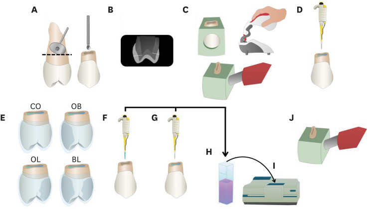
ePub Objectives This study aimed to evaluate the bleaching efficacy and hydrogen peroxide permeability in the pulp chamber by the at-home bleaching gel in protocols applied on different dental surfaces.
Materials and Methods Forty premolars were randomly into 4 groups: control group no bleaching, only application on the buccal surface (OB), only application on the lingual surface (OL) and application in buccal and lingual surfaces, simultaneously (BL). At-home bleaching gel (White Class 7.5%) was used for the procedure. The bleaching efficacy was evaluated with a digital spectrophotometer (color change in CIELAB [Δ
E ab] and CIEDE 2000 [ΔE 00] systems and Whitening Index for Dentistry [ΔWID]). The hydrogen peroxide permeability in the pulp chamber (µg/mL) was assessed using UV-Vis spectrophotometry and data were analyzed for a 1-way analysis of variance and Tukey’s test (α = 0.05).Results All groups submitted to bleaching procedure showed bleaching efficacy when measured with Δ
E ab and ΔE 00 (p > 0.05). Therefore, when analyzed by ΔWID, a higher bleaching efficacy were observed for the application on the groups OB and BL (p = 0.00003). Similar hydrogen peroxide permeability was found in the pulp chambers of the teeth undergoing different protocols (p > 0.05).Conclusions The application of bleaching gel exclusively on the OB is sufficient to achieve bleaching efficacy, when compared to BL. Although the OL protocol demonstrated lower bleaching efficacy based on the ΔWID values, it may still be of interest and relevant in certain clinical scenarios based on individual needs, requiring clinical trials to better understand its specificities.
-
Citations
Citations to this article as recorded by
- Effect of whitening pens on hydrogen peroxide permeability in the pulp chamber, color change and surface morphology
Laryssa Mylenna Madruga Barbosa, Gabrielle Gomes Centenaro, Deisy Cristina Ferreira Cordeiro, Maria Alice de Matos Rodrigues, Letícia Condolo, Michael Willian Favoreto, Alessandra Reis, Alessandro D. Loguercio
Journal of Dentistry.2025; 154: 105595. CrossRef - Evaluation of bleaching efficiency of carbamide peroxide applied on different dental surfaces: An in vitro study
R. Gokulnath, R. S. Mohan Kumar, A. Jayasenthil, R. Anjana, G. Sree Vidya
Journal of Conservative Dentistry and Endodontics.2025; 28(4): 366. CrossRef - Characterization and effects on enamel of low-concentration bleaching gels containing hyaluronic acid, NF_TiO2 nanoparticles and irradiated with violet LED light
Marcos Roberto Lima Benati, Matheus Kury, Priscila Borges Gobbo de Melo, Iago César Ribeiro Teles Matos, Roberta Tarkany Basting, Rosanna Tarkany Basting, Fernando Luis Esteban Florez, Vanessa Cavalli
Clinical Oral Investigations.2025;[Epub] CrossRef - Impact of bleaching on white spot lesions: hydrogen peroxide permeability and color alteration
Laryssa Mylenna Madruga Barbosa, Bruno Baracco, Taynara S. Carneiro, Michael Willian Favoreto, Michel Wendlinger, Daniel Jiménez-Díez, Laura Ceballos, Alessandro D. Loguercio
Clinical Oral Investigations.2025;[Epub] CrossRef - Efficacy of a buccal and lingual at‐home bleaching protocol—A randomized, split‐mouth, single‐blind controlled trial
Heloisa Forville, Laís Giacomini Bernardi, Michael Willian Favoreto, Felipe Coppla, Taynara de Souza Carneiro, Fabiana Madalozzo Coppla, Alessandro D. Loguercio, Alessandra Reis
Journal of Esthetic and Restorative Dentistry.2024; 36(9): 1301. CrossRef - REANATOMIZAÇÃO DE DENTE CONOIDE ASSOCIADA A ESTÉTICA VERMELHA: RELATO DE CASO
Ana Karolayne Sousa de Morais, Daniele Fernanda Sousa Barros, Daniel Messias Limeira, Rhana Leticia de Oliveira Faria, Roberta Furtado Carvalho, Sandna Nolêto de Araújo, Laura Barbosa Santos Di Milhomem
Revista Contemporânea.2024; 4(10): e6299. CrossRef - Effect of the reduction in the exposure time to at-home bleaching gel on color change and tooth sensitivity: A systematic review and meta-analysis
Priscila Borges Gobbo de Melo, Letícia Vasconcelos Silva Souza, Lucianne Cople Maia, Guido Artemio Marañón-Vásquez, Matheus Kury, Vanessa Cavalli
Clinical Oral Investigations.2024;[Epub] CrossRef
- Effect of whitening pens on hydrogen peroxide permeability in the pulp chamber, color change and surface morphology
- 4,932 View
- 85 Download
- 5 Web of Science
- 7 Crossref

- Effects of dentin surface preparations on bonding of self-etching adhesives under simulated pulpal pressure
- Chantima Siriporananon, Pisol Senawongse, Vanthana Sattabanasuk, Natchalee Srimaneekarn, Hidehiko Sano, Pipop Saikaew
- Restor Dent Endod 2022;47(1):e4. Published online December 28, 2021
- DOI: https://doi.org/10.5395/rde.2022.47.e4
-
Abstract
PDFPubReaderePub
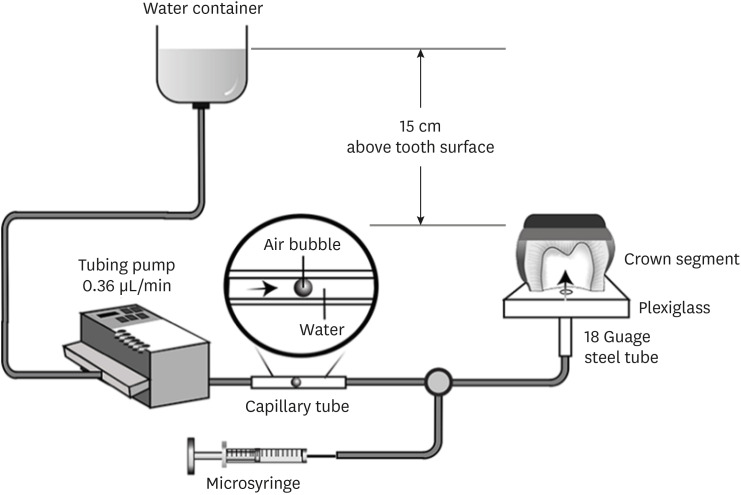
Objectives This study evaluated the effects of different smear layer preparations on the dentin permeability and microtensile bond strength (µTBS) of 2 self-etching adhesives (Clearfil SE Bond [CSE] and Clearfil Tri-S Bond Universal [CTS]) under dynamic pulpal pressure.
Materials and Methods Human third molars were cut into crown segments. The dentin surfaces were prepared using 4 armamentaria: 600-grit SiC paper, coarse diamond burs, superfine diamond burs, and carbide burs. The pulp chamber of each crown segment was connected to a dynamic intra-pulpal pressure simulation apparatus, and the permeability test was done under a pressure of 15 cmH2O. The relative permeability (%P) was evaluated on the smear layer-covered and bonded dentin surfaces. The teeth were bonded to either of the adhesives under pulpal pressure simulation, and cut into sticks after 24 hours water storage for the µTBS test. The resin-dentin interface and nanoleakage observations were performed using a scanning electron microscope. Statistical comparisons were done using analysis of variance and
post hoc tests.Results Only the method of surface preparation had a significant effect on permeability (
p < 0.05). The smear layers created by the carbide and superfine diamond burs yielded the lowest permeability. CSE demonstrated a higher µTBS, with these values in the superfine diamond and carbide bur groups being the highest. Microscopic evaluation of the resin-dentin interface revealed nanoleakage in the coarse diamond bur and SiC paper groups for both adhesives.Conclusions Superfine diamond and carbide burs can be recommended for dentin preparation with the use of 2-step CSE.
-
Citations
Citations to this article as recorded by- Effect of smear layer pretreatment with EDTA and sodium hypochlorite on the dentin bond durability of universal adhesives
Thanawat Ruaydee, Chantida Pawaputanon Na Mahasarakham, Vanthana Sattabanasuk, Pipop Saikaew
Frontiers in Dental Medicine.2026;[Epub] CrossRef - The effect of different adhesive strategies and diamond burs on dentin bond strength of universal resin cements
Chavakorn Atsavathavornset, Pipop Saikaew, Choltacha Harnirattisai, Hidehiko Sano
Clinical Oral Investigations.2025;[Epub] CrossRef - Universal adhesive systems in dentistry: A narrative review
Svetlana N. Razumova, Anzhela S. Brago, Oxana R. Ruda, Zoya A. Guryeva, Elvira V. Adzhieva
Russian Journal of Dentistry.2024; 28(5): 512. CrossRef - Delayed light activation of resin composite affects the bond strength of adhesives under dynamic simulated pulpal pressure
Nattaporn Sukprasert, Choltacha Harnirattisai, Pisol Senawongse, Hidehiko Sano, Pipop Saikaew
Clinical Oral Investigations.2022; 26(11): 6743. CrossRef
- Effect of smear layer pretreatment with EDTA and sodium hypochlorite on the dentin bond durability of universal adhesives
- 4,044 View
- 60 Download
- 3 Web of Science
- 4 Crossref
- Physicochemical properties, cytotoxicity and penetration into dentinal tubules of sodium hypochlorite with and without surfactants
- Hernán Coaguila-Llerena, Isadora Barbieri, Mário Tanomaru-Filho, Renato de Toledo Leonardo, Ana Paula Ramos, Gisele Faria
- Restor Dent Endod 2020;45(4):e47. Published online September 10, 2020
- DOI: https://doi.org/10.5395/rde.2020.45.e47
-
Abstract
PDFPubReaderePub
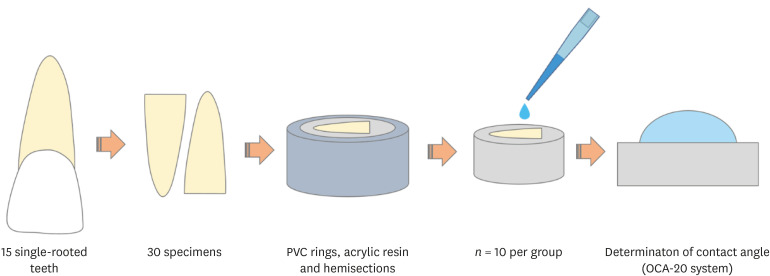
Objectives The aim of this study was to assess the physicochemical properties, cytotoxicity and penetration into dentinal tubules of ChlorCid™ Surf (3% sodium hypochlorite [NaOCl] with surfactant) in comparison to ChlorCid™ (3% NaOCl without surfactant).
Materials and Methods The physicochemical properties evaluated were pH, surface tension, free available chlorine (FAC) and contact angle. Cytotoxicity was evaluated in L929 fibroblasts exposed to the solutions by 3-(4,5-dimethylthiazol-2-yl)-2,5-diphenyl tetrazolium bromide and neutral red assays. Assessment of penetration into dentinal tubules was performed by staining single-rooted permanent human teeth with crystal violet (
n = 9), which were irrigated with the solutions and analyzed in cervical, middle and apical segments. Data were analyzed by one-way analysis of variance (ANOVA) and Tukey'spost -test, 2-way ANOVA and Bonferroni'spost -test ort -test (α = 0.05).Results ChlorCid™ Surf and ChlorCid™ FAC values were close to those indicated by the manufacturer. ChlorCid™ Surf showed lower surface tension and contact angle on dentin, and higher pH than ChlorCid™ (
p < 0.05). The penetration of ChlorCid™ Surf was higher in cervical and middle segments, compared with ChlorCid™ (p < 0.05). There was no difference in irrigant cytotoxicity (p > 0.05).Conclusions ChlorCid™ Surf showed lower surface tension, lower contact angle on root canal dentin, higher penetration into dentinal tubules and more alkaline pH, compared with ChlorCid™. However, both solutions showed similar cytotoxicity and FAC content.
-
Citations
Citations to this article as recorded by- Physicochemical and Biological Properties of the “All-In-One” Endodontic Irrigant Triton
Jesus Aranda, Elda Olivia Nobre de Souza, Arturo Javier Aranda Garcia, Renato de Toledo Leonardo, Ana Paula Ramos, Giampiero Rossi-Fedele, Gisele Faria
Journal of Endodontics.2026; 52(3): 421. CrossRef - Single‐ or two‐visits endodontic treatment: Impact of irrigants on surface roughness, microhardness, topography, and fracture resistance of root dentin
Yasmin Padoin, Sidnei Flores de Pellegrin, Duvan Cala Castillo, William Lemos Bevilaqua, Gabriel Kalil Rocha Pereira, Guilherme Pauletto
European Journal of Oral Sciences.2026;[Epub] CrossRef - Effects of penetration enhancers on the performance of irrigants for root canal disinfection
Yi Luo, Runze Liu, Pei Liu, Mengting Duan, Wei Fan, Bing Fan
Clinical Oral Investigations.2025;[Epub] CrossRef - Influence of post space disinfection protocols on the push-out bond strength of fiber posts luted with self-adhesive cement
Satheesh B. Haralur, Salem Ali Alqahtani, Khalid Salem Alqahtani, Mohammed A. Al-Qarni, Saeed M. AlQahtani
AIP Advances.2025;[Epub] CrossRef - Research methods assessing sodium hypochlorite cytotoxicity: A scoping review
Hernán Coaguila-Llerena, Luana Raphael da Silva, Gisele Faria
Heliyon.2024; 10(1): e23060. CrossRef - Amelioration in the sodium hypochlorite as root canal irrigant – A review
Preety Sehrawat
International Dental Journal of Student's Research.2024; 12(2): 65. CrossRef - Sonic-assisted antibacterial photodynamic therapy: a strategy for enhancing lateral canal disinfection
Yanhuang Wang, Lishan Lei, Jing Huang, Zhiyu Cai, Xiaojing Huang
BMC Oral Health.2024;[Epub] CrossRef - A Comparative Evaluation of Contact Angle and Depth of Penetration of Sodium Hypochlorite With Various Surfactants: An In Vitro Study
Shubhashini N, Krithika D, Akhilesh Gowda , Shruthi Nagaraja , Rhea S Mathew, Nivaskumar G A, Vinaychandra R
Cureus.2024;[Epub] CrossRef - Antibacterial efficacy of silver nanoparticles, sodium hypochlorite, chlorhexidine, and hypochlorous acid on dentinal surfaces infected with Enterococcus faecalis
Aysenur Oncu, Berkan Celikten, Betül Aydın, Gulin Amasya, Erkan Tuncay, Gamze Guney Eskiler, Leyla Açık, Fatma Semra Sevimay
Microscopy Research and Technique.2024; 87(9): 2094. CrossRef - Advances in the Role of Sodium Hypochlorite Irrigant in Chemical Preparation of Root Canal Treatment
Chen Cai, Xuan Chen, Yang Li, Qianzhou Jiang, Yeliz Guven
BioMed Research International.2023;[Epub] CrossRef - Effect of sodium hypochlorite-based formulations on the adhesion interface after fiber post cementation
Joatan Lucas de Sousa Gomes COSTA, Tatiane Miranda MANZOLI, João Felipe BESEGATO, Joissi Ferrari ZANIBONI, Eliane Cristina Gulin DE OLIVEIRA, Lucas David GALVANI, Andréa Abi Rached DANTAS, Luis Geraldo VAZ, Milton Carlos KUGA
Dental Materials Journal.2023; 42(6): 878. CrossRef - Physicochemical properties and penetration into dentinal tubules of calcium hypochlorite with surfactants
Hernán Coaguila-Llerena, Julia da Silva Toledo, Ana Paula Ramos, Gisele Faria
Brazilian Dental Journal.2022; 33(2): 1. CrossRef
- Physicochemical and Biological Properties of the “All-In-One” Endodontic Irrigant Triton
- 3,252 View
- 40 Download
- 12 Crossref
Basic Research
- Real-time measurement of dentinal fluid flow during desensitizing agent application
- Sun-Young Kim, Eun-Joo Kim, In-Bog Lee
- J Korean Acad Conserv Dent 2010;35(5):313-320. Published online September 30, 2010
- DOI: https://doi.org/10.5395/JKACD.2010.35.5.313
-
Abstract
PDFPubReaderePub
Objectives The aim of this study was to examine changes in the dentinal fluid flow (DFF) during desensitizing agent application and to compare permeability after application among the agents.
Materials and Methods A Class 5 cavity was prepared to exposure cervical dentin on an extracted human premolar which was connected to a sub-nanoliter fluid flow measuring device (NFMD) under 20 cm water pressure. DFF was measured from before application of desensitizing agent (Seal&Protect, SP; SuperSeal, SS; BisBlock, BB; Gluma desensitizer, GL; Bi-Fluoride 12, BF) through application procedure to 5 min after application.
Results DFF rate after each desensitizing agent application was significantly reduced when compared to initial DFF rate before application (
p < 0.05). SP showed a greater reduction in DFF rate than GL and BF did (p < 0.05). SS and BB showed a greater reduction in DFF rate than BF did (p < 0.05).Conclusions Characteristic DFF aspect of each desensitizing agent was shown in NFMD during the application procedure.
-
Citations
Citations to this article as recorded by- CPNE7 Induces Biological Dentin Sealing in a Dentin Hypersensitivity Model
S.H. Park, Y.S. Lee, D.S. Lee, J.C. Park, R. Kim, W.J. Shon
Journal of Dental Research.2019; 98(11): 1239. CrossRef
- CPNE7 Induces Biological Dentin Sealing in a Dentin Hypersensitivity Model
- 1,684 View
- 2 Download
- 1 Crossref
Original Articles
- Quantitative comparison of permeability in the adhesive interface of four adhesive systems
- Juhea Chang, Keewook Yi, Hae-Young Kim, In Bog Lee, Byeong Hoon Cho, Ho-Hyun Son
- J Korean Acad Conserv Dent 2009;34(1):51-60. Published online January 31, 2009
- DOI: https://doi.org/10.5395/JKACD.2009.34.1.051
-
Abstract
PDFPubReaderePub
The purpose of this study was to perform quantitative comparisons of water permeable zones in both the adhesive and the hybrid layer before and after thermocycling in order to assess the integrity of the bonding interface. Twenty eight flat dentin surfaces were bonded with a light-cured composite resin using one of four commercial adhesives [OptiBond FL (OP), AdheSE (AD), Clearfil SE Bond (CL), and Xeno III (XE)]. These were sectioned into halves and subsequently cut to yield 2-mm thick specimens; one specimen for control and the other subjected to thermocycling for 10,000 cycles. After specimens were immersed in ammoniacal silver nitrate for 24 h and exposed to a photo developing solution for 8 h, the bonded interface was analyzed by scanning electron microscopy (SEM) and wavelength dispersive spectrometry (WDS) at five locations per specimen. Immediately after bonding, the adhesive layer of OP showed the lowest silver uptake, followed by CL, AD, and XE in ascending order (p < 0.0001); the hybrid layer of CL had the lowest silver content among the groups (p = 0.0039). After thermocycling, none of the adhesives manifested a significant increase of silver in either the adhesive or the hybrid layer. SEM demonstrated the characteristic silver penetrated patterns within the interface. It was observed that integrity of bonding was well maintained in OP and CL throughout the thermocycling process. Adhesive-tooth interfaces are vulnerable to hydrolytic degradation and its permeability varies in different adhesive systems, which may be clinically related to the restoration longevity.
- 1,649 View
- 6 Download
- The effect of bonding resin on bond strength of dual-cure resin cements
- Duck-Su Kim, Sang-Hyuk Park, Gi-Woon Choi, Kyung-Kyu Choi
- J Korean Acad Conserv Dent 2007;32(5):426-436. Published online September 30, 2007
- DOI: https://doi.org/10.5395/JKACD.2007.32.5.426
-
Abstract
PDFPubReaderePub
The objective of this study is to evaluate the effect of an additional application of bonding resin on the bond strength of resin luting cements in both the light-cure (LC) and self-cure (SC) modes by means of the µTBS tests.
Three combinations of One-Step Plus with Choice, Single Bond with Rely X ARC, and One-Up Bond F with Bistite II were used. D/E resin and Pre-Bond resin were used for the additional application. Twelve experimental groups were made. Three mandibular 3rd molars were used in each group. Indirect composite blocks were cemented on the tooth surface. 1 × 1 mm2 dentin-composite beam for µTBS testing were made and tested.
When total-etching dentin adhesives were used, an additional application of the bonding resin increased the bond strength (
P < 0.05). However, this additional application didn't influence the bond strength of self-etching dentin adhesives (P > 0.05).In conclusion, the results suggest that an additional application of the bonding resin increases bond strength and enhances quality of bonding when using total-etching dentin adhesives.
-
Citations
Citations to this article as recorded by- Pull-out bond strength of a self-adhesive resin cement to NaOCl-treated root dentin: effect of antioxidizing agents
Maryam Khoroushi, Marzieh Kachuei
Restorative Dentistry & Endodontics.2014; 39(2): 95. CrossRef - Effects of dentin moisture on the push-out bond strength of a fiber post luted with different self-adhesive resin cements
Sevinç Aktemur Türker, Emel Uzunoğlu, Zeliha Yılmaz
Restorative Dentistry & Endodontics.2013; 38(4): 234. CrossRef - Microtensile bond strength of self-etching and self-adhesive resin cements to dentin and indirect composite resin
Jae-Gu Park, Young-Gon Cho, Il-Sin Kim
Journal of Korean Academy of Conservative Dentistry.2010; 35(2): 106. CrossRef - Effect of curing methods of resin cements on bond strength and adhesive interface of post
Mun-Hong Kim, Hae-Jung Kim, Young-Gon Cho
Journal of Korean Academy of Conservative Dentistry.2009; 34(2): 103. CrossRef - Effect of a desensitizer on dentinal bond strength in cementation of composite resin inlay
Sae-Hee Han, Young-Gon Cho
Journal of Korean Academy of Conservative Dentistry.2009; 34(3): 223. CrossRef
- Pull-out bond strength of a self-adhesive resin cement to NaOCl-treated root dentin: effect of antioxidizing agents
- 2,791 View
- 7 Download
- 5 Crossref
- Effect of the additional application of a resin layer on dentin bonding using single-step adhesives
- Seung-Mo Choi, Sang-Hyuk Park, Kyung-Kyu Choi, Sang-Jin Park
- J Korean Acad Conserv Dent 2007;32(4):313-326. Published online July 31, 2007
- DOI: https://doi.org/10.5395/JKACD.2007.32.4.313
-
Abstract
PDFPubReaderePub
The purpose of this study was to prove that an intermediate resin layer (IRL) can increase the bond strength to dentin by reducing the permeability of single-step adhesives.
Flat dentin surfaces were created on buccal and lingual side of freshly extracted third molar using a low-speed diamond saw under copious water flow. Approximately 2.0 mm thick axially sectioned dentin slice was abraded with wet #600 SiC paper. Three single-step self-etch adhesives; Adper Prompt L-Pop (3M ESPE, St Paul, MN, USA), One-Up Bond F (Tokuyama Corp, Tokyo, Japan) and Xeno III (Dentsply, Konstanz, Germany) were used in this study. Each adhesive groups were again subdivided into ten groups by; whether IRL was used or not; whether adhesives were cured with light before application of IRL or not; the mode of composite application.
The results of this study were as follows;
1. Bond strength of single-step adhesives increased by an additional coating of intermediate resin layer, and this increasement was statistically signigicant when self-cured composite was used (p < 0.001).
2. When using IRL, there were no difference on bond strengths regardless the curing procedure of single-step adhesives.
3. There were no significant difference on bond strengths between usage of AB2 or SM as an IRL.
4. The thickness of hybrid layer was correlated with the acidity of adhesive used, and the nanoleakage represented by silver deposits and grains was examined within hybrid and adhesive layer in most of single-step adhesives.
5. Neither thickness of hybrid layer nor nanoleakage were related to bond strength.
-
Citations
Citations to this article as recorded by- Quantitative comparison of permeability in the adhesive interface of four adhesive systems
Juhea Chang, Keewook Yi, Hae-Young Kim, In Bog Lee, Byeong Hoon Cho, Ho-Hyun Son
Journal of Korean Academy of Conservative Dentistry.2009; 34(1): 51. CrossRef
- Quantitative comparison of permeability in the adhesive interface of four adhesive systems
- 2,082 View
- 2 Download
- 1 Crossref
 First
First Prev
Prev

